

Betta fish anatomy at a glance
A betta is a small, slender bony fish, up to about 6.5 cm long, with an upturned mouth, one dorsal fin set far back, a very long anal fin, long thin pelvic (ventral) fins, and a pair of labyrinth organs above the gills that let it breathe air. Its body plan is otherwise that of a typical spiny-rayed fish: gills under a bony gill cover, a swim bladder, a four-part heart, and a short gut suited to eating insects and small animals.
Wild bettas have short fins. Wild Betta splendens live in still, shallow water in the Mekong basin of Southeast Asia: floodplains, canals, rice paddies and ponds (FishBase; Watson et al.). They are dark and fairly short-finned. Bettas were bred for fighting in Thailand for centuries, with reports going back to the 14th century (Kwon et al. 2022), and ornamental breeders later turned these short-finned fish into the long-finned veiltails, halfmoons and crowntails sold today. Wild B. splendens are listed as Vulnerable by the IUCN.
Betta fish diagram (labeled)
These labeled diagrams match the 3D model. The first shows the external anatomy of a male with every fin named; the second shows the internal organs with the body wall removed.
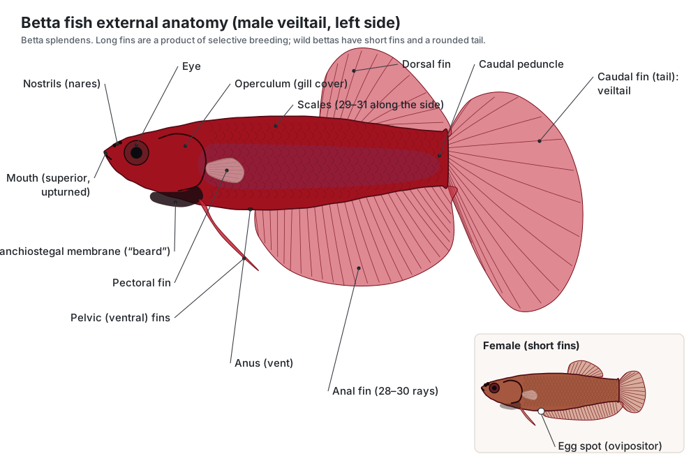
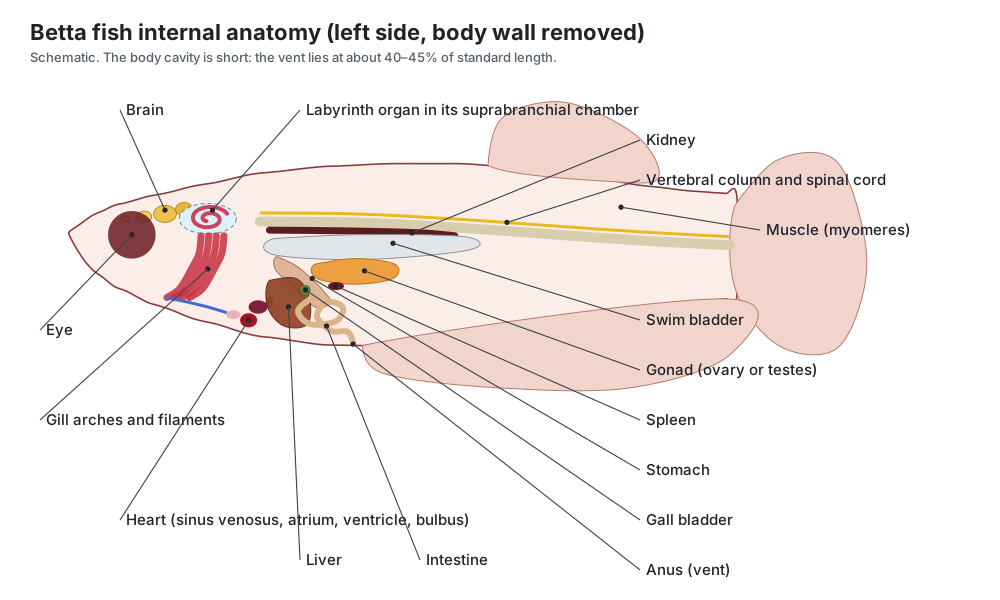
External anatomy and betta fin names
A betta has seven fins: three single fins on the midline (dorsal, caudal and anal) and two pairs (pectoral and pelvic). Hobbyists often call the pelvic fins ventral fins. Each fin is a thin membrane held up by jointed bony fin rays.
| Fin | Hobby name | Number | Betta anatomy |
|---|---|---|---|
| Dorsal fin | Top fin | 1 (median) | 10–14 rays; short base set far back on the body |
| Caudal fin | Tail | 1 (median) | Rounded in wild fish; very long in show varieties |
| Anal fin | Bottom fin | 1 (median) | 28–30 rays; runs from the vent almost to the tail |
| Pectoral fins | Side fins | 2 (paired) | 11–12 rays; small, clear, always moving |
| Pelvic fins | Ventral fins | 2 (paired) | Long, narrow, sickle-shaped, with a filament tip |
Ray counts: Tan & Ng (2005), wild-caught B. splendens.
- Mouth (superior, upturned): Picks food from just under and at the water surface, and gulps air for the labyrinth organ. Males also use it to blow mucus-coated bubbles for the nest and to carry eggs.
- Nostrils (nares): Smell only. They do not connect to the mouth and are not used for breathing.
- Eye: Colour vision for spotting prey and rivals. Fish have no eyelids.
- Operculum (gill cover): Protects the gills and pumps water over them. During a display a betta 'flares': it swings both gill covers out sideways to look bigger.
- Branchiostegal membrane (the “beard”): Seals the gill chamber from below during breathing. When a betta flares, the membrane is pushed out and down and shows as a dark ruff under the head.
- Scales: Flexible armour that protects the body while letting it bend.
- Caudal peduncle: Transfers the pull of the trunk muscles to the tail fin.
- Anus (vent): Releases faeces.
- Egg spot (ovipositor): The opening through which the female releases her eggs.
Betta fin types
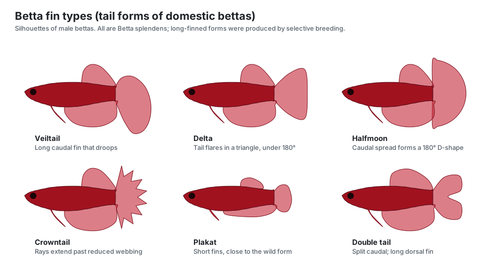
Betta "types" are named mostly by the shape of the tail. All are the same species; they differ in fin length and webbing, which breeders have selected for. Genetic studies show that several fin traits are controlled by single major genes, and that different fins can be changed independently.
| Type | What it looks like |
|---|---|
| Plakat | Short fins and a round or slightly pointed tail. Closest to the wild form and to the original fighting fish of Thailand. |
| Veiltail | Long caudal fin that droops downward like a veil. The most common long-finned pet betta. |
| Delta / super delta | Tail flares in a triangle with straight edges; spread less than 180° (super delta: nearly 180°). |
| Halfmoon | Caudal fin spreads to a full 180° D-shape when the fish flares. |
| Crowntail | Fin rays extend beyond much-reduced webbing, giving a spiky crown edge. Webbing of each fin is controlled by separate loci (Kwon et al. 2022). |
| Double tail | Caudal fin split into two lobes, usually with a broader dorsal fin. Recessive; linked to a deletion near zic1/zic4 (Wang et al. 2021). |
Very long fins come at a cost: they add drag and tear easily, which can make swimming harder for show bettas than for short-finned plakats.
The labyrinth organ: how bettas breathe air
The betta labyrinth organ is a pair of air-breathing organs, one on each side of the head, above the gills and under the gill cover. Each sits in an air space called the suprabranchial chamber. The organ itself is a set of thin, folded bony plates that grow from the first epibranchial (the upper bone of the first gill arch), covered with thin, blood-rich respiratory epithelium. The folds give a large surface for taking oxygen from a bubble of air (Tate et al. 2017).
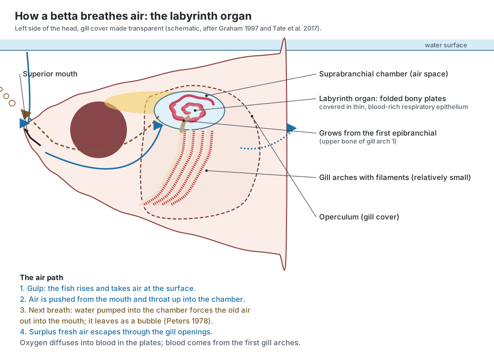
How a breath works. The betta swims to the surface and gulps air. On the next breath, water pumped into the chamber pushes the old air forward into the mouth, where it leaves as a bubble; fresh air is then pushed up into the chamber, and any surplus escapes through the gill openings (Peters 1978; Graham 1997). Blood from the first gill arches flows through the labyrinth and picks up oxygen before returning to the heart.
Do bettas have to breathe air? Bettas breathe air routinely, but studies class B. splendens as a facultative air breather: in well-oxygenated water it can meet its needs with its gills, unlike the obligate air-breathing blue gourami (Mendez-Sanchez & Burggren 2014, 2019). The popular claim that a betta always "drowns" without air is too simple. In practice, warm, still aquarium water is often low in oxygen, and displaying males use much more oxygen, so a betta must always be able to reach the surface easily. Bettas are not born with a working labyrinth; it develops during the first weeks of life.
Betta blood also binds oxygen extremely tightly (half-saturated at only about 2.9 mmHg), a trait of fishes that live in low-oxygen water (Mendez-Sanchez & Burggren 2019).
- Labyrinth organ: Takes oxygen from gulped air. The bony plates grow from the first epibranchial (the upper bone of the first gill arch), and their folds give a large surface of thin respiratory epithelium close to blood capillaries.
- Suprabranchial chamber: Holds the gulped air bubble against the labyrinth. On the next breath, water pumped into the chamber pushes the old air out into the mouth; it leaves as a bubble. Fresh air is then gulped, and surplus air escapes through the gill openings (Peters 1978; Graham 1997).
Betta gills
Betta gills are like those of other bony fish: four gill arches on each side, each with two rows of red gill filaments and short gill rakers. Because the labyrinth chamber takes up space above them, labyrinth fishes have relatively small gills compared with fishes that breathe only water. Among labyrinth fishes, though, bettas have fairly large gills: young B. splendens have about three times the gill area of the blue gourami at the same stage, which fits their ability to live on water-breathing alone (Mendez-Sanchez & Burggren 2019).
- Gill arches: Support the gill filaments and the blood vessels that serve them.
- Gill filaments: Take oxygen from water and release carbon dioxide and ammonia.
- Gill rakers: Keep food from passing out through the gills.
Betta internal anatomy
A betta's internal organs are packed into a short body cavity behind the head, because the vent lies far forward, at about 40–45% of the body length. From front to back you find the heart under the throat, the liver, a small stomach, the looped intestine, the gonad, and above them the swim bladder and the dark red kidney against the backbone. The brain sits in the skull close to the labyrinth chambers.
- Stomach: Stores and starts digesting prey.
- Intestine: Digests food and absorbs nutrients. A short gut suits a diet of insects and small animals.
- Liver: Makes bile, stores energy and processes nutrients and toxins.
- Gall bladder: Stores bile for fat digestion.
- Spleen: Makes and stores blood cells; part of the immune system.
- Swim bladder: Controls buoyancy, so the fish can hover without effort.
- Kidney: Makes large amounts of dilute urine to remove water that enters through the gills in fresh water.
Betta stomach size. A betta's stomach is a small sac. You may read that it is "the size of its eye"; that is a feeding rule of thumb, not a measured fact. The useful point stands: a betta is a small fish, and a few small food items once or twice a day is enough.
Heart. Like other fish, a betta has a single circulation and a heart with four parts in series: sinus venosus, atrium, ventricle and bulbus arteriosus. It pumps oxygen-poor blood to the gills and labyrinth.
- Sinus venosus: First of the heart's four parts in series.
- Atrium: Fills the ventricle.
- Ventricle: Pumps blood forward to the gills and labyrinth.
- Bulbus arteriosus: Smooths each pulse of blood.
- Dorsal aorta: Carries oxygenated blood to the organs and muscles.
Male vs female betta anatomy
Male and female bettas differ most in their fins, colour and beard. Use the Male/Female switch on the model to compare them. Sex becomes easy to tell at about 3–4 months of age (Watson et al.). In ornamental bettas, sex is mainly set by the dmrt1 gene, with males as the XY sex (Kwon et al. 2022).
| Feature | Male | Female |
|---|---|---|
| Fins | Long; caudal, dorsal and anal flow behind the body | Short; tail rounded |
| Colour | Bright and iridescent | Usually duller |
| Beard | Large and dark, very obvious when flaring | Small and pale |
| Body shape | Slimmer | Rounder belly, especially when full of eggs |
| Egg spot | None in adults | White ovipositor between ventral and anal fins |
| Pattern when breeding | Darkens | Often vertical bars when ready to spawn |
| Gonads | Paired testes | Ovary |
| Behaviour | Builds and guards the bubble nest; very aggressive to other males | Releases eggs during the embrace |
The egg spot is a useful clue but not perfect: it can be hard to see on pale fish, and young males can show a similar spot.
Flaring and the betta beard
The betta beard is the branchiostegal membrane, a skin fold under the gill covers held up by thin branchiostegal rays. When a betta flares, it swings both gill covers out sideways, pushes the beard down and spreads its fins to look larger. Males flare at rival males and even at their own reflection.
Flaring is costly. With the gill covers held open, water does not flow well over the gills, so the fish briefly puts itself into a low-oxygen state. Displaying males therefore breathe air more often; the oxygen they take in per breath stays the same, so they make more trips to the surface (reviewed by Tate et al. 2017).
Bubble nests and reproduction
Male bettas build bubble nests: they gulp air at the surface, mix it with mucus in the mouth, and blow sticky bubbles that float together, often among plants (Tate et al. 2017). The female releases eggs during the spawning embrace, when the male wraps his body around her and fertilises the eggs as they are released (Watson et al.). The male then places the eggs in the nest and guards the eggs and young. The nest keeps eggs at the surface, where oxygen exchange is best in warm, still water. Larvae use up their yolk sac about 3 days after hatching.
- Ovary: Produces eggs, released in batches during the spawning embrace.
- Testes: Produce sperm.
- Egg spot (ovipositor): The opening through which the female releases her eggs.
Betta colours and scales
Betta colour comes from chromatophores (colour cells) in the skin. Black-brown melanophores hold melanin, yellow-red cells hold pteridine and carotenoid pigments, and iridophores contain stacks of reflective crystals that produce blue, green and metallic sheens. Genetic studies of pet bettas found strong selection on genes for melanin (mitfa), red pigments (xdh, bco1l) and iridophore development (alkal2l), and an albino form caused by a change near mitfa (Wang et al. 2021; Kwon et al. 2022).
Bettas have thin, overlapping scales, with 29–31 in a row along the side of the body (Tan & Ng 2005). Wild males have red parallel bars on the gill cover.
Health-related betta anatomy
Knowing the anatomy helps explain common pet problems. These notes are general; for diagnosis and treatment, see a veterinarian who treats fish.
Swim bladder problems. The swim bladder controls buoyancy. A betta that floats, sinks or swims tilted is often said to have "swim bladder disease", but that describes a sign, not a cause. In fish, buoyancy problems can come from the gut (for example, a swollen stomach or intestine pressing on the bladder), from infection or fluid inside the swim bladder, from deformity or from injury (Sirri et al. 2020; Merck Veterinary Manual). A vet can use X-rays to see the swim bladder and tell these apart.
Fin damage and fin rot. Long betta fins are thin membranes over fragile rays, so they tear easily and are slow to heal. Fin damage can come from biting by tank mates or from sharp decorations. "Fin rot" is the name for fins that fray and recede from the edges; bacteria such as Aeromonas, Pseudomonas and Flavobacterium columnare are common in diseased aquarium fish, and outbreaks are strongly linked to poor water quality (Merck Veterinary Manual). Fin rays can regrow if the base of the fin is healthy.
Breathing. Because bettas take air at the surface, they need open surface water.
Betta fish dissection
Bettas are pets, not lab specimens, and are far too small for a class dissection. To learn fish dissection, use the step-by-step virtual perch dissection; the organs you find in a perch match those of a betta, apart from the labyrinth organ. Use the 3D model above to explore the betta's internal organs without harming an animal.
Betta vs a typical bony fish
Compared with a generic bony fish such as the yellow perch on our fish anatomy page, a betta has the same basic organs but several special features:
| Feature | Betta | Yellow perch |
|---|---|---|
| Breathing | Gills plus a pair of labyrinth organs for air | Gills only |
| Mouth | Superior (upturned), small | Terminal, large |
| Dorsal fin | One, short-based, set far back | Two: spiny and soft |
| Anal fin | Very long (28–30 rays) | Short (II, 6–8) |
| Body cavity | Short; vent at about 40–45% of standard length | Long; vent about two-thirds of the way back |
| Size | Up to about 6.5 cm total length | Usually 10–25 cm |
| Habitat | Still, shallow, warm, often low-oxygen water | Lakes and slow rivers |
Betta fish anatomy labeled: every part and its function
External
- Skin and colour cells (Integument, chromatophores)
- A thin living skin with a mucus coat. Colour cells (chromatophores) in it give bettas their reds, blues and blacks. Protects the body and holds the pigment cells. Melanophores make black-brown melanin, xanthophores and erythrophores hold yellow-red pigments, and iridophores contain reflective crystals that make blue and metallic sheens.
- Scales
- Thin, overlapping bony scales in the skin, about 29–31 along the side of the body. Flexible armour that protects the body while letting it bend.
- Snout
- The short front of the head, above the mouth and in front of the eyes. Carries the nostrils.
- Mouth (superior, upturned)
- A small mouth that opens upward at the top of the snout: a superior mouth. Picks food from just under and at the water surface, and gulps air for the labyrinth organ. Males also use it to blow mucus-coated bubbles for the nest and to carry eggs.
- Nostrils (nares) (Nares)
- Two small openings on each side of the snout leading to a blind smell pit. Smell only. They do not connect to the mouth and are not used for breathing.
- Cheek
- The side of the head below and behind the eye. Covers the jaw muscles.
- Operculum (gill cover) (Gill cover)
- The bony flap on each side of the head that covers the gills and the labyrinth organ. Protects the gills and pumps water over them. During a display a betta 'flares': it swings both gill covers out sideways to look bigger.
- Branchiostegal membrane (the “beard”) (Beard, gill membrane)
- The skin fold under the gill covers, supported by the branchiostegal rays. Hobbyists call it the betta's 'beard'. Seals the gill chamber from below during breathing. When a betta flares, the membrane is pushed out and down and shows as a dark ruff under the head.
- Lateral line system
- Sense organs that detect water movement, in canals and pits on the head and along the body. Senses nearby water movement and low-frequency vibration: prey, predators and rivals.
- Dorsal fin (Top fin)
- The single fin on the back, set far back on the body above the rear of the anal fin. Stability against rolling. Raised wide during displays.
- Caudal fin (tail fin) (Tail)
- The tail fin. In the veiltail shown, it is long and droops like a veil; in wild bettas it is short and rounded. Propulsion and steering. Very long show tails add drag and tear easily.
- Caudal peduncle
- The short, deep 'wrist' of the tail just in front of the tail fin. Transfers the pull of the trunk muscles to the tail fin.
- Anal fin (Bottom fin)
- A very long fin under the body, running from just behind the vent almost to the tail. Keeps the fish upright and stable; it is spread during displays and wraps the female during spawning.
- Pectoral fins (Side fins)
- Small, clear, rounded paired fins just behind the gill covers. Fine steering, hovering and backing up. They beat almost constantly when a betta hovers.
- Pelvic fins (ventral fins) (Ventral fins)
- A pair of long, narrow fins under the throat, ending in a thin filament. Balance and touch. In males they are often held forward during displays.
- Anus (vent)
- The opening of the gut, far forward on the belly. Releases faeces.
- Urogenital pore
- The small opening just behind the anus for urine and eggs or sperm. Releases urine and gametes.
- Egg spot (ovipositor) (Ovipositor, genital papilla) · females only
- A small white bump between the ventral and anal fins of females, where eggs come out. The opening through which the female releases her eggs.
Skeletal
- Fin rays (lepidotrichia)
- Thin, jointed bony rods that hold up each fin membrane. Stiffen the fins while letting them bend and fold.
- Pterygiophores
- Small internal bones that anchor the dorsal- and anal-fin rays inside the body. Support and move the median fins.
- Skull (neurocranium)
- The bony box around the brain and inner ears. Protects the brain and sense organs; anchors jaws and gill arches.
- Premaxilla (upper jaw)
- The main tooth-bearing bone of the short upper jaw. Forms the upper lip of the upturned mouth.
- Dentary (lower jaw)
- The tooth-bearing bone of the lower jaw, which rises to the front of the upturned mouth. Closes on prey; small teeth help grip insect larvae.
- Opercle
- The largest bone of the gill cover. Forms most of the gill cover; it swings outward when a betta flares.
- Preopercle
- The L-shaped front bone of the gill cover. Front frame of the gill cover.
- Branchiostegal rays
- Curved bony rods that support the branchiostegal membrane (the beard). Spread the gill membrane; they fan out when the beard is shown.
- Abdominal (trunk) vertebrae
- The vertebrae above the body cavity, carrying ribs. Support the body and anchor the ribs.
- Caudal vertebrae
- The vertebrae of the tail, with haemal arches below them. Bend from side to side to drive the tail.
- Ribs
- Thin bones curving down around the body cavity. Protect the organs and support the body wall.
- Pectoral girdle (cleithrum)
- The bony arch behind the gill cover that carries the pectoral fins. Supports the pectoral fins and forms the back wall of the gill chamber.
- Pelvic girdle
- Small bones in the belly that carry the ventral fins. Anchor the pelvic fins.
- Hypural plate
- Flattened bones at the end of the backbone that carry the tail-fin rays. Support the caudal fin.
Muscular
- Epaxial muscle
- The upper block of swimming muscle above the backbone. Bends the body for swimming.
- Hypaxial muscle
- The lower block of swimming muscle below the backbone. Bends the body and supports the body wall.
- Myomeres
- W-shaped muscle blocks separated by thin sheets (myosepta). Contract in a wave from head to tail.
Nervous
- Olfactory bulbs
- Small front lobes of the brain that process smell. Smell.
- Telencephalon (forebrain)
- The forebrain. Learning, memory and social behaviour such as aggression.
- Optic lobes (optic tectum)
- The large paired visual centres of the brain. Process vision. Bettas rely mostly on sight to judge rivals and mates.
- Cerebellum
- A lobe at the back of the brain. Coordinates swimming and balance.
- Medulla oblongata
- The brainstem, which continues as the spinal cord. Controls breathing rhythm and heart rate, and receives hearing and lateral-line input.
- Spinal cord
- The main nerve cord, running inside the vertebral arches. Carries signals between brain and body; generates the swimming rhythm.
Sensory
- Eye
- Large eyes high on the sides of the head. Colour vision for spotting prey and rivals. Fish have no eyelids.
- Iris
- The coloured ring around the pupil. Shades the lens.
- Lens
- A hard, round lens. Focuses light; it moves back and forth to focus.
- Olfactory rosette
- Folded smell tissue inside each nasal pit. Detects chemicals in the water.
- Inner ear
- Fluid-filled canals and sacs inside the skull. Balance and hearing.
- Otoliths (ear stones)
- Small stones of calcium carbonate in the inner ear. Sense gravity, acceleration and sound.
Gills, labyrinth & swim bladder
- Labyrinth organ (Labyrinth apparatus, suprabranchial organ)
- A maze of thin, folded bony plates covered in blood-rich skin, used to breathe air. Takes oxygen from gulped air. The bony plates grow from the first epibranchial (the upper bone of the first gill arch), and their folds give a large surface of thin respiratory epithelium close to blood capillaries.
- Suprabranchial chamber (Labyrinth chamber)
- An air-filled cavity above the gills that holds the labyrinth organ. Holds the gulped air bubble against the labyrinth. On the next breath, water pumped into the chamber pushes the old air out into the mouth; it leaves as a bubble. Fresh air is then gulped, and surplus air escapes through the gill openings (Peters 1978; Graham 1997).
- Gill arches
- Four bony arches on each side that carry the gills. Support the gill filaments and the blood vessels that serve them.
- Gill filaments
- Red, feathery strips on the outer side of each arch. Take oxygen from water and release carbon dioxide and ammonia.
- Gill rakers
- Short projections on the inner side of the arches. Keep food from passing out through the gills.
- Swim bladder (Gas bladder, air bladder)
- A gas-filled sac along the top of the body cavity. Controls buoyancy, so the fish can hover without effort.
- Gas gland
- A blood-rich patch on the swim-bladder wall that secretes gas into it. Adds gas to the swim bladder.
Digestive
- Jaw teeth
- Rows of small, pointed teeth on the jaws. Grip small live prey; fighting males also bite fins and scales.
- Mouth cavity (buccal cavity)
- The space inside the mouth. Takes in food, water and air. Males mix air with mucus here to blow nest bubbles.
- Pharynx
- The throat, with the gill slits on each side. Passes food back and air up to the labyrinth chambers.
- Oesophagus (Esophagus)
- A short tube from the throat to the stomach. Carries food to the stomach.
- Stomach
- A small, stretchy sac at the front of the body cavity. Stores and starts digesting prey.
- Intestine
- A short, looped tube from the stomach to the vent. Digests food and absorbs nutrients. A short gut suits a diet of insects and small animals.
- Liver
- A large brownish organ at the front of the body cavity. Makes bile, stores energy and processes nutrients and toxins.
- Gall bladder
- A small green sac of bile. Stores bile for fat digestion.
Circulatory
- Sinus venosus
- Thin-walled sac that collects blood returning to the heart. First of the heart's four parts in series.
- Atrium
- The thin-walled receiving chamber. Fills the ventricle.
- Ventricle
- The thick-walled pumping chamber. Pumps blood forward to the gills and labyrinth.
- Bulbus arteriosus
- An elastic bulb at the front of the heart. Smooths each pulse of blood.
- Ventral aorta
- The artery from the heart forward to the gill arches. Carries oxygen-poor blood to the gills.
- Afferent branchial arteries
- Arteries that carry blood up each gill arch. Deliver oxygen-poor blood to the filaments and, via the first arches, to the labyrinth.
- Efferent branchial arteries
- Arteries that collect blood leaving the gills. Carry oxygenated blood to the body.
- Dorsal aorta
- The main artery to the body. Carries oxygenated blood to the organs and muscles.
- Spleen
- A small dark red organ in the body cavity. Makes and stores blood cells; part of the immune system.
Excretory
- Head kidney
- The front part of the kidney, behind the skull. Makes blood and immune cells; holds hormone (interrenal) tissue.
- Kidney
- A dark red strip under the backbone. Makes large amounts of dilute urine to remove water that enters through the gills in fresh water.
Reproductive
- Ovary · females only
- The egg-producing organ of the female. Produces eggs, released in batches during the spawning embrace.
- Oviduct · females only
- A short duct from the ovary to the egg spot. Carries eggs out.
- Testes · males only
- Paired, small, creamy-white organs in males. Produce sperm.
- Sperm ducts · males only
- Ducts from the testes to the urogenital pore. Release sperm onto the eggs during the embrace. Fertilisation is external.
Frequently asked questions
What are the parts of a betta fish?
Externally: an upturned mouth, large eyes, nostrils, gill covers with a 'beard' (branchiostegal membrane) underneath, scales, and six kinds of fin: dorsal, caudal (tail), anal, paired pectorals and paired pelvic (ventral) fins. Internally: gills, a pair of labyrinth organs for breathing air, a heart, liver, small stomach, short intestine, swim bladder, kidney, gonads and brain.
What are the names of betta fish fins?
Bettas have one dorsal fin (top fin) set far back, a caudal fin (tail), a very long anal fin (bottom fin), two pectoral fins (small clear side fins) and two pelvic fins, which hobbyists call ventral fins.
What is the labyrinth organ in a betta?
A pair of air-breathing organs above the gills, one under each gill cover. Each is a set of thin folded bony plates, growing from the first gill arch and covered in blood-rich skin, inside an air chamber. A betta gulps air at the surface and takes oxygen from it there.
Can betta fish drown?
Not in the sense usually claimed. Studies class Betta splendens as a facultative air breather: in well-oxygenated water it can get by on its gills. But bettas breathe air routinely, and in warm, still, low-oxygen water, which is typical of small tanks, they depend on it. Always give a betta easy access to the surface.
Do betta fish have gills?
Yes. Bettas have four gill arches on each side, like other bony fish, but the gills are relatively small because the labyrinth chamber sits above them.
How big is a betta's stomach?
It is small, a sac at the front of the short body cavity. The common saying that it is 'the size of its eye' is a rule of thumb for portion size, not a measurement. Feed a few small items once or twice a day.
What is a betta's beard?
The beard is the branchiostegal membrane, a skin fold under the gill covers. When a betta flares, it swings its gill covers out and pushes this membrane down so it shows as a dark ruff. Males have larger beards than females.
How can you tell a male from a female betta?
Adult males usually have much longer fins, brighter colour and a bigger beard. Females have shorter fins, a rounder belly, often vertical bars when ready to spawn, and a small white egg spot (ovipositor) between the ventral and anal fins. Sexing becomes reliable at around 3–4 months.
Do wild bettas have long fins?
No. Wild Betta splendens have short fins and a rounded tail. Long-finned forms such as the veiltail and halfmoon are the result of selective breeding by people.
What is swim bladder disease in bettas?
It is a name for a sign: a fish that floats, sinks or swims tilted because it cannot control buoyancy. Possible causes include gut problems, infection, fluid in the swim bladder, deformity and injury. A vet who treats fish can examine the fish and take X-rays to find the cause.
References
- Tan, H.H. & Ng, P.K.L. (2005). The fighting fishes (Teleostei: Osphronemidae: genus Betta) of Singapore, Malaysia and Brunei. Raffles Bulletin of Zoology Suppl. 13: 43–99 (meristics and proportions of B. splendens, Table 7).
- Froese, R. & Pauly, D. (eds). FishBase: Betta splendens, Siamese fighting fish (size, range, habitat, diet, vertebrae, IUCN status).
- Tate, M., McGoran, R.E., White, C.R. & Portugal, S.J. (2017). Life in a bubble: the role of the labyrinth organ in determining territory, mating and aggressive behaviours in anabantoids. Journal of Fish Biology 91: 723–749.
- Mendez-Sanchez, J.F. & Burggren, W.W. (2014). Environmental modulation of the onset of air breathing and survival of Betta splendens and Trichopodus trichopterus. Journal of Fish Biology 84: 794–807.
- Mendez-Sanchez, J.F. & Burggren, W.W. (2019). Hypoxia-induced developmental plasticity of larval growth, gill and labyrinth organ morphometrics in two anabantoid fish. Journal of Morphology 280: 193–204.
- Mendez-Sanchez, J.F. & Burggren, W.W. (2019). Very high blood oxygen affinity and large Bohr shift differentiates the air-breathing Siamese fighting fish (Betta splendens) from the blue gourami. Comparative Biochemistry and Physiology A 229: 45–51.
- Wang, L. et al. (2021). Genomic basis of striking fin shapes and colors in the fighting fish. Molecular Biology and Evolution 38: 3383–3396.
- Kwon, Y.M. et al. (2022). Genomic consequences of domestication of the Siamese fighting fish. Science Advances 8: eabm4950.
- Ladich, F. & Yan, H.Y. (1998). Correlation between auditory sensitivity and vocalization in anabantoid fishes. Journal of Comparative Physiology A 182: 737–746.
- Watson, C., DiMaggio, M.A., Hill, J.E., Tuckett, Q. & Yanong, R. Evolution, Culture, and Care for Betta splendens. UF/IFAS Extension FA212.
- Merck Veterinary Manual. Bacterial diseases of fish (Aeromonas, Pseudomonas, Flavobacterium columnare; role of water quality).
- Sirri, R., Mandrioli, L., Zamparo, S. et al. (2020). Swim bladder disorders in koi carp (Cyprinus carpio). Animals 10: 1974.
- Graham, J.B. (1997). Air-Breathing Fishes: Evolution, Diversity, and Adaptation. Academic Press.
More interactive anatomy models
- Fish anatomyExternal and internal anatomy of a bony fish: fins, gills, swim bladder, heart and brain.
- Perch dissectionGuided yellow perch (fish) dissection, from scales to the swim bladder.
- Human anatomy1,000+ bones, muscles, organs, vessels and nerves from real anatomical data.
- Octopus anatomyThree hearts, a ring-shaped brain, eight arms and a beak, in 3D.
- Squid anatomyPen, ten arms, giant axons, ink sac and three hearts of the longfin squid.
- Squid dissectionStep-by-step guided squid dissection with every structure labeled.