

Squid anatomy at a glance
A squid is a cephalopod mollusc with a long, torpedo-shaped mantle, a pair of fins at the back, a short head with two large eyes, and ten appendages: eight arms and two longer tentacles. Inside, it has a thin internal shell called the pen (gladius), two gills, three hearts, blue copper-based blood, an ink sac, and the famous giant axons that fire its escape jet.
This page models the longfin inshore squid, Doryteuthis pealeii (formerly Loligo pealeii), the squid most often dissected in North American classrooms and a species J. Z. Young worked on at Woods Hole in his pioneering studies of squid giant nerve fibres. Adults are usually 10–20 cm in mantle length. It belongs to the family Loliginidae, the "myopsid" squids, whose eyes are covered by a clear cornea.
Orientation. In cephalopods the arms are treated as anterior (front), the mantle tip and fins as posterior (back), the funnel side as ventral (belly), and the pen side as dorsal (back). Arms are numbered I (dorsal) to IV (ventral) on the left and right sides.
Squid external anatomy (labeled)
The external anatomy of a squid comprises the mantle, fins, collar, head, eyes, funnel, eight arms with stalked suckers, two tentacles ending in clubs, and the mouth with its beak. The diagram shows the ventral side, which is the side with the funnel.
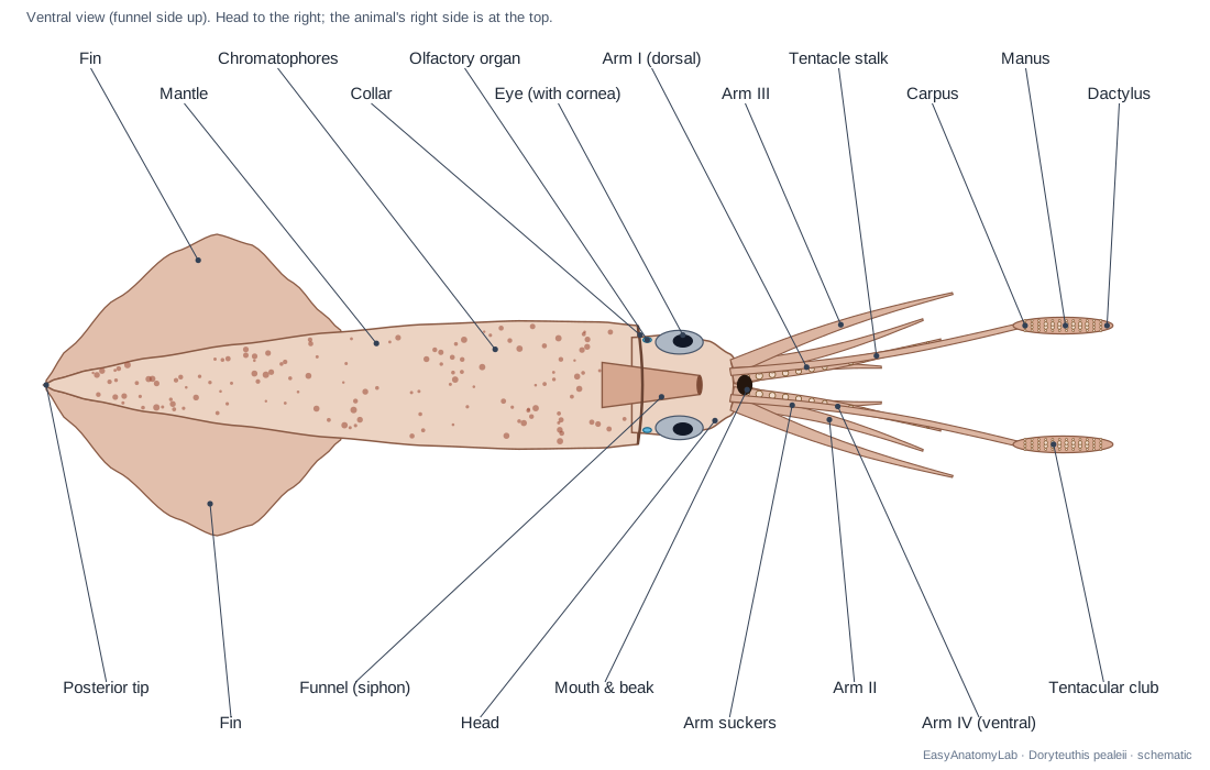
- Mantle: The long, torpedo-shaped muscular tube that forms the body and encloses the organs and the mantle cavity. Mantle muscles contract to squeeze water out through the funnel (jet propulsion) and relax to draw water in over the gills for breathing.
- Fins: A pair of muscular fins that together form a rhombus (diamond shape) at the posterior end of the mantle. Fine control of position, slow swimming, steering and stabilising during jetting.
- Collar (Mantle opening): The free anterior edge of the mantle, which encircles the 'neck' behind the head. The gap between collar and head is the inhalant opening: water enters the mantle cavity here and is then sealed in when the mantle contracts.
- Head (Cephalic region): The short region between the collar and the arms that carries the eyes and houses the brain. Protects the brain inside the cephalic cartilage and carries the eyes, olfactory organs and arms.
- Funnel (Siphon): A muscular cone on the ventral side through which water is expelled. Jet propulsion and steering, and the exit for exhaled water, ink, faeces, urine, eggs and spermatophores.
- Arms: Eight muscular arms, each with two rows of stalked suckers along its inner (oral) surface. Hold and manipulate prey after the tentacles have caught it, and help steer.
- Arm suckers: Cup-shaped suckers on short stalks, in two alternating rows on each arm. Grip prey by suction and by the toothed ring inside each cup.
- Sucker rings (Chitinous ring teeth): Hard, toothed rings that line the opening of each squid sucker. The teeth dig into the prey's skin so it cannot slip free.
- Tentacles (Tentacle stalks): Two long, retractile feeding tentacles with bare stalks and a sucker-bearing club at the tip. Shoot out in a fraction of a second to seize prey, then pull it back to the arms.
- Chromatophores: Tiny elastic pigment sacs in the skin that make the coloured dots of a squid. Muscles stretch each sac open to show its colour (red, brown, yellow) in a fraction of a second, for camouflage and signalling.
Squid internal anatomy (labeled)
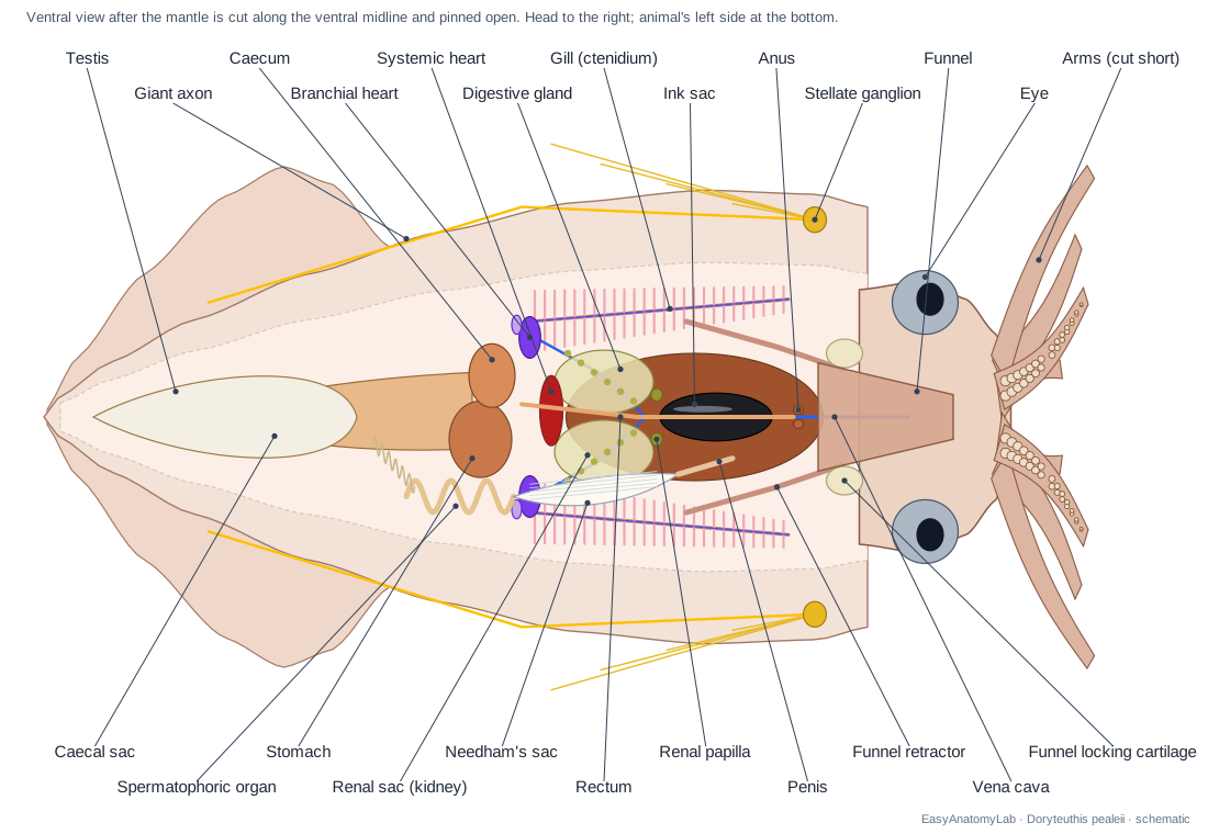
The internal anatomy of a squid is seen by cutting the mantle along the ventral midline. In this classic view the two feathery gills lie along the sides, the three hearts sit near the gill bases, the silvery ink sac lies along the rectum in the middle, the digestive gland lies deeper and to the front, and the gonad fills the posterior end. The pen runs along the dorsal side, under everything else.
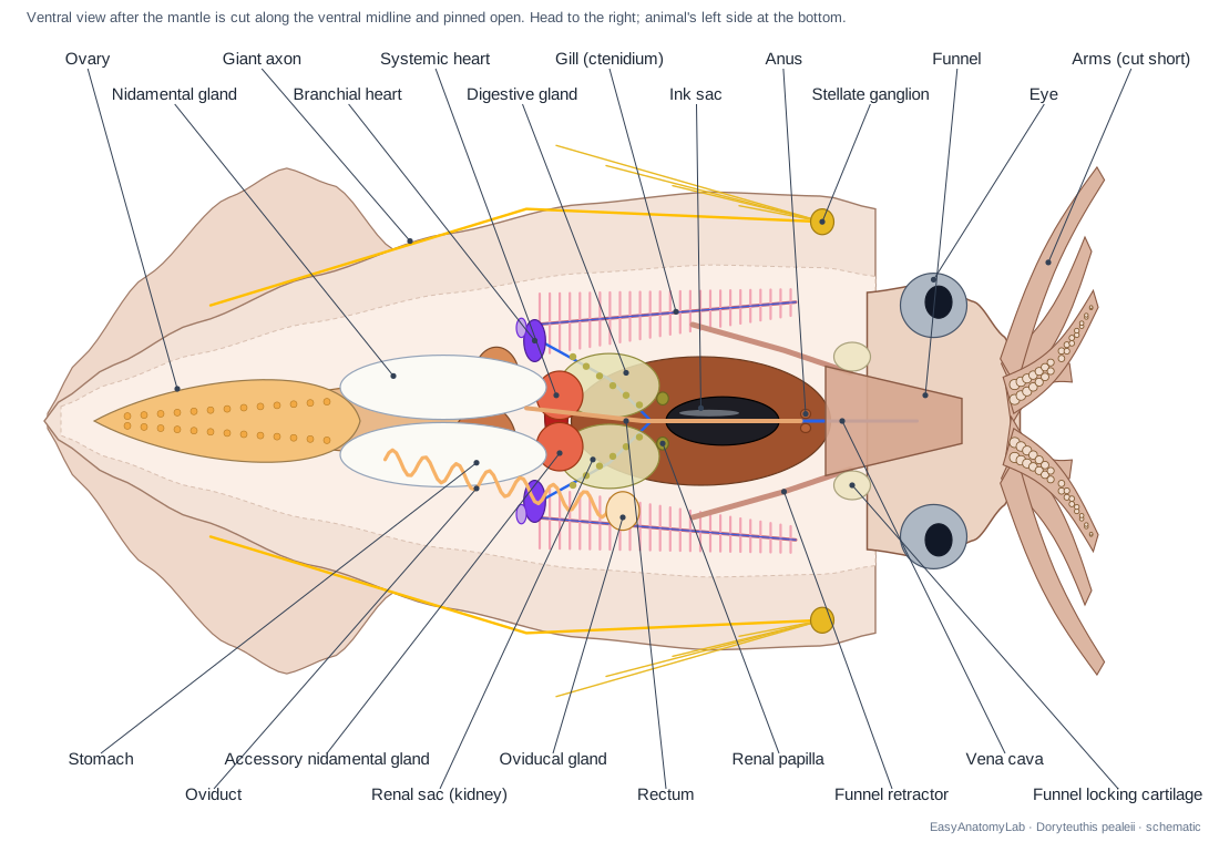
Organ systems found inside the mantle, from front to back on the ventral side: funnel and funnel retractor muscles; anus and rectum; ink sac; renal papillae and renal sacs (kidneys); accessory nidamental glands (females); the three hearts at the gill bases; nidamental glands or the male duct system; stomach and caecum; caecal sac; and the gonad.
Male vs female squid anatomy
Male and female squid differ mainly inside: a female has two large white nidamental glands, accessory nidamental glands, an ovary and a single oviduct; a male has a testis and a duct system that packages sperm into spermatophores, stored in Needham's sac. Externally, a mature male's left ventral arm is modified as the hectocotylus.
| Feature | Female | Male |
|---|---|---|
| Gonad (posterior) | Ovary: grape-like cluster of eggs | Testis: compact, white |
| Main ventral glands | 2 large white nidamental glands + red-orange accessory nidamental glands | None; caecal sac visible |
| Ducts (left side) | Single oviduct with oviducal gland | Vas deferens, spermatophoric organ, Needham's sac, penis |
| Gametes seen | Ripe eggs in the oviduct | Thread-like white spermatophores |
| External sign | Seminal receptacle on ventral buccal membrane | Hectocotylus: left arm IV with papillae |
| Size (D. pealeii) | Smaller on average | Grow faster and larger |
During mating, large "consort" males hold the female side by side and use the hectocotylus to place spermatophores in her mantle cavity near the oviduct opening. Smaller "sneaker" males dart in head-to-head and attach spermatophores near the seminal receptacle on her buccal membrane. Females store sperm and can lay fertilised eggs for weeks.
Squid anatomy diagram: every part and its function
Every structure in the 3D model, grouped by organ system. This is the text version of the labeled diagram above.
External
- Mantle
- The long, torpedo-shaped muscular tube that forms the body and encloses the organs and the mantle cavity. Mantle muscles contract to squeeze water out through the funnel (jet propulsion) and relax to draw water in over the gills for breathing.
- Fins
- A pair of muscular fins that together form a rhombus (diamond shape) at the posterior end of the mantle. Fine control of position, slow swimming, steering and stabilising during jetting.
- Collar (Mantle opening)
- The free anterior edge of the mantle, which encircles the 'neck' behind the head. The gap between collar and head is the inhalant opening: water enters the mantle cavity here and is then sealed in when the mantle contracts.
- Mantle cavity
- The water-filled space between the mantle wall and the visceral mass. Holds the gills and receives the anus, kidney openings, ink and gonoducts, so wastes and gametes are flushed out with the exhaled jet.
- Head (Cephalic region)
- The short region between the collar and the arms that carries the eyes and houses the brain. Protects the brain inside the cephalic cartilage and carries the eyes, olfactory organs and arms.
- Arms
- Eight muscular arms, each with two rows of stalked suckers along its inner (oral) surface. Hold and manipulate prey after the tentacles have caught it, and help steer.
- Arm suckers
- Cup-shaped suckers on short stalks, in two alternating rows on each arm. Grip prey by suction and by the toothed ring inside each cup.
- Sucker rings (Chitinous ring teeth)
- Hard, toothed rings that line the opening of each squid sucker. The teeth dig into the prey's skin so it cannot slip free.
- Tentacles (Tentacle stalks)
- Two long, retractile feeding tentacles with bare stalks and a sucker-bearing club at the tip. Shoot out in a fraction of a second to seize prey, then pull it back to the arms.
- Carpus (Wrist of the tentacular club)
- The base of the tentacular club, with small suckers and knobs. Its small suckers and knobs can lock the two clubs together so they strike as one unit.
- Manus (Hand of the tentacular club)
- The broad middle part of the club, with four rows of suckers; the middle rows are enlarged. Main gripping surface that holds struck prey.
- Dactylus (Finger of the tentacular club)
- The tapering tip of the club with small suckers in four rows. Adds extra grip at the tip of the strike.
- Buccal membrane
- A thin, pointed collar of skin around the mouth, inside the ring of arms. Helps hold food at the mouth; in females it carries the seminal receptacle.
- Funnel (Siphon)
- A muscular cone on the ventral side through which water is expelled. Jet propulsion and steering, and the exit for exhaled water, ink, faeces, urine, eggs and spermatophores.
- Chromatophores
- Tiny elastic pigment sacs in the skin that make the coloured dots of a squid. Muscles stretch each sac open to show its colour (red, brown, yellow) in a fraction of a second, for camouflage and signalling.
Pen, cartilage & muscle
- Pen (Gladius)
- A thin, transparent, feather-shaped internal shell made of chitin and protein. Stiffens the mantle, gives the muscles an attachment and keeps the body streamlined.
- Cephalic cartilage (Cranium)
- A cartilage capsule that encloses the brain and the two statocysts. Protects the brain and forms the hollow chambers of the statocysts, like a simple skull.
- Nuchal cartilage
- A cartilage plate on the dorsal 'neck' that locks the head to the mantle. A sliding joint that keeps head and mantle aligned during jetting.
- Funnel locking cartilages
- Two oval, grooved cartilage sockets on the sides of the funnel base. Snap onto the mantle locking cartilages like press studs, sealing the collar so the jet only leaves through the funnel.
- Mantle locking cartilages
- Two ridges on the inner surface of the mantle that fit into the funnel locking cartilages. The 'male' half of the funnel–mantle lock.
- Funnel retractor muscles
- Paired muscle bands that run from the funnel back to the pen. Pull the funnel back and hold it in place against the force of the jet.
- Head retractor muscles
- Paired muscles that anchor the head to the pen. Pull the head back into the mantle opening and support it.
Nervous
- Supraesophageal mass (Upper brain)
- The dorsal part of the brain, above the esophagus. Higher centres for learning, memory and decision-making (vertical and frontal lobes).
- Subesophageal mass (Lower brain)
- The ventral part of the brain, below the esophagus. Motor centres for the arms, funnel, mantle and chromatophores; its palliovisceral lobe contains the first-order giant neurons that start the escape jet.
- Optic lobes
- Two large kidney-shaped lobes of the brain directly behind the eyes. Process visual information; they are the largest parts of the squid brain.
- Optic glands
- Tiny endocrine glands on the optic tracts. Release hormones that trigger sexual maturation.
- Pallial nerves
- Paired nerves that run from the brain back to the stellate ganglia. Carry motor commands, including the second-order giant fibres, from the brain to the mantle.
- Stellate ganglia
- Two star-shaped nerve centres on the inner wall of the mantle. Relay the brain's commands to the mantle muscles through the stellar nerves; here the second-order giant fibres synapse on the giant-axon cells.
- Stellar nerves
- Nerves that fan out from each stellate ganglion over the inner mantle wall. Activate the circular muscles of the mantle so the whole mantle contracts at once.
- Giant axon
- The largest nerve fibre in the squid: up to about 0.5–1 mm across, inside the longest, most posterior stellar nerve. A wide axon conducts impulses very fast, so all mantle muscles contract together for the escape jet.
- Axial nerve cords
- A nerve cord running down the centre of each arm and tentacle. Coordinate sucker and arm movements locally.
Sensory
- Eyes
- Two large camera-type eyes, one on each side of the head. Vision for hunting, schooling and avoiding predators.
- Cornea (Corneal membrane)
- A clear membrane of skin that covers the eye of loliginid squids. Seals the eye from seawater and protects the lens.
- Lens
- A hard, spherical lens that focuses light on the retina. Focuses images; moves forward and back to change focus.
- Statocysts
- A pair of fluid-filled balance organs inside the cephalic cartilage. Sense gravity, acceleration and rotation, like the vertebrate inner ear.
- Olfactory organs
- Small ciliated pits that detect chemicals in the water. Smell: detection of dissolved chemicals.
Digestive
- Buccal mass (Buccal bulb)
- A muscular ball at the centre of the arms that holds the beak and radula. Works the beak and radula to bite and swallow food.
- Upper beak
- The smaller, dorsal jaw of the beak, with a hooked tip. Bites prey into small pieces with the lower beak, like a parrot's beak upside down.
- Lower beak
- The larger, ventral jaw of the beak, which wraps around the upper beak. Cuts food with the upper beak.
- Radula
- A ribbon of tiny backward-pointing teeth on a tongue-like support. Pulls bitten food into the esophagus.
- Posterior salivary gland
- A gland behind the brain whose duct runs forward to the buccal mass. Secretes saliva with enzymes and toxins that start digestion and subdue prey.
- Esophagus
- A narrow tube that carries food from the buccal mass to the stomach. Moves food by muscular contractions.
- Stomach
- A muscular sac where food is ground and digestion begins. Mixes food with enzymes from the digestive gland.
- Caecum
- A chamber beside the stomach where most nutrients are absorbed. Sorts food particles and absorbs digested nutrients.
- Caecal sac
- A long, thin-walled extension of the caecum reaching towards the posterior end. Extra absorptive surface for digestion.
- Digestive gland (Liver, hepatopancreas)
- A large, elongated brown-orange gland in the anterior visceral mass. Makes digestive enzymes, absorbs nutrients and stores reserves.
- Digestive duct appendages (Pancreas)
- Small glandular outgrowths on the ducts of the digestive gland. Secrete enzymes and absorb fluid; often called the 'pancreas'.
- Intestine & rectum
- The short intestine that runs forward from the stomach to the anus. Carries waste forward to the anus.
- Anus
- The opening of the rectum into the mantle cavity. Releases faeces (and ink) to be flushed out of the funnel.
- Ink sac
- A silvery, pear-shaped sac filled with black ink (melanin). Stores ink that is squirted out to confuse predators.
- Ink duct
- The duct that carries ink from the ink sac into the rectum near the anus. Releases ink, which leaves through the anus and funnel.
Circulatory
- Systemic heart
- The main heart, which pumps oxygenated blood from the gills to the body. Drives blood through the aortas to the head, mantle and organs.
- Branchial hearts (Gill hearts)
- Two small hearts, one at the base of each gill. Pump deoxygenated blood from the veins through the gills.
- Branchial heart appendages (Pericardial glands)
- Small glandular caps on each branchial heart. Filter the blood to make primary urine, which passes into the excretory system.
- Anterior aorta (Cephalic aorta)
- The main artery to the head. Supplies the brain, eyes, buccal mass and arms.
- Posterior aorta
- The artery to the posterior body. Supplies the mantle, fins and gonad.
- Vena cava
- The main vein, which brings blood back from the head and splits towards the branchial hearts. Returns deoxygenated blood to the branchial hearts; its branches carry the renal appendages.
- Afferent branchial vessels
- Vessels that carry blood from each branchial heart into a gill. Deliver deoxygenated blood to the gill lamellae.
- Efferent branchial vessels
- Vessels that carry oxygenated blood from each gill to the systemic heart. Return oxygenated blood to the systemic heart.
Respiratory
- Gills (Ctenidia)
- Two long, feather-like gills in the mantle cavity. Take up oxygen from water pumped over them by the mantle.
- Branchial glands
- Strips of glandular tissue along the attached edge of each gill. Thought to form blood cells and immune cells, and help make haemocyanin.
Excretory
- Renal sacs (Kidneys, nephridia)
- Thin-walled kidney sacs on the ventral side of the visceral mass. Collect urine and remove nitrogen waste (mostly ammonia) from the blood.
- Renal appendages
- Spongy, 'cottage-cheese-like' outgrowths of the veins inside the renal sacs. Secrete waste from the blood into the urine.
- Renal papillae
- Two small nipple-like openings of the kidneys. Release urine into the mantle cavity.
Reproductive
- Hectocotylus · males only
- The modified left ventral arm (arm IV) of the male, used to transfer spermatophores. Picks up spermatophores from the penis and places them inside the female's mantle cavity near the oviduct opening.
- Seminal receptacle · females only
- A small pouch on the ventral buccal membrane of females that stores sperm. Receives spermatophores from males that mate head-to-head; stored sperm can fertilise eggs as they pass the arms during egg-laying.
- Ovary · females only
- A single ovary filled with developing eggs, at the posterior end of the body. Produces eggs, which are released into the body (coelomic) cavity and enter the oviduct.
- Oviduct · females only
- A single coiled duct on the left side that carries ripe eggs forward. Stores ripe eggs before spawning.
- Oviducal gland · females only
- A gland at the end of the oviduct. Coats each egg with the first jelly layer.
- Nidamental glands · females only
- Two large, white, oval glands on the ventral side of the female's visceral mass. Secrete the thick jelly that forms the egg capsules ('egg fingers').
- Accessory nidamental glands · females only
- Glands in front of the nidamental glands that turn mottled red-orange in mature females. House symbiotic bacteria that are added to the egg jelly and help protect the eggs from fungi and microbes.
- Testis · males only
- A single, compact testis at the posterior end of the body. Produces sperm.
- Vas deferens (Sperm duct) · males only
- A thin coiled duct that carries sperm from the testis. Transports sperm.
- Spermatophoric organ (Spermatophoric gland complex) · males only
- A glandular, coiled organ that packages sperm into spermatophores. Builds each spermatophore: a sperm mass, a cement body and an ejaculatory apparatus in a tough tube.
- Needham's sac (Spermatophoric sac) · males only
- A long, white storage sac packed with spermatophores. Stores finished spermatophores until mating.
- Spermatophores · males only
- Long, thin, white sperm packets, lined up like tiny threads. Each one turns inside out when released (the spermatophoric reaction) and sticks to the female as a sperm bulb (spermatangium).
- Penis (Terminal organ) · males only
- The muscular end of the male duct, opening into the mantle cavity. Delivers spermatophores to the hectocotylus, which carries them to the female.
Squid nervous system and brain
The squid brain is a ring of fused ganglia around the esophagus, protected by the cephalic cartilage and flanked by two large optic lobes; from it, pallial nerves run to the stellate ganglia, whose giant axons drive the mantle.
- Supraesophageal mass (Upper brain): The dorsal part of the brain, above the esophagus. Higher centres for learning, memory and decision-making (vertical and frontal lobes).
- Subesophageal mass (Lower brain): The ventral part of the brain, below the esophagus. Motor centres for the arms, funnel, mantle and chromatophores; its palliovisceral lobe contains the first-order giant neurons that start the escape jet.
- Optic lobes: Two large kidney-shaped lobes of the brain directly behind the eyes. Process visual information; they are the largest parts of the squid brain.
- Stellate ganglia: Two star-shaped nerve centres on the inner wall of the mantle. Relay the brain's commands to the mantle muscles through the stellar nerves; here the second-order giant fibres synapse on the giant-axon cells.
- Giant axon: The largest nerve fibre in the squid: up to about 0.5–1 mm across, inside the longest, most posterior stellar nerve. A wide axon conducts impulses very fast, so all mantle muscles contract together for the escape jet.
The squid giant axon. The giant fibre system has three relays: giant cells in the brain, second-order fibres in the pallial nerve, and third-order giant axons in the stellar nerves. Third-order axons are largest in the longest, most posterior nerves, up to about 0.5–1 mm across, so the far end of the mantle contracts at the same time as the near end. Hodgkin and Huxley used these axons to work out how the action potential works.
Squid eye anatomy
The squid eye is a camera-type eye with a cornea, iris, rigid spherical lens and retina; it evolved independently of the vertebrate eye and has no blind spot, because the nerve fibres leave from behind the retina.
- Eyes: Two large camera-type eyes, one on each side of the head. Vision for hunting, schooling and avoiding predators.
- Cornea (Corneal membrane): A clear membrane of skin that covers the eye of loliginid squids. Seals the eye from seawater and protects the lens.
- Lens: A hard, spherical lens that focuses light on the retina. Focuses images; moves forward and back to change focus.
- Statocysts: A pair of fluid-filled balance organs inside the cephalic cartilage. Sense gravity, acceleration and rotation, like the vertebrate inner ear.
Loliginid squids such as Doryteuthis have the eye sealed behind a transparent corneal membrane (myopsid eye). Open-ocean (oegopsid) squids, including the giant and Humboldt squids, have no cornea, so seawater bathes the lens.
Squid heart and circulatory system
A squid has three hearts: two branchial hearts pump blood through the gills, and the systemic heart pumps the oxygenated blood through the anterior and posterior aortas to the body. The blood is blue because it carries oxygen on copper-based haemocyanin.
- Systemic heart: The main heart, which pumps oxygenated blood from the gills to the body. Drives blood through the aortas to the head, mantle and organs.
- Branchial hearts (Gill hearts): Two small hearts, one at the base of each gill. Pump deoxygenated blood from the veins through the gills.
- Branchial heart appendages (Pericardial glands): Small glandular caps on each branchial heart. Filter the blood to make primary urine, which passes into the excretory system.
- Vena cava: The main vein, which brings blood back from the head and splits towards the branchial hearts. Returns deoxygenated blood to the branchial hearts; its branches carry the renal appendages.
- Gills (Ctenidia): Two long, feather-like gills in the mantle cavity. Take up oxygen from water pumped over them by the mantle.
Squid beak, mouth and digestive system
The squid mouth sits at the centre of the arms and contains a parrot-like beak of two chitinous jaws and a toothed radula; food passes through the esophagus, which threads through the brain, to the stomach and caecum, helped by the digestive gland.
- Upper beak: The smaller, dorsal jaw of the beak, with a hooked tip. Bites prey into small pieces with the lower beak, like a parrot's beak upside down.
- Lower beak: The larger, ventral jaw of the beak, which wraps around the upper beak. Cuts food with the upper beak.
- Radula: A ribbon of tiny backward-pointing teeth on a tongue-like support. Pulls bitten food into the esophagus.
- Esophagus: A narrow tube that carries food from the buccal mass to the stomach. Moves food by muscular contractions.
- Stomach: A muscular sac where food is ground and digestion begins. Mixes food with enzymes from the digestive gland.
- Caecum: A chamber beside the stomach where most nutrients are absorbed. Sorts food particles and absorbs digested nutrients.
- Digestive gland (Liver, hepatopancreas): A large, elongated brown-orange gland in the anterior visceral mass. Makes digestive enzymes, absorbs nutrients and stores reserves.
- Ink sac: A silvery, pear-shaped sac filled with black ink (melanin). Stores ink that is squirted out to confuse predators.
Squid pen (gladius)
The squid pen, or gladius, is a feather-shaped internal shell of chitin and protein that runs the length of the dorsal mantle. It is all that is left of the external shell of the squid's ancestors, and it gives the mantle muscles a stiff support.
Squid tentacle and arm anatomy
Squid have eight arms with suckers along their whole length and two longer tentacles with suckers only on the club. The club has three regions: the carpus (base), manus (hand) and dactylus (tip). Every squid sucker sits on a short stalk and is reinforced by a hard, toothed sucker ring.
- Carpus (Wrist of the tentacular club): The base of the tentacular club, with small suckers and knobs. Its small suckers and knobs can lock the two clubs together so they strike as one unit.
- Manus (Hand of the tentacular club): The broad middle part of the club, with four rows of suckers; the middle rows are enlarged. Main gripping surface that holds struck prey.
- Dactylus (Finger of the tentacular club): The tapering tip of the club with small suckers in four rows. Adds extra grip at the tip of the strike.
- Hectocotylus: The modified left ventral arm (arm IV) of the male, used to transfer spermatophores. Picks up spermatophores from the penis and places them inside the female's mantle cavity near the oviduct opening.
Squid vs octopus anatomy
Squid and octopuses are both cephalopods with three hearts, blue blood, a beak, ink and a ring-shaped brain, but their bodies are built for different lives. See the octopus anatomy model to compare.
| Feature | Squid (Doryteuthis) | Octopus (Octopus vulgaris) |
|---|---|---|
| Appendages | 8 arms + 2 tentacles | 8 arms, no tentacles |
| Suckers | Stalked, with toothed chitinous rings | Sessile (no stalk), no rings |
| Shell | Internal pen (gladius) | None (vestigial stylets only) |
| Body shape | Long mantle with fins | Rounded mantle, no fins |
| Lifestyle | Fast open-water swimmer, schools | Bottom-living, crawls, dens |
| Male mating arm | Left arm IV (hectocotylus) | Right arm 3 (hectocotylus) |
| Egg care | Egg capsules left on the seabed | Female guards eggs until they hatch |
Giant squid anatomy
The giant squid, Architeuthis dux, has the same basic body plan as the longfin squid but on a vast scale: specimens of around 12–13 m total length are reported, most of it the two very long feeding tentacles. It has the largest eyes known in any animal, about 27 cm across, used to spot sperm whales in the dark. Its suckers have finely serrated rings, its tissues contain ammonium ions that help with buoyancy, and, as an oegopsid squid, its eyes have no cornea.
Colossal squid anatomy
The colossal squid, Mesonychoteuthis hamiltoni, is shorter than the giant squid but much heavier, making it the heaviest known invertebrate (the Te Papa specimen weighed about 495 kg). Its tentacle clubs carry two rows of sharp, swivelling hooks as well as suckers, its arms have hooks too, and its beak is the largest of any squid. It is a glass squid (family Cranchiidae) of the Southern Ocean.
Humboldt squid anatomy
The Humboldt or jumbo squid, Dosidicus gigas, is a flying squid (family Ommastrephidae) of the eastern Pacific, reaching over 1 m in mantle length. Compared with the longfin squid it has an open (oegopsid) eye, an inverted-T-shaped funnel locking cartilage, sharply toothed suckers, small light organs in its skin, and a chromatophore system that flashes the whole body red and white.
Vampire squid anatomy
The vampire squid, Vampyroteuthis infernalis, is not a true squid. It is the only living member of the order Vampyromorphida, more closely related to octopuses. It has eight webbed arms lined with soft cirri, two thin retractile filaments stored in pockets, large ear-like fins, light organs, no ink sac, and an internal gladius. Adults are only about 20 cm in mantle length.
Squid species compared
| Species | Group | Size | Distinctive anatomy |
|---|---|---|---|
| Longfin inshore squid (Doryteuthis pealeii) | Myopsida, Loliginidae | 10–20 cm ML (up to ~50 cm) | Corneal eye, rhomboid fins, toothed sucker rings, no photophores |
| Giant squid (Architeuthis dux) | Oegopsida, Architeuthidae | ~12–13 m total length | Largest eyes (~27 cm), very long tentacles, ammonium buoyancy |
| Colossal squid (Mesonychoteuthis hamiltoni) | Oegopsida, Cranchiidae | ~495 kg, ML up to ~2.5 m | Swivelling hooks on clubs and arms, largest beak |
| Humboldt squid (Dosidicus gigas) | Oegopsida, Ommastrephidae | >1 m ML | Inverted-T funnel lock, photophores, red-white flashing |
| Vampire squid (Vampyroteuthis infernalis) | Vampyromorphida (not a squid) | ~20 cm ML | Webbed arms with cirri, 2 retractile filaments, no ink sac |
Squid anatomy and physiology: how the parts work together
Breathing and jetting use the same pump: the mantle expands to draw water in at the collar, then contracts; the collar locks onto the funnel cartilages so the water passes over the gills and out of the funnel. Escape is triggered by the giant fibre system, which makes the whole mantle contract at once. Feeding starts with the tentacles striking prey, the arms holding it and the beak biting it into pieces small enough to pass through the brain ring. Excretion happens as the branchial heart appendages filter blood and the renal appendages secrete ammonia into the renal sacs. Camouflage comes from nerve-controlled chromatophores, and ink provides a last defence.
Squid anatomy quiz
Test yourself, then try the full worksheet with answer key on the squid dissection guide.
Which side of a squid carries the funnel?
The ventral side.
What is the squid's internal shell called?
The pen, or gladius.
How many hearts does a squid have?
Three: two branchial hearts and one systemic heart.
Which arm is the hectocotylus in Doryteuthis?
The left ventral arm (left arm IV) of mature males.
Name the three parts of a tentacular club.
Carpus, manus and dactylus.
Which white glands identify a female squid?
The nidamental glands.
What does the stellate ganglion control?
The mantle muscles, through the stellar nerves and giant axons.
What colour is squid blood, and why?
Blue, because oxygen is carried by copper-based haemocyanin.
Frequently asked questions
How many hearts does a squid have?
Three. Two branchial (gill) hearts pump blood through the two gills, and one systemic heart pumps the oxygenated blood to the rest of the body.
How many arms and tentacles does a squid have?
Ten appendages: eight arms with two rows of suckers along their length, and two longer tentacles with suckers only on the club at the tip.
Does a squid have a bone or a shell?
It has no bones. A squid has a thin, flexible internal shell called the pen or gladius, made of chitin and protein, running down the dorsal side of the mantle. The beak and sucker rings are its other hard parts.
Why is squid blood blue?
Squid blood carries oxygen with haemocyanin, a copper-based protein that turns blue when it binds oxygen.
Where is a squid's brain?
In the head, between the eyes, protected by the cephalic cartilage. It forms a ring around the esophagus, with large optic lobes on each side.
Where is the ink sac in a squid?
On the ventral side of the digestive gland, alongside the rectum. When you open the mantle from the ventral side it is the silvery-black, pear-shaped sac in the middle, near the anus.
How can you tell a male squid from a female squid?
Inside, a female has two large white nidamental glands, and a male has a white Needham's sac full of spermatophores on its left side. Externally, a mature male's left arm IV is modified as a hectocotylus.
Why does the squid have a giant axon?
Wider axons conduct impulses faster. Squid giant axons, up to about 1 mm wide, make every part of the mantle contract at the same moment, giving a powerful escape jet.
What is the difference between squid and octopus anatomy?
A squid has a long mantle with fins, an internal pen, eight arms plus two tentacles and toothed, stalked suckers. An octopus has a rounded mantle without fins or pen, eight arms and no tentacles, and suckers without stalks or rings.
References
- Williams, L. W. (1909). The Anatomy of the Common Squid, Loligo pealii, Lesueur. Leiden: E. J. Brill (Biodiversity Heritage Library).
- Doryteuthis Naef, 1912. Tree of Life Web Project.
- Keynes, R. D. (2005). J. Z. and the discovery of squid giant nerve fibres. Journal of Experimental Biology 208: 179–180.
- Maxwell, M. R. & Hanlon, R. T. (2000). Female reproductive output in the squid Loligo pealeii. Marine Ecology Progress Series 199: 159–170.
- Dunlap, P. V. et al. Bacterial pigmentation of the accessory nidamental gland of Loligo pealei (abstract). WHOI Sea Grant.
- Gilly Lab, Hopkins Marine Station (Stanford). Kids and Squids: A Humboldt Squid Dissection Guide for Educators.
- Ommastrephidae (flying squids). Tree of Life Web Project.
- Vampyroteuthidae: Vampyroteuthis infernalis. Tree of Life Web Project.
- Museum of New Zealand Te Papa Tongarewa. Colossal squid (Mesonychoteuthis hamiltoni).
- Animal Diversity Web. Architeuthis dux (giant squid).
- Animal Diversity Web. Dosidicus gigas (Humboldt squid).
- Nilsson, D.-E. et al. (2012). A unique advantage for giant eyes in giant squid. Current Biology 22: 683–688.
More interactive anatomy models
- Octopus anatomyThree hearts, a ring-shaped brain, eight arms and a beak, in 3D.
- Squid dissectionStep-by-step guided squid dissection with every structure labeled.
- Human anatomy1,000+ bones, muscles, organs, vessels and nerves from real anatomical data.
- Fish anatomyExternal and internal anatomy of a bony fish: fins, gills, swim bladder, heart and brain.
- Perch dissectionGuided yellow perch (fish) dissection, from scales to the swim bladder.
- Betta fish anatomyLabyrinth organ, long fins and bubble nests of the Siamese fighting fish.