

Anatomy of an octopus at a glance
An octopus is a cephalopod mollusc with three body regions: the mantle, a muscular sac that holds the organs; the head, which carries the eyes and brain; and eight arms that surround the mouth and beak. It has no bones. It has three hearts, blue copper-based blood, and about 500 million neurons, two-thirds of them in its arms.
By convention, the arms are the front (anterior) of the body, the tip of the mantle is the back (posterior), and the funnel side is the underside (ventral). The arms are numbered L1–L4 and R1–R4 from the dorsal side to the ventral side.
Octopus external anatomy: labeled diagram
The outside of an octopus shows the mantle, head, two eyes, the funnel (siphon), the mantle opening, eight arms lined with suckers, and the web of skin between the arm bases. The rounded “head” you see is actually the mantle; the true head sits between the mantle and the arms.
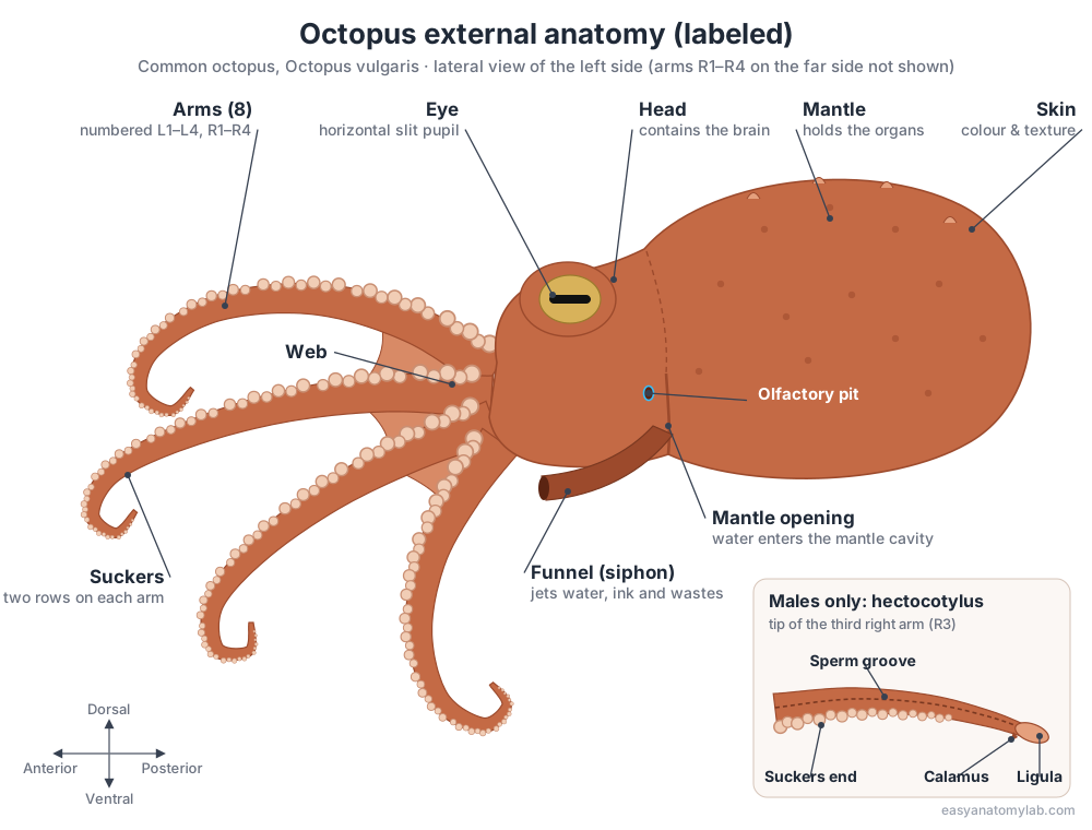
Males can be told apart by the hectocotylus: the tip of the third right arm (R3) has no suckers and ends in a spoon-shaped ligula. Each eye has a horizontal slit pupil, and a small olfactory pit behind and below each eye detects smells carried in the water.
Octopus internal anatomy diagram
Inside the mantle, the visceral mass holds the digestive gland, stomach, caecum, ink sac, three hearts, kidneys and gonad, while the ventral mantle cavity holds the two gills. The brain sits in the head inside a cartilaginous cranium, wrapped around the esophagus.
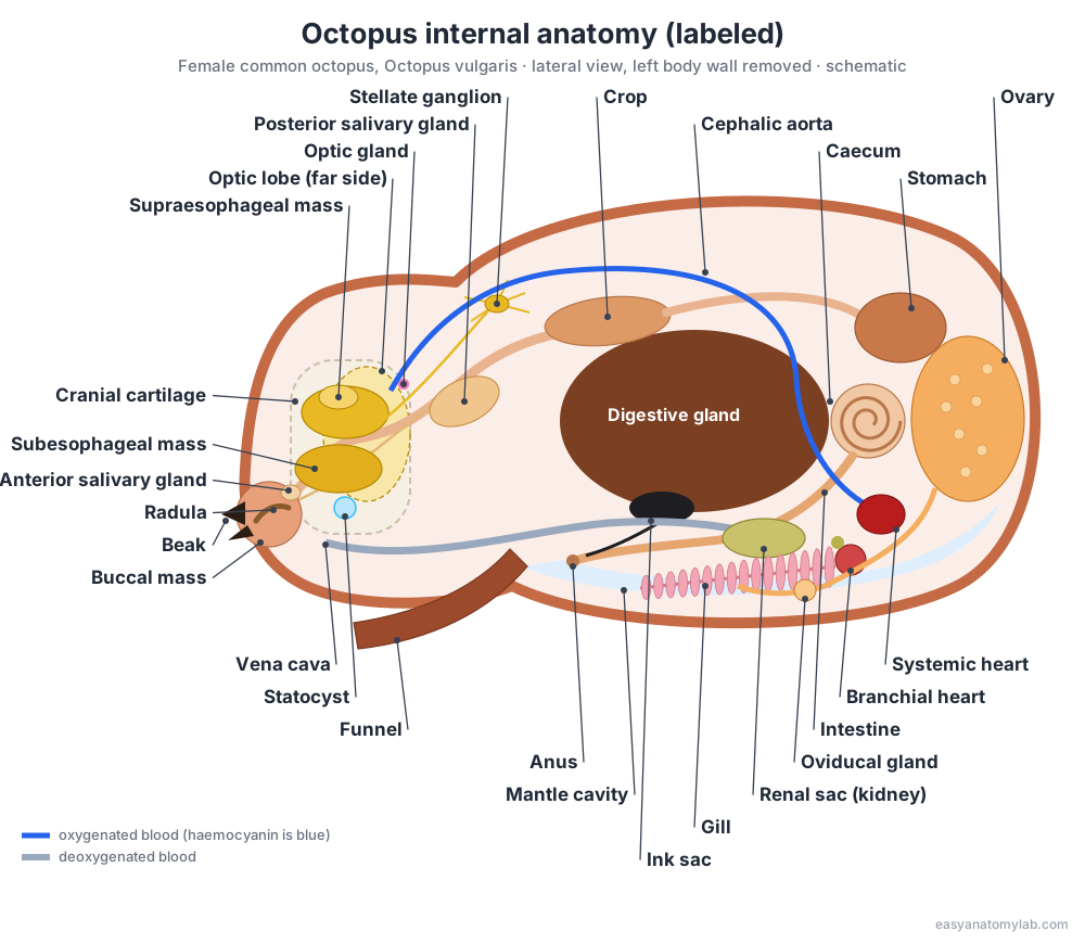
Octopus anatomy: every labeled part and its function
This is the text version of the 3D model: every structure, grouped by body system, with its location and function.
External
- Mantle
- The muscular, sac-like body that holds the internal organs and the mantle cavity. Rhythmic contraction pumps water over the gills for breathing; a strong contraction forces water out of the funnel for jet propulsion.
- Head (Cephalic region)
- The region between the mantle and the arms that carries the eyes and houses the brain. Protects the central brain inside a cartilaginous cranium and supports the two large eyes.
- Funnel (Siphon)
- A muscular tube on the ventral side that expels water from the mantle cavity. Jet propulsion and steering, and the exit route for exhaled water, ink, faeces, urine and eggs or sperm.
- Arms
- Eight flexible, boneless arms lined with suckers, used for crawling, exploring and capturing prey. Locomotion, manipulation of objects, and catching and holding prey. Each arm is a muscular hydrostat, moved by muscles working against each other without any skeleton.
- Suckers
- Muscular suction cups along the underside (oral surface) of each arm, in two rows in Octopus vulgaris. Grip surfaces and prey, and sense chemicals and touch. Octopuses effectively 'taste by touch'.
- Web (Interbrachial membrane)
- A thin sheet of skin joining the bases of neighbouring arms. Spread like an umbrella to trap prey underneath, for example when the octopus pounces ('parachutes') onto a crab.
Nervous & endocrine
- Supraesophageal mass (Upper (dorsal) brain)
- The part of the central brain lying above (dorsal to) the esophagus. Higher control: learning, memory and decision-making. The vertical lobe and frontal lobes are the main learning and memory centres.
- Subesophageal mass (Lower (ventral) brain)
- The part of the central brain lying below (ventral to) the esophagus. Lower motor centres: the brachial lobe (arms), the pedal lobe (funnel and eye muscles), the palliovisceral lobe (mantle, breathing, ink release and the hearts) and the chromatophore lobes (skin colour).
- Optic lobes
- Two large kidney-shaped lobes that process visual information. Process visual input, store visual memories and help control body patterning for camouflage.
- Optic glands
- Small endocrine glands that control sexual maturation and the end of life after reproduction. Act much like the vertebrate pituitary gland. Their secretions trigger maturation of the gonads.
- Stellate ganglia
- A pair of star-shaped nerve centres on the inner wall of the mantle. Receive the pallial nerves from the brain and send stellar nerves out to the mantle muscles that drive breathing and jet propulsion.
- Axial nerve cords
- A large nerve cord running down the centre of each arm. Coordinate arm and sucker movement locally. A ganglion for each sucker lets the arm carry out reflexes with little input from the brain.
Sensory
- Eyes
- Two large camera-type eyes with a lens, iris and retina, and a horizontal slit pupil. Vision. Sharp, high-resolution sight is used to hunt, avoid predators and recognise objects.
- Statocysts
- A pair of fluid-filled balance organs embedded in the cranial cartilage. Sense gravity, linear acceleration (via the statolith on the macula) and rotation (via the crista), like the vertebrate inner ear.
- Olfactory organs (Olfactory pits)
- A pair of small ciliated pits that detect odours carried in the water. Distance chemoreception ('smell'). An olfactory nerve runs from each pit to the olfactory lobe of the brain, next to the optic gland.
Digestive
- Beak
- Hard, parrot-like jaws of chitin and protein at the centre of the arms. Bites and tears prey into pieces small enough to swallow.
- Buccal mass
- The muscular bulb around the mouth that holds the beak, radula and salivary papilla. Works the beak and the radula, a ribbon of tiny chitinous teeth that rasps food and moves it to the esophagus.
- Radula
- A ribbon of tiny, backward-pointing chitinous teeth on the floor of the buccal cavity. Rasps and licks food torn off by the beak and pulls it back into the esophagus, like a conveyor belt.
- Anterior salivary glands
- A pair of small glands on the buccal mass. Secrete mucus that lubricates food as it is swallowed.
- Posterior salivary glands
- A pair of large glands that produce venom and digestive enzymes. Saliva carried to the buccal mass by a duct paralyses prey and begins digesting it.
- Esophagus
- The tube that carries food from the buccal mass to the crop. Moves swallowed food into the crop and stomach.
- Crop
- An expanded section of the esophagus that stores food. Holds swallowed food so the octopus can eat a lot quickly and digest it later.
- Stomach
- A muscular, gizzard-like chamber where food is mixed and broken down. Muscular churning mixes food with enzymes from the digestive gland and starts digestion.
- Caecum
- A coiled, spiral pouch connected to the stomach. Sorts digested particles, sends fine material to the digestive gland, and absorbs nutrients.
- Digestive gland ('Liver' / hepatopancreas)
- The largest organ of the visceral mass, which makes enzymes and absorbs nutrients. Secretes digestive enzymes, absorbs and stores nutrients, and helps remove wastes.
- Intestine & anus
- The intestine carries waste from the stomach to the anus, which opens into the mantle cavity. Absorbs remaining nutrients and forms faeces, which are flushed out of the funnel with exhaled water.
- Ink sac
- A pouch that makes and stores dark ink, released for defence. Ink is shot out through the funnel as a dark cloud or a blob (pseudomorph) that distracts predators while the octopus escapes.
Circulatory
- Systemic heart
- The main heart, which pumps oxygenated blood to the whole body. Receives oxygenated blood from both gills through two auricles and pumps it out through the aortas from one ventricle.
- Branchial hearts
- Two accessory hearts, one at the base of each gill. Pump deoxygenated blood returning from the body through the gill capillaries.
- Cephalic aorta
- The main artery carrying oxygenated blood forward to the head and arms. Supplies the brain, eyes, buccal mass and arms.
- Vena cava
- The large vein returning deoxygenated blood from the head and arms. Brings blood back to the branchial hearts so it can be pumped through the gills.
Respiratory
- Gills (Ctenidia)
- A pair of feather-like gills inside the mantle cavity. Gas exchange. Water enters the mantle cavity through the mantle opening, flows over the gills and leaves through the funnel.
Excretory
- Branchial heart appendages (Pericardial glands)
- A small gland attached to each branchial heart. Filters blood into the pericardial coelom, making the primary urine that is then modified in the renal sacs.
- Renal sacs (Kidneys)
- Paired kidney sacs that remove nitrogen waste from the blood. Renal appendages around the veins modify the filtrate from the branchial heart appendages. Urine is released into the mantle cavity through the renal papillae.
Reproductive
- Hectocotylus · males only
- The modified tip of the male's third right arm (R3), used to pass spermatophores to the female. Spermatophores travel along a groove on the arm to the spoon-shaped tip (ligula), which is inserted into the female's mantle cavity.
- Gonad (Ovary / testis)
- A single reproductive gland, an ovary in females or a testis in males. Produces eggs or sperm. It enlarges a lot as the animal matures.
- Oviducts & oviducal glands · females only
- Paired ducts that carry eggs from the ovary to the mantle cavity. The oviducal gland on each duct coats the eggs and stores sperm received during mating (spermathecae).
- Spermatophoric complex (Needham's sac & terminal organ) · males only
- The male duct system that packages sperm into spermatophores. Spermatophoric glands build sperm packets (spermatophores), Needham's sac stores them, and the terminal organ passes them to the hectocotylus.
Octopus nervous system and brain: the “nine brains”
An octopus has one central brain, not nine. The brain is a ring of lobes around the esophagus, flanked by two large optic lobes. The “nine brains” idea counts the axial nerve cord of each of the eight arms as a mini-brain, because about two-thirds of the octopus’s roughly 500 million neurons are in the arms and each arm can control many movements on its own.
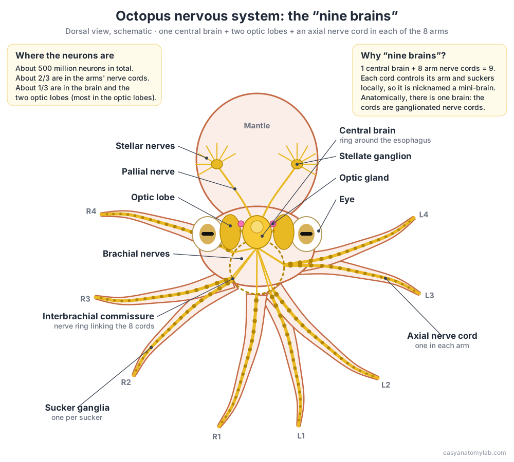
- Supraesophageal mass: Higher control: learning, memory and decision-making. The vertical lobe and frontal lobes are the main learning and memory centres.
- Subesophageal mass: Lower motor centres: the brachial lobe (arms), the pedal lobe (funnel and eye muscles), the palliovisceral lobe (mantle, breathing, ink release and the hearts) and the chromatophore lobes (skin colour).
- Optic lobes: Process visual input, store visual memories and help control body patterning for camouflage.
- Optic glands: Act much like the vertebrate pituitary gland. Their secretions trigger maturation of the gonads.
- Axial nerve cords: Coordinate arm and sucker movement locally. A ganglion for each sucker lets the arm carry out reflexes with little input from the brain.
- Stellate ganglia: Receive the pallial nerves from the brain and send stellar nerves out to the mantle muscles that drive breathing and jet propulsion.
Each arm cord has a ganglion for every sucker and is linked to the other cords by a nerve ring near the arm bases. The brain sends broad commands (“reach there”); the arm works out much of the detail itself. This is why a severed arm can still reach and grasp for a while.
Octopus circulatory system: three hearts and blue blood
An octopus has three hearts. Two branchial hearts, one at the base of each gill, pump blood through the gills. The systemic heart receives the oxygenated blood through two auricles and pumps it around the body through the aortas.
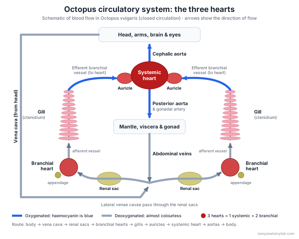
The blood is blue because it carries oxygen on haemocyanin, a copper-based protein dissolved in the plasma, rather than on iron-based haemoglobin in red cells. Haemocyanin carries less oxygen than haemoglobin, and the extra branchial hearts help push blood through the gills. The circulation is closed: blood stays inside vessels. When an octopus jets, pressure in the mantle stops blood returning to the systemic heart, which briefly stops beating (Wells et al., 1987).
Octopus respiratory system: gills
An octopus breathes with two feather-like gills (ctenidia) inside the mantle cavity. The mantle muscles pump water in through the mantle opening, across the gills, and out through the funnel.
- Each gill is made of folded lamellae that give a large surface for gas exchange.
- Blood flows through the gill in the opposite direction to the water (countercurrent exchange).
- The gills have no cilia; all water movement comes from the mantle muscles.
- A branchial heart at the base of each gill pumps deoxygenated blood through it.
Octopus digestive system: beak to anus
Food passes from the beak and buccal mass, down the esophagus (which runs through the brain), into the crop for storage, then to the stomach, caecum and digestive gland, and finally out through the intestine and anus into the mantle cavity, where exhaled water flushes it out of the funnel.
- Beak: two chitinous jaws that bite prey into pieces small enough to pass through the brain.
- Buccal mass and radula: the radula, a ribbon of tiny teeth, pulls food back into the esophagus. The toothed salivary papilla can drill through shells.
- Salivary glands: the small anterior pair make mucus; the large posterior salivary glands make venom and enzymes that paralyse and start digesting prey.
- Esophagus and crop: the crop stores food so the octopus can eat quickly and digest later.
- Stomach and caecum: the stomach mixes food with enzymes; the spiral caecum sorts particles and absorbs nutrients.
- Digestive gland: the largest organ, often called the “liver”. It makes enzymes and absorbs and stores nutrients.
- Intestine, ink sac and anus: the ink duct joins the rectum, so ink and faeces leave by the same route.
Octopus reproductive system: male vs female
Both sexes have a single gonad at the back of the visceral mass. Males have a testis, a spermatophoric complex that packs sperm into spermatophores, and a hectocotylus arm to hand them over. Females have an ovary and paired oviducts with oviducal glands that store sperm.
| Male | Female | |
|---|---|---|
| Gonad | Testis | Ovary |
| Ducts | Spermatophoric complex: spermatophoric glands, Needham’s sac (storage), terminal organ | Paired oviducts with oviducal glands (sperm storage, egg coating) |
| External sign | Hectocotylus: third right arm (R3) with a sucker-less ligula tip | All eight arms are normal |
| Product | Spermatophores, long tubes packed with sperm | Eggs, laid in strings (festoons) in a den |
| After breeding | Declines and dies | Broods the eggs without feeding, dies around hatching |
Sexual maturation is switched on by the optic glands. In brooding females, removing these glands made them stop brooding, resume feeding and live for months longer (Wodinsky, 1977).
Octopus jet propulsion diagram
An octopus swims by jet propulsion: it fills its mantle cavity with water, then contracts the mantle to force the water out of the funnel, which pushes the body the opposite way, usually mantle-first.
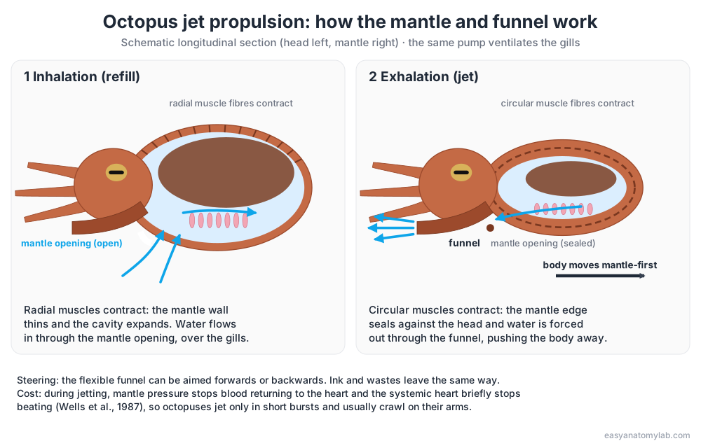
Radial muscles in the mantle wall expand the cavity; circular muscles squeeze it. The funnel can be pointed to steer. The same pumping, done gently, is how the octopus breathes. Jetting is fast but expensive, so octopuses usually crawl.
Octopus life cycle diagram
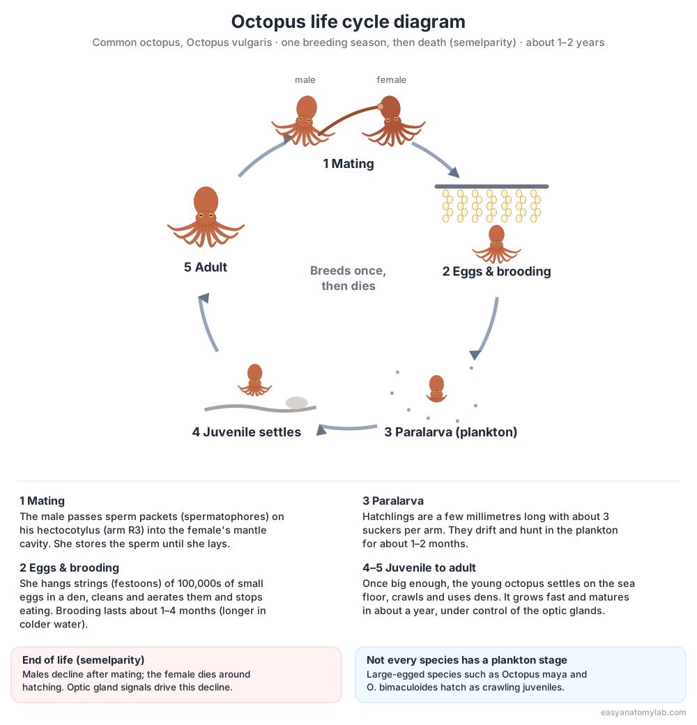
The common octopus lives about one to two years and breeds only once. After mating, the female lays strings of eggs in a den and guards them until they hatch into tiny planktonic paralarvae, which later settle on the sea floor as juveniles. The female dies around the time her eggs hatch.
Octopus species compared: blue-ringed, dumbo and giant Pacific
All octopuses share the same basic anatomy shown on this page, but some species differ in striking ways. Dumbo octopuses belong to a separate group, the cirrate octopods, with fins and finger-like cirri on their arms.
| Species | Size | Habitat | Notable anatomy |
|---|---|---|---|
| Common octopus (Octopus vulgaris) | Total length usually under 1 m | Shallow coastal seas, rocky reefs | The reference species on this page; incirrate (no fins or cirri), two rows of suckers, ink sac |
| Giant Pacific octopus (Enteroctopus dofleini) | Largest octopus; most under about 32 kg (70 lb) | Cold North Pacific, mainly near shore | Lives about 3–5 years; the female broods for about 6 months without eating |
| Blue-ringed octopuses (Hapalochlaena) | About 12–20 cm | Tide pools and reefs, Japan to Australia | Tetrodotoxin (TTX) venom; flashing blue rings |
| Dumbo octopuses (Grimpoteuthis) | Mostly about 20–30 cm | Deep sea, recorded to 6,957 m | Cirrate: ear-like fins, cirri along the arms, internal shell supporting the fins, no ink sac |
Blue-ringed octopus anatomy
Blue-ringed octopuses carry tetrodotoxin (TTX), a nerve poison that can kill a human, in their posterior salivary glands and other tissues. Where the TTX comes from is still debated: bacteria able to make TTX have been found in the salivary glands, but this has not been proven to be the source (Whitelaw et al., 2020). The blue rings are iridophores (light-reflecting cells) that are hidden or exposed by muscles, so the rings can flash in under a second (Mäthger et al., 2012).
Dumbo octopus anatomy
Dumbo octopuses are cirrate octopods. Two ear-like fins on the mantle drive swimming, supported by a U-shaped internal shell. Their arms carry a single row of suckers alternating with finger-like cirri, and a deep web joins the arms. They lack an ink sac and, unlike the common octopus, cannot change colour. One was filmed at 6,957 m in the Java Trench, one of the deepest cephalopod sightings (Jamieson & Vecchione, 2020).
Giant Pacific octopus anatomy
The giant Pacific octopus is the largest octopus species. Its anatomy is the same as the common octopus, on a larger scale. It lives about three to five years, longer than most octopuses, and the female broods her eggs for about six months without eating before she dies.
Frequently asked questions
How many hearts does an octopus have?
Three. Two branchial hearts, one at the base of each gill, pump blood through the gills. One systemic heart then pumps the oxygenated blood to the rest of the body.
Does an octopus really have nine brains?
Not anatomically. An octopus has one central brain, ring-shaped around its esophagus, with two large optic lobes. The 'nine brains' idea counts each arm's axial nerve cord as a mini-brain: about two-thirds of the roughly 500 million neurons are in the arms, and each arm can run many movements locally.
Why is octopus blood blue?
Octopus blood carries oxygen with haemocyanin, a copper-based protein dissolved in the blood. It turns blue when it binds oxygen and is almost colourless without it, unlike our iron-based, red haemoglobin.
Does an octopus have bones?
No. Its largest hard part is the chitinous beak. The brain is protected by a cartilaginous cranium, two tiny stylets are all that is left of the ancestral shell, and the arms are muscular hydrostats with no skeleton.
How does an octopus breathe?
With two gills inside the mantle cavity. The mantle muscles draw water in through the mantle opening, pass it over the gills, where oxygen enters the blood, and push it out through the funnel.
How can you tell a male octopus from a female?
Look at the third right arm. In males its tip is modified into the hectocotylus, a sucker-less, spoon-shaped tip (the ligula) used to pass sperm packets to the female. Internally, males have a testis and a spermatophoric complex; females have an ovary and paired oviducts.
Are octopus arms tentacles?
No. Octopuses have eight arms with suckers along their whole length. Tentacles are the two longer feeding appendages of squid and cuttlefish, which carry suckers only on the club at the tip.
Where is an octopus's mouth?
On the underside, at the centre where the arms meet. The beak sits inside the mouth, and behind it the radula and the esophagus, which passes through the brain.
How long do octopuses live?
Not long. The common octopus lives about one to two years, and even the giant Pacific octopus only about three to five. Most octopuses breed once and then die; the female dies around the time her eggs hatch.
References
- Wells, M. J. (1978). Octopus: Physiology and Behaviour of an Advanced Invertebrate. Chapman & Hall / Springer.
- Young, J. Z. (1971). The Anatomy of the Nervous System of Octopus vulgaris. Oxford: Clarendon Press. (Reviewed in Nature 236, 1972.)
- Hochner, B. (2012). An embodied view of octopus neurobiology. Current Biology 22, R887–R892.
- Wells, M. J. et al. (1987). Blood flow and pressure changes in exercising octopuses (Octopus vulgaris). Journal of Experimental Biology 131, 175–187.
- Wodinsky, J. (1977). Hormonal inhibition of feeding and death in Octopus: control by optic gland secretion. Science 198, 948–951.
- Wang, Z. Y. & Ragsdale, C. W. (2018). Multiple optic gland signaling pathways implicated in octopus maternal behaviors and death. Journal of Experimental Biology 221, jeb185751.
- Polese, G., Bertapelle, C. & Di Cosmo, A. (2016). Olfactory organ of Octopus vulgaris: morphology, plasticity, turnover and sensory characterization. Biology Open 5, 611–619.
- Durante et al. (2024). A step-by-step guide to ageing octopus (stylet and beak increment analysis). Marine and Freshwater Research.
- Whitelaw, B. L. et al. (2020). Adaptive venom evolution and toxicity in octopods is driven by extensive novel gene formation, expansion, and loss. GigaScience 9, giaa120.
- Mäthger, L. M. et al. (2012). How does the blue-ringed octopus (Hapalochlaena lunulata) flash its blue rings? Journal of Experimental Biology 215, 3752–3757.
- Jamieson, A. J. & Vecchione, M. (2020). First in situ observation of Cephalopoda at hadal depths (Octopoda: Opisthoteuthidae: Grimpoteuthis sp.). Marine Biology 167, 82.
- SeaDoc Society. Giant Pacific octopus facts.
More interactive anatomy models
- Squid anatomyPen, ten arms, giant axons, ink sac and three hearts of the longfin squid.
- Squid dissectionStep-by-step guided squid dissection with every structure labeled.
- Human anatomy1,000+ bones, muscles, organs, vessels and nerves from real anatomical data.
- Fish anatomyExternal and internal anatomy of a bony fish: fins, gills, swim bladder, heart and brain.
- Perch dissectionGuided yellow perch (fish) dissection, from scales to the swim bladder.
- Betta fish anatomyLabyrinth organ, long fins and bubble nests of the Siamese fighting fish.