

Squid dissection overview
A squid dissection takes about 45 minutes: you examine and sex the squid from the outside, remove the beak, open the mantle along the ventral (funnel) side, identify the gills, three hearts, ink sac and reproductive organs, then remove the ink sac and the pen. The usual lab specimen is the longfin inshore squid, sold frozen or preserved. If you cannot take part in a hands-on dissection, the 3D model above follows exactly the same steps as a virtual squid dissection.
For the anatomy behind each structure, see the interactive squid anatomy page.
Materials (squid dissection kit)
- One squid per pair of students (fresh-frozen market squid or a preserved specimen), thawed
- Dissecting tray lined with paper towel
- Small sharp scissors (fine-pointed) and a scalpel or craft knife
- Forceps and a blunt probe; 2–4 dissecting pins
- Hand lens or dissecting microscope (for sucker rings and the lens)
- Ruler (for mantle length), white paper for the ink-writing activity
- Disposable nitrile gloves, safety glasses, apron; paper towels and a waste bag
Squid for dissection: whole, uncleaned squid with the head and arms on. Market squid from the seafood counter work as well as supply-house specimens and do not contain preservative.
Safety
- Wear gloves and eye protection. Preserved specimens may contain fixative: work in a ventilated room and rinse the squid first.
- Cut away from your body and your partner's hands. Lift the mantle with forceps before cutting so the scissor tip stays shallow.
- Anyone with a shellfish or seafood allergy should not handle the specimen; use the virtual dissection instead.
- Do not eat any part of a lab specimen. Bag all tissue for disposal and wash hands and tools afterwards.
How to dissect a squid: step by step
These are the same steps as the guided panel next to the 3D model. Structures to find in each step are listed in bold.
Orient the squid and measure it
- Rinse the squid and lay it in the tray. The darker side with the fins lying flat is dorsal (back); the paler side with the funnel is ventral (belly).
- Identify the three body regions: the long mantle, the short head with the eyes, and the ring of arms and tentacles.
- Find the funnel (siphon) under the head and the collar, the free edge of the mantle.
- Measure mantle length (ML) along the dorsal midline from the collar to the tip of the mantle. Scientists use ML, not total length, because tentacles stretch.
- Use a hand lens to look at the coloured dots on the skin: these are chromatophores.
Find: Mantle, Fins, Head, Funnel, Collar, Chromatophores
Count the arms and tentacles
- Spread the appendages out. Count eight arms and two longer tentacles (10 in total).
- Number the arms I (dorsal) to IV (ventral) on each side. The tentacles arise between arms III and IV.
- Compare an arm and a tentacle: arms have suckers along their whole length; tentacles have a bare stalk and suckers only on the club.
- On one club, find the carpus (base, small suckers and knobs), the manus (wide middle, four rows of suckers) and the dactylus (tip).
- Pull off one large sucker and look at it under a hand lens: the hard, toothed sucker ring sits inside the cup on a short stalk.
Find: Arms, Arm suckers, Sucker rings, Tentacles, Carpus, Manus, Dactylus
Examine the head and eyes
- Find the two large eyes. In this squid each eye is covered by a clear corneal membrane (a 'closed', myopsid eye).
- Look just behind and below each eye for the small olfactory pit.
- Spread the arms to see the mouth at the centre, surrounded by the thin, pointed buccal membrane.
Find: Eyes, Cornea, Olfactory organs, Buccal membrane
Sex the squid from the outside
- Look at the left ventral arm (left arm IV). In a mature male the outer third or so is the hectocotylus: its suckers are tiny or replaced by fleshy papillae.
- In a female, look on the ventral buccal membrane, just below the beak, for the seminal receptacle where sperm may be stored.
- Record your prediction (male or female). You will check it when you open the mantle. Use the Female/Male switch on the model to compare.
Caution: Young or immature squid may show no clear external difference; the internal organs are the reliable test.
Find: Hectocotylus, Seminal receptacle, Buccal membrane
Remove the beak and buccal mass
- Push the arms back to expose the round buccal mass at the centre of the arm crown.
- Cut around the base of the arms to free the buccal mass, or squeeze it until the beak pops out.
- Separate the two halves of the beak: the larger lower beak wraps around the smaller upper beak. It looks like a parrot's beak upside down.
- Inside the buccal mass, behind the beak, find the radula, a tiny ribbon of teeth. Use a hand lens.
Caution: The beak is sharp at its tip. Rinse and keep it as a specimen; it dries hard.
Find: Buccal mass, Upper beak, Lower beak, Radula
Open the mantle (ventral incision)
- Turn the squid ventral side up (funnel on top), head towards you.
- Find where the mantle snaps onto the funnel. Unhook the funnel locking cartilages from the mantle locking cartilages with your fingers.
- Lift the collar with forceps, insert the scissor tip just under the mantle, and cut along the ventral midline from the collar to the tip. Keep the blade shallow so you do not cut the organs.
- Fold the mantle flaps out to each side and pin them. You are now looking into the mantle cavity.
- Note the funnel retractor muscles running back from the funnel.
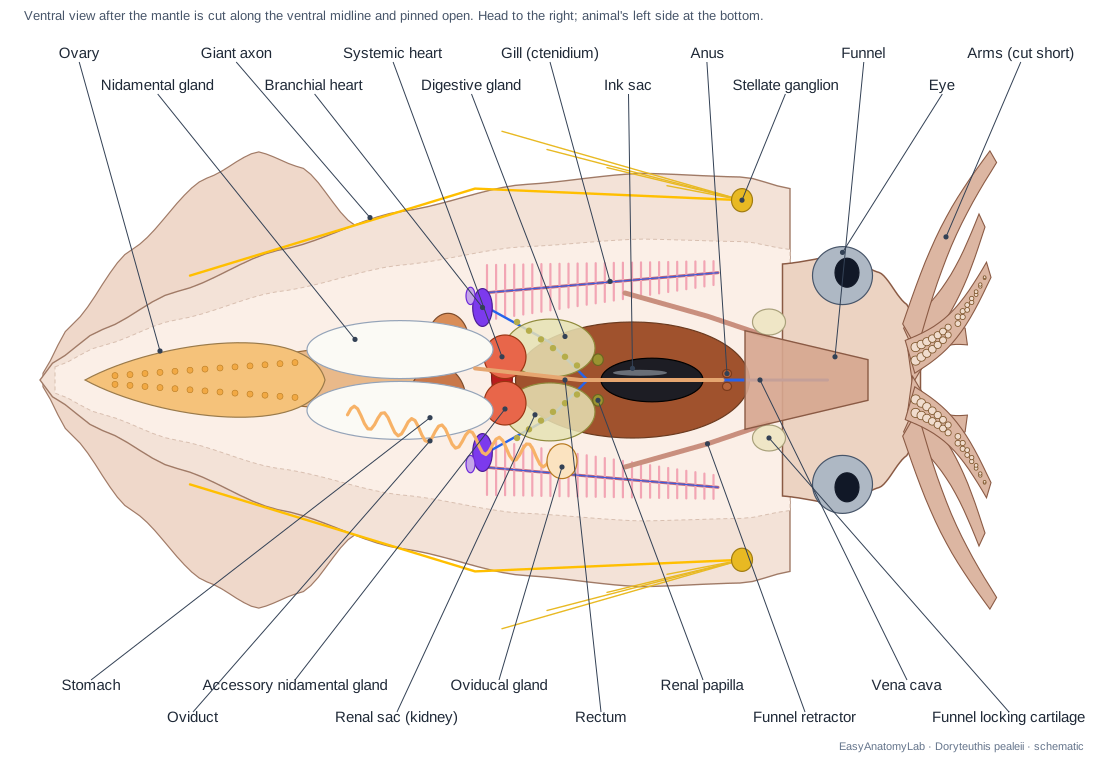
Caution: Cut slightly to one side of the midline if your squid is a female: the large white nidamental glands lie right under the ventral mantle.
Find: Funnel locking cartilages, Mantle locking cartilages, Mantle cavity, Funnel retractor muscles
Identify the sex from the reproductive organs
- Female: two large, white, oval nidamental glands lie on the ventral side; in front of them are the accessory nidamental glands (mottled red-orange when mature). The ovary is at the posterior end and the coiled oviduct, with the oviducal gland, runs forward on the left.
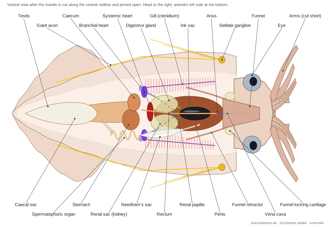
- Male: a long, white Needham's sac on the animal's left side is packed with thread-like spermatophores; it leads to the penis near the left gill. Behind it is the coiled spermatophoric organ, and the compact white testis is at the posterior end.
- Was your external prediction correct? Swap squid with a group that has the other sex. Use the Female/Male switch to see both.
Find: Nidamental glands, Accessory nidamental glands, Ovary, Oviduct, Oviducal gland, Needham's sac, Spermatophores, Spermatophoric organ, Penis, Testis
Find the gills and the three hearts
- Find the two feathery gills along the sides of the mantle cavity. Their bases are posterior; their tips point towards the head.
- At the base of each gill is a small branchial (gill) heart, often with a pale appendage attached.
- Between the two branchial hearts, on the midline, find the systemic heart. Females: gently move the accessory nidamental glands aside.
- Trace the vessels: the vena cava and its branches bring blood to the branchial hearts; efferent vessels take oxygenated blood from the gills to the systemic heart.
Find: Gills, Branchial hearts, Branchial heart appendages, Systemic heart, Vena cava, Afferent branchial vessels, Efferent branchial vessels
Find the anus, kidneys and ink sac
- Follow the rectum forward along the ventral midline to the anus, just behind the funnel.
- On each side of the rectum, a little behind the anus, look for the small renal papillae (kidney openings). The thin renal sacs lie behind them.
- Find the silvery-black ink sac lying along the rectum on the digestive gland. Its duct joins the rectum near the anus.
- Free the ink sac with forceps and lift it out without bursting it. Pierce it over a dish and dip the tip of the pen (step 12) or a toothpick in the ink to write your name.
Caution: Ink stains clothes and skin. Keep the sac over the tray.
Find: Intestine & rectum, Anus, Renal papillae, Renal sacs, Renal appendages, Ink sac, Ink duct
Trace the digestive system
- Find the large digestive gland ('liver'), brown or orange, in the front half of the visceral mass.
- Behind it, near the gill bases, find the muscular stomach and, beside it, the caecum. The thin-walled caecal sac extends towards the posterior end.
- At the back of the digestive gland look for the small digestive duct appendages ('pancreas').
- Follow the narrow esophagus forward along the dorsal side of the digestive gland toward the head. It passes through the brain.
Find: Digestive gland, Stomach, Caecum, Caecal sac, Digestive duct appendages, Esophagus, Posterior salivary gland
Find the stellate ganglia and giant axons
- Push the viscera aside and look on the inner surface of the mantle, just behind the collar, near the dorsal side. On each side is a star-shaped stellate ganglion.
- Nerves fan out from it across the mantle wall. The last, longest stellar nerve runs back toward the fins; it contains the largest giant axon.
- Trace the pallial nerve forward from the ganglion towards the head.
Caution: The nerves are thin and pale. Use a hand lens and good light.
Find: Stellate ganglia, Stellar nerves, Giant axon, Pallial nerves
Remove the pen (gladius)
- Remove or push aside the remaining organs to see the inner dorsal wall of the mantle.
- Feel along the dorsal midline for a stiff strip. Make a shallow cut along it near the collar end and grip the tip of the pen with forceps.
- Pull it out slowly. It is thin, clear and feather-shaped, with a central rib (rachis): the squid's internal shell.
- Use the pointed end of the pen as a quill with the ink from step 9.
Find: Pen, Nuchal cartilage, Head retractor muscles
Dissect the eye and find the brain
- Cut one eye out of the head with scissors and make a small cut through its front.
- Squeeze gently to release the lens: a hard, clear-to-amber ball. Place it on printed text and look through it.
- Look into the eye cup for the dark retina.
- Carefully shave the cartilage between the eyes from the dorsal side to find the pale brain and, at the sides, the large optic lobes. The statocysts lie in the cartilage below the brain.
Find: Eyes, Lens, Cornea, Optic lobes, Supraesophageal mass, Subesophageal mass, Cephalic cartilage, Statocysts
Squid dissection labeled: female
In a dissected female squid, the most obvious organs after opening the mantle are the two large white nidamental glands. The labeled diagram shows what you should see in the ventral view.
Squid dissection labeled: male
In a dissected male squid, look on the animal's left for the long white Needham's sac full of spermatophores, leading to the penis near the left gill.
Male vs female squid dissection
You can tell a male from a female squid only reliably once the mantle is open. Females have nidamental glands (white), accessory nidamental glands (mottled red-orange in mature animals), an ovary and an oviduct, sometimes full of eggs. Males have a testis, a coiled spermatophoric organ, Needham's sac with spermatophores and a penis, and the left arm IV is a hectocotylus. Compare specimens with another group so everyone sees both sexes.
Squid ink sac and eye dissection
The ink sac is the silvery-black, pear-shaped sac lying along the rectum on the ventral side of the digestive gland. Lift it out intact, then pierce it over a dish to write with the ink using the squid's own pen as a quill. For the squid eye dissection, cut out one eye, slit the front and squeeze out the hard, spherical lens; it magnifies print and dries into a clear-to-amber bead.
Squid dissection worksheet: lab questions with answer key
Use these as pre-lab or post-lab questions. Open each answer only after you have written your own.
1. How many arms and tentacles does a squid have, and how do they differ?
Eight arms and two tentacles. Arms have two rows of suckers along their whole length. Tentacles are longer, have a bare stalk, and carry suckers only on the club at the tip (carpus, manus and dactylus).
2. How can you tell the dorsal side from the ventral side?
The dorsal side is darker (more chromatophores) and has the fins and the pen; the ventral side is paler and carries the funnel.
3. What is the function of the funnel (siphon)?
It expels water from the mantle cavity for jet propulsion and steering, and carries out ink, waste and gametes.
4. How many hearts did you find, and what does each do?
Three. Two branchial hearts at the gill bases pump blood through the gills; the systemic heart between them pumps oxygenated blood to the body.
5. What structures tell you the sex of the squid?
Female: large white nidamental glands, accessory nidamental glands, ovary and oviduct; seminal receptacle on the buccal membrane. Male: testis, spermatophoric organ, Needham's sac with spermatophores, penis, and a hectocotylized left arm IV.
6. Where is the ink sac, and what is ink used for?
On the ventral side of the digestive gland, beside the rectum, opening near the anus. Ink forms a cloud or a decoy blob that confuses predators.
7. What is the pen (gladius) and why is it important for classification?
It is a reduced internal shell made of chitin and protein. Having a shell, even a reduced internal one, shows that squid are molluscs.
8. Why are the squid's nerve fibres to the mantle so wide?
Wide (giant) axons conduct impulses faster, so the whole mantle contracts at the same time for a fast escape jet.
9. What does the beak do, and what does the radula do?
The beak bites prey into small pieces; the radula, a ribbon of small teeth behind it, pulls the pieces into the esophagus.
10. Why must a squid bite its food into small pieces?
The esophagus passes through the middle of the ring-shaped brain, so large pieces could press on the brain.
11. How is water moved over the gills?
The mantle expands to draw water in around the collar, then contracts; the collar seals against the funnel locking cartilages, so the water passes over the gills and out of the funnel.
12. Name two ways a squid differs from an octopus.
A squid has fins, an internal pen and two tentacles in addition to eight arms, and its suckers are stalked with toothed rings. An octopus has no fins, no pen, no tentacles, and its suckers have no rings.
Giant squid dissection
Giant squid (Architeuthis dux) are only dissected by museum scientists, when a dead specimen washes ashore or is caught by fishers. The procedure is the same in principle: the mantle is opened ventrally to examine the gills, hearts, gonads and stomach contents, and the beak and statoliths are removed for identification and ageing. Classroom dissections of large squid usually use the Humboldt squid (Dosidicus gigas).
Frequently asked questions
What kind of squid is used for dissection?
In North America it is usually the longfin inshore squid, Doryteuthis (formerly Loligo) pealeii, bought frozen or preserved from biological suppliers or seafood markets. Market squid (Doryteuthis opalescens) from the Pacific coast is very similar.
Which way do you cut a squid open for dissection?
Along the ventral midline (the funnel side), from the collar to the tip of the mantle, keeping the scissors shallow. This gives the classic textbook view of the gills, hearts, ink sac and gonads.
Can you write with squid ink?
Yes. Remove the ink sac intact, pierce it over a dish, and use the pointed end of the squid's own pen as a quill or a toothpick.
Is there a virtual squid dissection?
Yes. The 3D model on this page follows the same 13 steps as the lab, so students who opt out of handling specimens can locate every structure.
How do you tell a male squid from a female squid in a dissection?
Open the mantle: females have two large white nidamental glands, and males have a white Needham's sac full of thread-like spermatophores on the left side.
What does the inside of a squid look like?
From the ventral side you see two feathery gills along the sides, three hearts near the gill bases, the silvery ink sac in the middle, and the reproductive organs behind them. The brown digestive gland lies deeper, and the clear pen runs along the back.
References
- Gilly Lab, Hopkins Marine Station (Stanford). Kids and Squids: A Humboldt Squid Dissection Guide for Educators.
- Biology Corner. Virtual Squid Dissection.
- Williams, L. W. (1909). The Anatomy of the Common Squid, Loligo pealii, Lesueur. Leiden: E. J. Brill (Biodiversity Heritage Library).
- Biodidac (University of Ottawa). Loligo sp., internal anatomy dissection photographs.
- Maxwell, M. R. & Hanlon, R. T. (2000). Female reproductive output in the squid Loligo pealeii. Marine Ecology Progress Series 199: 159–170.
- Doryteuthis Naef, 1912. Tree of Life Web Project.
More interactive anatomy models
- Octopus anatomyThree hearts, a ring-shaped brain, eight arms and a beak, in 3D.
- Squid anatomyPen, ten arms, giant axons, ink sac and three hearts of the longfin squid.
- Human anatomy1,000+ bones, muscles, organs, vessels and nerves from real anatomical data.
- Fish anatomyExternal and internal anatomy of a bony fish: fins, gills, swim bladder, heart and brain.
- Perch dissectionGuided yellow perch (fish) dissection, from scales to the swim bladder.
- Betta fish anatomyLabyrinth organ, long fins and bubble nests of the Siamese fighting fish.