

Perch dissection overview
A fish dissection on a yellow perch takes one or two lab periods. You examine the outside, then open the body cavity from the left side, identify the organs in place, and remove them in layers until you reach the swim bladder and kidney along the backbone. The heart, brain and eye come last. This page works as a perch dissection guide for the wet lab and as a virtual fish dissection for students who opt out. For the anatomy itself, see the fish anatomy model.
Materials and safety
- Preserved yellow perch (one per pair of students)
- Dissecting tray or pan
- Fine scissors (one sharp and one blunt tip if possible)
- Scalpel or single-edge razor blade
- Forceps and blunt probe; dissecting needles or pins
- Hand lens or dissecting microscope; glass slide and cover slip
- Ruler (mm) for measurements
- Nitrile gloves, safety goggles and a lab apron
- Paper towels and a labelled bag or container for disposal
- Wear gloves and eye protection: preservative can irritate skin and eyes. Work in a ventilated room.
- Cut away from your body and your partner's hands. Point scissor tips up when cutting the body wall.
- The dorsal, anal and pelvic fin spines and the opercular spine are sharp. Handle the fish by the body.
- Never eat or drink in the lab. Wash hands after the dissection.
- Dispose of tissue as your teacher directs, not in the sink.
Pre-lab questions
Which fins are paired and which are single (median)?
Paired: pectoral and pelvic. Median: spiny dorsal, soft dorsal, caudal and anal.
What organ lets a bony fish float at a chosen depth?
The swim bladder.
Which way does water flow through a fish when it breathes?
In through the mouth, over the gill filaments, and out under the operculum.
What does the lateral line detect?
Water movement and low-frequency vibration.
External examination of the perch
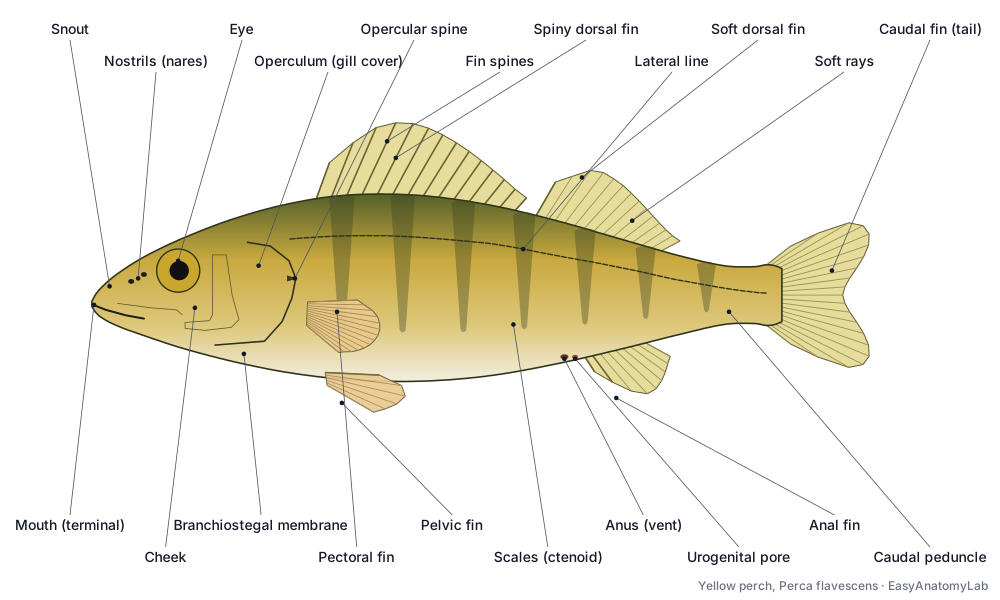
Before cutting, lay the perch on its right side with its head to your left. Measure it, then find the terminal mouth, nares, eyes, operculum, lateral line and eight fins. Count fin spines and rays: a yellow perch typically has 13–15 spines in the first dorsal fin, I–II spines and 12–15 rays in the second dorsal, and II spines and 6–8 rays in the anal fin. Pull one scale from above the lateral line and count its yearly rings (annuli) under a microscope to estimate age.
How to dissect a perch: step by step
1. External anatomy
- Lay the perch on its right side in the tray, head to your left, so its left side faces up.
- Measure the total length (snout to tail tip) in millimetres.
- Find the terminal mouth, the two nostrils (nares) on each side of the snout, and the eyes. Notice there are no eyelids.
- Find the operculum (gill cover) and its free rear edge, and the lateral line running along the side.
- Name the fins: spiny dorsal, soft dorsal, caudal, anal, and the paired pectoral and pelvic fins.
- On the belly, find the anus and, just behind it, the urogenital pore, both in front of the anal fin.
Find: Mouth (terminal), Nostrils (nares), Eye, Operculum (gill cover), Lateral line, Spiny dorsal fin, Soft dorsal fin, Caudal fin (tail fin), Anal fin, Pectoral fins, Pelvic fins (thoracic), Caudal peduncle, Anus (vent), Urogenital pore.
2. Fins, spines and soft rays
- Spread each fin with a probe. Spines are stiff, sharp and unsegmented; soft rays are flexible, segmented and branched near the tip.
- Count the spines and rays of each fin and write a fin formula, e.g. anal fin II, 7.
- Note the pelvic fins sit under the pectorals (thoracic position) and each has 1 spine and 5 soft rays.
- Note the caudal fin is homocercal (equal upper and lower lobes) and shallowly forked.
⚠ Fin spines are sharp. Spread fins with a probe, not your fingers.
Find: Fin spines, Soft rays (lepidotrichia), Spiny dorsal fin, Soft dorsal fin, Anal fin, Pelvic fins (thoracic), Caudal fin (tail fin).
3. Scales and age rings
- With forceps, pull one scale from the side of the body between the two dorsal fins, above the lateral line.
- Place it on a slide with a drop of water and a cover slip, and look at it under low power.
- Find the tiny teeth (ctenii) on the exposed rear edge: these are ctenoid scales.
- Count the dark bands of closely spaced rings (annuli). Each marks one winter, so the count estimates the fish's age in years.
Find: Scales (ctenoid), Lateral line, Skin and mucus layer.
4. Mouth, jaws and teeth
- Open the mouth wide. Run a gloved finger along the jaws to feel the bands of small, brush-like teeth.
- Find the stiff 'tongue' on the floor of the mouth. Try to move it; it has no muscles of its own.
- Look to the back of the mouth to see the gill arches and gill rakers from the inside.
- Find the upper jaw bones (premaxilla and maxilla) and the lower jaw (dentary).
Find: Buccal (mouth) cavity, Teeth, Tongue (basihyal), Premaxilla, Maxilla, Lower jaw (dentary), Gill rakers, Pharynx.
5. Remove the operculum: gills
- Lift the operculum and cut it away close to its attachment with scissors.
- Count the gill arches on this side (4). Each arch carries two rows of red gill filaments.
- Remove one arch. Under the hand lens, compare the red filaments (outer side) with the white, bony gill rakers (inner side).
- Look for the small, red pseudobranch on the inner surface of the operculum near its top.
Find: Operculum (gill cover), Opercle, Preopercle, Subopercle, Interopercle, Gill arches, Gill filaments, Gill rakers, Pseudobranch.
6. Open the body cavity
- Insert the tip of the scissors into the anus, blade tip pointing up, and cut forward along the belly midline to the isthmus between the gill covers. Keep the cut shallow.
- From the anus, cut up the left side to just below the lateral line.
- Make a second vertical cut just behind the operculum, from the belly up to the same level.
- Join the tops of the two vertical cuts with a cut along the side, just below the lateral line.
- Lift the flap of body wall, free it from the organs with the probe, and remove it. You will cut through the thin ribs and see the muscle in cross-section.
⚠ Keep the scissor tips pointing up so you do not cut into the organs underneath.
Find: Hypaxial muscle, Ribs (pleural ribs), Myomeres and myosepta, Epaxial muscle.
7. Organs in place
- Before moving anything, sketch the organs as they lie.
- Find the large reddish-brown liver at the front of the body cavity, behind the heart.
- Find the gonad: an orange, granular ovary (female) or a pair of smooth, white testes (male).
- Find the stomach and intestine below the gonad, and the silvery swim bladder along the top of the cavity.
- Look for the dark red spleen near the stomach and the yellow mesenteric fat around the intestine.
Find: Liver, Ovary, Testes, Stomach, Intestine, Swim bladder (physoclistous), Spleen, Mesenteric fat, Gall bladder.
8. The heart
- Extend the midline cut forward between the pectoral fins to open the pericardial cavity in the throat.
- Find the dark red, triangular ventricle, the larger thin-walled atrium above it, and the thin sinus venosus behind.
- Find the pale, pear-shaped bulbus arteriosus in front of the ventricle, leading into the ventral aorta towards the gills.
- Trace the path: sinus venosus → atrium → ventricle → bulbus arteriosus → ventral aorta → gills.
Find: Pericardial cavity, Sinus venosus, Atrium, Ventricle, Bulbus arteriosus, Ventral aorta, Afferent branchial arteries.
9. Gonads: male or female?
- Decide the sex of your perch. An ovary is a single, large, orange-yellow sac full of visible eggs. Testes are a pair of smooth, creamy-white strips.
- Compare with a group that has a fish of the other sex (use the Female/Male switch in the viewer).
- Follow the gonad back to its duct and the urogenital opening.
- Remove the gonad to see the digestive tract more clearly.
Find: Ovary, Oviduct (gonoduct), Testes, Sperm ducts, Urogenital pore.
10. Digestive tract
- Find the oesophagus behind the gills and follow it to the stomach.
- Find the three short, thick pyloric caeca where the stomach joins the intestine.
- Follow the intestine to the anus. Lift the liver and find the small green gall bladder.
- Cut the oesophagus and the intestine near the anus and lift out the whole tract. Slit open the stomach and look at what the fish ate.
⚠ Do not cut the gall bladder; bile stains everything green.
Find: Oesophagus (esophagus), Stomach, Pyloric caeca, Intestine, Liver, Gall bladder, Pancreas (diffuse), Spleen, Hepatic portal vein.
11. Swim bladder
- With the gut removed, the thin, silvery swim bladder lies along the roof of the body cavity.
- Note that it has no duct to the oesophagus: the perch is physoclistous.
- Look for the red gas gland on its lower front wall. The oval, which removes gas, is on the upper rear wall.
- Puncture the swim bladder with a probe and watch it collapse.
Find: Swim bladder (physoclistous), Gas gland and rete mirabile, Oval.
12. Kidney
- Remove the swim bladder. The long, dark red-brown strip pressed against the vertebral column is the kidney.
- Follow it forward to the head kidney behind the gills, and back to the ureters and the small urinary bladder.
- Notice the dorsal aorta running beside the kidney under the vertebral column.
Find: Kidney (trunk kidney), Head kidney, Ureters (mesonephric ducts), Urinary bladder, Dorsal aorta, Cardinal veins.
13. Brain
- Turn the fish dorsal side up. With a scalpel, shave thin slices off the top of the skull, starting between the eyes and working back.
- When the skull opens, pick away the bone and fatty tissue with forceps.
- From front to back identify the olfactory bulbs, telencephalon (cerebrum), the large optic lobes, the cerebellum and the medulla oblongata, which leads into the spinal cord.
⚠ Shave the bone in thin layers. Pressing hard will crush the brain.
Find: Olfactory bulbs, Telencephalon (cerebrum), Optic lobes (optic tectum), Cerebellum, Medulla oblongata, Spinal cord, Optic nerves.
14. Eye
- Remove one eye by cutting around it with fine scissors, then cut the optic nerve behind it.
- Cut the eye in half around its equator. The jelly-like vitreous body will come out with the lens.
- Find the hard, round (spherical) lens. In a preserved fish it is white and opaque.
- Look inside the back half for the retina, and the dark red, horseshoe-shaped choroid gland behind it.
Find: Eye, Cornea, Iris, Lens (spherical), Retina, Choroid gland (choroid rete), Optic nerves.
Perch dissection labeled: internal anatomy
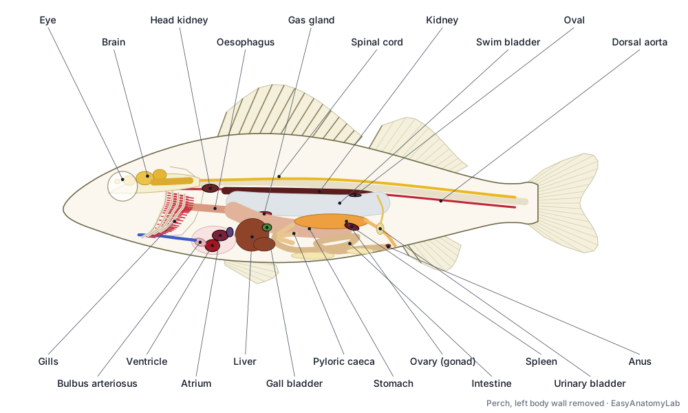
With the left body wall removed, a labeled perch dissection shows the liver at the front of the body cavity, the gonad and gut below the silvery swim bladder, and the kidney against the backbone. Compare this labeled diagram with your specimen.
Male vs female perch dissection
You can only reliably sex a perch by opening it. A female has one large, orange-yellow, granular ovary; in spring it may fill half the body cavity and you can see individual eggs. A male has a pair of long, smooth, creamy-white testes. Switch the model between Female and Male, and compare your fish with a group that has the other sex.
Fish gill, heart, brain and eye dissection
Gills: cut away the operculum and remove one of the four arches; red filaments face out, white rakers face in. Heart: extend the belly cut forward between the pectoral fins to open the pericardial cavity; the dark red ventricle lies below the thin atrium, with the pale bulbus arteriosus in front. Brain: shave the skull from above, starting between the eyes; the optic lobes are the largest part. Eye: remove it, cut it in half, and find the spherical lens and the retina.
Perch dissection lab questions and answer key
Answer these on your perch dissection worksheet, then click each question to check.
What type of scales does a perch have, and how can you tell?
Ctenoid scales. Their exposed rear edge carries tiny teeth (ctenii), which make the fish feel rough when stroked from tail to head.
How can you estimate the age of your perch from a scale?
Count the annuli: dark bands where the growth rings (circuli) are crowded together during slow winter growth. One annulus forms each year.
Write the fin formula for the anal fin of your perch. What do the Roman numerals mean?
Usually II, 6–8 (for example II, 7). Roman numerals count the spines; Arabic numerals count the soft rays.
Why are the gill filaments bright red?
They are packed with capillaries. Blood in the thin lamellae of the filaments picks up oxygen from the water and releases carbon dioxide.
What is the function of the gill rakers?
They point into the throat and stop food and debris from passing out over the delicate filaments.
How many chambers does the perch heart have? Name them in the order blood flows through.
One atrium and one ventricle (two chambers), plus the sinus venosus before them and the bulbus arteriosus after: sinus venosus → atrium → ventricle → bulbus arteriosus → ventral aorta → gills.
Does blood leaving the perch heart carry much oxygen?
No. The heart receives and pumps only deoxygenated blood, which goes to the gills first. Oxygenated blood then flows from the gills through the dorsal aorta to the body (single circulation).
What is the function of the pyloric caeca, and how many does a yellow perch have?
They add surface area for digesting and absorbing food and secrete enzymes. Yellow perch have three short, thick caeca.
What is the swim bladder for, and why is the perch called physoclistous?
It is a gas-filled buoyancy organ. Physoclistous means it is closed: there is no duct to the gut, so gas is added by the gas gland and removed at the oval.
Where is the kidney, and why is it easy to miss?
It is a long, dark red-brown strip pressed against the underside of the vertebral column, above the swim bladder. It lies outside the body-cavity lining and looks like a blood clot.
How did you decide whether your perch was male or female?
Females have a single large, orange-yellow ovary full of visible eggs. Males have a pair of smooth, creamy-white testes.
Which part of the perch brain is largest, and what does that suggest?
The optic lobes (optic tectum). Perch rely heavily on vision to hunt.
Why is the lens of a fish eye spherical?
Under water the cornea bends light very little, so the lens must do almost all the focusing. A round lens is the most powerful shape. Fish focus by moving the lens, not by changing its shape.
Frequently asked questions
How do you dissect a perch?
Examine the outside, remove a scale and the operculum, then insert scissors into the anus and cut forward along the belly to the isthmus. Make vertical cuts up from the anus and behind the gill cover, join them below the lateral line, and lift off the body wall. Identify the organs in place, then remove the gonad and gut to see the swim bladder and kidney. Finish with the heart, brain and eye.
How can you tell a male from a female perch in a dissection?
Females have one large, orange-yellow ovary full of visible eggs. Males have two smooth, creamy-white testes. The outside of the fish gives little clue outside the breeding season.
Why is the yellow perch used for fish dissection?
It is common, inexpensive and a typical spiny-rayed teleost, so its anatomy represents most living fishes. It is the fish most often used to teach the internal and external anatomy of a bony fish.
What organs can you see in a dissected fish?
Gills, heart, liver, gall bladder, stomach, pyloric caeca, intestine, spleen, gonad (ovary or testes), swim bladder, kidney, urinary bladder and, after opening the skull, the brain.
Is there a virtual fish dissection for students who opt out?
Yes. The 3D model on this page follows the same 14 steps as the wet lab, with every structure labeled, so students can complete the worksheet without dissecting a real fish.
References
- Scott, W.B. & Crossman, E.J. (1973). Freshwater Fishes of Canada. Fisheries Research Board of Canada Bulletin 184, pp. 755–761 (yellow perch: meristics, three pyloric caeca, ribbon spawning).
- New York State DEC, I FISH NY. Fish Dissection lesson plan.
- Joeris, L.S. Structure and growth of scales of yellow perch of Green Bay. Transactions of the American Fisheries Society.
- Prem, C. et al. (2000). Swim bladder gas gland cells produce surfactant: in vivo and in culture. Am. J. Physiol. 279: R2336 (perch, a physoclist).
- South Carolina DNR. Yellow perch (single ovary).
- Hickman, C.P. Jr. et al. Laboratory Studies in Integrated Principles of Zoology. McGraw-Hill (perch exercise).
- Helfman, G.S., Collette, B.B., Facey, D.E. & Bowen, B.W. (2009). The Diversity of Fishes, 2nd ed. Wiley-Blackwell.
More interactive anatomy models
- Fish anatomyExternal and internal anatomy of a bony fish: fins, gills, swim bladder, heart and brain.
- Betta fish anatomyLabyrinth organ, long fins and bubble nests of the Siamese fighting fish.
- Human anatomy1,000+ bones, muscles, organs, vessels and nerves from real anatomical data.
- Octopus anatomyThree hearts, a ring-shaped brain, eight arms and a beak, in 3D.
- Squid anatomyPen, ten arms, giant axons, ink sac and three hearts of the longfin squid.
- Squid dissectionStep-by-step guided squid dissection with every structure labeled.