

Anatomy of a fish at a glance
A bony fish has three body regions: the head (up to the rear edge of the gill cover), the trunk (which holds the body cavity), and the tail (behind the anus). It breathes with four pairs of gills under a bony operculum, floats with a gas-filled swim bladder, swims with W-shaped blocks of muscle, and steers with median fins (dorsal, caudal, anal) and paired fins (pectoral and pelvic). Its heart pumps blood in a single loop: heart → gills → body → heart.
The yellow perch is the fish most often dissected in school. It is a spiny-rayed teleost: its fins have true spines, its pelvic fins sit under the pectoral fins, and it has rough ctenoid scales. More than 96% of living fish species are teleosts, so what you learn on a perch applies to most fishes.
Orientation and terminology
Anterior = towards the snout; posterior = towards the tail; dorsal = the back; ventral = the belly; lateral = the side. Median fins lie on the midline; paired fins come in left and right pairs. A fin formula lists spines in Roman numerals and soft rays in Arabic numerals: the perch anal fin is II, 6–8.
Fish anatomy diagram (labeled)
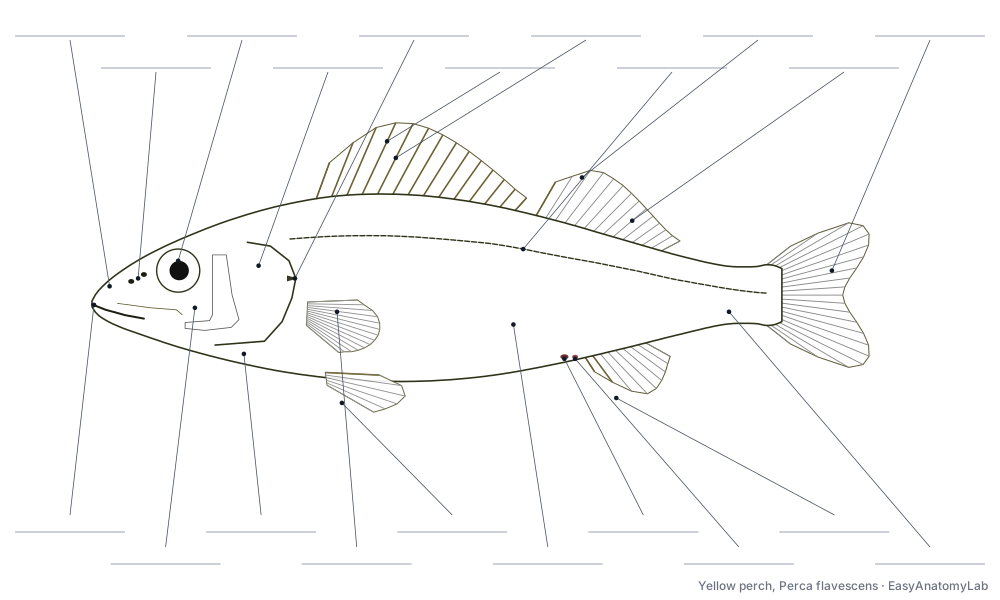
These labeled diagrams of a yellow perch match the 3D model. Use them as a fish anatomy worksheet: print the unlabeled version and label the fish anatomy yourself, or colour it in.
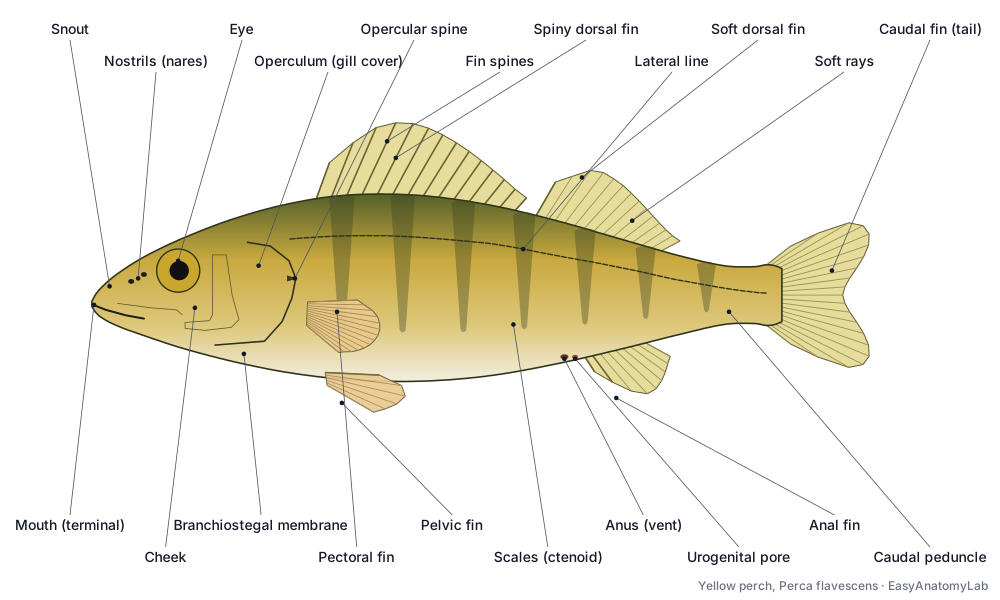
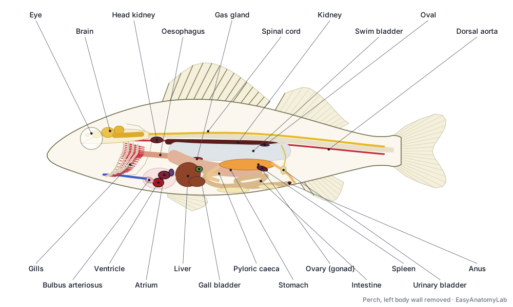
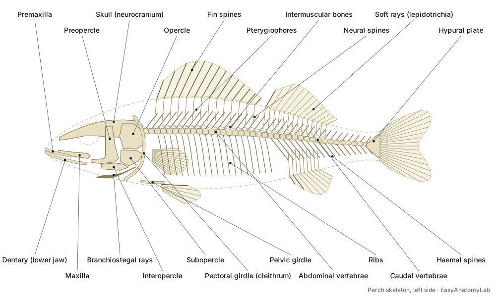
Worksheet and colouring versions: unlabeled fish outline (label the fish / colouring page).
External anatomy of a fish
The external anatomy of a fish includes the mouth, nostrils, eyes, operculum, lateral line, scales, vent and fins. The head ends at the free edge of the operculum. The lateral line is a row of pored scales that senses water movement. Perch scales are ctenoid (with tiny teeth on the rear edge) and record the fish's age as yearly rings.
- Mouth (terminal): Takes in food and the water used for breathing. The perch's protrusible upper jaw shoots forward as the mouth opens, sucking prey in.
- Nostrils (nares): Water flows in through the front opening, over the olfactory rosette, and out of the back one. They are for smell only.
- Eye: Vision in colour and dim light. Each eye sees a wide field to its own side.
- Operculum (gill cover): Protects the gills and works as a pump: as the mouth closes, the opercula swing out and then close, keeping water flowing one way over the gills.
- Lateral line: Senses water movement and low-frequency vibration. Fish use it to school, find prey, avoid obstacles and detect predators in the dark.
- Scales (ctenoid): Flexible armour that protects the body while still letting it bend.
- Caudal peduncle: Transfers the force of the trunk muscles to the tail fin.
- Anus (vent): Releases faeces.
- Urogenital pore: Releases urine from the urinary bladder and, in the breeding season, eggs or milt.
Fish fin anatomy
A perch has eight fins: two dorsal fins, a caudal fin and an anal fin on the midline, and paired pectoral and pelvic fins. Each fin is a thin membrane stretched over spines (stiff, unsegmented) and soft rays (flexible, segmented lepidotrichia), anchored inside the body by pterygiophores or girdle bones.
| Fin | Type | Yellow perch formula | Main job |
|---|---|---|---|
| Spiny dorsal | Median | XIII–XV (13–15 spines) | Defence, stability |
| Soft dorsal | Median | I–II, 12–15 | Stability, turning |
| Caudal (tail) | Median | 17 principal soft rays; homocercal, shallowly forked | Propulsion |
| Anal | Median | II, 6–8 | Stability |
| Pectoral (×2) | Paired | 13–15 soft rays | Steering, braking, hovering |
| Pelvic (×2) | Paired, thoracic | I, 5 | Braking, pitch control |
Fin counts: Scott & Crossman (1973).
Fish face anatomy
A fish's face is built from the snout, the jaws, the nostrils, the eyes, the cheek and the gill cover. The perch mouth is terminal; its upper jaw (premaxilla) slides forward when the mouth opens. Each side of the snout has two nostrils that lead to a blind smell pit, not to the throat. The gill cover is made of four bones (opercle, preopercle, subopercle, interopercle); the perch opercle ends in a flat spine, and the preopercle is saw-edged.
- Snout: Carries the nostrils and leads the body through the water.
- Premaxilla: Bites and grips prey. In perch it slides forward (protrudes) as the mouth opens.
- Maxilla: Swings forward when the mouth opens and pushes the premaxilla out.
- Lower jaw (dentary): Closes against the upper jaw to grip prey.
- Cheek: Covers the jaw-closing muscle (adductor mandibulae).
- Opercle: Forms most of the gill cover and ends in a flat spine in perch.
- Preopercle: Supports the gill cover and carries part of the head lateral line canal.
- Branchiostegal rays: Spread and fold the membrane during breathing, enlarging the gill chamber.
- Isthmus: Joins the head to the body on the underside and holds the hyoid and heart region.
Fish internal anatomy
The internal anatomy of a fish is packed into the body cavity behind the heart. In a perch opened from the left side you see, from front to back: the liver, the gonad (ovary or testes), the stomach with three pyloric caeca, the looping intestine, the dark spleen, and above everything the silvery swim bladder. The dark red kidney lies above the swim bladder, pressed against the backbone.
- Liver: Makes bile, stores fat and glycogen, processes nutrients from the gut, and makes yolk proteins for eggs.
- Gall bladder: Stores bile and releases it into the intestine to emulsify fats.
- Stomach: Stores prey and starts digestion with acid and the enzyme pepsin.
- Pyloric caeca: Increase the surface area for digestion and absorption of nutrients; they secrete digestive enzymes.
- Intestine: Digests food and absorbs nutrients and water.
- Pancreas (diffuse): Makes digestive enzymes and the hormones insulin and glucagon.
- Spleen: Makes and stores blood cells, removes old red blood cells and helps fight infection.
- Swim bladder (physoclistous): Controls buoyancy, so the fish can hover at any depth without swimming. Gas is added by the gas gland and removed at the oval.
- Kidney (trunk kidney): Filters blood and makes urine. In fresh water it produces large amounts of dilute urine to get rid of the water that soaks in through the gills.
- Urinary bladder: Stores urine and reabsorbs some salts before urine is released.
Swim bladder types. Perch, bass and most spiny-rayed fishes are physoclistous: the swim bladder is closed. A gas gland fed by a counter-current rete mirabile adds gas, and the oval removes it. Carp, koi, salmon and trout are physostomous: a pneumatic duct joins the swim bladder to the gut, so they can gulp or burp air.
Fish gill anatomy
Fish gills are four pairs of bony gill arches, each carrying two rows of red gill filaments on its outer side and stiff gill rakers on its inner side. Each filament is lined with microscopic plates (secondary lamellae) where blood flows opposite to the water. This counter-current flow keeps blood next to water that has more oxygen than it does, all along the lamella.
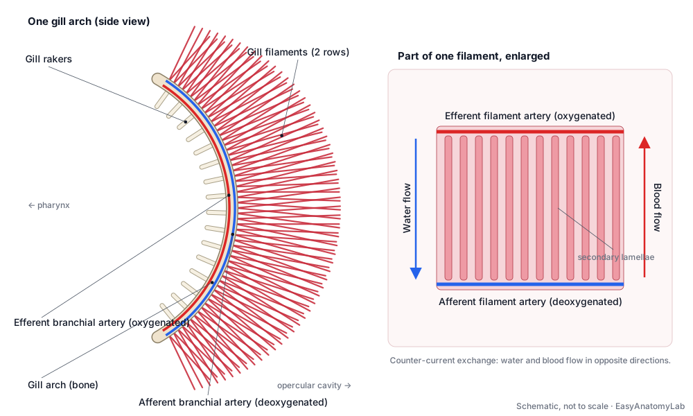
- Gill arches: Support the gill filaments and carry the branchial arteries.
- Gill filaments: Gas exchange. Each filament carries rows of microscopic plates (secondary lamellae) where blood takes up oxygen and releases carbon dioxide.
- Gill rakers: Strain food and keep food and debris away from the delicate filaments.
- Pseudobranch: Receives already oxygenated blood and sends it to the eye's choroid gland. It is not used for breathing.
- Afferent branchial arteries: Deliver deoxygenated blood to the gill filaments.
- Efferent branchial arteries: Collect oxygen-rich blood from the gills.
Fish heart anatomy
A fish heart has one atrium and one ventricle, so it is called two-chambered, but blood passes through four parts in a row: sinus venosus → atrium → ventricle → bulbus arteriosus. It lies in the pericardial cavity under the throat, just behind the gills. It pumps only deoxygenated blood, forward through the ventral aorta to the gills; oxygenated blood then flows to the body in the dorsal aorta. This is a single circulation, unlike the double circulation of mammals and birds.
- Sinus venosus: Receives blood from the cardinal veins (via the ducts of Cuvier) and hepatic veins and passes it to the atrium. It contains the pacemaker.
- Atrium: Fills the ventricle.
- Ventricle: Pumps blood forward through the bulbus arteriosus to the gills.
- Bulbus arteriosus: Smooths the pulse: it stretches as the ventricle beats and recoils between beats, protecting the fragile gills from pressure spikes.
- Ventral aorta: Distributes blood to the four pairs of afferent branchial arteries.
- Dorsal aorta: Supplies oxygenated blood to the muscles and organs.
- Cardinal veins: Return deoxygenated blood to the heart.
Fish brain anatomy
The fish brain is small and lies in a row inside the skull. From front to back: olfactory bulbs (smell), telencephalon (cerebrum: learning and memory), the large paired optic lobes (vision), the cerebellum (coordination) and the medulla oblongata (breathing, heart rate, hearing and lateral line), which continues as the spinal cord. In a sight-hunter like the perch, the optic lobes are the largest part.
- Olfactory bulbs: Process smell information from the olfactory rosettes in the nares.
- Telencephalon (cerebrum): Learning, memory and spatial behaviour, plus some smell processing.
- Optic lobes (optic tectum): Process vision and combine it with lateral-line and other senses to aim the fish at prey.
- Cerebellum: Coordinates swimming, balance and posture.
- Medulla oblongata: Controls breathing movements and heart rate, and receives input from the inner ear, lateral line and taste buds.
- Hypothalamus and pituitary: Controls hormones for growth, reproduction, stress and salt balance.
- Spinal cord: Carries signals between brain and body and runs swimming reflexes.
Fish eye anatomy
A fish eye has a flat cornea and a hard, perfectly round lens that bulges through the pupil. Under water the cornea barely bends light, so the spherical lens does the focusing. A fish focuses by moving the lens backwards and forwards with the retractor lentis muscle, instead of changing its shape as we do. Fish have no eyelids.
- Cornea: Lets light in and protects the eye.
- Iris: Frames the pupil. In most teleosts, unlike in sharks, the pupil can change size little or not at all.
- Lens (spherical): Focuses light on the retina. Fish focus by moving the lens forwards and backwards with the retractor lentis muscle, not by changing its shape.
- Retina: Rods detect dim light; cones detect colour. The signals leave through the optic nerve.
- Choroid gland (choroid rete): A counter-current net of blood vessels (rete mirabile) that raises oxygen pressure behind the retina, feeding its high oxygen demand.
- Optic nerves: Carry visual information.
Fish skeleton and bone anatomy
A bony fish skeleton has a skull (neurocranium) with jaws and gill-cover bones, a vertebral column of 38–41 hourglass-shaped vertebrae in perch, ribs around the body cavity, and fin supports. The pectoral girdle (cleithrum) is joined to the back of the skull, so fish have no neck. The tail fin is carried by the fan-shaped hypural plate. The small "pin bones" in a fillet are intermuscular bones in the muscle sheets.
- Skull (neurocranium): Protects the brain and sense organs and anchors the jaws, gill arches and pectoral girdle.
- Abdominal vertebrae: Support the trunk and protect the spinal cord; their ribs surround the body cavity.
- Caudal vertebrae: Form a flexible column for the side-to-side tail beat.
- Neural spines: Protect the spinal cord and anchor the epaxial muscles.
- Haemal spines: Protect the caudal artery and vein that run through the haemal arches, and anchor muscle.
- Ribs (pleural ribs): Support the body wall around the abdominal organs.
- Intermuscular bones (pin bones): Stiffen the myosepta and help transmit muscle force.
- Pectoral girdle (cleithrum): Supports the pectoral fins and forms the back wall of the gill chamber and front of the heart cavity.
- Pelvic girdle: Anchors the pelvic fins.
- Pterygiophores: Support the fin rays and give attachment to the small muscles that raise, lower and tilt them.
- Hypural plate: Supports the caudal fin rays.
Fish muscles
Most of a fish's body is swimming muscle, arranged in myomeres: W-shaped blocks separated by thin myosepta. A horizontal septum divides the epaxial (upper) from the hypaxial (lower) muscle. Most of it is white muscle for bursts of speed; a thin strip of red muscle along the side powers steady cruising.
Fish reproductive anatomy: male vs female
Female yellow perch have a single ovary, unusual among fishes: it begins as a pair and fuses early in development. In spring it is huge and orange, full of eggs that are laid as a gelatinous, accordion-folded ribbon. Males have a pair of smooth, white testes. Both release their eggs or sperm through a pore behind the anus, and fertilisation happens in the water. Use the Female/Male switch on the model to compare.
- Ovary: Produces eggs. Before spawning it can fill much of the body cavity.
- Oviduct (gonoduct): Carries eggs out of the body at spawning.
- Testes: Produce sperm (milt).
- Sperm ducts: Release sperm during spawning. Fertilisation is external, in the water.
- Urogenital pore: Releases urine from the urinary bladder and, in the breeding season, eggs or milt.
Fish tongue anatomy
Fish do have a tongue, but it is a stiff pad over the basihyal bone on the floor of the mouth. It has taste buds but no muscles of its own, so it cannot move like a human tongue. In trout and bonytongues it even carries teeth.
Bony fish vs cartilaginous fish anatomy
Cartilaginous fish (sharks, rays and chimaeras) differ from bony fish in several key ways: a skeleton of cartilage, 5–7 separate gill slits with no operculum, no swim bladder (a large oily liver gives buoyancy), tooth-like placoid scales, a heterocercal tail, a spiral valve in the intestine, and claspers on males for internal fertilisation. Compare them on the shark anatomy page.
How other fish compare
The perch is a good template, but fish anatomy varies a lot. These species often come up in class:
| Fish | Key anatomy | Swim bladder |
|---|---|---|
| Yellow perch | Two separate dorsal fins (spiny + soft), thoracic pelvic fins, ctenoid scales | Closed (physoclistous) |
| Largemouth bass | Spiny and soft dorsal fins joined by a deep notch; very large mouth | Closed (physoclistous) |
| Koi / common carp | No jaw teeth, pharyngeal teeth, two pairs of barbels, no stomach, Weberian apparatus | Open (physostomous), two chambers |
| Salmon | Adipose fin, abdominal pelvic fins, cycloid scales, many pyloric caeca | Open (physostomous) |
| Pufferfish | Four fused beak-like teeth, stretchy stomach for inflation, tetrodotoxin | Present |
| Deep-sea anglerfish | First dorsal spine forms a lure (illicium + glowing esca); dwarf males in some species fuse to females | Usually absent or reduced |
| Barreleye | Tubular eyes that rotate inside a transparent, fluid-filled head shield | — |
| Lionfish | Long venomous spines: 13 dorsal, 3 anal and 1 in each pelvic fin | Closed (physoclistous) |
Bass anatomy
Largemouth and smallmouth bass are sunfishes (Centrarchidae), close in body plan to perch: a spiny and a soft dorsal fin, thoracic pelvic fins, ctenoid scales and a closed swim bladder. Their two dorsal fins are joined by a deep notch rather than separate, and the largemouth's upper jaw reaches past the back of the eye.
Koi fish anatomy
Koi are ornamental common carp. They have no teeth in the jaws but grind food with pharyngeal teeth in the throat, two pairs of barbels on the upper jaw, and no true stomach. Their swim bladder has two chambers and stays open to the gut (physostomous). Small bones of the Weberian apparatus link the swim bladder to the inner ear, giving carp and their relatives (the Otophysi) excellent hearing. Perch have no Weberian apparatus.
Salmon anatomy
Salmon and trout have a small, rayless adipose fin between the dorsal fin and the tail; it may help sense or control flow over the tail. Their pelvic fins are abdominal (far back), their scales are smooth (cycloid), their swim bladder is physostomous, and they have dozens of pyloric caeca. Most salmon are anadromous: their kidneys and gills switch between saltwater and freshwater salt balance as they migrate.
Puffer fish anatomy
Pufferfish inflate by pumping water (or air) into a very stretchy stomach; they have no ribs or pelvic fins to stop the body swelling. Their four teeth are fused into a beak (Tetraodontidae means "four teeth"). Many species carry tetrodotoxin, a nerve poison concentrated in the liver, ovaries and skin.
Angler fish anatomy
In anglerfishes the first dorsal-fin spine has moved onto the snout as a fishing rod (illicium) tipped with a lure (esca); in deep-sea species the esca glows with symbiotic bacteria. In some deep-sea anglers tiny males fuse permanently to a female and share her blood supply, a form of sexual parasitism linked to changes in their immune system.
Barreleye fish anatomy
The barreleye (Macropinna microstoma) has green-lensed, tubular eyes inside a transparent, fluid-filled shield over its head. The eyes can rotate from looking straight up to looking forward when it feeds. The two dark spots above its mouth that look like eyes are actually its nostrils.
Lionfish anatomy
Lionfish are scorpionfishes with long, separated fin spines that carry venom: 13 dorsal spines, 3 anal spines and one in each pelvic fin. Their fan-like pectoral fins are used to herd prey.
Betta fish
Bettas share the perch body plan but have long, flowing fins, an upturned mouth and a labyrinth organ above the gills for breathing air. See betta fish anatomy.
Note: jellyfish and starfish are not fish at all; they are invertebrates (a cnidarian and an echinoderm). See jellyfish anatomy and starfish anatomy.
Fish anatomy labeled: every part and its function
External
- Skin and mucus layer (Integument, slime coat)
- The outer covering of the fish: a thin epidermis over the dermis, coated in mucus. Protects against injury and infection. Mucus (slime) cells in the epidermis secrete a slippery coat that reduces drag and traps bacteria and parasites.
- Scales (ctenoid) (Ctenoid scales)
- Thin, overlapping bony plates in the dermis. Perch scales are ctenoid: their exposed rear edge carries tiny teeth (ctenii). Flexible armour that protects the body while still letting it bend.
- Snout
- The front of the head, from the tip of the upper jaw to the front of the eye. Carries the nostrils and leads the body through the water.
- Mouth (terminal)
- The opening of the oral (buccal) cavity. A perch has a terminal mouth: it opens at the very front of the head. Takes in food and the water used for breathing. The perch's protrusible upper jaw shoots forward as the mouth opens, sucking prey in.
- Nostrils (nares) (Nares)
- Two small openings on each side of the snout that lead into a blind-ended nasal pit. Water flows in through the front opening, over the olfactory rosette, and out of the back one. They are for smell only.
- Cheek
- The side of the head below and behind the eye, in front of the preopercle. Covers the jaw-closing muscle (adductor mandibulae).
- Operculum (gill cover) (Gill cover)
- The bony flap on each side of the head that covers and protects the gills. Protects the gills and works as a pump: as the mouth closes, the opercula swing out and then close, keeping water flowing one way over the gills.
- Branchiostegal membrane
- A thin skin flap supported by the branchiostegal rays, along the lower edge of the gill cover. Seals the gill chamber from below so the mouth and opercula can pump water efficiently.
- Isthmus
- The narrow, fleshy part of the throat between the two gill chambers. Joins the head to the body on the underside and holds the hyoid and heart region.
- Lateral line
- A line of pored scales along each side of the body. Each pore opens into a canal lined with sense organs. Senses water movement and low-frequency vibration. Fish use it to school, find prey, avoid obstacles and detect predators in the dark.
- Spiny dorsal fin (First dorsal fin)
- The front dorsal fin, supported only by stiff, sharp spines. Defence (it can be raised to make the fish hard to swallow) and stability against rolling.
- Soft dorsal fin (Second dorsal fin)
- The rear dorsal fin, with one or two spines followed by flexible soft rays. Stabilises the fish and helps it turn and brake.
- Caudal fin (tail fin) (Tail fin)
- The tail fin. In a perch it is homocercal (upper and lower lobes equal) and shallowly forked. The main propulsive surface: side-to-side sweeps of the tail push the fish forward.
- Caudal peduncle
- The narrow part of the body just in front of the tail fin. Transfers the force of the trunk muscles to the tail fin.
- Anal fin
- A single fin on the belly behind the vent, with two spines followed by soft rays. Keeps the fish upright and stable, working with the dorsal fins.
- Pectoral fins
- The paired fins just behind the gill covers, equivalent to our arms. Steering, braking, hovering and slow manoeuvring.
- Pelvic fins (thoracic) (Ventral fins)
- The paired fins on the belly, equivalent to our legs. In perch they are thoracic: under the pectoral fins. Stability, braking and fine control of pitch and turning.
- Anus (vent) (Vent)
- The external opening of the intestine. Releases faeces.
- Urogenital pore (Urogenital opening)
- The opening for urine and eggs or sperm, just behind the anus. Releases urine from the urinary bladder and, in the breeding season, eggs or milt.
Skeletal
- Fin spines
- Stiff, sharp, unsegmented and unbranched fin supports. Defence and support. Spines can be locked upright.
- Soft rays (lepidotrichia) (Lepidotrichia)
- Flexible fin rays made of many small segments, usually branched near the tip. Support the fin membrane while letting the fin bend and change shape.
- Pterygiophores (Radials, fin supports)
- Internal rod-like bones that anchor the dorsal and anal fin rays inside the body. Support the fin rays and give attachment to the small muscles that raise, lower and tilt them.
- Skull (neurocranium) (Braincase, cranium)
- The bony box that holds the brain and inner ears. Protects the brain and sense organs and anchors the jaws, gill arches and pectoral girdle.
- Premaxilla
- The main tooth-bearing bone of the upper jaw. Bites and grips prey. In perch it slides forward (protrudes) as the mouth opens.
- Maxilla
- A flat bone behind the premaxilla. In perch it has no teeth and works as a lever. Swings forward when the mouth opens and pushes the premaxilla out.
- Lower jaw (dentary) (Mandible)
- The tooth-bearing bone of the lower jaw. Closes against the upper jaw to grip prey.
- Opercle
- The largest bone of the gill cover. Forms most of the gill cover and ends in a flat spine in perch.
- Preopercle
- A curved, L-shaped bone at the front of the gill cover. Supports the gill cover and carries part of the head lateral line canal.
- Subopercle
- A thin bone below the opercle. Completes the lower part of the gill cover.
- Interopercle
- A small bone along the lower front edge of the gill cover. Links the gill cover to the lower jaw, so opening the cover helps open the mouth.
- Branchiostegal rays
- Curved bony rods that fan out under the gill cover and support the branchiostegal membrane. Spread and fold the membrane during breathing, enlarging the gill chamber.
- Abdominal vertebrae (Precaudal vertebrae)
- The front vertebrae, above the body cavity. They carry ribs. Support the trunk and protect the spinal cord; their ribs surround the body cavity.
- Caudal vertebrae
- The rear vertebrae, behind the body cavity. They carry haemal spines instead of ribs. Form a flexible column for the side-to-side tail beat.
- Neural spines
- Spines on top of each vertebra, above the neural arch that encloses the spinal cord. Protect the spinal cord and anchor the epaxial muscles.
- Haemal spines
- Spines under the caudal vertebrae, below the haemal arch. Protect the caudal artery and vein that run through the haemal arches, and anchor muscle.
- Ribs (pleural ribs) (Pleural ribs)
- Thin curved bones that hang from the abdominal vertebrae. Support the body wall around the abdominal organs.
- Intermuscular bones (pin bones) (Pin bones, epineurals)
- Small, needle-like bones in the myosepta, not attached to the vertebrae at their outer end. Stiffen the myosepta and help transmit muscle force.
- Pectoral girdle (cleithrum) (Shoulder girdle)
- The bony arch behind the gill opening that carries the pectoral fins. Its largest bone is the cleithrum. Supports the pectoral fins and forms the back wall of the gill chamber and front of the heart cavity.
- Pelvic girdle (Basipterygium)
- A pair of flat bones (basipterygia) that support the pelvic fins. Anchors the pelvic fins.
- Hypural plate (Hypurals)
- Flattened bones at the end of the vertebral column that form a fan-shaped base for the tail fin rays. Supports the caudal fin rays.
Muscular
- Epaxial muscle
- The muscle mass above the horizontal septum: the thick 'back' fillet. Bends the body and tail from side to side for swimming and helps lift the head when feeding.
- Hypaxial muscle
- The muscle mass below the horizontal septum. Bends the body for swimming and forms the wall of the body cavity.
- Myomeres and myosepta (Muscle segments)
- Blocks of muscle (myomeres) separated by thin sheets of connective tissue (myosepta). Seen from the side they make a W-shaped (zig-zag) pattern. Each myomere spans several vertebrae because of its folded, cone-in-cone shape, which spreads the pull of each block along the body.
- Horizontal septum
- A sheet of connective tissue that divides the body muscle into epaxial and hypaxial halves. Separates the two muscle masses and anchors myosepta.
- Red muscle (Slow-twitch muscle)
- A thin wedge of dark red muscle just under the skin along the horizontal septum. Powers slow, steady cruising. It is rich in blood vessels, myoglobin and mitochondria, so it does not tire easily.
- Adductor mandibulae (jaw muscle) (Cheek muscle)
- The main jaw-closing muscle. Closes the mouth.
Nervous
- Olfactory bulbs (Olfactory lobes)
- The front-most part of the brain, which receives smell signals. Process smell information from the olfactory rosettes in the nares.
- Telencephalon (cerebrum) (Cerebrum, forebrain)
- A pair of small lobes behind the olfactory bulbs: the fish forebrain. Learning, memory and spatial behaviour, plus some smell processing.
- Optic lobes (optic tectum) (Optic lobes, mesencephalon)
- Two large rounded lobes on top of the midbrain, usually the biggest part of a perch brain. Process vision and combine it with lateral-line and other senses to aim the fish at prey.
- Cerebellum
- A single rounded lobe behind the optic lobes. Coordinates swimming, balance and posture.
- Medulla oblongata (Brainstem)
- The rear part of the brain, which narrows into the spinal cord. Controls breathing movements and heart rate, and receives input from the inner ear, lateral line and taste buds.
- Hypothalamus and pituitary
- The underside of the brain with the pituitary gland hanging below it. Controls hormones for growth, reproduction, stress and salt balance.
- Spinal cord (Nerve cord)
- The main nerve trunk running from the brain to the tail. Carries signals between brain and body and runs swimming reflexes.
- Optic nerves (Cranial nerve II)
- The nerves that carry signals from each retina to the brain. Carry visual information.
- Lateral line nerve (Posterior lateral line nerve)
- A nerve running under the lateral line, collecting signals from its neuromasts. Sends lateral-line signals to the medulla.
Sensory
- Eye
- A large camera eye on each side of the head. Fish have no eyelids. Vision in colour and dim light. Each eye sees a wide field to its own side.
- Cornea
- The clear front window of the eye. Lets light in and protects the eye.
- Iris
- The coloured ring around the pupil. Frames the pupil. In most teleosts, unlike in sharks, the pupil can change size little or not at all.
- Lens (spherical)
- A hard, round (spherical) lens that bulges through the pupil. Focuses light on the retina. Fish focus by moving the lens forwards and backwards with the retractor lentis muscle, not by changing its shape.
- Retina
- The light-sensitive layer lining the back of the eye. Rods detect dim light; cones detect colour. The signals leave through the optic nerve.
- Choroid gland (choroid rete) (Choroid rete mirabile)
- A horseshoe-shaped mass of capillaries behind the retina. A counter-current net of blood vessels (rete mirabile) that raises oxygen pressure behind the retina, feeding its high oxygen demand.
- Olfactory rosette (Nasal rosette)
- A rosette of folds lined with smell receptors inside each nasal pit. Detects dissolved chemicals: food, predators, mates and alarm substances from injured fish.
- Inner ear (Labyrinth)
- Three semicircular canals and three fluid-filled chambers, each with an otolith, in each side of the skull. Balance (which way is up, turning) and hearing.
- Otoliths (ear stones) (Sagitta, lapillus, asteriscus)
- Three pairs of calcium carbonate stones in the inner ear. The largest is the sagitta. Their inertia lets hair cells sense gravity, acceleration and sound vibrations.
Gills & swim bladder
- Gill arches (Branchial arches)
- Four curved bony arches on each side of the pharynx that carry the gills. Support the gill filaments and carry the branchial arteries.
- Gill filaments (Primary lamellae)
- Thin, bright red filaments in two rows on the back edge of each gill arch. Gas exchange. Each filament carries rows of microscopic plates (secondary lamellae) where blood takes up oxygen and releases carbon dioxide.
- Gill rakers
- Bony, finger-like projections on the inner edge of each gill arch. Strain food and keep food and debris away from the delicate filaments.
- Pseudobranch
- A small gill-like patch on the inner surface of the gill cover. Receives already oxygenated blood and sends it to the eye's choroid gland. It is not used for breathing.
- Swim bladder (physoclistous) (Gas bladder, air bladder)
- A thin-walled, gas-filled sac in the top of the body cavity. In perch it is closed (physoclistous): it has no duct to the gut. Controls buoyancy, so the fish can hover at any depth without swimming. Gas is added by the gas gland and removed at the oval.
- Gas gland and rete mirabile (Red body)
- A red, glandular patch in the swim bladder wall, fed by a rete mirabile: a 'wonderful net' of parallel capillaries. Secretes gas into the swim bladder. The gland releases lactic acid and carbon dioxide, which make haemoglobin give up oxygen (the Root effect); the counter-current rete multiplies the gas pressure.
- Oval (Ovale)
- A thin, well-vascularised patch in the swim bladder wall that can be opened or closed by a ring of muscle. Removes gas: when it is open, gas diffuses from the bladder into the blood and leaves through the gills.
Digestive
- Buccal (mouth) cavity (Oral cavity)
- The space inside the mouth, in front of the gills. Takes in food and water. By expanding and contracting it pumps water back over the gills.
- Teeth
- Bands of small, brush-like (villiform) teeth on the jaws and on the roof of the mouth. Grip slippery prey so it cannot escape. Perch swallow prey whole; they do not chew.
- Tongue (basihyal) (Basihyal)
- A small, stiff pad on the floor of the mouth, supported by the basihyal bone of the hyoid arch. Helps move food. It carries taste buds but has no muscles of its own and cannot move like our tongue.
- Pharynx
- The throat chamber between the gill arches. Water leaves it through the gill slits; food goes on to the oesophagus. Shared passage for food and breathing water.
- Pharyngeal teeth (Pharyngeal jaws)
- Toothed pads at the back of the throat, on modified gill-arch bones. A second set of jaws that grips prey and pushes it into the oesophagus.
- Oesophagus (esophagus) (Gullet)
- A short, muscular tube from the pharynx to the stomach. Carries swallowed food to the stomach.
- Stomach
- A thick-walled, stretchy sac. In perch it is long and ends in a blind pouch. Stores prey and starts digestion with acid and the enzyme pepsin.
- Pyloric caeca (Pyloric ceca)
- Finger-like blind pouches where the stomach joins the intestine. Yellow perch have three short, thick ones. Increase the surface area for digestion and absorption of nutrients; they secrete digestive enzymes.
- Intestine
- A narrow tube from the pyloric caeca to the anus. Digests food and absorbs nutrients and water.
- Liver
- The largest organ in the front of the body cavity, reddish-brown to tan. Makes bile, stores fat and glycogen, processes nutrients from the gut, and makes yolk proteins for eggs.
- Gall bladder
- A small green or yellow sac of bile attached to the liver. Stores bile and releases it into the intestine to emulsify fats.
- Pancreas (diffuse)
- Not a separate organ in perch, but small scattered patches of tissue in the mesentery. Makes digestive enzymes and the hormones insulin and glucagon.
- Mesenteric fat (Visceral fat)
- Yellow-white fat along the intestine and in the mesentery. Energy store.
Circulatory
- Pericardial cavity (Pericardium)
- A membrane-lined chamber that holds the heart. Protects the heart and lets it beat freely.
- Sinus venosus
- A thin-walled sac that collects returning (deoxygenated) blood. The first chamber of the fish heart. Receives blood from the cardinal veins (via the ducts of Cuvier) and hepatic veins and passes it to the atrium. It contains the pacemaker.
- Atrium (Auricle)
- A large, thin-walled chamber above the ventricle. Fills the ventricle.
- Ventricle
- The thick, muscular pumping chamber. In the dissection it is the dark red, triangular part of the heart. Pumps blood forward through the bulbus arteriosus to the gills.
- Bulbus arteriosus
- A pale, pear-shaped, elastic chamber in front of the ventricle. Smooths the pulse: it stretches as the ventricle beats and recoils between beats, protecting the fragile gills from pressure spikes.
- Ventral aorta
- The artery that carries deoxygenated blood forward from the heart to the gills. Distributes blood to the four pairs of afferent branchial arteries.
- Afferent branchial arteries
- Four pairs of arteries that carry blood up into the gill arches. Deliver deoxygenated blood to the gill filaments.
- Efferent branchial arteries
- Arteries that carry oxygenated blood out of the gill arches. Collect oxygen-rich blood from the gills.
- Dorsal aorta
- The main artery to the body, carrying oxygenated blood from the gills. Supplies oxygenated blood to the muscles and organs.
- Cardinal veins (Anterior and posterior cardinal veins, ducts of Cuvier)
- The main veins returning blood to the heart from the head (anterior cardinals) and body (posterior cardinals). Return deoxygenated blood to the heart.
- Hepatic portal vein
- A vein that carries nutrient-rich blood from the gut to the liver. Lets the liver process absorbed nutrients before they reach the rest of the body.
- Hepatic veins
- Short veins from the liver to the sinus venosus. Return blood from the liver to the heart.
- Spleen
- A small, dark red organ in the mesentery near the stomach and intestine. Makes and stores blood cells, removes old red blood cells and helps fight infection.
Excretory
- Head kidney (Anterior kidney)
- The front part of the kidney, behind the gills. In adult teleosts it no longer makes urine. It makes blood and immune cells and contains the fish's adrenal (interrenal and chromaffin) tissue.
- Kidney (trunk kidney) (Mesonephros)
- A long, dark red-brown strip pressed against the underside of the vertebral column. Filters blood and makes urine. In fresh water it produces large amounts of dilute urine to get rid of the water that soaks in through the gills.
- Ureters (mesonephric ducts) (Archinephric ducts)
- Ducts that carry urine from the kidneys to the urinary bladder. Drain urine.
- Urinary bladder
- A small, thin-walled sac at the back of the body cavity. Stores urine and reabsorbs some salts before urine is released.
Reproductive
- Ovary · females only
- In female yellow perch a single, large, orange-yellow organ full of eggs. Produces eggs. Before spawning it can fill much of the body cavity.
- Oviduct (gonoduct) · females only
- A short duct from the back of the ovary to the genital opening. Carries eggs out of the body at spawning.
- Testes · males only
- A pair of long, creamy-white organs in males. Produce sperm (milt).
- Sperm ducts · males only
- Ducts that carry sperm from the testes to the urogenital pore. Release sperm during spawning. Fertilisation is external, in the water.
Fish anatomy quiz
Test yourself, then click to check each answer.
Which fin pushes a fish forward?
The caudal (tail) fin.
What covers and protects the gills of a bony fish?
The operculum (gill cover).
How many pairs of gill arches does a perch have?
Four.
Name the four parts of the fish heart in the order blood flows.
Sinus venosus, atrium, ventricle, bulbus arteriosus.
Which organ lets a perch hover without swimming?
The swim bladder.
What does the lateral line detect?
Water movement and vibration.
Where is the fish kidney?
Along the underside of the vertebral column, above the swim bladder.
What are the finger-like pouches at the end of the stomach called?
Pyloric caeca.
Spines or soft rays: which are segmented?
Soft rays (lepidotrichia).
Which part of the brain is largest in a perch?
The optic lobes (optic tectum).
Doing a lab? Follow the step-by-step perch dissection guide with lab questions and an answer key.
Frequently asked questions
What are the main parts of a fish?
Externally: the head (snout, mouth, nostrils, eyes, operculum), the trunk with its lateral line and scales, and the fins: dorsal, caudal, anal, and the paired pectoral and pelvic fins. Internally: gills, heart, liver, stomach, pyloric caeca, intestine, swim bladder, kidneys, gonads, brain and spinal cord.
How many fins does a fish have?
It depends on the species. A yellow perch has 8: two dorsal fins, one caudal fin, one anal fin, two pectoral fins and two pelvic fins. Salmon and trout also have a small adipose fin.
How many chambers does a fish heart have?
Fish have one atrium and one ventricle, so the heart is often called two-chambered. In a row with them are the sinus venosus, which collects blood, and the bulbus arteriosus, which smooths the pulse on its way to the gills.
Do fish have lungs?
Most fish breathe only with gills. The swim bladder of ray-finned fishes evolved from the same pouch of the gut as lungs. A few fishes, such as lungfish, bichirs and gars, can breathe air with a lung or air bladder, and bettas breathe air with a labyrinth organ.
What does the swim bladder do?
It is a gas-filled sac that lets the fish float at a chosen depth without swimming. In perch it is closed (physoclistous); gas is added by the gas gland and removed at the oval. In carp and koi it also helps with hearing.
Do fish have a tongue?
Yes, but it is not like ours. A fish 'tongue' is a stiff pad on the floor of the mouth over the basihyal bone. It has no muscles of its own and cannot move independently.
Do fish have ears?
Fish have inner ears inside the skull, but no outer or middle ear. Each inner ear has three otoliths (ear stones) used for balance and hearing. The lateral line adds a sense of nearby water movement.
What is the difference between bony and cartilaginous fish?
Bony fish (like perch) have a bony skeleton, an operculum over the gills, a swim bladder and usually thin scales. Cartilaginous fish (sharks and rays) have a skeleton of cartilage, 5–7 exposed gill slits, no swim bladder (an oily liver keeps them buoyant) and tooth-like placoid scales.
How can you tell the age of a fish?
Count the yearly growth rings (annuli) on a scale or, more accurately, on an otolith (ear stone). Growth slows in winter, which leaves a narrow, dense band for each year.
References
- Scott, W.B. & Crossman, E.J. (1973). Freshwater Fishes of Canada. Fisheries Research Board of Canada Bulletin 184, pp. 755–761 (yellow perch).
- Froese, R. & Pauly, D. (eds). FishBase: Perca flavescens, yellow perch.
- Helfman, G.S., Collette, B.B., Facey, D.E. & Bowen, B.W. (2009). The Diversity of Fishes, 2nd ed. Wiley-Blackwell.
- Moyle, P.B. & Cech, J.J. (2004). Fishes: An Introduction to Ichthyology, 5th ed. Prentice Hall.
- Prem, C., Salvenmoser, W., Würtz, J. & Pelster, B. (2000). Swim bladder gas gland cells produce surfactant: in vivo and in culture. Am. J. Physiol. 279: R2336 (perch gas gland anatomy).
- Fänge, R. & Holmgren, S. (1982). Choline acetyltransferase activity in the fish swimbladder. J. Comp. Physiol. B (oval of the perch swim bladder).
- Lechner, W. & Ladich, F. (2008). Size matters: diversity in swimbladders and Weberian ossicles affects hearing in catfishes. J. Exp. Biol. 211: 1681–1689.
- Wainwright, P.C. & Turingan, R.G. (1997). Evolution of pufferfish inflation behavior. Evolution 51: 506–518.
- Noguchi, T. & Arakawa, O. (2008). Tetrodotoxin – distribution and accumulation in aquatic organisms, and cases of human intoxication. Marine Drugs 6: 220–242.
- Swann, J.B., Holland, S.J., Petersen, M., Pietsch, T.W. & Boehm, T. (2020). The immunogenetics of sexual parasitism. Science 369: 1608–1615.
- MBARI (2009). Researchers solve mystery of deep-sea fish with tubular eyes and transparent head (Macropinna microstoma).
- Reimchen, T.E. & Temple, N.F. (2004). Hydrodynamic and phylogenetic aspects of the adipose fin in fishes. Can. J. Zool. 82: 910–916.
- South Carolina DNR. Yellow perch (single ovary and egg ribbon).
- Iowa DNR BioNet. Common carp, Cyprinus carpio (barbels, pharyngeal teeth).
More interactive anatomy models
- Perch dissectionGuided yellow perch (fish) dissection, from scales to the swim bladder.
- Betta fish anatomyLabyrinth organ, long fins and bubble nests of the Siamese fighting fish.
- Human anatomy1,000+ bones, muscles, organs, vessels and nerves from real anatomical data.
- Octopus anatomyThree hearts, a ring-shaped brain, eight arms and a beak, in 3D.
- Squid anatomyPen, ten arms, giant axons, ink sac and three hearts of the longfin squid.
- Squid dissectionStep-by-step guided squid dissection with every structure labeled.