

Bivalve anatomy at a glance
A clam is a bivalve mollusc: a soft, headless body enclosed by two hinged shell valves. The body has four main parts. The mantle lines the shell and secretes it. The gills hang in the mantle cavity and both breathe and filter food. The muscular foot digs. The visceral mass holds the gut, digestive gland, gonad, heart and kidneys. Two adductor muscles close the shell, and an elastic hinge ligament opens it.
Bivalves have no head, no jaws and no radula (the rasping tongue of snails). Water enters through the incurrent siphon, passes through the gills and leaves through the excurrent siphon, carrying food in and wastes out. The hard clam is a typical member of the venus clam family (Veneridae) and is used in most school and university labs, so the anatomy here applies to bivalves in general, with differences noted below.
Bivalve shell anatomy: how to orient a clam shell
The hinge is dorsal, the opening edge is ventral, and the beaks point to the anterior end. In Mercenaria the umbo (beak) sits in front of the middle of the hinge and curves forward, over a small heart-shaped depression called the lunule. Behind the beaks, the dark brown hinge ligament lies in a long, flattened area called the escutcheon. The siphons come out at the posterior end.
Left vs right valve: hold the shell with the hinge up and the anterior end (lunule) pointing to your left. The valve facing you is the left valve; the one facing away is the right valve. Hard clams are equivalve, so you must use orientation, not shape, to tell them apart.
Inside each valve you can see the anterior and posterior adductor muscle scars, joined by the pallial line where the mantle edge attaches. Near the posterior scar the line bends into a short V, the pallial sinus, which makes room for the siphons. The hinge plate carries three cardinal teeth under the beak and a lateral tooth behind them. A dark violet border near the posterior margin is the trademark of Mercenaria: Indigenous peoples of the Atlantic coast cut it into purple wampum beads.
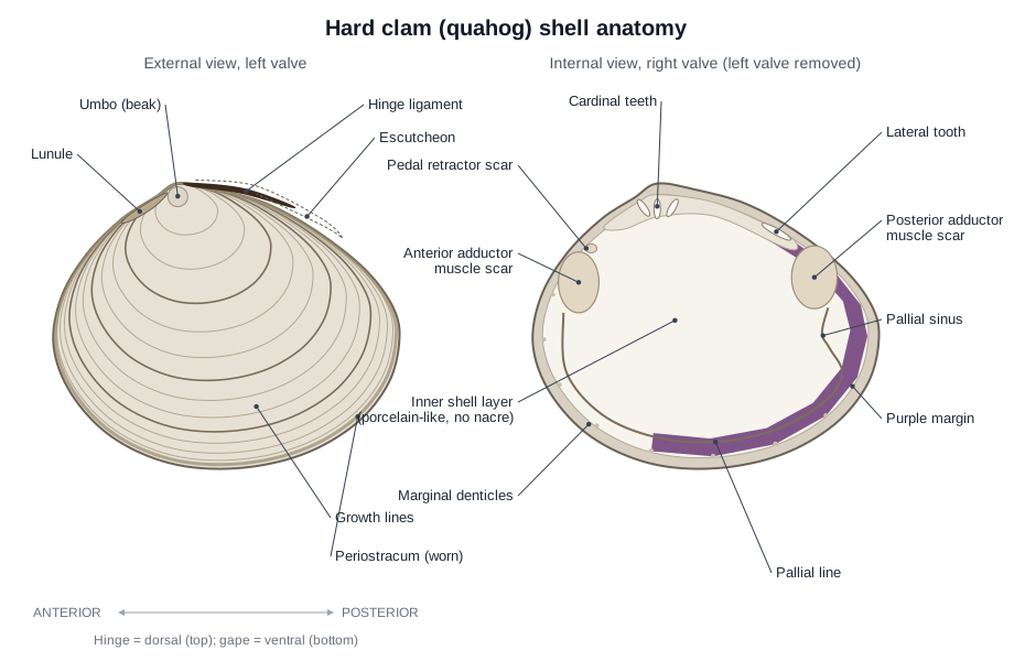
The shell has three layers: a thin protein periostracum on the outside (mostly worn away in quahogs), a chalky outer calcareous layer, and a smooth inner layer. Unlike freshwater mussels and pearl oysters, the hard clam has no nacre (mother-of-pearl); its inner layer is porcelain-like and white.
Bivalve external anatomy
From the outside you can see the two valves, the umbo, hinge ligament, lunule, escutcheon and growth lines, and in a live clam the siphons and foot. The short siphons are fused together. The lower, wider incurrent siphon has a fringe of short sensory tentacles; the upper excurrent siphon carries water out. The white, blade- or plough-shaped foot extends from the anterior-ventral gape. Inside the shell edge you can see the frilled mantle edge.
Bivalve internal anatomy: the classic lab view
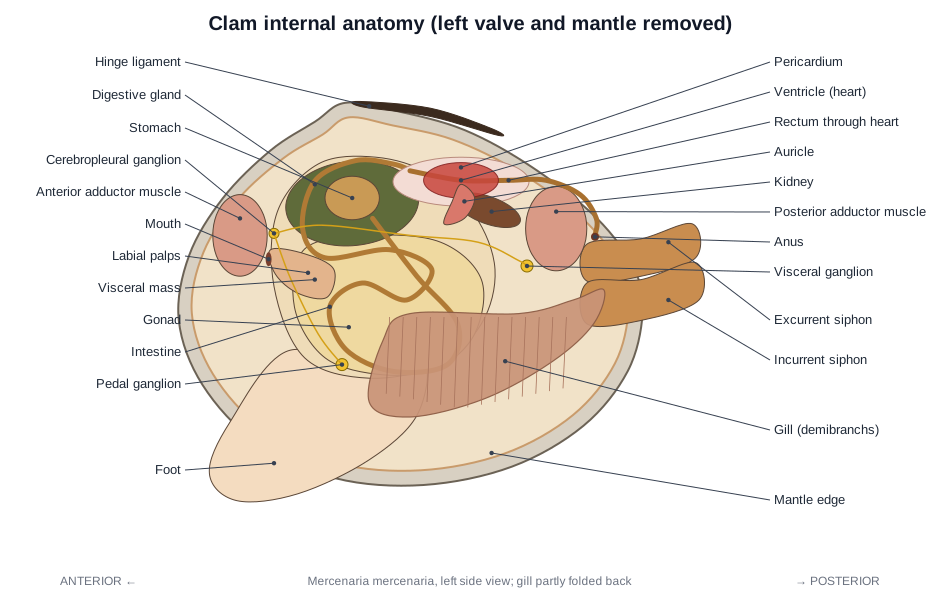
With the left valve and left mantle lobe removed, you see the gills, labial palps, foot, visceral mass, both adductor muscles and the heart. The large, ridged, brownish gills cover much of the side of the body. Each gill has an outer and an inner plate (demibranch). At their front ends are the flap-like labial palps, which surround the mouth just behind the anterior adductor. Below the gills lies the foot, and above it the visceral mass, filled with creamy gonad and the green-brown digestive gland. Just under the hinge is the thin pericardium around the heart, with the brown kidneys below it.
Clam anatomy diagram: every labeled part
These are all 61 structures in the 3D model, grouped by system. Click any of them in the viewer above to find it.
Shell
- Left valve (Left shell)
- One of the two hinged halves of the shell, on the animal's left side. Protects the soft body. In the classic lab view it is removed so the left side of the animal can be studied.
- Right valve (Right shell)
- The valve on the animal's right side; the soft body rests in it after the left valve is removed. Protects the right side of the body and carries the muscle scars, pallial line and hinge teeth you study on the shell interior.
- Umbo (Beak)
- The swollen, oldest part of each valve, next to the hinge. Marks where the shell began to grow; every growth line is centred on it.
- Hinge ligament (Ligament)
- A dark brown, elastic, protein band that joins the two valves along the dorsal edge. Acts like a spring that pulls the valves open. The adductor muscles work against it to close the shell, so a dead clam gapes.
- Cardinal teeth (Hinge teeth)
- Short, strong teeth directly below the beak on the hinge plate. Interlock with sockets in the opposite valve so the valves cannot slide past each other when they close.
- Lateral tooth (Posterior lateral tooth)
- A long, low tooth on the hinge plate behind the cardinal teeth. Adds a second interlock that stops the valves twisting.
- Lunule
- A heart-shaped depression on the dorsal surface in front of the beaks. An external landmark that identifies the anterior end of the shell.
- Escutcheon
- A long, flattened area on the dorsal surface behind the beaks. An external landmark that identifies the posterior end of the shell.
- Growth lines (Concentric ridges, annual rings)
- Concentric ridges and bands on the outside of the shell, centred on the umbo. Record the growth history. The mantle adds new shell at the margin; when growth slows in winter or during spawning, a band forms.
- Periostracum
- The thin, outermost protein (conchiolin) layer of the shell. Protects the calcium carbonate from dissolving and is the template on which new shell is laid.
- Outer shell layer (Prismatic layer, ostracum)
- The chalky outer calcareous layer, made of aragonite crystals. Forms most of the shell's bulk and strength. Secreted by the outer fold of the mantle edge.
- Inner shell layer (Lamellar layer, hypostracum)
- The smooth, glossy, porcelain-like inner layer of the shell. Laid down by the whole outer surface of the mantle, so the shell thickens throughout life.
- Anterior adductor muscle scar
- The oval mark where the anterior adductor muscle attaches to the shell. Shows where the muscle pulls on the valve; useful for orienting an empty shell.
- Posterior adductor muscle scar
- The oval mark where the posterior adductor muscle attaches. Shows the attachment of the posterior adductor and, joined to it, the posterior pedal retractor.
- Pallial line
- A thin line on the inner shell surface, parallel to the ventral margin. Marks where the pallial (mantle edge) muscles attach the mantle to the shell.
- Pallial sinus
- A V-shaped notch in the pallial line near the posterior end. Marks the space and muscle attachment for the siphons when they are pulled in.
- Purple margin (Violet border, wampum)
- The dark violet band on the inside of the shell near the posterior and ventral edges. A pigment in the shell. It has no known body function but is a reliable identification feature of Mercenaria.
- Marginal denticles (Crenulations)
- Tiny teeth along the inside edge of the valve margin. Interlock when the valves close, stopping them shearing sideways.
Mantle & body
- Left mantle lobe (Mantle, pallium)
- A thin sheet of tissue that lines the left valve and encloses the mantle cavity. Secretes the shell, forms the mantle cavity that holds the gills, and its edge senses the surroundings.
- Right mantle lobe (Mantle)
- The mantle sheet lining the right valve. Secretes the right valve and forms the right wall of the mantle cavity.
- Mantle edge (Mantle margin, mantle folds)
- The thickened, frilled free margin of each mantle lobe. Bivalve mantle edges have three folds: the outer fold secretes the shell and periostracum, the middle fold is sensory, and the inner fold is muscular and controls water flow.
- Incurrent siphon (Inhalant siphon, ventral siphon)
- The lower (ventral) of the two siphons; water and food enter the clam through it. Draws in seawater carrying oxygen and phytoplankton. Its fringe of tentacles keeps out large particles.
- Excurrent siphon (Exhalant siphon, dorsal siphon)
- The upper (dorsal) siphon, which carries water out of the clam. Expels water that has passed through the gills, together with faeces, nitrogen waste and, in the breeding season, eggs or sperm.
- Foot
- A large, white, muscular, blade- or plough-shaped organ used for digging. Burrowing: blood pumped into the foot makes its tip swell into an anchor, then the pedal retractor muscles pull the shell down after it.
- Visceral mass
- The soft central body above the foot that holds the gonad, digestive gland and coiled intestine. Contains most of the organs. In a lab, slicing it lengthways shows the gonad and intestine.
Muscles
- Anterior adductor muscle
- One of two large muscles that close the shell; this is the front one. Contracts to pull the valves shut against the spring of the hinge ligament.
- Posterior adductor muscle
- The rear adductor muscle that closes the shell. Works with the anterior adductor to close the shell.
- Catch muscle (Catch (smooth) portion of the adductors)
- The white, smooth-muscle part of each adductor that holds the shell closed for hours. Uses a 'catch' mechanism to stay contracted with very little energy, so the clam can stay shut during low tide or when attacked.
- Anterior pedal retractor muscle (Anterior foot retractor)
- A paired muscle band running from the shell to the foot at the front. Pulls the foot back into the shell and, during digging, pulls the shell down towards the anchored foot.
- Posterior pedal retractor muscle (Posterior foot retractor)
- A paired muscle running from the shell near the posterior adductor to the foot. Retracts the foot and pulls the shell forward and down while burrowing.
Digestive
- Mouth
- A simple slit-like opening between the labial palps. Takes in strings of food particles bound in mucus, delivered by the labial palps.
- Labial palps
- Two pairs of soft, triangular flaps on each side of the mouth. Ciliated ridges on their inner faces sort particles from the gills: food goes to the mouth, while sand and excess material are rejected as pseudofaeces.
- Esophagus (Oesophagus)
- A short tube from the mouth to the stomach. Carries food strings into the stomach by ciliary action.
- Stomach
- A sac in the upper visceral mass where food is mixed, sorted and partly digested. Ciliated sorting areas send fine particles into the digestive gland and coarse particles to the intestine.
- Gastric shield
- A hard, chitin-like plate lining part of the stomach wall. A bearing surface that the rotating crystalline style rubs against, grinding food and wearing away the style's tip.
- Crystalline style
- A clear, flexible, jelly-like rod of enzymes that rotates in the gut. Rotated by cilia, it winds the mucus food string into the stomach. As its tip wears away it releases digestive enzymes (amylase, cellulase).
- Style sac
- A tube-shaped pouch off the stomach that makes and turns the crystalline style. Its ciliated, glandular wall secretes the style and spins it.
- Digestive gland (Digestive diverticula, 'liver')
- A greenish-brown mass of branching tubules around the stomach. Absorbs fine food particles and digests them inside its cells; it also stores food reserves.
- Intestine (Midgut)
- A long tube that coils through the visceral mass. Absorbs nutrients and packs undigested material into faecal pellets.
- Rectum
- The last part of the gut, which passes straight through the heart. Carries faecal pellets to the anus.
- Anus
- The opening at the end of the rectum. Releases faeces into the exhalant water stream so they are carried out of the shell.
Gills
- Outer demibranch (Lateral demibranch, gill)
- The outer of the two gill plates on each side; together the two plates form one gill (ctenidium). Exchanges gases and filters food. Cilia pump water through tiny pores into internal water tubes and up to the exhalant chamber.
- Inner demibranch (Medial demibranch, gill)
- The inner gill plate on each side, next to the visceral mass. Same as the outer demibranch: breathing and filter feeding.
- Food groove (Ventral marginal groove)
- A ciliated groove along the lower free edge of the gill. Carries mucus-bound food particles forward along the gill edge to the palps, like a conveyor belt.
Circulatory
- Ventricle (Heart)
- The single muscular pumping chamber of the heart, wrapped around the rectum. Pumps blood forward into the anterior aorta and backward into the posterior aorta.
- Auricles (Atria)
- Two thin-walled chambers, one on each side of the ventricle. Collect oxygenated blood returning from the gills and pass it into the ventricle.
- Pericardium (Pericardial cavity)
- A thin membrane enclosing the fluid-filled cavity around the heart. Lets the heart beat freely; fluid filtered into it from the heart passes on to the kidneys.
- Anterior aorta
- The artery leaving the front of the ventricle. Supplies the foot, visceral mass, anterior mantle and anterior adductor.
- Posterior aorta
- The artery leaving the rear of the ventricle, with a swelling called the bulbus arteriosus. Supplies the posterior adductor, the mantle and the siphons.
Excretory
- Kidneys (Nephridia, organ of Bojanus)
- A pair of brown, spongy kidneys below the pericardium. Take filtrate from the pericardial cavity, reabsorb useful substances and release nitrogen waste (mostly ammonia) as urine into the exhalant chamber.
- Pericardial glands (Keber's organ)
- Reddish-brown glandular tissue on the wall of the pericardium. Filter blood into the pericardial cavity and store waste, working with the kidneys.
Nervous & sensory
- Cerebropleural ganglia (Cerebral ganglia)
- A pair of small nerve centres beside the mouth, linked by a commissure over the esophagus. Control the palps, anterior adductor and mantle, and coordinate the other ganglia.
- Pedal ganglia
- Fused nerve centres at the base of the foot. Control the muscles of the foot.
- Visceral ganglia (Parietovisceral ganglia)
- The largest ganglia, fused into an orange mass in front of the posterior adductor. Control the gills, posterior adductor, siphons, posterior mantle and heart.
- Cerebrovisceral connectives (Visceral nerves)
- A pair of long nerve cords from the cerebropleural to the visceral ganglia. Carry signals between the front and back of the body.
- Cerebropedal connectives
- A pair of nerves linking the cerebropleural ganglia to the pedal ganglia. Coordinate foot movements with the rest of the body.
- Statocysts
- A pair of tiny balance organs. Sense gravity, so the clam can orient itself while burrowing.
- Osphradium
- A small patch of sensory cells in the exhalant chamber. Thought to sense chemicals or particles in the water passing out of the gills.
- Siphonal tentacles (Pallial tentacles)
- Short sensory tentacles fringing the siphon openings, best developed on the incurrent siphon. Sense touch and light, triggering the siphons to withdraw, and screen out large particles.
Reproductive
- Gonad (Ovary or testis)
- The reproductive tissue that fills much of the visceral mass around the intestine. Produces eggs (in females) or sperm (in males).
- Gonoducts & genital pore (Genital papilla)
- Short ducts carrying eggs or sperm from the gonad to a small pore on each side. Release eggs or sperm into the exhalant water stream, which carries them out of the excurrent siphon.
How a clam feeds: filter feeding with gills and palps
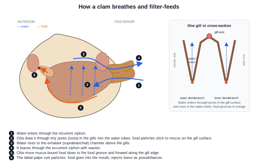
A clam is a filter feeder: its gills strain phytoplankton and other fine particles from the water it pumps through the mantle cavity. Cilia on the gill filaments create the water current. Particles caught on the gill surface are wrapped in mucus and carried by cilia down to the food grooves along the lower gill edges, then forward to the labial palps. The palps sort the particles: food goes into the mouth, and rejected material is bound in mucus and expelled as pseudofaeces.
Inside the stomach, the clear crystalline style rotates against the hard gastric shield, winding in the mucus food string and releasing digestive enzymes. Fine particles pass into the digestive gland to be digested inside its cells; the rest go to the intestine and leave as faecal pellets through the anus, into the outgoing water.
How a clam breathes
Clams breathe with their gills, using the same water current that brings their food. Oxygen diffuses from the water into the blood flowing through the gill filaments, and the thin mantle also takes up oxygen. Oxygenated blood returns from the gills to the two auricles of the heart. When the shell is shut, for example at low tide, a clam can switch to anaerobic metabolism for a while.
Burrowing with the foot
A clam burrows by pushing its foot into the sediment, swelling the tip with blood as an anchor, and pulling the shell down after it. The foot has no skeleton: it is a hydrostatic organ stiffened by blood pressure. Contraction of the adductors squeezes blood into the foot, then the anterior and posterior pedal retractor muscles pull the shell towards the anchored foot. Repeating the cycle rocks the clam into the sand. Hard clams live just below the surface, from a few centimetres to about 15 cm deep, with their short siphons reaching the water.
The clam heart and the rectum
A clam's heart has three chambers, one ventricle and two auricles, and the rectum passes straight through the ventricle. The heart sits dorsally, just under the hinge, in a fluid-filled sac called the pericardium. The ventricle pumps blood forward through the anterior aorta and backward through the posterior aorta. Bivalves have an open circulatory system: blood leaves the arteries and flows through open spaces in the tissues before returning through the kidneys and gills to the auricles.
Below the pericardium lie the paired kidneys (nephridia, or organ of Bojanus), which take fluid from the pericardial cavity and release urine into the exhalant chamber. Reddish pericardial glands (Keber's organ) on the pericardial wall also help filter the blood.
Nervous system and senses
Clams have no brain; they have three pairs of ganglia linked by nerve cords. The cerebropleural ganglia lie beside the mouth, the pedal ganglia at the base of the foot, and the visceral ganglia on the front of the posterior adductor muscle. Senses are simple: balance organs (statocysts) in the foot, an osphradium in the exhalant chamber, and touch- and light-sensitive cells on the siphon tentacles and mantle edge.
Reproduction
Most adult hard clams are either male or female, and they release eggs and sperm into the water. The gonad fills much of the visceral mass around the intestine and opens through small genital pores into the exhalant chamber, so gametes leave through the excurrent siphon. Males and females look the same: you need a microscope to see eggs or sperm. Young quahogs usually mature first as males, and many later become females. Fertilised eggs develop into a swimming veliger larva that settles after one to a few weeks.
Shell growth and age: reading the growth lines
A clam grows by adding new shell at the margin, so each growth line marks a former edge of the shell. The mantle edge lays down new shell during the growing season; in winter or during spawning, growth slows and a dark band forms. Counting these annual bands, usually in a cut section through the shell, gives the clam's age. Hard clams grow fastest in their first few years, so later bands are packed tightly near the edge. One quahog from Massachusetts was aged at about 106 years this way.
Clam vs mussel vs oyster vs scallop: comparing bivalves
All bivalves share the same basic plan (two valves, mantle, gills, foot and adductors), but they differ in muscles, foot and lifestyle.
| Bivalve | Adductor muscles | Foot | Siphons | Lifestyle | Distinctive feature |
|---|---|---|---|---|---|
| Hard clam (Mercenaria) | Two, similar size | Large, blade-shaped; burrows | Short, fused | Burrows shallowly in sand and mud | Lunule, purple inner margin, no nacre |
| Blue mussel (Mytilus) | Two; anterior one very small | Small; spins byssal threads | None (exhalant opening only) | Attached to rocks by byssus | Elongated shell, umbo at the front tip |
| Oyster (Crassostrea) | One, central | Absent in adults | None | Cemented to hard surfaces by the left valve | Unequal valves; cupped left, flat right |
| Scallop (Pecten, Argopecten) | One, very large (the part people eat) | Small | None | Lies free on the bottom and swims by clapping its valves | Dozens of mirror eyes on the mantle edge |
| Freshwater mussel (Unionidae) | Two | Large; burrows | None: incurrent and excurrent openings in the mantle edge | Lives half-buried in river and lake beds | Pearly nacre; larvae (glochidia) are brooded in the female's gills and attach to fish |
Bivalve anatomy of a scallop: what is different
A scallop has a single, very large adductor muscle, rows of eyes on its mantle edge, and swims instead of burrowing. It claps its valves together to jet water out near the hinge, so it moves in a series of jerks. The adductor has a large quick part for swimming and a smaller catch part for staying closed; the white “scallop” sold as food is this muscle. Scallops have dozens of small eyes, up to about 200 in some species, that focus light with a curved mirror of guanine crystals rather than a lens. They have no siphons and only a small foot.
Frequently asked questions
Do clams have a brain?
No. A clam has no brain or head. Its nervous system is three pairs of ganglia (small nerve centres): cerebropleural ganglia by the mouth, pedal ganglia in the foot and visceral ganglia by the posterior adductor muscle, joined by nerve cords.
Do clams have a heart?
Yes. A clam has a three-chambered heart, one ventricle and two auricles, in the pericardial cavity just under the hinge. Unusually, the rectum passes straight through the ventricle.
Do clams have eyes?
Hard clams have no eyes. Like many burrowing clams they can sense light and shadow with cells in the siphons and mantle edge, and pull the siphons in when disturbed. Scallops, by contrast, have dozens of mirror eyes along the mantle edge.
How do clams breathe?
Cilia on the gills pump water in through the incurrent siphon, across the gills and out through the excurrent siphon. Oxygen diffuses into the blood in the gills and through the mantle surface.
How do clams eat?
Clams are filter feeders. The gills trap phytoplankton and other particles in mucus, cilia carry them to the labial palps, the palps sort food from sand, and the mouth swallows the food.
Are clams alive when you buy them?
Fresh clams in the shell are sold alive. A live clam keeps its shell shut, or closes it when tapped, because the adductor muscles are working against the hinge ligament. A clam that gapes and does not close is dead.
Can clams feel pain?
This is not settled. Clams react to harmful stimuli with reflexes, such as closing the shell and pulling in the siphons, which shows they detect damage (nociception). But they have no brain, and there is no good evidence yet of the learned avoidance or trade-off behaviour used as evidence of pain in octopuses and crabs. Scientific reviews that led to legal protection in the UK covered cephalopods and decapod crustaceans, not bivalves.
How long do clams live?
Hard clams commonly live several decades. By counting annual growth bands in the shell, researchers aged one Mercenaria from Massachusetts at about 106 years.
How do you tell the left valve from the right valve?
Find the anterior end first: the beaks point towards it and the heart-shaped lunule is there. Hold the shell with the hinge up and the anterior end pointing to your left. The valve facing you is the left valve.
Dissecting a clam?
Follow the step-by-step clam dissection guide with the same 3D model, a materials list, and lab questions with an answer key.
References
- Fox, R. Invertebrate Anatomy OnLine: Mercenaria mercenaria, hard clam (lab exercise). Lander University.
- Stanley, J.G. (1985). Species profiles: life histories and environmental requirements of coastal fishes and invertebrates (Mid-Atlantic): hard clam. U.S. Fish and Wildlife Service Biological Report 82(11.41).
- Baker, S. & Sturmer, L. Taxonomy, anatomy and biology of the hard clam. University of Florida IFAS Shellfish Aquaculture Extension.
- Helm, M.M. & Bourne, N. (2004). Hatchery culture of bivalves: a practical manual, Part 2: Basic bivalve biology. FAO Fisheries Technical Paper 471.
- University of California Museum of Paleontology. Bivalvia: morphology.
- Loosanoff, V.L. (1937). Development of the primary gonad and sexual phases in Venus mercenaria Linnaeus. Biological Bulletin 72: 389–405.
- Ridgway, I.D. et al. (2011). New species longevity record for the northern quahog (= hard clam), Mercenaria mercenaria. Journal of Shellfish Research 30(1).
- Harding, J.M. (2007). Northern quahog (hard clam) Mercenaria mercenaria age-at-length relationships. Journal of Shellfish Research 26(1).
- Suzuki, T. (1985). Comparative anatomy of the excretory organs and heart of bivalves. Bulletin of the National Research Institute of Aquaculture 7: 59–82.
- Palmer, B.A. et al. (2017). The image-forming mirror in the eye of the scallop. Science 358: 1172–1175.
- Birch, J. et al. (2021). Review of the evidence of sentience in cephalopod molluscs and decapod crustaceans. LSE Consulting.
- MarLIN, Marine Biological Association. Hard-shell clam (Mercenaria mercenaria).
More interactive anatomy models
- Clam dissectionGuided clam dissection with the visceral mass and organs labeled.
- Human anatomy1,000+ bones, muscles, organs, vessels and nerves from real anatomical data.
- Octopus anatomyThree hearts, a ring-shaped brain, eight arms and a beak, in 3D.
- Squid anatomyPen, ten arms, giant axons, ink sac and three hearts of the longfin squid.
- Squid dissectionStep-by-step guided squid dissection with every structure labeled.
- Fish anatomyExternal and internal anatomy of a bony fish: fins, gills, swim bladder, heart and brain.