

Clam dissection overview
In a clam dissection you identify the shell features, open the shell by cutting the two adductor muscles, remove the left valve, and then study the mantle, gills, foot, visceral mass and heart. The standard lab clam is the hard clam or quahog, a marine bivalve. The whole lab takes about 45–60 minutes. If you cannot or choose not to dissect an animal, use the 3D model above as a virtual clam dissection: it follows the same steps and covers every structure on a typical worksheet.
Materials and safety
- Preserved hard clam (quahog), or a fresh clam from a fish market
- Dissecting tray and pins (or a shallow dish)
- Scalpel, fine scissors, blunt probe and forceps
- Hand lens or dissecting microscope; compound microscope, slides, cover slips
- Disposable gloves, safety glasses and a lab coat or apron
- Ruler (mm), paper towels, and a sheet or worksheet for drawings
- Wear gloves and safety glasses. Preserved specimens may contain irritating preservative; work in a ventilated room.
- Scalpels are sharp: cut away from yourself and keep fingers out of the line of the blade.
- Do not pry a closed shell with a scalpel; the blade can snap. Use a blunt wedge.
- Never eat any part of a lab specimen. Dispose of tissue as your teacher directs and wash your hands afterwards.
Pre-lab questions
- To which phylum and class does the clam belong?
- What holds the shell closed, and what opens it?
- How does a clam get its food?
External anatomy of the clam: before you cut
Start by orienting the shell: hinge = dorsal, gape = ventral, beaks and lunule = anterior, ligament and siphons = posterior. Then hold the clam hinge-up with the anterior end to your left; the valve facing you is the left valve. Find the umbo, hinge ligament, lunule, escutcheon and concentric growth lines, and estimate the clam's age from the darker annual bands. See the labeled shell diagram for the external and internal shell features.
How to dissect a clam: step by step
1. Orient the shell: dorsal, ventral, anterior, posterior
- Rinse the clam and place it on the tray. The hinge, with its dark brown ligament, is the dorsal side; the opposite edge, where the valves open, is ventral.
- Find the umbo (beak), the swollen oldest part of each valve beside the hinge.
- The beaks point forward. Just in front of them is the small heart-shaped lunule: that end is anterior. The long, flat escutcheon behind the beaks marks the posterior end.
- Note the concentric growth lines around the umbo. They are crowded near the margin, where growth is slow in older clams.
Label: Umbo, Hinge ligament, Lunule, Escutcheon, Growth lines, Periostracum
2. Identify the left and right valves
- Hold the clam with the hinge up and the anterior end (lunule) pointing to your left.
- The valve facing you is the LEFT valve; the one facing away is the RIGHT valve.
- Measure the shell length (anterior to posterior), height (umbo to ventral margin) and width (across both valves) in millimetres and record them.
Label: Left valve, Right valve, Umbo, Lunule
3. Observe the siphons, foot and mantle edge
- Look between the valves at the posterior end. The short, fused siphons may be visible: the lower one is the incurrent siphon, the upper one the excurrent siphon.
- Look at the anterior-ventral gape for the white, blade-shaped foot.
- Note the frilled mantle edge just inside the shell margin. In preserved clams the siphons and foot are usually pulled in.
Label: Incurrent siphon, Excurrent siphon, Siphonal tentacles, Foot, Mantle edge
4. Open the shell by cutting the adductor muscles
- If the valves are closed, slide a blunt probe or butter knife into the gape at the posterior end (or ask your teacher to wedge it open).
- Keep the blade of the scalpel flat against the inside of the LEFT valve. Slide it towards the anterior-dorsal corner and cut the anterior adductor muscle close to the shell.
- Repeat at the posterior-dorsal corner to cut the posterior adductor muscle. The valves will spring apart, pulled open by the hinge ligament.
Caution: Always cut away from your hand and keep the scalpel blade against the shell. Never force a blade into a tightly closed shell: wedge it first.
Label: Anterior adductor muscle, Posterior adductor muscle, Catch muscle, Hinge ligament
5. Remove the left valve and examine the mantle
- Gently peel the left mantle lobe off the inside of the left valve with the blunt end of a probe, working from the hinge outwards.
- Lift the left valve away by breaking it at the hinge. Place the clam in a tray with the right valve down.
- The thin sheet now covering the body is the left mantle lobe. Find its thickened edge and trace it back to the siphons.
Label: Left mantle lobe, Mantle edge, Right mantle lobe, Incurrent siphon, Excurrent siphon
6. Lift the mantle: gills and mantle cavity
- Fold the left mantle lobe back (or cut it away along the pallial line) to open the mantle cavity.
- The large, ridged, brown plates are the gills. Each gill has two plates: the outer and inner demibranchs.
- Look for the food groove along the lower edge of the gill. With a hand lens you can see the vertical ridges (plicae) and the many filaments.
- Cut a small piece of gill, mount it in water and view it under a microscope to see the filaments and beating cilia.
Label: Outer demibranch, Inner demibranch, Food groove, Labial palps
7. Labial palps, mouth and foot
- Fold the gills back. At the anterior end, between the anterior adductor and the gills, find two pairs of flap-like labial palps.
- Part the palps to find the slit-like mouth just behind the anterior adductor.
- Note the muscular foot below the visceral mass and the pedal retractor muscles that run from the shell to the foot.
Label: Labial palps, Mouth, Foot, Visceral mass, Anterior pedal retractor muscle, Posterior pedal retractor muscle
8. Open the pericardium: heart and rectum
- Just below the hinge, find the thin, dark-lined pericardium. Carefully cut it open with fine scissors.
- Inside is the heart: one ventricle and two thin auricles (atria), one on each side.
- Trace the tube that runs straight through the ventricle. This is the rectum: in clams the gut passes through the heart.
- Below the pericardium find the brown, spongy kidneys (nephridia).
Caution: The pericardium and heart are very thin. Use fine scissors and lift the membrane with forceps before cutting.
Label: Pericardium, Ventricle, Auricles, Rectum, Anterior aorta, Posterior aorta, Kidneys, Pericardial glands
9. Cut the visceral mass: gonad, digestive gland and intestine
- Hold the foot and slice the visceral mass and foot in half lengthways (a sagittal cut), from the ventral edge of the foot up towards the hinge.
- The creamy tissue filling most of the visceral mass is the gonad (ovary or testis).
- The darker green-brown tissue near the top is the digestive gland, around the stomach.
- Find the coiled intestine cut in cross-section among the gonad, and the stomach with the style sac running down from it. A clear, jelly-like crystalline style may be present in fresh clams.
- Smear a little gonad on a slide and look under the microscope for eggs or sperm to sex your clam.
Label: Gonad, Digestive gland, Stomach, Intestine, Style sac, Crystalline style, Gastric shield, Esophagus
10. Follow the rectum to the anus
- Follow the rectum back from the heart, over the posterior adductor muscle.
- The anus opens on the back of the posterior adductor, beside the excurrent siphon, so wastes leave with the outgoing water.
- Look for the small orange visceral ganglia on the front face of the posterior adductor.
Label: Rectum, Anus, Posterior adductor muscle, Excurrent siphon, Visceral ganglia
11. Find the nervous system (advanced)
- Clams have no brain. Find the three pairs of ganglia: the cerebropleural ganglia beside the mouth, the pedal ganglia at the base of the foot and the visceral ganglia on the posterior adductor.
- Under a dissecting microscope, trace the thin cerebrovisceral connectives that link the front and back ganglia.
Label: Cerebropleural ganglia, Pedal ganglia, Visceral ganglia, Cerebrovisceral connectives, Cerebropedal connectives, Statocysts, Osphradium
12. Clean the shell: interior features
- Scrape the soft parts out of the right valve and rinse it.
- Find the two adductor muscle scars, the pallial line joining them and the V-shaped pallial sinus near the posterior scar.
- Find the cardinal teeth under the umbo and the lateral tooth behind them, the violet border near the posterior margin, and the tiny denticles on the inner edge.
- Look at the broken or cut edge of the left valve to see the chalky outer layer and the smooth inner layer.
Label: Anterior adductor muscle scar, Posterior adductor muscle scar, Pallial line, Pallial sinus, Cardinal teeth, Lateral tooth, Purple margin, Marginal denticles, Outer shell layer, Inner shell layer
Clam dissection labeled: what you should find
A labeled clam dissection shows the adductor muscles at each end, the gills across the side of the body, the labial palps by the mouth, the foot below and the visceral mass above it, with the heart under the hinge.
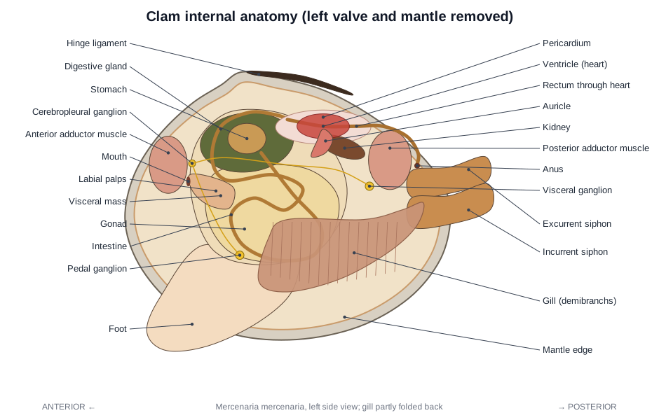
The labeled visceral mass: after the sagittal cut, the visceral mass shows creamy gonad surrounding cut loops of intestine, with the green-brown digestive gland and stomach near the top and the style sac running down from the stomach. The clam heart is the small ventricle in the pericardium under the hinge; the rectum runs straight through it.
Freshwater vs marine clam dissection
Freshwater mussels and marine clams have the same organs, but differ in shell and siphons. Freshwater mussels (family Unionidae) have a thick, dark periostracum, a pearly nacreous inner shell and no true siphons, only incurrent and excurrent openings in the mantle edge. Females may have swollen gills full of glochidia larvae. Marine clams like the quahog have fused siphons, a pallial sinus in the shell and a white, non-pearly inner layer. Either works for this guide.
Male vs female clams
You cannot tell a male clam from a female by looking. Both have the same organs; only the gonad tissue differs. Smear a small piece of gonad on a slide: round eggs mean female, tiny moving sperm mean male. Hard clams are usually separate sexes, though young clams often mature first as males.
Clam dissection lab questions
- How can you tell the anterior end of a clam shell from the posterior end?
- How do you identify the left valve?
- Why must you cut the adductor muscles to open the clam?
- What do the growth lines on the shell tell you?
- Name the two siphons and give the direction of water flow through each.
- What are the two functions of the gills?
- What is unusual about the relationship between the heart and the digestive tract?
- What is the function of the labial palps?
- What organ fills most of the visceral mass, and how can you tell if your clam is male or female?
- Why is the anus located near the excurrent siphon?
- What is the crystalline style and what does it do?
- How does the foot help the clam burrow?
Clam dissection answer key
Pre-lab questions
- To which phylum and class does the clam belong? Phylum Mollusca, class Bivalvia (also called Pelecypoda).
- What holds the shell closed, and what opens it? The anterior and posterior adductor muscles close it. The elastic hinge ligament springs it open when the muscles relax.
- How does a clam get its food? It is a filter feeder: water enters through the incurrent siphon, the gills trap food particles in mucus, and cilia carry them to the labial palps and mouth.
Analysis questions
- How can you tell the anterior end of a clam shell from the posterior end? The beaks (umbos) point towards the anterior end, and the heart-shaped lunule is in front of them. The ligament, escutcheon and siphons are posterior.
- How do you identify the left valve? Hold the shell with the hinge up and the anterior end to your left. The valve facing you is the left valve.
- Why must you cut the adductor muscles to open the clam? They hold the valves shut against the pull of the ligament. Once both are cut, nothing holds the shell closed and it springs open.
- What do the growth lines on the shell tell you? They record growth over time. Annual bands can be counted to estimate the clam's age.
- Name the two siphons and give the direction of water flow through each. The incurrent (inhalant) siphon, the lower one, brings water in. The excurrent (exhalant) siphon, the upper one, carries water out.
- What are the two functions of the gills? Gas exchange (breathing) and filter feeding: they trap food particles and move them to the palps.
- What is unusual about the relationship between the heart and the digestive tract? The rectum passes straight through the ventricle of the heart.
- What is the function of the labial palps? They sort particles brought by the gills, passing food into the mouth and rejecting sand as pseudofaeces.
- What organ fills most of the visceral mass, and how can you tell if your clam is male or female? The gonad. Males and females look the same; you must check a smear of gonad under the microscope for eggs or sperm.
- Why is the anus located near the excurrent siphon? So faeces leave with the outgoing water and do not contaminate the incoming water used for feeding and breathing.
- What is the crystalline style and what does it do? A clear rod of digestive enzymes in the style sac. It rotates against the gastric shield, winding in the food string and releasing enzymes as its tip wears away.
- How does the foot help the clam burrow? The foot probes into the sediment, swells with blood at its tip to form an anchor, and the pedal retractor muscles then pull the shell down after it.
Frequently asked questions
What clam is used for dissection?
Most schools use the hard clam or quahog, Mercenaria mercenaria, a thick-shelled marine clam from the western Atlantic. Freshwater mussels (Unionidae) are sometimes used instead; their internal anatomy is very similar.
How do you open a clam for dissection?
Wedge the valves apart slightly, then slide a scalpel along the inside of one valve and cut the anterior and posterior adductor muscles close to the shell. The hinge ligament then springs the valves open.
Where is the heart in a clam?
Just under the hinge, in a thin sac called the pericardium. The heart has one ventricle and two auricles, and the rectum runs straight through the ventricle.
What is the visceral mass of a clam?
The soft central body above the foot. It contains the gonad, digestive gland, stomach and coiled intestine. Cutting it in half lengthways shows these organs.
What is the difference between a freshwater and a marine clam dissection?
The organs are the same, but freshwater mussels have no true siphons (just incurrent and excurrent openings in the mantle edge), a thick dark periostracum and a pearly nacreous inner shell. Females may hold glochidia larvae in their gills. Marine clams like Mercenaria have fused siphons and a non-pearly inner shell.
Can I do a virtual clam dissection instead?
Yes. The interactive model on this page follows the same steps as a real dissection, so students who opt out of dissecting an animal can still identify every structure and answer the lab questions.
Learn the anatomy first
The clam anatomy page explains how each organ works, with labeled shell and internal anatomy diagrams and a comparison of clams, mussels, oysters and scallops.
References
- Fox, R. Invertebrate Anatomy OnLine: Mercenaria mercenaria, hard clam (lab exercise). Lander University.
- Stanley, J.G. (1985). Species profiles: life histories and environmental requirements of coastal fishes and invertebrates (Mid-Atlantic): hard clam. U.S. Fish and Wildlife Service Biological Report 82(11.41).
- Baker, S. & Sturmer, L. Taxonomy, anatomy and biology of the hard clam. University of Florida IFAS Shellfish Aquaculture Extension.
- Helm, M.M. & Bourne, N. (2004). Hatchery culture of bivalves: a practical manual, Part 2: Basic bivalve biology. FAO Fisheries Technical Paper 471.
- University of California Museum of Paleontology. Bivalvia: morphology.
- Loosanoff, V.L. (1937). Development of the primary gonad and sexual phases in Venus mercenaria Linnaeus. Biological Bulletin 72: 389–405.
More interactive anatomy models
- Clam anatomyBivalve anatomy: shell, adductor muscles, gills, foot, siphons and heart.
- Human anatomy1,000+ bones, muscles, organs, vessels and nerves from real anatomical data.
- Octopus anatomyThree hearts, a ring-shaped brain, eight arms and a beak, in 3D.
- Squid anatomyPen, ten arms, giant axons, ink sac and three hearts of the longfin squid.
- Squid dissectionStep-by-step guided squid dissection with every structure labeled.
- Fish anatomyExternal and internal anatomy of a bony fish: fins, gills, swim bladder, heart and brain.