

Jellyfish anatomy at a glance
A jellyfish has no brain, no heart, no blood, no bones, no lungs and no anus. Its body is a bell of jelly (mesoglea) covered by two thin cell layers, with a single mouth on the underside that leads into a stomach and a network of canals. Roughly 95% of the animal is water. Signals travel through a nerve net, and eight small sense organs (rhopalia) around the bell edge sense light and gravity and set the swimming rhythm.
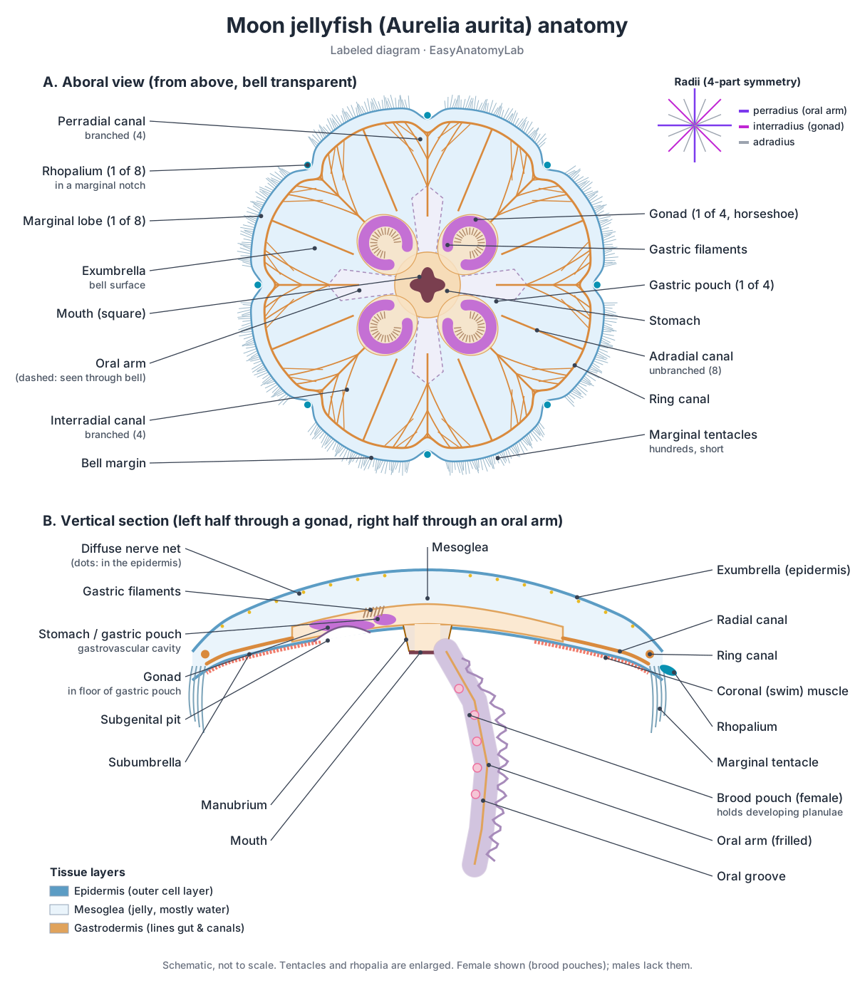
The moon jellyfish, Aurelia aurita, is the jellyfish in most biology textbooks. It is a “true jellyfish” (class Scyphozoa) in the phylum Cnidaria, the group that also includes corals, sea anemones and hydras. Its body has four-part (tetramerous) radial symmetry: four oral arms, four gastric pouches, four gonads and 4 × 2 = 8 rhopalia. Biologists name the radii: perradii run through the four mouth corners and oral arms, interradii run through the gonads halfway between them, and adradii lie halfway between a perradius and an interradius.
Two surfaces are used for orientation. The exumbrella (aboral side) is the domed top of the bell. The subumbrella (oral side) is the concave underside, where the mouth is. Note that the name Aurelia aurita covers a complex of several very similar species; the anatomy here applies to all of them.
Body layers: epidermis, mesoglea and gastrodermis
Jellyfish are built from only two cell layers with jelly in between. Animals like this are called diploblastic.
- Epidermis (Ectoderm): The outer cell layer covering the whole jellyfish, inside and out of the bell. Protection, secretion of mucus, and the home of most nerve cells, sensory cells, stinging cells and the swimming muscle cells.
- Mesoglea (Jelly layer): The thick, transparent jelly that fills the bell between the two cell layers. Acts as an elastic hydrostatic skeleton: it springs back after each muscle contraction, reopening the bell without any muscles doing the work.
- Gastrodermis (Endoderm): The inner cell layer lining the stomach, gastric pouches and canals. Secretes digestive enzymes, absorbs food particles for intracellular digestion and moves fluid with its cilia and flagella.
Jellyfish have no skeleton. The springy mesoglea works as a hydrostatic skeleton: it is squeezed by the swimming muscle and then recoils, reopening the bell.
Labeled jellyfish diagram
The main parts of a jellyfish are the bell, tentacles, oral arms, mouth, stomach and gastric pouches, gonads, radial and ring canals, nerve net and rhopalia. The labeled diagram below shows a moon jellyfish from above and in vertical section. Every part labeled here can also be clicked in the 3D model.
External (bell & arms)
- Exumbrella (Aboral surface of the bell)
- The smooth, domed upper (outer) surface of the bell. Covers and protects the bell. In Aurelia its mucus-coated surface also traps small plankton, which cilia carry towards the bell margin.
- Subumbrella (Oral surface of the bell)
- The concave underside of the bell, facing the mouth. Carries the swimming muscles. When they contract, the subumbrella pulls in and pushes water out from under the bell.
- Marginal lobes (Marginal lappets, velar lobes)
- The eight broad, scalloped lobes that make up the edge of the bell. The flexible edge of each lobe flaps outward and inward with each stroke, helping the bell 'row' through the water and steer.
- Rhopalial lappets
- Pairs of small flaps on either side of each rhopalium. Shelter the delicate rhopalium between them.
- Bell margin (Umbrella margin)
- The thin outer edge of the bell, where exumbrella meets subumbrella. Carries the fringe of tentacles and the eight rhopalia. Mucus with trapped food collects here and is picked up by the oral arms.
- Marginal tentacles (Fringing tentacles)
- Hundreds of short, fine tentacles forming a fringe around the bell margin. Capture small plankton with their stinging cells as water flows past the bell margin during swimming.
- Oral arms (Mouth arms)
- Four long, frilly arms hanging from the corners of the mouth. Collect food from the tentacles and bell margin and pass it to the mouth. In females, fertilised eggs develop on the oral arms.
- Oral arm frills (Frilled margins)
- The ruffled, curtain-like edges along both sides of each oral arm. Increase the surface area for catching and holding prey and mucus strings, and border the oral groove.
Body layers
- Mesoglea (Jelly layer)
- The thick, transparent jelly that fills the bell between the two cell layers. Acts as an elastic hydrostatic skeleton: it springs back after each muscle contraction, reopening the bell without any muscles doing the work.
- Epidermis (Ectoderm)
- The outer cell layer covering the whole jellyfish, inside and out of the bell. Protection, secretion of mucus, and the home of most nerve cells, sensory cells, stinging cells and the swimming muscle cells.
- Gastrodermis (Endoderm)
- The inner cell layer lining the stomach, gastric pouches and canals. Secretes digestive enzymes, absorbs food particles for intracellular digestion and moves fluid with its cilia and flagella.
Sensory (rhopalia)
- Rhopalia (Tentaculocysts, marginal sense organs)
- Eight small, club-shaped sense organs set in notches around the bell margin. Sense gravity (balance) and light, and contain the pacemakers that set the rhythm of swimming.
- Statocyst (Lithocyst)
- The heavy, crystal-filled tip of each rhopalium. Gravity sensing. Its weight bends the rhopalium when the bell tilts, which tells the jellyfish which way is up.
- Statoliths
- Tiny crystals packed inside the statocyst. Add weight to the rhopalium tip so that it responds to gravity.
- Pigment-cup ocellus (Cup eye)
- A small cup of pigment cells with light-sensitive cells inside. Detects light and its direction. It is thought to be the main light sensor of the rhopalium.
- Pigment-spot ocellus (Eyespot)
- A flat patch of pigment cells on the rhopalium. A simple light-detecting spot; its exact role is less clear than that of the cup ocellus.
- Touch plate
- A patch of ciliated sensory cells on the upper side of the rhopalium. Works with the statocyst as a gravity sensor: when the rhopalium tilts, its cilia press on or move away from the hood.
- Rhopalial hood
- A small fold of the bell that roofs over each rhopalium. Protects the rhopalium and provides the surface that the touch plate contacts.
- Sensory pits (Olfactory pits)
- Small ciliated depressions near the base of each rhopalium, one on the exumbrellar side and one on the subumbrellar side. Thought to be chemoreceptors (chemical senses); their function has not been fully demonstrated.
Nervous
- Diffuse nerve net (DNN)
- A fine mesh of small nerve cells spread through the epidermis of the whole body. Carries slow, local signals (touch, feeding responses) and relays sensory input to the rhopalia to adjust swimming.
- Motor nerve net (Giant fibre nerve net, MNN)
- A second, separate nerve net of large neurons in the subumbrella. Spreads each swimming signal from the rhopalia rapidly across the bell, so the whole swim muscle contracts at once.
- Rhopalial ganglia (Marginal centres, pacemakers)
- Clusters of nerve cells in each rhopalium that act as swimming pacemakers. Generate the rhythmic signals for each swim pulse and integrate gravity and light information.
Gastrovascular (digestive)
- Mouth
- A single, square-shaped opening in the centre of the underside. Takes in food and also expels undigested remains: the gut has only one opening.
- Manubrium
- A short tube hanging from the centre of the subumbrella, ending in the mouth. Connects the mouth to the central stomach.
- Oral grooves
- A ciliated groove running along the inner face of each oral arm. Cilia carry captured food and mucus strings up the arm into the mouth.
- Stomach (Central stomach)
- The central chamber of the gastrovascular cavity. Receives food from the mouth and begins digestion with enzymes.
- Gastric pouches
- Four interradial chambers branching off the central stomach. Digest food and send nutrient-rich fluid out into the radial canals.
- Gastric filaments (Phacellae, gastric cirri)
- Rows of small, finger-like filaments inside each gastric pouch. Carry stinging cells and gland cells that kill struggling prey and secrete digestive enzymes.
- Adradial canals (Unbranched radial canals)
- Eight straight, unbranched canals running from the stomach to the ring canal. Carry food-laden fluid outward from the gastric pouches to the ring canal.
- Perradial canals (Branched radial canals)
- Four branched canals running from the stomach to the perradial rhopalia. Return fluid from the ring canal to the stomach and distribute food along the way.
- Interradial canals (Branched radial canals)
- Four branched canals running from the gastric pouches to the interradial rhopalia. Return fluid from the ring canal towards the stomach and distribute food.
- Ring canal (Circular canal, marginal canal)
- A canal running right around the bell, just inside the margin. Distributes nutrients to the bell margin, tentacles and rhopalia, and links the outflowing and returning canals.
Muscular
- Coronal muscle (Swim muscle, circular muscle)
- A broad ring of circular muscle on the underside of the bell. Contracts to squeeze the bell and push water out from underneath it: the power stroke of swimming.
- Radial muscles
- Muscle fibres on the subumbrella running from the centre towards the margin. Help shape and fold the bell margin, for example during turning and feeding.
Reproductive
- Gonads (Ovaries / testes)
- Four horseshoe-shaped reproductive organs, easily seen through the bell. Produce eggs (in females) or sperm (in males). The gametes are released into the gastric pouch and leave through the mouth.
- Subgenital pits (Subgenital porticoes, subgenital ostia)
- Four round pockets opening on the underside of the bell beneath the gonads. Thin the tissue below each gonad; they are thought to help with respiration and with supplying water to the gonads. They do not release gametes.
- Brood pouches (Brood sacs) · females only
- Small pockets along the oral arms of females where fertilised eggs develop. Hold fertilised eggs until they develop into swimming planula larvae, which are then released.
- Planula larvae · females only
- Tiny, oval, ciliated larvae brooded on the oral arms of females. Swim away, settle on a hard surface and grow into a polyp (the scyphistoma), starting the asexual part of the life cycle.
Stinging cells
- Cnidocytes (Stinging cells, nematocytes)
- Specialised cells that each contain one stinging capsule. Fire their capsule to catch prey and defend the jellyfish.
- Nematocyst (Cnida, stinging capsule)
- The stinging capsule inside a cnidocyte, holding a coiled, venom-filled tubule. When triggered, the pressurised capsule bursts open, water rushes in, and the tubule shoots out, turns inside out and pierces or entangles prey, injecting venom.
- Tubule (Thread, filament)
- The long, hollow thread coiled inside the nematocyst capsule. Penetrates or wraps around prey and delivers venom through its tip.
- Cnidocil (Trigger)
- A hair-like trigger projecting from the surface of the cnidocyte. Senses touch. Firing usually needs both touch and chemical cues from prey, so the jellyfish rarely wastes capsules.
- Operculum (Capsule lid)
- The lid closing the top of the nematocyst capsule. Flips open when the capsule fires, letting the tubule shoot out.
Jellyfish digestive system (gastrovascular cavity)
A jellyfish digests food in a single gastrovascular cavity with one opening, the mouth. The same cavity digests food and carries it around the body, so it does the work of both a gut and a circulatory system.
Moon jellyfish eat small plankton. Prey is stung by the marginal tentacles and the oral arms. Particles also stick to mucus on the bell surface, are carried by cilia to the bell margin, and are picked up by the oral arms. Food travels up the oral grooves to the mouth, then into the stomach and four gastric pouches, where gastric filaments add enzymes and stinging cells. Digestion starts in the cavity and is finished inside gastrodermal cells.
From the pouches, cilia drive fluid outward through 8 unbranched adradial canals to the ring canal at the margin, and back through 8 branched canals (4 perradial and 4 interradial). Waste leaves through the mouth.
- Mouth: A single, square-shaped opening in the centre of the underside. Takes in food and also expels undigested remains: the gut has only one opening.
- Manubrium: A short tube hanging from the centre of the subumbrella, ending in the mouth. Connects the mouth to the central stomach.
- Oral grooves: A ciliated groove running along the inner face of each oral arm. Cilia carry captured food and mucus strings up the arm into the mouth.
- Stomach (Central stomach): The central chamber of the gastrovascular cavity. Receives food from the mouth and begins digestion with enzymes.
- Gastric pouches: Four interradial chambers branching off the central stomach. Digest food and send nutrient-rich fluid out into the radial canals.
- Gastric filaments (Phacellae, gastric cirri): Rows of small, finger-like filaments inside each gastric pouch. Carry stinging cells and gland cells that kill struggling prey and secrete digestive enzymes.
- Adradial canals (Unbranched radial canals): Eight straight, unbranched canals running from the stomach to the ring canal. Carry food-laden fluid outward from the gastric pouches to the ring canal.
- Perradial canals (Branched radial canals): Four branched canals running from the stomach to the perradial rhopalia. Return fluid from the ring canal to the stomach and distribute food along the way.
- Interradial canals (Branched radial canals): Four branched canals running from the gastric pouches to the interradial rhopalia. Return fluid from the ring canal towards the stomach and distribute food.
- Ring canal (Circular canal, marginal canal): A canal running right around the bell, just inside the margin. Distributes nutrients to the bell margin, tentacles and rhopalia, and links the outflowing and returning canals.
Jellyfish nervous system and rhopalia
Jellyfish have no brain. Instead, they have nerve nets plus small clusters of neurons in the rhopalia. A moon jellyfish has two nerve nets: a slow, diffuse nerve net spread through the whole epidermis, and a fast motor nerve net over the swim muscle. Each of the eight rhopalia contains a pacemaker. The fastest pacemaker fires the motor nerve net, and the whole bell contracts at once. If all rhopalia are removed, the bell stops pulsing on its own.
- Diffuse nerve net (DNN): A fine mesh of small nerve cells spread through the epidermis of the whole body. Carries slow, local signals (touch, feeding responses) and relays sensory input to the rhopalia to adjust swimming.
- Motor nerve net (Giant fibre nerve net, MNN): A second, separate nerve net of large neurons in the subumbrella. Spreads each swimming signal from the rhopalia rapidly across the bell, so the whole swim muscle contracts at once.
- Rhopalial ganglia (Marginal centres, pacemakers): Clusters of nerve cells in each rhopalium that act as swimming pacemakers. Generate the rhythmic signals for each swim pulse and integrate gravity and light information.
Each rhopalium is a tiny, club-shaped sense organ. It ends in a crystal-weighted statocyst for balance and carries two simple eyes (ocelli): a pigment-cup ocellus on its oral side and a pigment-spot ocellus on its aboral side. So a moon jellyfish has 16 simple eyes, none of which can form an image.
- Rhopalia (Tentaculocysts, marginal sense organs): Eight small, club-shaped sense organs set in notches around the bell margin. Sense gravity (balance) and light, and contain the pacemakers that set the rhythm of swimming.
- Statocyst (Lithocyst): The heavy, crystal-filled tip of each rhopalium. Gravity sensing. Its weight bends the rhopalium when the bell tilts, which tells the jellyfish which way is up.
- Statoliths: Tiny crystals packed inside the statocyst. Add weight to the rhopalium tip so that it responds to gravity.
- Touch plate: A patch of ciliated sensory cells on the upper side of the rhopalium. Works with the statocyst as a gravity sensor: when the rhopalium tilts, its cilia press on or move away from the hood.
- Rhopalial hood: A small fold of the bell that roofs over each rhopalium. Protects the rhopalium and provides the surface that the touch plate contacts.
- Pigment-cup ocellus (Cup eye): A small cup of pigment cells with light-sensitive cells inside. Detects light and its direction. It is thought to be the main light sensor of the rhopalium.
- Pigment-spot ocellus (Eyespot): A flat patch of pigment cells on the rhopalium. A simple light-detecting spot; its exact role is less clear than that of the cup ocellus.
- Sensory pits (Olfactory pits): Small ciliated depressions near the base of each rhopalium, one on the exumbrellar side and one on the subumbrellar side. Thought to be chemoreceptors (chemical senses); their function has not been fully demonstrated.
How jellyfish move: jet propulsion and passive energy recapture
A jellyfish swims by contracting the ring of muscle under its bell, which pushes water out from under the bell and drives the animal forward. The elastic mesoglea then springs the bell open again, with no opposing muscle.
- Power stroke: the coronal (swim) muscle contracts. The bell narrows and water is forced out of the subumbrellar cavity.
- Recovery stroke: the muscle relaxes and the mesoglea's elastic recoil reopens the bell, drawing water back in.
- Passive energy recapture: as the bell refills, a ring-shaped vortex (a “stopping vortex”) forms under it and pushes on the subumbrella, giving a second, free push. Gemmell et al. (2013) found that this lets moon jellyfish travel about 30% further per stroke and cuts their cost of transport by about 48%, making them among the most energy-efficient swimmers measured.
Flat, oblate jellyfish like Aurelia are often called “rowers” because the flexible bell margin paddles the water, while tall, bullet-shaped species rely more on pure jet propulsion. Box jellyfish boost their jet with a velarium that narrows the bell opening.
- Coronal muscle (Swim muscle, circular muscle): A broad ring of circular muscle on the underside of the bell. Contracts to squeeze the bell and push water out from underneath it: the power stroke of swimming.
- Radial muscles: Muscle fibres on the subumbrella running from the centre towards the margin. Help shape and fold the bell margin, for example during turning and feeding.
Jellyfish stinging cells: cnidocytes and nematocysts
Jellyfish catch prey with stinging cells called cnidocytes, each holding a capsule (nematocyst) with a coiled, venom-filled tubule. When the cnidocil trigger is touched, usually together with chemical cues from prey, the lid (operculum) flips open and the tubule shoots out, turning inside out as it goes, and pierces or wraps around the prey.
Nematocyst discharge is one of the fastest movements in biology. High-speed filming of capsules from the cnidarian Hydra showed discharge in about 700 nanoseconds, with accelerations of up to about 5.4 million g (Nüchter et al., 2006). Each cnidocyte fires only once and is then replaced. The moon jellyfish sting is mild to people, while box jellyfish venom can be life-threatening.
- Cnidocytes (Stinging cells, nematocytes): Specialised cells that each contain one stinging capsule. Fire their capsule to catch prey and defend the jellyfish.
- Nematocyst (Cnida, stinging capsule): The stinging capsule inside a cnidocyte, holding a coiled, venom-filled tubule. When triggered, the pressurised capsule bursts open, water rushes in, and the tubule shoots out, turns inside out and pierces or entangles prey, injecting venom.
- Tubule (Thread, filament): The long, hollow thread coiled inside the nematocyst capsule. Penetrates or wraps around prey and delivers venom through its tip.
- Operculum (Capsule lid): The lid closing the top of the nematocyst capsule. Flips open when the capsule fires, letting the tubule shoot out.
- Cnidocil (Trigger): A hair-like trigger projecting from the surface of the cnidocyte. Senses touch. Firing usually needs both touch and chemical cues from prey, so the jellyfish rarely wastes capsules.
Jellyfish reproduction and life cycle diagram
The jellyfish life cycle alternates between a sexual medusa stage and an asexual polyp stage. For the moon jellyfish, the steps are: medusa → eggs and sperm → planula larva → polyp (scyphistoma) → strobila → ephyra → young medusa → adult medusa.
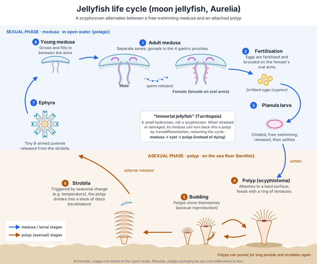
- Adult medusae have separate sexes. Gonads in the gastric pouches make eggs or sperm. Males release sperm into the water.
- Fertilisation and brooding: eggs are fertilised and kept in brood pouches on the female's oral arms, where they develop.
- Planula larva: a tiny, oval, ciliated larva is released, swims and then settles on a hard surface.
- Polyp (scyphistoma): the planula becomes a small polyp with a ring of tentacles. It feeds and can make more polyps by budding (asexual reproduction).
- Strobila: triggered mainly by seasonal temperature change, the polyp divides crosswise into a stack of discs. This is strobilation; one polyp can release many ephyrae (up to about 30 have been reported for Aurelia).
- Ephyra: each disc swims away as a tiny eight-armed juvenile with a rhopalium at the tip of each arm. It grows, the gaps between the arms fill in, and it becomes an adult medusa.
- Gonads (Ovaries / testes): Four horseshoe-shaped reproductive organs, easily seen through the bell. Produce eggs (in females) or sperm (in males). The gametes are released into the gastric pouch and leave through the mouth.
- Subgenital pits (Subgenital porticoes, subgenital ostia): Four round pockets opening on the underside of the bell beneath the gonads. Thin the tissue below each gonad; they are thought to help with respiration and with supplying water to the gonads. They do not release gametes.
- Brood pouches (Brood sacs): Small pockets along the oral arms of females where fertilised eggs develop. Hold fertilised eggs until they develop into swimming planula larvae, which are then released.
- Planula larvae: Tiny, oval, ciliated larvae brooded on the oral arms of females. Swim away, settle on a hard surface and grow into a polyp (the scyphistoma), starting the asexual part of the life cycle.
Immortal jellyfish life cycle (Turritopsis)
The “immortal jellyfish” can turn its adult medusa back into a polyp. In Turritopsis (a small hydrozoan, not a true jellyfish), a damaged, stressed or ageing medusa can shrink into a cyst and transform into a new polyp colony instead of dying. Its specialised cells change into other cell types, a process called transdifferentiation (Piraino et al., 1996, who studied the species then called T. nutricula; the reversing species is now usually named T. dohrnii). In the wild, most still die from predators or disease.
Polyp vs medusa
A polyp is the attached, tube-shaped body form; a medusa is the free-swimming bell. Both have the same two cell layers, a single mouth and tentacles with stinging cells. They are essentially the same body plan turned upside down.
| Feature | Polyp (scyphistoma) | Medusa (jellyfish) |
|---|---|---|
| Shape | Tube or vase | Bell or umbrella |
| Mouth & tentacles | Point up | Point down |
| Lifestyle | Attached to the sea floor | Free-swimming |
| Mesoglea | Thin | Thick jelly |
| Reproduction | Asexual (budding, strobilation) | Sexual (eggs and sperm) |
| Size (Aurelia) | A few millimetres | Usually 25–40 cm across |
Box jellyfish anatomy
Box jellyfish (class Cubozoa) have a cube-shaped bell, tentacles on four corner pedalia, a velarium, and 24 eyes on four rhopalia. Species include the Australian sea wasp Chironex fleckeri, whose sting can kill a person, and the small Caribbean Tripedalia cystophora, the best-studied species for vision.
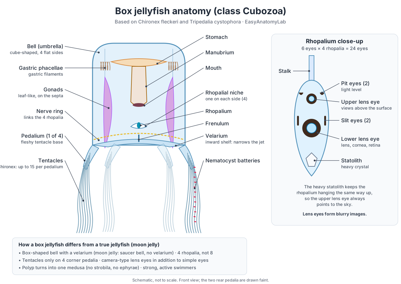
- Pedalia: fleshy, blade-like tentacle bases at the four lower corners of the bell. Chironex has one branched pedalium per corner with up to about 15 tentacles, which can stretch to about 3 m; Tripedalia (“three pedalia”) has three small pedalia per corner, each with a single tentacle.
- Velarium: a shelf of tissue that folds inward around the bell opening. It narrows the opening, so each contraction makes a faster jet. Moon jellyfish have no velarium.
- Rhopalia: four, one in a niche on each flat side of the bell, linked by a nerve ring. Each has six eyes: an upper and a lower lens eye (camera-type eyes with a lens, cornea and retina), two pit eyes and two slit eyes. The lenses are only about 0.1 mm across and form deliberately blurry images (Nilsson et al., 2005). A heavy statolith keeps each rhopalium hanging upright, so the upper lens eyes always look up through the water surface; Tripedalia uses them to find mangrove canopy and stay near its prey (Garm et al., 2011).
- Life cycle: a box jellyfish polyp does not strobilate. Each polyp transforms completely into a single small medusa (Werner, 1983).
Comparing jellyfish: moon, box, lion's mane, immortal and more
“Jellyfish” is a common name for several different groups. True jellyfish are scyphozoans; box jellyfish and many small hydromedusae are separate classes of cnidarians, and comb jellies are not cnidarians at all.
| Animal | Group | Key anatomy |
|---|---|---|
| Moon jellyfish (Aurelia) | Scyphozoa (true jellyfish) | Flat bell 25–40 cm; hundreds of short tentacles; 4 frilly oral arms; 4 horseshoe gonads; 8 rhopalia. |
| Box jellyfish (Chironex, Tripedalia) | Cubozoa | Cube-shaped bell; 4 pedalia; velarium; 24 eyes incl. lens eyes; polyp becomes one medusa. |
| Lion's mane (Cyanea capillata) | Scyphozoa | Largest known jellyfish: bell can exceed 2 m; 8 clusters of many long tentacles, reported up to about 36 m. |
| Cannonball jelly (Stomolophus meleagris) | Scyphozoa (Rhizostomeae) | Firm, rounded bell up to about 20 cm; no marginal tentacles; oral arms fused into a thick central column whose branches carry many small mouth openings (mouthlets). |
| Upside-down jelly (Cassiopea) | Scyphozoa (Rhizostomeae) | Lies bell-down on the sea floor with oral arms up; hosts photosynthetic algae; releases stinging mucus with 'cassiosomes'. |
| Immortal jellyfish (Turritopsis) | Hydrozoa | Tiny (a few mm); has a velum like other hydromedusae; medusa can revert to a polyp. |
| Comb jelly | Ctenophora (not a jellyfish) | 8 rows of beating cilia (comb rows); sticky colloblasts instead of stinging cells; no polyp stage. |
Are comb jellies jellyfish? No. Comb jellies (phylum Ctenophora) look similar but have their own body plan: they swim with eight rows of fused cilia that scatter light into rainbows, catch prey with sticky colloblast cells, and lack cnidocytes and a medusa/polyp cycle.
Simple jellyfish diagram for kids
A simple jellyfish diagram needs only five labels: bell, tentacles, oral arms, mouth and stomach. The bell is the see-through umbrella it swims with. The tentacles around the edge sting and catch tiny food. The oral arms hanging in the middle pass the food to the mouth on the underside. The stomach is in the middle of the bell. Older students can add the four gonads (the four rings you can see through the bell) and the rhopalia (sense organs) around the edge.
Frequently asked questions
What are the main parts of a jellyfish?
A jellyfish (medusa) has a bell made of jelly-like mesoglea between two cell layers, a fringe of tentacles at the bell margin, a mouth on the underside that leads to a stomach and gastric pouches, oral arms hanging from the mouth, gonads in the gastric pouches, a network of canals, a nerve net and small sense organs (rhopalia) around the bell edge.
Does a jellyfish have a brain or a heart?
No. Jellyfish have no brain, heart, blood, bones, lungs or gills. Signals travel through a nerve net, with small clusters of nerve cells in the rhopalia acting as pacemakers. Nutrients are spread by the canals of the gastrovascular system, and oxygen simply diffuses through the thin tissues.
How many stomachs does a jellyfish have?
One. A moon jellyfish has a single central stomach that opens into four gastric pouches. The four pouches are often mistaken for separate stomachs.
How do jellyfish eat and get rid of waste?
Prey is caught by stinging cells on the tentacles and oral arms, passed along the oral arms to the mouth, and digested in the stomach and gastric pouches. Undigested remains leave through the same opening, the mouth, because jellyfish have no anus.
How do jellyfish move?
By contracting the circular swim muscle under the bell, which pushes water out (jet propulsion or 'rowing'). The elastic mesoglea then reopens the bell. Moon jellyfish also gain extra push from a vortex ring that forms under the bell as it refills, which lets them travel about 30% further per stroke.
How many eyes does a jellyfish have?
A moon jellyfish has 16 simple eyes (ocelli): two on each of its eight rhopalia. They detect light but cannot form images. Box jellyfish have 24 eyes, six on each of four rhopalia, including two lens eyes per rhopalium that form blurry images.
What is the difference between a polyp and a medusa?
Both are body forms of cnidarians. A polyp is a tube-shaped, usually attached stage with the mouth and tentacles facing up. A medusa is a free-swimming bell with the mouth and tentacles facing down. In jellyfish, the polyp reproduces asexually and the medusa reproduces sexually.
Is a comb jelly a jellyfish?
No. Comb jellies belong to a separate phylum, Ctenophora. They swim with eight rows of beating cilia (comb rows), catch prey with sticky cells (colloblasts) rather than stinging cells, and have no polyp stage.
Are jellyfish really 95% water?
About that. The bulk of a jellyfish is mesoglea, a collagen gel that is mostly water; the whole animal is commonly given as roughly 95% water.
References
- Brusca, R.C., Moore, W. & Shuster, S.M. (2016). Invertebrates, 3rd ed. Sinauer: chapter on phylum Cnidaria.
- Ruppert, E.E., Fox, R.S. & Barnes, R.D. (2004). Invertebrate Zoology: A Functional Evolutionary Approach, 7th ed. Brooks/Cole: Cnidaria (Scyphozoa, Cubozoa).
- Gemmell, B.J. et al. (2013). Passive energy recapture in jellyfish contributes to propulsive advantage over other metazoans. PNAS 110(44): 17904–17909.
- Nakanishi, N., Hartenstein, V. & Jacobs, D.K. (2009). Development of the rhopalial nervous system in Aurelia sp.1 (Cnidaria, Scyphozoa). Development Genes and Evolution 219: 301–317.
- Pallasdies, F. et al. (2019). From single neurons to behavior in the jellyfish Aurelia aurita. eLife 8: e50084.
- Lucas, C.H. (2001). Reproduction and life history strategies of the common jellyfish, Aurelia aurita, in relation to its ambient environment. Hydrobiologia 451: 229–246.
- Nüchter, T. et al. (2006). Nanosecond-scale kinetics of nematocyst discharge. Current Biology 16(9): R316–R318.
- Nilsson, D.-E. et al. (2005). Advanced optics in a jellyfish eye. Nature 435: 201–205.
- Garm, A., Oskarsson, M. & Nilsson, D.-E. (2011). Box jellyfish use terrestrial visual cues for navigation. Current Biology 21: 798–803.
- Werner, B. (1983). Die Metamorphose des Polypen von Tripedalia cystophora (Cubozoa, Carybdeidae) in die Meduse. Helgoländer Meeresuntersuchungen 36: 257–276.
- Piraino, S., Boero, F., Aeschbach, B. & Schmid, V. (1996). Reversing the life cycle: medusae transforming into polyps and cell transdifferentiation in Turritopsis nutricula. Biological Bulletin 190: 302–312.
- Ames, C.L. et al. (2020). Cassiosomes are stinging-cell structures in the mucus of the upside-down jellyfish Cassiopea xamachana. Communications Biology 3: 67.
- Kramp, P.L. (1961), genus diagnosis of Aurelia, via The Scyphozoan (UC Merced).
More interactive anatomy models
- Human anatomy1,000+ bones, muscles, organs, vessels and nerves from real anatomical data.
- Octopus anatomyThree hearts, a ring-shaped brain, eight arms and a beak, in 3D.
- Squid anatomyPen, ten arms, giant axons, ink sac and three hearts of the longfin squid.
- Squid dissectionStep-by-step guided squid dissection with every structure labeled.
- Fish anatomyExternal and internal anatomy of a bony fish: fins, gills, swim bladder, heart and brain.
- Perch dissectionGuided yellow perch (fish) dissection, from scales to the swim bladder.