

Anatomy of a shark at a glance
A shark is a cartilaginous fish (class Chondrichthyes): its skeleton is cartilage, not bone. Its body has three regions: the head, from the snout to the last gill slit; the trunk, which holds the body cavity; and the tail, behind the cloaca. It breathes with gills that open through separate gill slits, has no swim bladder, stays buoyant with an oily liver, and digests food in a short intestine with a spiral valve.
This page uses the spiny dogfish, Squalus acanthias, as the reference shark. It is a small shark, usually 70–100 cm long, found in temperate seas worldwide, and it is the standard laboratory shark. Its anatomy is typical of sharks, so what you learn here applies to great whites, hammerheads and other species, with the differences listed further down.
Directions: anterior is towards the snout, posterior towards the tail, dorsal the back and ventral the belly. Left and right are always the shark's own left and right.
Shark external anatomy: labeled diagram
The external anatomy of a shark includes the snout, two nostrils, two eyes, a spiracle behind each eye, five gill slits on each side, the mouth, two dorsal fins, paired pectoral and pelvic fins, a heterocercal caudal fin, the lateral line and the cloaca. Males also have two claspers.
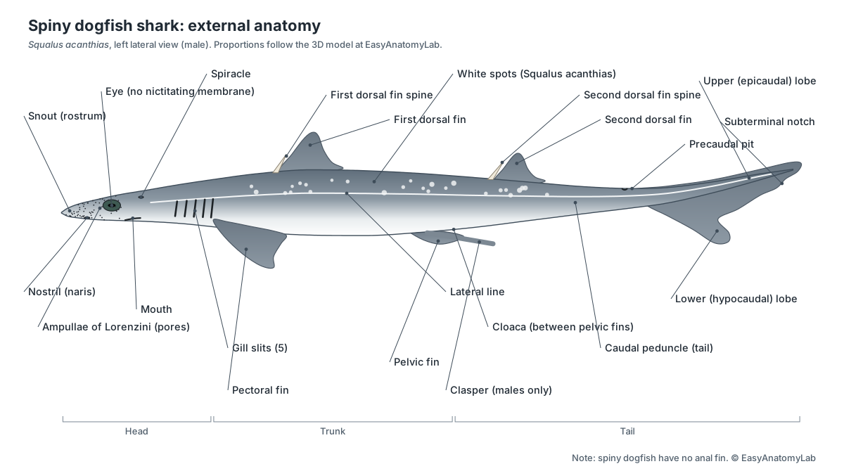
Three features identify a spiny dogfish: a sharp spine in front of each dorsal fin, no anal fin, and small white spots on its grey back. The skin is covered with tiny tooth-like placoid scales (dermal denticles), which make it feel like sandpaper when stroked from tail to head.
| Fin | Paired? | Main job |
|---|---|---|
| Pectoral fins | Yes | Steering, braking and lift |
| Pelvic fins | Yes | Stability; form the claspers in males |
| First and second dorsal fins | No | Stop rolling; each has a spine in Squalus |
| Anal fin | No | Absent in spiny dogfish; present in most other sharks |
| Caudal (tail) fin | No | Thrust; the larger upper lobe also gives lift |
Shark internal anatomy: dogfish shark diagram
Inside a shark, the body cavity is filled mostly by a huge two-lobed liver. Between its lobes lie the J-shaped stomach, the spleen, the pancreas and the valvular intestine with its spiral valve, which ends at the rectum, rectal gland and cloaca. The kidneys lie flat against the roof of the cavity, the gonads sit at its front, and the heart sits in a separate pericardial cavity under the gills.
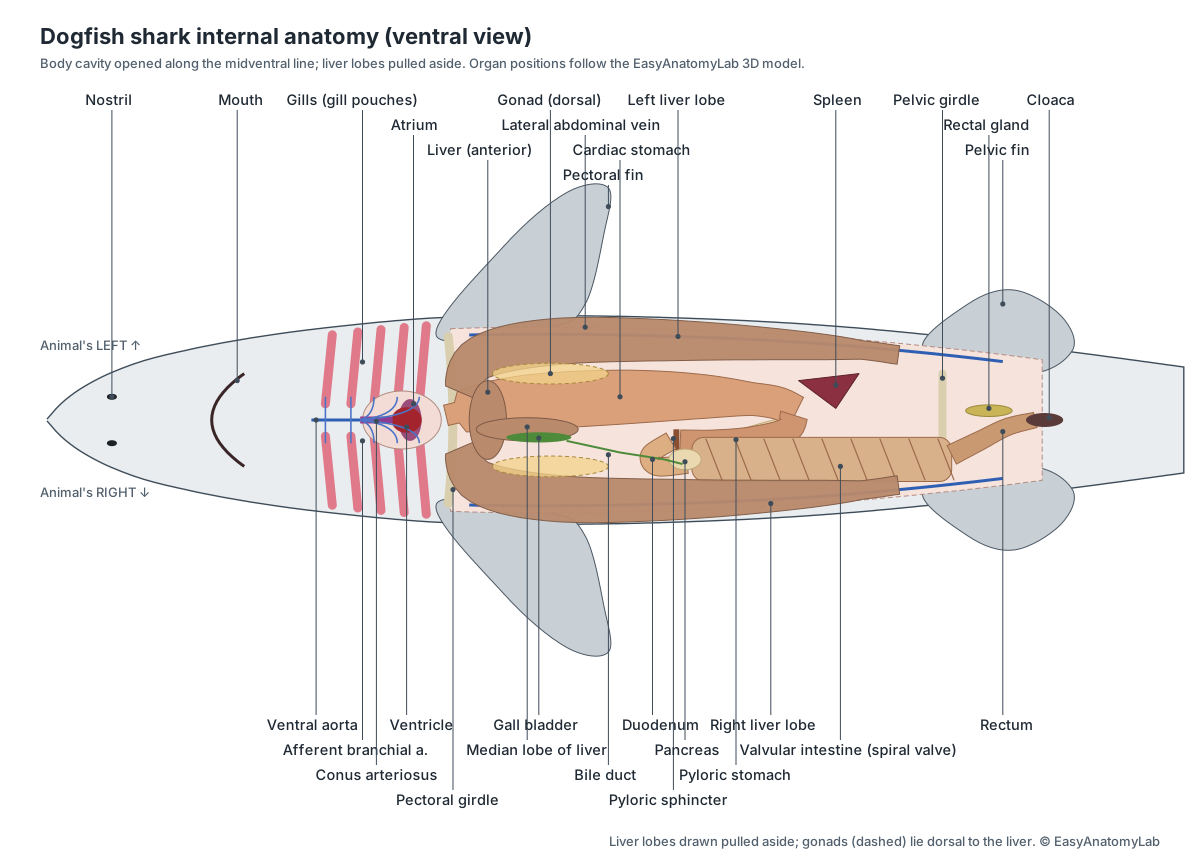
For a step-by-step guide to finding these organs in a real specimen, see our dogfish shark dissection guide.
Shark anatomy labeled: every part and its function
This is the text version of the 3D model: every labeled structure of the spiny dogfish, grouped by body system, with what it does.
External
- Snout (Rostrum (external))
- The pointed front of the head, in front of the eyes and mouth. Streamlines the head and carries dense clusters of electroreceptor pores (ampullae of Lorenzini) and the olfactory organs.
- Head (Cephalic region)
- The region from the snout to the last gill slit, flattened on its dorsal and ventral surfaces. Holds the brain, eyes, nostrils, jaws and gills. The flat underside helps generate lift as the shark swims.
- Trunk (Body wall, skin with placoid scales)
- The spindle-shaped middle of the body, covered by tough skin set with tiny tooth-like placoid scales (dermal denticles). Protects the organs and anchors the swimming muscles. The denticles reduce drag and turbulence as the shark swims.
- Tail (Caudal region, caudal peduncle)
- The muscular, laterally compressed region behind the cloaca that drives the caudal fin. Side-to-side waves of the tail muscles produce thrust for swimming.
- Nostrils (Nares, external nares)
- Paired openings on the underside of the snout that lead into blind olfactory sacs. Water flows in and out of each olfactory sac for smelling. A skin flap divides each opening into an incurrent and an excurrent part.
- First dorsal fin (Anterior dorsal fin)
- The front fin on the back, small and triangular in the spiny dogfish. Keeps the shark stable and stops it rolling.
- Second dorsal fin (Posterior dorsal fin)
- The smaller rear fin on the back, behind the level of the pelvic fins. Adds stability and helps prevent rolling and yawing.
- Dorsal fin spines
- A sharp spine in front of each dorsal fin, the feature that gives the spiny dogfish its name. Defence. A captured dogfish arches its back to drive a spine into an attacker. Glands at the spine base secrete a mild venom.
- Pectoral fins
- Large paired fins behind the head, low on the sides. Steering, braking and lift. They act like an aircraft's wings and help keep the front of the body up.
- Pelvic fins
- Small paired fins on the underside, either side of the cloaca. Stability. In males the inner part of each pelvic fin forms a clasper.
- Caudal fin (Tail fin, heterocercal tail)
- An asymmetrical (heterocercal) tail fin: the vertebral column bends up into the larger upper lobe. The main source of thrust. Because the upper lobe is larger and stiffer, each beat also pushes the tail down and the head up, helping a shark without a swim bladder keep its height.
- Precaudal pit
- A small notch on the top of the caudal peduncle, just in front of the caudal fin. Marks the base of the tail fin. It is an identification feature used in shark keys.
- Cloaca (Vent)
- A shared chamber between the pelvic fins into which the intestine, kidneys and reproductive ducts all open. Releases faeces, urine and eggs or sperm. A urinary papilla (female) or urogenital papilla (male) projects from its dorsal wall.
- Abdominal pores
- A pair of tiny openings beside the cloaca that lead into the body cavity. Connect the body cavity to the outside; their exact role is uncertain, possibly releasing excess coelomic fluid.
Skeletal (cartilage)
- Chondrocranium (Cartilaginous skull, neurocranium)
- A single piece of cartilage that encloses the brain and the organs of smell, sight and hearing. Protects the brain and sense organs and supports the snout. The jaws hang from it by ligaments and the hyomandibular cartilage.
- Palatoquadrate cartilage (Upper jaw)
- The paired cartilage of the upper jaw, bearing the upper teeth. Forms the upper jaw. It is not fused to the skull, so the jaws can protrude when the shark bites.
- Meckel's cartilage (Lower jaw, mandibular cartilage)
- The paired cartilage of the lower jaw, bearing the lower teeth. Forms the lower jaw. It is closed by the powerful adductor mandibulae muscle.
- Hyomandibular cartilage (Hyomandibula)
- The upper element of the hyoid arch, running from the otic capsule to the angle of the jaws. Suspends the jaws from the skull and lets them swing forward during a bite.
- Ceratohyal & basihyal (Lower hyoid arch, 'tongue')
- The lower elements of the hyoid arch, joined in the midline by the plate-like basihyal. Supports the floor of the mouth and the front wall of the first gill pouch. The basihyal forms the shark's simple, immovable 'tongue'.
- Branchial arches (Gill arches)
- Five pairs of jointed cartilage arches that support the gills and pharynx. Support the gill septa and gill rakers, and open and close the pharynx during breathing.
- Vertebral column (Backbone, spine)
- A chain of about 100–110 cartilage vertebrae from the skull into the upper lobe of the tail. Supports the body, protects the spinal cord (in the neural arches) and the caudal vessels (in the haemal arches of the tail).
- Ribs
- Short, slender cartilage rods on the trunk vertebrae. Stiffen the body wall and give attachment to the muscles.
- Pectoral girdle (Scapulocoracoid)
- A U-shaped cartilage bar that crosses the belly behind the gills and rises up each side. Supports the pectoral fins and forms the back wall of the pericardial cavity.
- Pelvic girdle (Puboischiadic bar)
- A straight transverse cartilage bar in the belly wall in front of the cloaca. Supports the pelvic fins and, in males, the claspers.
- Fin cartilages (Basals & radials (pterygiophores))
- Rows of cartilage plates and rods that support the base of each fin. Give the fins a stiff base and transmit muscle forces to the fin.
- Ceratotrichia (Dermal fin rays)
- Fine, flexible fibres of elastin-like protein that stiffen the outer part of every fin. Support the thin outer fin while keeping it flexible.
Muscular
- Epaxial muscles (Dorsal muscle mass)
- The thick block of swimming muscle above the horizontal septum. Bends the body and tail from side to side and lifts the head and tail.
- Hypaxial muscles (Ventral muscle mass)
- The swimming muscle below the horizontal septum, forming the flanks and belly wall. Bends the body sideways and supports the body cavity.
- Myomeres & myosepta (Muscle segments, horizontal septum)
- Zig-zag (W-shaped) blocks of muscle separated by sheets of connective tissue (myosepta). Each myomere pulls on the myosepta and vertebral column. Contracting them in a wave from head to tail produces swimming.
- Adductor mandibulae (Jaw-closing muscle)
- The large muscle at the corner of the mouth that closes the jaws. Snaps the lower jaw up against the upper jaw during a bite.
Nervous
- Olfactory bulbs & tracts
- Paired swellings pressed against the olfactory sacs, joined to the forebrain by long stalks (tracts). Receive the olfactory nerve fibres and process smell before relaying it to the forebrain.
- Telencephalon (Cerebrum, cerebral hemispheres)
- The paired cerebral hemispheres at the front of the brain. Integrates smell with other senses and helps control behaviour.
- Diencephalon (Thalamus region, with epiphysis and hypophysis)
- A small region behind the hemispheres with a thin roof and the pineal stalk. Relays sensory signals and controls hormones: the epiphysis (pineal) senses light and the hypophysis (pituitary) hangs from its floor.
- Optic lobes (Optic tectum, mesencephalon)
- A pair of rounded lobes forming the roof of the midbrain. Process vision and coordinate eye and body movements with what the shark sees.
- Cerebellum (Metencephalon)
- A large oval lobe on top of the hindbrain. Coordinates swimming and balance.
- Medulla oblongata (Myelencephalon)
- The rear of the brain, which narrows into the spinal cord. Controls breathing and heart rate, and is the origin of most cranial nerves (V–X).
- Spinal cord
- The nerve cord running from the medulla through the neural arches of the vertebrae. Carries signals between brain and body and generates the rhythmic swimming pattern.
- Optic nerve (II)
- The thick nerve from each eye to the brain. Carries visual signals from the retina to the optic lobes.
- Trigeminal & facial nerves (V, VII)
- Large nerves from the side of the medulla to the snout, jaws and the ampullae of Lorenzini. The trigeminal carries touch from the head and controls the jaw muscles; branches of the facial (and lateral line) nerves supply the ampullae and head canals.
- Vagus nerve (X) (With lateral line nerve)
- A large nerve from the medulla to the gills, heart and gut, with a long lateral line branch. Controls the gill arches, slows the heart and supplies the stomach. The lateral line branch carries lateral line signals to the brain.
Sensory
- Eyes
- Large, lidless camera-type eyes on the sides of the head. Vision in dim light. A reflective layer behind the retina (tapetum lucidum) bounces light back through the photoreceptors.
- Olfactory sacs (Olfactory organs, nasal capsules)
- Blind pouches behind each nostril, lined with folded sensory tissue (olfactory lamellae). Smell. Odour molecules in the water stimulate receptor cells, which send signals to the olfactory bulbs.
- Inner ear (Membranous labyrinth)
- Fluid-filled sacs and three semicircular canals embedded in the otic capsules of the skull. Balance and hearing. Semicircular canals sense rotation; the sacculus, utriculus and lagena sense gravity, acceleration and low-frequency sound.
- Endolymphatic pores
- Two tiny pores on top of the head, between the spiracles. External openings of the endolymphatic ducts, which lead to the inner ear.
- Lateral line
- A canal of pressure-sensing organs (neuromasts) running along each side from head to tail. Detects water movement, vibrations and pressure changes from prey, predators and obstacles.
- Ampullae of Lorenzini (Electroreceptors)
- Jelly-filled canals that open as pores on the snout and head and end in sensory bulbs. Electroreception: detecting the weak electric fields produced by the muscles and nerves of prey, even prey buried in sand.
Digestive
- Mouth (Oral opening, labial pockets)
- A broad, curved slit on the underside of the head. Takes in food and the water used for breathing. A labial pocket at each corner gives the lower jaw room to move.
- Teeth
- Rows of flat, blade-like teeth with an oblique cusp, similar in both jaws, forming a cutting edge. Cut prey into pieces. Spiny dogfish slice rather than tear.
- Pharynx (Throat)
- The wide chamber behind the mouth, perforated by the spiracles and the five pairs of internal gill slits. Passes food to the esophagus and water out through the gills.
- Esophagus
- A short, muscular tube from the pharynx to the stomach. Moves food into the stomach.
- Cardiac stomach (Proximal limb of stomach)
- The large first limb of the J-shaped stomach. Stores food and begins digestion with acid and enzymes. Internal folds (rugae) let it stretch.
- Pyloric stomach (Distal limb of stomach)
- The narrow second limb of the stomach, which turns forward after the U-bend. Mixes food and passes it to the intestine in small amounts.
- Pyloric sphincter (Pyloric valve)
- A ring of thickened circular muscle at the end of the stomach. Controls the release of partly digested food into the intestine.
- Duodenum
- The short first section of the intestine after the pyloric sphincter. Receives bile from the bile duct and enzymes from the pancreas.
- Valvular intestine (Spiral intestine, ileum)
- A wide, straight section of intestine with spiral markings on its surface (shown here with a window cut in its wall). The main site of digestion and absorption.
- Spiral valve
- A corkscrew-shaped fold of the intestinal lining inside the valvular intestine. Greatly increases the surface for absorption and slows food so it stays longer in contact with the gut lining.
- Rectum (Colon)
- The narrow last part of the intestine. Carries faeces to the cloaca.
- Liver
- A huge, oily liver with long right and left lobes and a small median lobe that holds the gall bladder. Produces bile, stores energy as oil and gives buoyancy. Sharks have no swim bladder; low-density liver oil offsets their weight.
- Gall bladder & bile duct
- A long, green sac along the right edge of the median liver lobe, with its duct. Stores bile and releases it into the intestine to emulsify fats.
- Pancreas
- A pale gland with two lobes: a slender dorsal lobe and a flat oval ventral lobe, joined by a narrow bridge. Makes digestive enzymes (released into the intestine) and the hormones insulin and glucagon.
Circulatory
- Spleen
- A dark red, triangular organ at the bend of the stomach. Makes and stores blood cells and filters the blood. It belongs to the lymphatic system.
- Pericardial cavity (Pericardium)
- A separate, pear-shaped part of the body cavity that holds the heart. Gives the heart room to beat. Its stiff walls help suck blood into the heart (aspiration).
- Sinus venosus
- A thin-walled triangular sac that collects all the blood returning to the heart. Receives venous blood from the ducts of Cuvier and hepatic sinuses and passes it to the atrium.
- Atrium (Auricle)
- A large, thin-walled chamber that bulges out on either side of the ventricle. Fills the ventricle. Valves stop blood flowing back into the sinus venosus.
- Ventricle
- The thick-walled, muscular pumping chamber of the heart. Pumps deoxygenated blood forward into the conus arteriosus and on to the gills.
- Conus arteriosus
- A muscular tube in front of the ventricle, lined with rows of pocket valves. Contracts after the ventricle and its valves prevent backflow, smoothing the pressure delivered to the gills.
- Ventral aorta
- The artery that carries blood forward from the heart under the gills. Distributes deoxygenated blood to the afferent branchial arteries.
- Afferent branchial arteries
- Five pairs of arteries that take blood from the ventral aorta into the gills. Deliver deoxygenated blood to the capillaries of the gill lamellae.
- Efferent branchial arteries
- Four pairs of arteries that collect oxygenated blood from the gills. Carry oxygen-rich blood to the dorsal aorta and, via carotid arteries, to the head.
- Dorsal aorta (Continues as the caudal artery)
- The main artery of the body, running back just under the vertebral column. Carries oxygenated blood to the body and organs.
- Coeliac artery
- The first large branch of the dorsal aorta in the body cavity. Supplies the stomach, liver, pancreas, intestine and gonads.
- Mesenteric arteries (Anterior mesenteric, lienogastric & posterior mesenteric)
- Three arteries from the aorta to the intestine, spleen, stomach and rectal gland. The anterior mesenteric supplies the intestine, the lienogastric the spleen and stomach, and the posterior mesenteric the rectal gland.
- Hepatic portal vein
- A large vein from the gut to the liver. Brings nutrient-rich blood from the gut to the liver for processing before it returns to the heart.
- Cardinal veins & sinuses
- Large, thin-walled veins that return blood from the head (anterior) and body (posterior). Return deoxygenated blood to the ducts of Cuvier.
- Common cardinal veins (Ducts of Cuvier)
- Short, wide veins that carry all body blood into the sides of the sinus venosus. Collect blood from the cardinal and lateral abdominal veins.
- Lateral abdominal veins
- A pair of veins along the inner side wall of the body cavity. Drain the body wall and pelvic fins into the ducts of Cuvier.
Respiratory
- Gill slits (External gill slits)
- Five vertical slits on each side of the head. Outlets for water that has passed over the gills.
- Spiracle
- A small opening behind each eye, the remnant of a first gill slit. Lets water enter the pharynx without passing through the mouth, which matters most in bottom-resting sharks and rays. It contains a small pseudobranch.
- Gills (demibranchs) (Gill filaments, lamellae, interbranchial septa)
- Rows of filaments on the walls of each gill pouch where oxygen enters the blood. Gas exchange. Blood in the lamellae flows opposite to the water (countercurrent exchange), taking up oxygen efficiently.
- Gill rakers
- Short cartilaginous projections on the inner edges of the gill arches. Stop food particles from entering the gill pouches.
Excretory & salt balance
- Kidneys (Opisthonephros (mesonephros))
- Two long, ribbon-like kidneys along the roof of the body cavity, outside the peritoneum. Filter the blood, make urine and keep the body's urea. Sharks keep blood urea high so their body fluids match seawater.
- Archinephric ducts (Wolffian / mesonephric ducts, urinary ducts)
- Ducts along the kidneys that carry urine to the cloaca. In females the archinephric duct is the urinary duct. In males it carries sperm instead, and a separate accessory urinary duct drains the kidney.
- Rectal gland (Digitiform gland)
- A small, finger-shaped gland attached to the dorsal side of the rectum. Salt balance: secretes a concentrated sodium chloride solution into the rectum, removing excess salt taken in with food and seawater.
- Urinary papilla · females only
- A fleshy projection in the cloaca with a pore at its tip that releases urine. Releases urine from the urinary sinus into the cloaca.
Reproductive
- Claspers (Myxopterygia, pterygopodia) · males only
- A pair of rod-like copulatory organs formed from the inner edge of the male's pelvic fins. One clasper is inserted into the female's cloaca. Sperm runs along a groove on the clasper, flushed by seawater from the siphon sac.
- Ovaries · females only
- Paired ovaries holding eggs of various sizes, the largest full of yellow yolk. Produce eggs and sex hormones.
- Ostium tubae (Ostium of the oviducts) · females only
- A single funnel shared by both oviducts that opens into the body cavity. Catches eggs released from the ovaries into the body cavity.
- Oviducts (Müllerian ducts) · females only
- Paired tubes from the ostium to the uteri. Carry eggs from the ostium to the shell glands and uteri. Fertilisation occurs in the upper oviduct.
- Shell glands (Nidamental / oviducal glands) · females only
- A small swelling on each oviduct a short way behind the ostium. Store sperm and secrete a thin membrane that wraps several fertilised eggs together into a 'candle'.
- Uteri · females only
- The enlarged rear part of each oviduct, where the young develop. House the developing pups for a gestation of about 18–24 months, one of the longest of any vertebrate.
- Embryos (pups) with yolk sacs · females only
- Developing pups in the uteri of a pregnant female, each attached to a yolk sac. Grow on the yolk in their yolk sac (lecithotrophy), with no placenta.
- Testes · males only
- Paired, pale, elongated testes at the front of the body cavity. Produce sperm and testosterone.
- Epididymis (With efferent ductules) · males only
- A coiled mass of tubules at the front of each kidney that receives sperm from the testis. Collects and matures sperm.
- Ductus deferens (Vas deferens (archinephric duct)) · males only
- A tightly coiled white tube along the ventral surface of each kidney. Carries sperm towards the cloaca.
- Seminal vesicles · males only
- The straight, widened rear end of each ductus deferens. Store sperm and add secretions that package it.
- Sperm sacs · males only
- Blind pouches branching from the urogenital sinus beside the seminal vesicles. Store sperm before mating.
- Urogenital papilla · males only
- A fleshy projection in the male cloaca through which both sperm and urine leave. Releases sperm and urine from the urogenital sinus.
- Siphon sacs · males only
- Paired muscular sacs under the belly skin in front of the pelvic fins. Fill with seawater before mating and squirt it down the clasper groove to flush sperm into the female.
Shark skeleton: do sharks have bones?
No. A shark's skeleton is made entirely of cartilage. Some parts, such as the vertebrae and jaws, are strengthened by a layer of calcified tiles called tesserae, so they feel hard, but sharks have no true bone. Cartilage is lighter than bone, which helps a fish without a swim bladder stay afloat.
- Chondrocranium: Protects the brain and sense organs and supports the snout. The jaws hang from it by ligaments and the hyomandibular cartilage.
- Palatoquadrate cartilage: Forms the upper jaw. It is not fused to the skull, so the jaws can protrude when the shark bites.
- Meckel's cartilage: Forms the lower jaw. It is closed by the powerful adductor mandibulae muscle.
- Hyomandibular cartilage: Suspends the jaws from the skull and lets them swing forward during a bite.
- Branchial arches: Support the gill septa and gill rakers, and open and close the pharynx during breathing.
- Vertebral column: Supports the body, protects the spinal cord (in the neural arches) and the caudal vessels (in the haemal arches of the tail).
- Pectoral girdle: Supports the pectoral fins and forms the back wall of the pericardial cavity.
- Pelvic girdle: Supports the pelvic fins and, in males, the claspers.
- Ceratotrichia: Support the thin outer fin while keeping it flexible.
Because cartilage rarely fossilises, most fossil sharks are known only from their teeth, spines and vertebrae.
Shark tooth anatomy
Shark teeth are modified placoid scales: a core of dentine covered by a hard enamel-like layer, with a pulp cavity, set in rows on the jaw cartilages rather than in sockets. New teeth form on the inside of the jaw and move outward to replace worn ones throughout life.
Spiny dogfish teeth are small and similar in both jaws, each with one oblique cusp, so together they form a continuous cutting edge for slicing prey. Other sharks have teeth shaped for their diet: serrated triangles in great white sharks, hooked cockscomb-shaped teeth in tiger sharks, and hundreds of tiny, non-functional teeth in the filter-feeding whale shark.
Shark muscle anatomy
Most of a shark's body is swimming muscle arranged in zig-zag blocks called myomeres, separated by sheets of connective tissue (myosepta). A horizontal septum divides the epaxial muscles above from the hypaxial muscles below. Waves of contraction along the myomeres bend the body and drive the tail.
- Epaxial muscles: Bends the body and tail from side to side and lifts the head and tail.
- Hypaxial muscles: Bends the body sideways and supports the body cavity.
- Myomeres & myosepta: Each myomere pulls on the myosepta and vertebral column. Contracting them in a wave from head to tail produces swimming.
- Adductor mandibulae: Snaps the lower jaw up against the upper jaw during a bite.
Shark heart anatomy and circulation
A shark has one heart with four chambers in a row: the sinus venosus, atrium, ventricle and conus arteriosus. It sits in the pericardial cavity just in front of the pectoral girdle and pumps only oxygen-poor blood, forward to the gills.
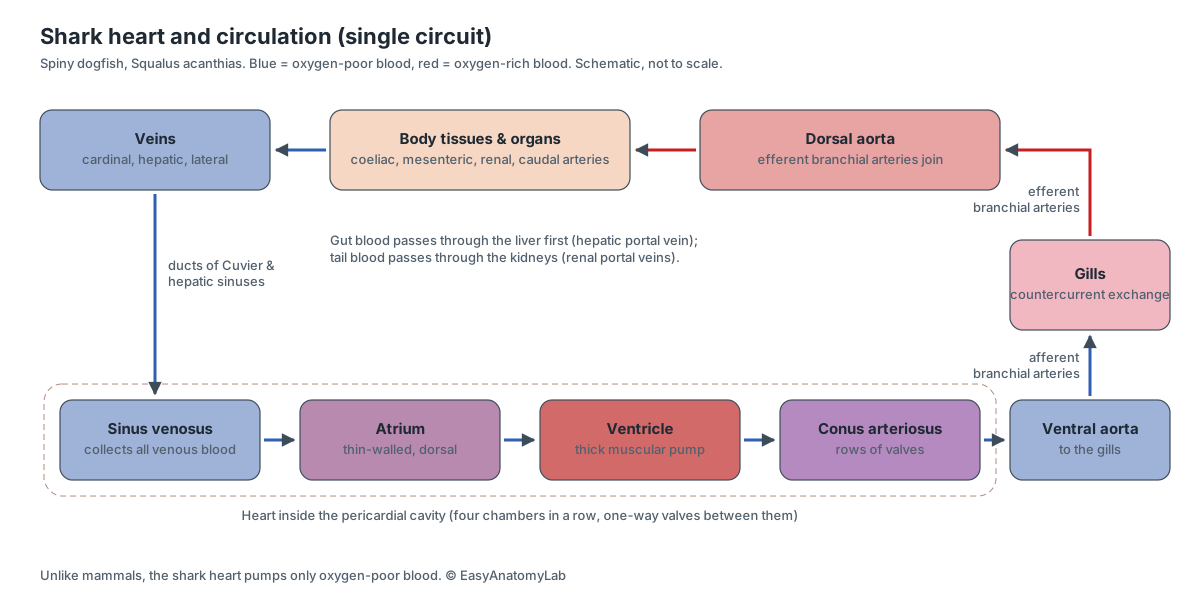
Blood leaves the conus through the ventral aorta, passes up five pairs of afferent branchial arteries into the gills, picks up oxygen, and is collected by efferent branchial arteries into the dorsal aorta, which supplies the body. Veins and large thin-walled sinuses return it to the sinus venosus. This is a single circuit, unlike the double circulation of mammals.
Shark gills and breathing
Sharks breathe with gills inside five pairs of gill pouches. Water enters the mouth (and spiracles), flows over the gill filaments, and leaves through the gill slits. In the gill lamellae, blood flows opposite to the water (countercurrent exchange), which extracts oxygen efficiently.
In spiny dogfish, a half-gill (hemibranch) lines the hyoid arch and full gills (holobranchs) sit on gill arches 1–4, giving nine demibranchs per side. Gill rakers on the inner edges of the arches keep food out of the gill pouches. Dogfish can pump water with their mouth and throat, so, unlike some open-ocean sharks, they do not have to swim to breathe.
Shark digestive system: liver, stomach and spiral valve
Food passes from the mouth and pharynx down the short esophagus into the J-shaped stomach, through the pyloric sphincter and duodenum into the valvular intestine, then out through the rectum and cloaca. The liver and pancreas add bile and enzymes in the duodenum.
- Liver: Produces bile, stores energy as oil and gives buoyancy. Sharks have no swim bladder; low-density liver oil offsets their weight.
- Cardiac stomach: Stores food and begins digestion with acid and enzymes. Internal folds (rugae) let it stretch.
- Pyloric stomach: Mixes food and passes it to the intestine in small amounts.
- Pancreas: Makes digestive enzymes (released into the intestine) and the hormones insulin and glucagon.
- Valvular intestine: The main site of digestion and absorption.
- Spiral valve: Greatly increases the surface for absorption and slows food so it stays longer in contact with the gut lining.
The spiral valve lets a short intestine absorb food efficiently. CT scans of 22 shark families (Leigh et al., 2021) showed that its spiral shape also makes fluid flow mostly in one direction, like the valve Nikola Tesla designed in 1920.
Salt and water balance: kidneys and rectal gland
Sharks keep their body fluids about as concentrated as seawater by retaining urea and TMAO in the blood, rather than by drinking and pumping out salt like bony fish. The long, ribbon-like kidneys conserve urea, and the rectal gland secretes excess sodium chloride into the rectum. Burger and Hess discovered this gland's salt-secreting role in the spiny dogfish in 1960.
Shark senses and brain
Sharks combine smell, hearing, vision, the lateral line and electroreception to find prey. The ampullae of Lorenzini detect weak electric fields from prey, the lateral line senses water movement, and large olfactory sacs give an acute sense of smell.
- Olfactory bulbs & tracts: Receive the olfactory nerve fibres and process smell before relaying it to the forebrain.
- Telencephalon: Integrates smell with other senses and helps control behaviour.
- Diencephalon: Relays sensory signals and controls hormones: the epiphysis (pineal) senses light and the hypophysis (pituitary) hangs from its floor.
- Optic lobes: Process vision and coordinate eye and body movements with what the shark sees.
- Cerebellum: Coordinates swimming and balance.
- Medulla oblongata: Controls breathing and heart rate, and is the origin of most cranial nerves (V–X).
Spiny dogfish eyes have no movable eyelids and no nictitating membrane (third eyelid). Requiem and other ground sharks, such as tiger, bull and lemon sharks, close a nictitating membrane over the eye when they bite.
Male vs female shark anatomy: reproductive system
You can tell male and female sharks apart from the outside: males have two claspers on the inner edges of the pelvic fins, and females do not. Fertilisation is internal. Inside, both sexes have paired gonads at the front of the body cavity, but their ducts differ.
| Male shark anatomy | Female shark anatomy | |
|---|---|---|
| Gonads | Paired testes | Paired ovaries with large yolky eggs |
| Ducts | Epididymis, coiled ductus deferens, seminal vesicle, sperm sac | Single ostium, paired oviducts, shell glands, uteri |
| External | Claspers with a groove; siphon sacs under the belly skin | No claspers |
| Cloaca | Urogenital papilla (sperm and urine) | Urinary papilla, with an oviduct opening on each side |
| Role | Inserts a clasper into the female's cloaca | Carries the young for about 18–24 months |
Spiny dogfish are aplacental viviparous (older texts say ovoviviparous): the pups develop inside the uteri, living on the yolk in their yolk sacs, and are born alive. Gestation lasts about 18–24 months, one of the longest known in any vertebrate, and litters range from 1 to about 20 pups, each 18–30 cm long at birth (FishBase). Switch the 3D model to Female to see the uteri and pups.
Great white shark anatomy vs the spiny dogfish
The great white shark (Carcharodon carcharias) has the same basic organs as a dogfish, but it is built for fast, open-water hunting of large prey. It is a regional endotherm: networks of blood vessels (retia mirabilia) keep its swimming muscles, eyes and brain warmer than the water, and its red swimming muscle commonly runs several degrees above water temperature.
- A nearly symmetrical, crescent-shaped (lunate) tail with keels on the caudal peduncle, instead of the dogfish's heterocercal tail.
- Large, serrated, triangular teeth for cutting flesh; no dorsal fin spines; an anal fin is present.
- No nictitating membrane: it rolls its eyes backward when it strikes.
- Spiracles are tiny or absent, and it relies on swimming to ventilate its gills (ram ventilation).
Hammerhead shark anatomy: the cephalofoil
A hammerhead shark's wide, flattened head is called the cephalofoil. It spreads the eyes, nostrils and ampullae of Lorenzini across a wide surface. Juvenile scalloped hammerheads search a wider strip of seabed for buried prey than sandbar sharks with the same electrosensitivity (Kajiura & Holland, 2002), and the widely spaced eyes give hammerheads larger binocular overlap than requiem sharks: 48° in the winghead and 34° in the scalloped hammerhead, versus about 10° in the lemon shark (McComb et al., 2009).
Inside, hammerheads have the same organ systems as the dogfish. Unlike the dogfish, they nourish their young through a yolk-sac placenta.
Whale shark anatomy: filter feeding
The whale shark (Rhincodon typus), the largest living fish, feeds on plankton. Its mouth is at the front of the head (terminal), not underneath, and its teeth are tiny and play no part in feeding. Instead, water passes through filter pads that block the pharynx in front of the gills: Motta et al. (2010) described 20 pads covered with a mesh of openings averaging about 1.2 mm, which they suggested work by cross-flow filtration so they do not clog.
Basking shark mouth and gill rakers
The basking shark (Cetorhinus maximus), the second-largest fish, is also a filter feeder. It swims slowly with its huge mouth open, and water flows out through enormous gill slits that almost encircle the head. Long, bristle-like gill rakers on the gill arches strain out zooplankton. These are the same structures that, in the dogfish, are short knobs that simply keep food out of the gill pouches.
Shark species anatomy compared
Every shark shares the cartilaginous skeleton, gills, spiral valve, rectal gland and ampullae of Lorenzini. This table lists the anatomical features that set common species apart from the spiny dogfish.
| Species | Distinctive anatomy |
|---|---|
| Spiny dogfish (Squalus acanthias) | Dorsal fin spines, no anal fin, white spots, no nictitating membrane; pups grow on yolk for 18–24 months |
| Great white (Carcharodon carcharias) | Warm swimming muscles (regional endothermy), lunate tail with keels, serrated triangular teeth |
| Hammerheads (Sphyrna) | Cephalofoil with widely spaced eyes and electroreceptors; yolk-sac placenta |
| Whale shark (Rhincodon typus) | Terminal mouth, filter pads over the gills, tiny teeth; largest fish |
| Basking shark (Cetorhinus maximus) | Huge gill slits and long bristle-like gill rakers for passive filter feeding |
| Tiger shark (Galeocerdo cuvier) | Cockscomb-shaped serrated teeth that can cut turtle shells; nictitating membrane; striped juveniles |
| Bull shark (Carcharhinus leucas) | Kidneys and rectal gland adjust so it can live in fresh water; small eyes; placenta |
| Lemon shark (Negaprion brevirostris) | Two dorsal fins of nearly equal size, yellowish skin; can rest on the bottom and pump water over its gills |
| Thresher sharks (Alopias) | Upper tail lobe about as long as the body, used to slap and stun schooling fish (Oliver et al., 2013) |
| Goblin shark (Mitsukurina owstoni) | Long flat snout; jaws shoot forward up to about 9% of body length (Nakaya et al., 2016) |
| Nurse shark (Ginglymostoma cirratum) | Barbels by the nostrils, small mouth for suction feeding, large spiracles; rests on the bottom |
| Leopard shark (Triakis semifasciata) | Slender houndshark with dark saddles and spots; anal fin present and no fin spines |
Shark anatomy worksheet: test yourself
Use the 3D model or the diagrams above to answer these. Answers are on the dissection page.
- Which external feature tells you a shark is male?
- How many gill slits does a spiny dogfish have on each side?
- Name the four chambers of the shark heart in order.
- Which organ gives a shark buoyancy instead of a swim bladder?
- What structure inside the intestine increases its surface area?
- Which gland removes excess salt?
- Name two sense organs that sharks have and humans do not.
Full lab questions with an answer key are in the shark dissection guide.
Frequently asked questions
Do sharks have bones?
No. A shark's skeleton is made entirely of cartilage. Parts of it are strengthened by a surface layer of calcified tiles (tesserae) and calcified vertebrae, so it can feel hard, but it is not bone. The only hard tissues are the teeth, scales and spines, which are made of dentine.
How many gill slits does a shark have?
Most sharks, including the spiny dogfish, have five pairs of gill slits. A few primitive species, such as sixgill and sevengill sharks, have six or seven pairs. Sharks also have a spiracle behind each eye, the remnant of an extra gill slit.
How many hearts does a shark have?
One. A shark's heart has four chambers in a row: sinus venosus, atrium, ventricle and conus arteriosus. It pumps blood in a single circuit: to the gills, then to the body, then back to the heart.
How do sharks stay afloat without a swim bladder?
Sharks have no swim bladder. They get lift from a large, oil-rich liver (about 10–12% of body weight in spiny dogfish), a light cartilage skeleton, and lift from the pectoral fins and the heterocercal tail as they swim.
What is the spiral valve in a shark?
The spiral valve is a corkscrew-shaped fold inside the shark's intestine (the valvular intestine). It greatly increases the area for absorbing food and slows food down, so a short intestine can digest efficiently.
What does the rectal gland do?
The rectal gland removes excess salt. It secretes a concentrated sodium chloride solution into the rectum, which leaves through the cloaca. It was discovered in the spiny dogfish in 1960.
How can you tell a male shark from a female?
Males have a pair of claspers, rod-like extensions of the pelvic fins used to transfer sperm. Females have no claspers. Inside, males have testes and coiled sperm ducts; females have ovaries, oviducts and uteri.
Do sharks lay eggs or give birth?
Both, depending on the species. The spiny dogfish gives birth to live young after an 18–24 month pregnancy; the pups grow on yolk inside the mother (aplacental viviparity). Some sharks, like hammerheads and bull sharks, have a placenta, and others, such as catsharks and horn sharks, lay egg cases.
What are the ampullae of Lorenzini?
They are electroreceptors: jelly-filled canals that open as pores on the shark's snout and end in sensory bulbs. They detect the tiny electric fields made by the muscles of prey, even when the prey is hidden in sand.
References
- Griffin, L. E. (1922). A Guide for the Dissection of the Dogfish (Squalus acanthias), 3rd ed. Portland: Reed College. Project Gutenberg edition.
- Froese, R. & Pauly, D. (eds). Squalus acanthias, Picked dogfish. FishBase.
- Florida Museum of Natural History. Shark biology.
- Burger, J. W. & Hess, W. N. (1960). Function of the rectal gland in the spiny dogfish. Science 131, 670–671.
- Bradley, S. E. & Burger, J. W. (1965). Dynamics of sulfabromophthalein secretion by the liver of the spiny dogfish, Squalus acanthias. Bulletin of the Mount Desert Island Biological Laboratory.
- Leigh, S. C., Summers, A. P., Hoffmann, S. L. & German, D. P. (2021). Shark spiral intestines may operate as Tesla valves. Proceedings of the Royal Society B 288, 20211359.
- Kajiura, S. M. & Holland, K. N. (2002). Electroreception in juvenile scalloped hammerhead and sandbar sharks. Journal of Experimental Biology 205, 3609–3621.
- McComb, D. M., Tricas, T. C. & Kajiura, S. M. (2009). Enhanced visual fields in hammerhead sharks. Journal of Experimental Biology 212, 4010–4018.
- Motta, P. J. et al. (2010). Feeding anatomy, filter-feeding rate, and diet of whale sharks Rhincodon typus during surface ram filter feeding off the Yucatan Peninsula, Mexico. Zoology 113, 199–212.
- Oliver, S. P. et al. (2013). Thresher sharks use tail-slaps as a hunting strategy. PLoS ONE 8, e67380.
- Nakaya, K. et al. (2016). Slingshot feeding of the goblin shark Mitsukurina owstoni. Scientific Reports 6, 27786.
- Martin, R. A. ReefQuest Centre for Shark Research: Fuel economy of the white shark (regional endothermy).
More interactive anatomy models
- Shark dissectionGuided spiny dogfish shark dissection with labeled internal anatomy.
- Human anatomy1,000+ bones, muscles, organs, vessels and nerves from real anatomical data.
- Octopus anatomyThree hearts, a ring-shaped brain, eight arms and a beak, in 3D.
- Squid anatomyPen, ten arms, giant axons, ink sac and three hearts of the longfin squid.
- Squid dissectionStep-by-step guided squid dissection with every structure labeled.
- Fish anatomyExternal and internal anatomy of a bony fish: fins, gills, swim bladder, heart and brain.