

Before you start: materials and safety
You need a preserved dogfish, a dissecting tray, basic tools and protective equipment. Double-injected specimens (arteries filled with red latex, veins with blue) make the blood vessels much easier to trace.
Materials
- Preserved spiny dogfish (Squalus acanthias), ideally a double-injected specimen (arteries red, veins blue)
- Large dissecting tray with wax or a cutting mat
- Scalpel, sharp scissors (one blunt-tipped blade is best), forceps and a blunt probe
- Bone or heavy scissors, or a strong scalpel, for the pectoral girdle and cartilage
- Dissecting pins, string or rubber bands to hold the body wall open
- Hand lens or dissecting microscope (for denticles, gill lamellae and teeth)
- Nitrile gloves, safety glasses and a lab coat or apron
- Paper towels, a wash bottle of water and a labeled bag for storing the specimen
Safety
- Wear gloves and eye protection. Specimens are stored in preservative; work in a ventilated room and rinse the shark in water before you start.
- Watch the dorsal fin spines: they are sharp enough to pierce gloves even in a preserved shark.
- Cut away from your hands and from your lab partner. Lift the body wall with forceps before cutting so you do not slice the organs underneath.
- Never eat or drink in the lab, and wash your hands when you finish.
- Dispose of tissue and sharps as your teacher directs.
Dogfish shark dissection labeled diagram
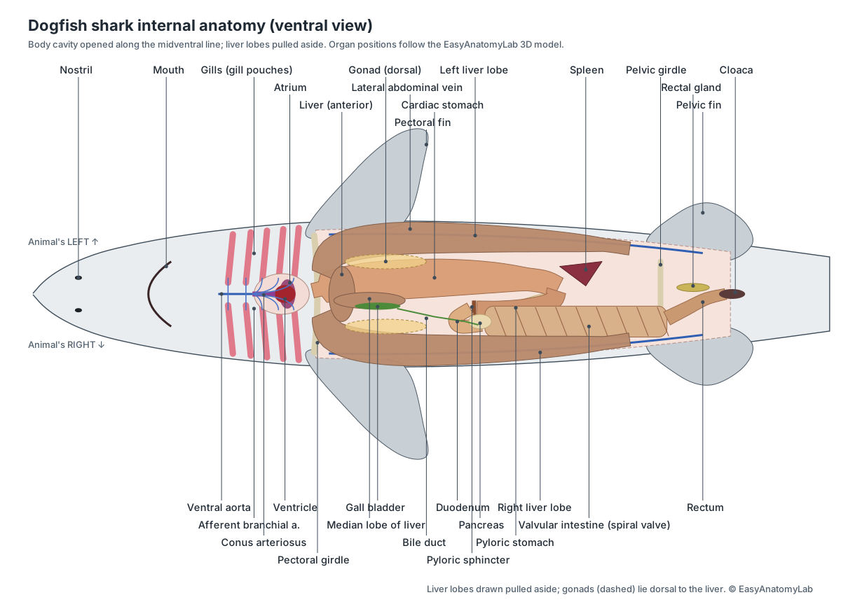
This is what you should see after opening the body cavity and pulling the liver lobes aside: the stomach, spleen, pancreas and valvular intestine between the lobes, the heart in the throat, and the rectal gland near the cloaca.
Spiny dogfish shark dissection: the procedure
The order below follows classic dogfish lab manuals: external anatomy first, then the body cavity and digestive organs, the kidneys and reproductive organs, the heart and gills through the throat, and finally the brain from the top of the head.
1. External anatomy
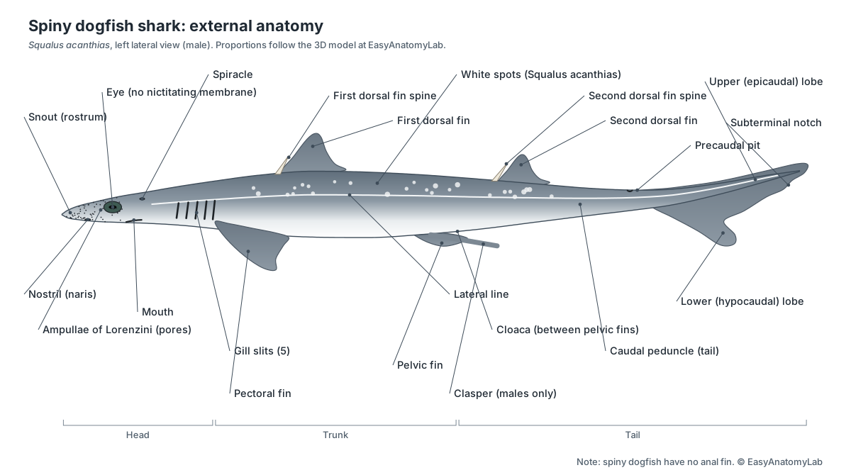
- Rinse the shark and lay it on its side in the tray. Identify the head, trunk and tail regions.
- Find the two dorsal fins and the spine in front of each, the paired pectoral and pelvic fins, and the heterocercal caudal fin. Note: Squalus has no anal fin.
- Locate the eyes, the spiracle behind each eye, and the five gill slits on each side.
- Trace the pale lateral line along the side of the body from head to tail.
- Stroke the skin from head to tail and back. The roughness is caused by placoid scales (dermal denticles). Scrape a few onto a slide and view them under the microscope.
Caution: The dorsal spines are sharp. Keep your hands away from the front edge of the dorsal fins.
Identify: Snout, Eyes, Spiracle, Gill slits, First dorsal fin, Dorsal fin spines, Second dorsal fin, Pectoral fins, Pelvic fins, Caudal fin, Precaudal pit, Lateral line, Trunk.
2. Ventral surface: sexing the shark
- Turn the shark on its back. Find the cloaca between the pelvic fins.
- Check the inner edge of the pelvic fins. Rod-like claspers mean the shark is male; no claspers means it is female. (Use the Female/Male toggle on the model to compare.)
- With a probe, find the small abdominal pore on each side of the cloaca.
- Look into the cloaca for the urinary papilla (female) or urogenital papilla (male).
Identify: Cloaca, Claspers, Pelvic fins, Abdominal pores.
3. Head: nostrils and sensory pores
- On the underside of the snout, find the two nostrils. Probe one: it ends blindly in an olfactory sac and does not connect to the mouth.
- Look for the dark pores of the ampullae of Lorenzini scattered over the snout. Squeeze gently near a cluster to see jelly come out of a pore.
- On top of the head, between the spiracles, find the two tiny endolymphatic pores.
Identify: Nostrils, Ampullae of Lorenzini, Endolymphatic pores, Lateral line, Olfactory sacs.
4. Mouth, teeth and jaws
- Open the mouth and examine the teeth with a hand lens. Note the rows of replacement teeth behind the functional row.
- Feel the upper jaw (palatoquadrate) and lower jaw (Meckel's cartilage). They are not fused to the skull.
- Cut through the corner of the mouth on one side, back through the gill slits, to open the pharynx. Find the gill rakers on the inner edges of the gill arches and the internal gill slits.
Caution: Cut through the corner of the jaw with heavy scissors, keeping your fingers clear of the teeth.
Identify: Mouth, Teeth, Palatoquadrate cartilage, Meckel's cartilage, Pharynx, Gill rakers, Branchial arches.
5. Opening the body cavity
- With the shark on its back, lift the body wall with forceps just in front of the cloaca and make a small cut slightly to one side of the midline.
- Extend the cut forward to the pectoral girdle, keeping the scissor tips up so you do not damage the organs. Avoid cutting through the pectoral girdle for now.
- Make two cross-cuts on each side, one just behind the pectoral fins and one just in front of the pelvic fins, and fold back the flaps. Pin them open.
- Note the lateral abdominal veins on the inside of the body wall, and the shiny peritoneum lining the cavity. Rinse out any preservative.
Caution: Keep the scissor tips pointing up. Cutting too deep will damage the liver and intestine.
Identify: Liver, Lateral abdominal veins, Pectoral girdle, Pelvic girdle.
6. Liver and gall bladder
- The large, oily liver fills much of the cavity. Identify its long right and left lobes and the small median lobe between them.
- Find the green gall bladder along the right edge of the median lobe and follow the bile duct towards the intestine.
- Lift the liver to one side to see the organs underneath. If the specimen is a mature female, look for the single ostium on the front of the liver.
Identify: Liver, Gall bladder & bile duct, Hepatic portal vein, Ostium tubae.
7. Digestive tract
- Follow the gut from front to back: the short esophagus, the large cardiac stomach, the U-bend and the narrower pyloric stomach.
- Feel the firm pyloric sphincter at the end of the stomach, then the short duodenum that turns right and back.
- Find the pancreas: a slender dorsal lobe along the stomach and a flat ventral lobe on the duodenum.
- Find the dark, triangular spleen at the bend of the stomach and the wide valvular intestine with spiral lines on its surface.
Identify: Esophagus, Cardiac stomach, Pyloric stomach, Pyloric sphincter, Duodenum, Pancreas, Spleen, Valvular intestine, Rectum.
8. Stomach contents and the spiral valve
- Cut open the cardiac stomach along its length and rinse out the contents. Record any prey you find (fish, squid, crabs). Note the folds (rugae) of the lining.
- Cut along one side of the valvular intestine and fold back the wall to expose the spiral valve. Count the turns and note whether the spiral changes direction.
- Find the rectal gland on the dorsal side of the rectum. Cut across it to see its central duct.
Identify: Cardiac stomach, Spiral valve, Valvular intestine, Rectum, Rectal gland.
9. Kidneys and salt balance
- Push the digestive organs aside (or remove the gut after cutting the esophagus and rectum, if your teacher allows).
- Find the long, dark kidneys against the roof of the body cavity. Peel back the peritoneum: the kidneys lie outside it.
- Find the archinephric (urinary) duct along the medial edge of each kidney and the dorsal aorta between the kidneys.
- Review the rectal gland: it, not the kidney, removes most excess salt.
Identify: Kidneys, Archinephric ducts, Dorsal aorta, Rectal gland, Cardinal veins & sinuses.
10. Female reproductive system
- Switch the model to Female. Find the paired ovaries dorsal to the front of the liver; large yellow eggs show the female is mature.
- Find the single ostium on the front of the liver and follow each oviduct over the liver and back along the body wall.
- Find the small shell gland on each oviduct, then the enlarged uteri at the rear of the cavity.
- In a pregnant dogfish, open one uterus carefully. You may find a 'candle' of eggs or pups, each with a yolk sac. Measure the pups and count them.
Identify: Ovaries, Ostium tubae, Oviducts, Shell glands, Uteri, Embryos (pups) with yolk sacs, Urinary papilla.
11. Male reproductive system
- Switch the model to Male. Find the paired testes dorsal to the front of the liver.
- Follow the tightly coiled ductus deferens along the ventral surface of each kidney, from the epididymis to the straight seminal vesicle.
- Find the sperm sacs near the cloaca and the urogenital papilla inside it.
- Reflect the belly skin in front of the pelvic fins to find the siphon sacs, and trace a clasper groove to its opening.
Identify: Testes, Epididymis, Ductus deferens, Seminal vesicles, Sperm sacs, Urogenital papilla, Siphon sacs, Claspers.
12. Heart and pericardial cavity
- Extend the midventral cut forward through the pectoral girdle (use heavy scissors) and through the throat muscles towards the lower jaw.
- Open the pericardial cavity, a separate chamber in front of the transverse septum. Identify the ventricle (ventral), the atrium (dorsal, bulging at the sides), the sinus venosus (dorsal and posterior) and the conus arteriosus (anterior).
- Follow the ventral aorta forward from the conus and find the afferent branchial arteries branching to the gill arches.
- Find the ducts of Cuvier (common cardinal veins) entering the sinus venosus from each side.
Caution: The pectoral girdle is tough cartilage. Cut slowly and away from your body.
Identify: Pericardial cavity, Ventricle, Atrium, Sinus venosus, Conus arteriosus, Ventral aorta, Afferent branchial arteries, Common cardinal veins.
13. Gills and branchial circulation
- Cut through the gill slits on one side and open the gill pouches. Count the demibranchs: one on the hyoid arch and two on each of gill arches 1–4.
- Remove one gill arch and look at it under a lens: filaments, lamellae, the cartilage arch, gill rays and gill rakers.
- Find the efferent branchial arteries collecting blood from the gills and joining to form the dorsal aorta.
Identify: Gills (demibranchs), Gill rakers, Branchial arches, Afferent branchial arteries, Efferent branchial arteries, Dorsal aorta, Pharynx, Spiracle.
14. Musculature and skeleton
- Remove a strip of skin from the side of the tail. Note the zig-zag myomeres separated by myosepta.
- Cut across the tail behind the cloaca. Find the epaxial and hypaxial muscles, the horizontal septum, the vertebral column with the spinal cord above and the caudal artery and vein below.
- Notice that the skeleton is cartilage, not bone: it can be cut with a scalpel.
Identify: Myomeres & myosepta, Epaxial muscles, Hypaxial muscles, Vertebral column, Spinal cord, Ribs, Fin cartilages, Ceratotrichia.
15. Brain and cranial nerves
- With the shark on its belly, remove the skin from the top of the head between the eyes and spiracles.
- Shave thin slices of cartilage off the top of the chondrocranium until the brain shows through. Work slowly: the brain is soft.
- Identify, from front to back: olfactory bulbs and tracts, telencephalon, diencephalon, optic lobes, cerebellum and medulla oblongata.
- Find the optic nerves running to the eyes and the large trigeminal-facial and vagus nerves. If time allows, dissect out the inner ear beside the medulla.
Caution: Remove cartilage in thin shavings. Deep cuts will destroy the brain.
Identify: Chondrocranium, Olfactory bulbs & tracts, Telencephalon, Diencephalon, Optic lobes, Cerebellum, Medulla oblongata, Optic nerve (II), Trigeminal & facial nerves (V, VII), Vagus nerve (X), Inner ear, Spinal cord.
Male vs female: what changes in the dissection
Only the reproductive steps differ. Check the pelvic fins first: claspers mean a male.
- Female: ovaries with yellow eggs at the front of the cavity, one ostium on the front of the liver, paired oviducts with small shell glands, and two uteri at the back. Pregnant females may hold a "candle" of eggs or pups with yolk sacs.
- Male: pale testes at the front of the cavity, a tightly coiled ductus deferens on each kidney, seminal vesicles and sperm sacs near the cloaca, siphon sacs under the belly skin, and the claspers.
Many lab dogfish are pregnant females. If yours is, measure and count the pups and record whether each still has a yolk sac: you are looking at one stage of a pregnancy that lasts nearly two years.
Shark dissection lab questions and answer key
Answer these during or after the dissection. Click a question to reveal the answer.
How did you determine the sex of your shark?
By checking for claspers on the inner edge of the pelvic fins. Males have claspers; females do not.
How many gill slits does a spiny dogfish have on each side, and what is the spiracle?
Five gill slits on each side. The spiracle, behind each eye, is a remnant of an extra gill slit that lets water into the pharynx.
Name two features that identify Squalus acanthias as a spiny dogfish.
A spine in front of each of its two dorsal fins, and no anal fin. It also has small white spots on its back.
Why is the shark's liver so large, and how does it help the shark?
It stores oil (including squalene) that is less dense than seawater. This gives buoyancy, because sharks have no swim bladder, and it also stores energy.
What is the function of the spiral valve?
It increases the surface area for absorbing digested food and slows the passage of food through the short intestine.
What does the rectal gland do?
It secretes a concentrated salt (NaCl) solution into the rectum, removing excess salt from the blood.
List the four chambers of the shark heart in the order blood flows through them.
Sinus venosus → atrium → ventricle → conus arteriosus. Blood then goes to the ventral aorta and the gills.
Is the blood in the ventral aorta oxygen-rich or oxygen-poor? Explain.
Oxygen-poor. The shark has a single circuit: the heart pumps deoxygenated blood forward to the gills, and only blood leaving the gills (efferent arteries, dorsal aorta) is oxygen-rich.
What is the difference between a holobranch and a hemibranch?
A holobranch has gill filaments on both faces of the septum; a hemibranch (demibranch) has them on one face only. Squalus has a hemibranch on the hyoid arch and holobranchs on arches 1–4.
What were the stomach contents of your shark, and what do they tell you about its diet?
Answers vary. Common finds are fish, squid, crabs and shrimps, showing that spiny dogfish are opportunistic predators.
How does a spiny dogfish reproduce?
Fertilisation is internal (using claspers). Young develop in the uteri on yolk for about 18–24 months and are born alive (aplacental viviparity).
Name the brain regions from front to back.
Olfactory bulbs and tracts, telencephalon (cerebrum), diencephalon, optic lobes (midbrain), cerebellum and medulla oblongata.
Do sharks have bones? What evidence did you see?
No. The skeleton is cartilage, which you could cut with a scalpel. Some parts, such as the vertebrae and jaws, are calcified but are not bone.
Virtual shark dissection
The 3D viewer above is a complete virtual dogfish dissection. Each step sets the skin and body wall, hides the layers you would have moved aside, and lists the structures to find; click any one for its location and function. Students who opt out of a physical dissection can work through the same steps and answer the same lab questions. For the full anatomy, including species comparisons and the shark skeleton, see our shark anatomy page.
Frequently asked questions
What shark is used in dissection labs?
Usually the spiny dogfish, Squalus acanthias, a small shark that is common, inexpensive and has anatomy typical of sharks. You can recognise it by the spine in front of each dorsal fin and the lack of an anal fin.
Where do you make the first cut in a shark dissection?
On the belly. With the shark on its back, make a midventral incision slightly to one side of the midline, from just in front of the cloaca forward to the pectoral girdle, then make cross-cuts to fold back the body wall.
What will I find inside a pregnant dogfish shark?
The two uteri at the back of the body cavity may hold a 'candle' of eggs wrapped in a thin membrane, or pups attached to yellow yolk sacs. Spiny dogfish carry their young for about 18–24 months, so pups of many sizes are found in lab specimens.
Is there a virtual shark dissection?
Yes. The 3D model on this page follows the same 15 steps as a real dissection. Each step removes the right layers and highlights the structures to find, so students who opt out of a physical dissection can complete the same lab. It is also a free alternative to plastic 4D anatomy kits.
How do you find the shark's heart?
The heart is not in the main body cavity. It lies in the pericardial cavity in the throat, in front of the pectoral girdle. Extend the midventral cut forward through the pectoral girdle and throat muscles to open it.
How do you expose the shark brain?
Work from the dorsal side of the head. Remove the skin between the eyes, then shave thin slices of cartilage from the top of the chondrocranium until the brain shows. Deep cuts will destroy it, because the brain is soft.
References
- Griffin, L. E. (1922). A Guide for the Dissection of the Dogfish (Squalus acanthias), 3rd ed. Portland: Reed College. Project Gutenberg edition.
- Paradise Valley Community College (Maricopa), BIO145 Lab 4: dogfish shark digestive system.
- Savalli, U. BIO 370 Vertebrate Zoology: shark dissection structures checklist.
- Froese, R. & Pauly, D. (eds). Squalus acanthias. FishBase.
- Burger, J. W. & Hess, W. N. (1960). Function of the rectal gland in the spiny dogfish. Science 131, 670–671.
- Leigh, S. C. et al. (2021). Shark spiral intestines may operate as Tesla valves. Proceedings of the Royal Society B 288, 20211359.
More interactive anatomy models
- Shark anatomyCartilaginous skeleton, spiral valve, gill slits and ampullae of Lorenzini.
- Human anatomy1,000+ bones, muscles, organs, vessels and nerves from real anatomical data.
- Octopus anatomyThree hearts, a ring-shaped brain, eight arms and a beak, in 3D.
- Squid anatomyPen, ten arms, giant axons, ink sac and three hearts of the longfin squid.
- Squid dissectionStep-by-step guided squid dissection with every structure labeled.
- Fish anatomyExternal and internal anatomy of a bony fish: fins, gills, swim bladder, heart and brain.