

Sea star anatomy at a glance
A sea star (starfish) is an echinoderm with a central disc and usually five arms, a skeleton of tiny calcite plates (ossicles) in its skin, and a unique water vascular system that works its hundreds of tube feet. It has no head, no brain and no heart-driven blood circulation. Its mouth is in the middle of the underside, and it eats by pushing its cardiac stomach out of its mouth.
This page models Asterias rubens, the common sea star of the North Atlantic. The sea stars sold for school dissections in North America are usually the closely related Forbes sea star, Asterias forbesi. Their internal anatomy is the same at the level taught in class, so this diagram works for both.
- Size: usually 10–30 cm across (A. rubens); about 15 cm (A. forbesi).
- Symmetry: pentaradial (five-part) as adults, bilateral as larvae.
- Body cavity: a large fluid-filled coelom fills the arms and disc.
- Each arm holds two digestive glands (pyloric caeca), two gonads and four rows of tube feet.
Are starfish fish?
No. Starfish are not fish. They are echinoderms (phylum Echinodermata, class Asteroidea), invertebrates related to sea urchins, sand dollars, brittle stars and sea cucumbers. They have no backbone, fins, gills, scales or jaws. That is why many biologists and aquariums prefer the name sea star. Both names refer to the same animals.
Orientation: oral and aboral sides, arms and the madreporite
Sea stars have no front or back, so they are described by two surfaces. The oral surface is the underside with the mouth. The aboral surface is the upper side, away from the mouth. The midline of each arm is a radius (or ambulacrum); the space between two arms is an interradius.
The only part that breaks the five-part symmetry on the outside is the madreporite, which sits in one interradius. Many lab manuals use it to number the arms: with the aboral side up and the madreporite nearest you, the arm to its left is arm I, the arm to its right is arm II, and arms III, IV and V follow counter-clockwise. The two arms that flank the madreporite are called the bivium, and the other three the trivium.
Sea star diagram: external anatomy
The labeled sea star diagrams below show the external anatomy from above (aboral view) and below (oral view). On the aboral surface you can find the central disc, the five arms, the madreporite, the tiny anus, spines, pedicellariae and papulae. On the oral surface you can find the mouth, the peristomial membrane, five ambulacral grooves with four rows of tube feet each, the ambulacral spines, and an eyespot at each arm tip.
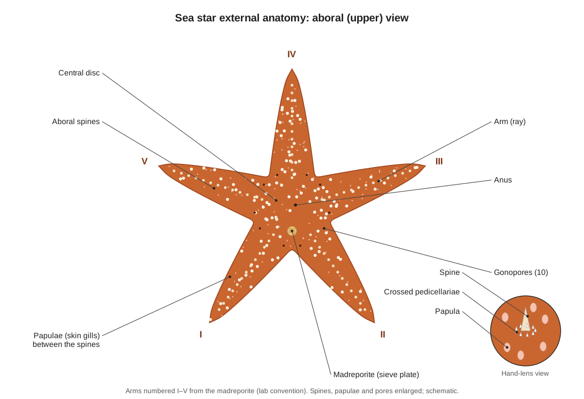
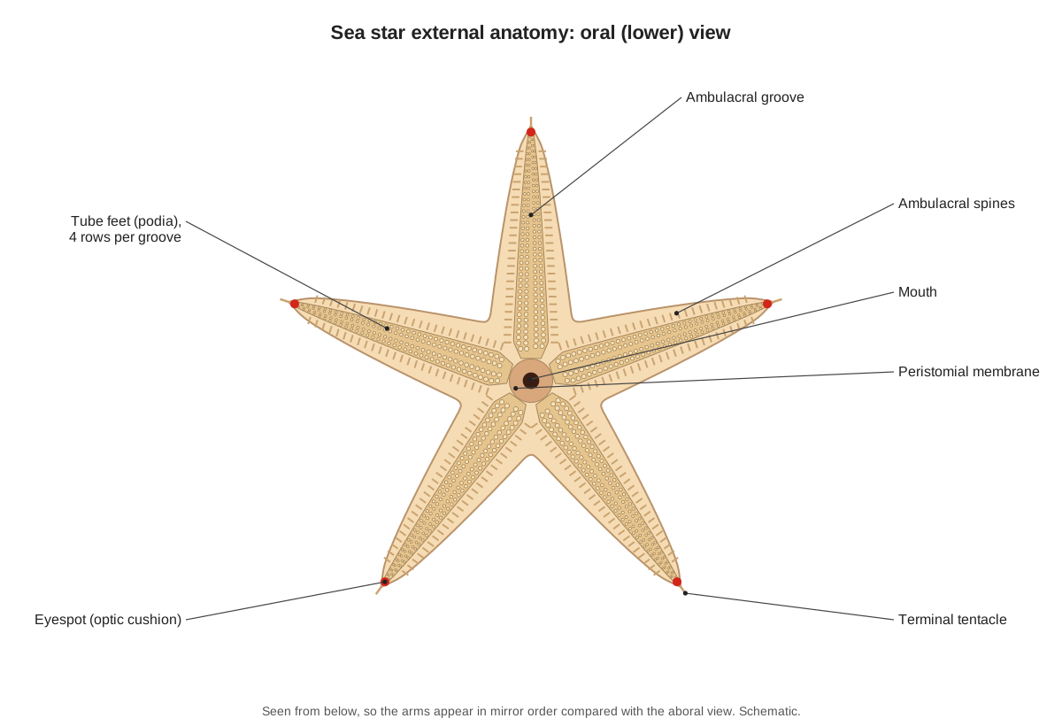
Pedicellariae are tiny pincers that keep the skin free of settling larvae and debris. Asterias has two kinds: small crossed pedicellariae in wreaths around each spine, and larger straight pedicellariae scattered between them. Papulae (skin gills) are thin, finger-like bulges of the body wall between the spines; they are the main place where the sea star takes in oxygen.
Sea star internal anatomy diagram
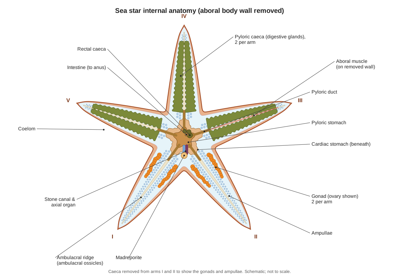
Inside, a sea star is mostly a fluid-filled body cavity (coelom) holding the gut, the gonads and the water vascular system. When the aboral body wall is removed, the first organs you see are the green-brown pyloric caeca (two per arm). In the disc lie the small pyloric stomach and, beneath it, the large cardiac stomach. The gonads sit at the base of each arm under the caeca, and rows of bead-like ampullae line the floor of each arm.
Sea star water vascular system diagram
The water vascular system is a network of fluid-filled canals that works the tube feet. Fluid passes from the madreporite down the stone canal to the ring canal around the mouth, then along five radial canals, one in each arm. Short lateral canals, each with a one-way valve, lead to the ampullae and tube feet.
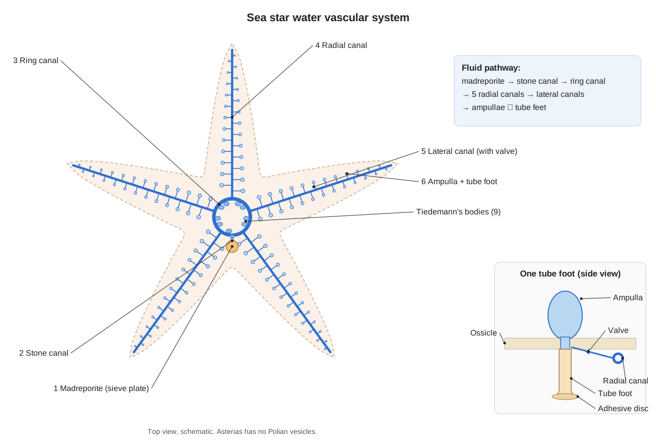
Asterias has nine small Tiedemann's bodies on the inner edge of the ring canal (in pairs between the arms, with one missing where the stone canal joins). Unlike many other sea stars, it has no Polian vesicles.
How tube feet work
Each tube foot is a hollow, muscular tube joined to a bulb, the ampulla, inside the arm. When the ampulla squeezes, the valve in the lateral canal closes and fluid is forced into the tube foot, which gets longer. The tip then sticks to the surface. Muscles in the wall of the tube foot shorten it, pushing the fluid back into the ampulla.
Tube feet grip mainly by chemistry, not suction. Their tips have a duo-gland adhesive system: one set of gland cells secretes a glue made of many proteins, and another secretes a de-adhesive that lets the foot let go. The glue stays behind on the rock as a tiny footprint. Working together, hundreds of tube feet let a sea star climb rocks and pull open mussel shells.
Sea star digestive system: how sea stars eat
A sea star's digestive system runs from the mouth to a cardiac stomach, a pyloric stomach, ten pyloric caeca, a very short intestine with small rectal caeca, and the anus. Asterias feeds mainly on mussels, clams, barnacles and snails.
- The sea star humps over a mussel and grips both shell valves with its tube feet.
- A steady pull tires the mussel's adductor muscle until the shell opens slightly.
- Body-wall muscles raise the pressure in the coelom and push the thin cardiac stomach out through the mouth. A gap of about a millimetre is enough for a fold of stomach to enter.
- Enzymes digest the mussel inside its own shell.
- The soupy food passes to the pyloric stomach and into the pyloric caeca, where most digestion and absorption happen. Gastric ligaments and stomach muscles pull the cardiac stomach back in.
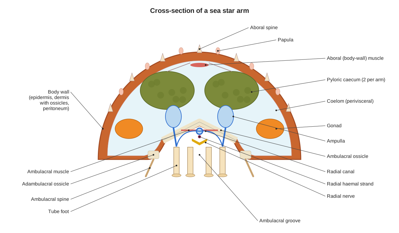
Cross-section of a sea star arm
A cross-section through an arm shows almost every organ system at once: the spiny body wall with papulae, two pyloric caeca hanging from the aboral wall, the gonads lower down near the arm base, the large coelom, the ampullae either side of the V-shaped ambulacral ossicles, and, in the groove below, the radial canal, the radial haemal strand, the radial nerve and the tube feet.
Nervous system and eyes
Sea stars have no brain. A nerve ring around the mouth links five radial nerves, one along the groove of each arm. The radial nerve is mainly sensory (ectoneural); a thin hyponeural layer on its inner side supplies the muscles, and small aboral nerves serve the upper body wall. A nerve net in the skin connects everything.
At the tip of every arm is a red eyespot (optic cushion) made of many tiny pigment-cup eyes, next to a sensory terminal tentacle. Studies on other sea stars show these eyes can form a coarse image, good enough to steer towards a reef. Sea stars often walk with their arm tips curled up so the eyes and tentacles can sample their surroundings.
Breathing, circulation and excretion
Sea stars breathe through their papulae and tube feet. Cilia keep coelomic fluid moving inside the body, carrying oxygen and food around. A small haemal system of spongy strands, centred on the axial organ beside the stone canal, runs alongside the water vascular system, but there is no true heart or blood with an oxygen-carrying pigment.
There are no kidneys. Ammonia, the main waste, simply diffuses out through the thin walls of the papulae and tube feet. Wandering cells called coelomocytes engulf particles and germs and can carry them out through the papulae.
Reproduction and the sea star life cycle diagram
Sea stars have separate sexes, but males and females look identical from the outside. Each arm has two gonads at its base: ovaries are usually orange and grainy, testes creamy white. In spring, adults release eggs and sperm through tiny gonopores near the arm bases, and fertilisation happens in the water. A moderate-sized female A. rubens can release about 2.5 million eggs.
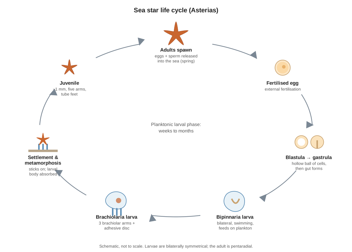
The embryo grows into a swimming, plankton-eating bipinnaria larva with bilateral symmetry and bands of cilia. It then becomes a brachiolaria larva, which has three short brachiolar arms and an adhesive disc. The brachiolaria settles on the sea floor, sticks on and metamorphoses into a tiny five-armed juvenile about a millimetre across, while the larval body is absorbed. The larval stage lasts from a few weeks (A. forbesi) to around three months (A. rubens).
Regeneration
Sea stars can regrow lost arms. An arm lost to a predator regrows over several months. In Asterias, a whole new animal can grow only from a piece that includes part of the central disc. A few sea stars, such as Linckia, can regrow a complete body from a single arm, forming a "comet" with one long arm and four small new ones.
Sea star vs brittle star, sea urchin, sand dollar and sea cucumber
All five groups are echinoderms with a water vascular system, tube feet and a calcite skeleton, but they look and live very differently. A brittle star diagram shows a small round disc with five long, thin, jointed arms that are clearly separate from it, no grooves on the underside, and no anus.
| Feature | Sea star | Brittle star | Sea urchin | Sand dollar | Sea cucumber |
|---|---|---|---|---|---|
| Body shape | Flat star; 5 arms merge into the disc | Small round disc; thin, snake-like arms sharply set off from it | Round, globe-shaped shell (test) | Flat, round disc | Soft, sausage-shaped |
| Skeleton | Loose mesh of ossicles; flexible | Arms armoured with jointed plates ('vertebrae') | Plates fused into a rigid test | Fused rigid test | Microscopic ossicles in leathery skin |
| Tube feet | Suckered; used to walk and grip | No suckers; used for feeding and sensing, not walking | Suckered, in 5 double rows | Short; some on 'petals' used for breathing | Around the mouth form feeding tentacles; others for crawling |
| How it moves | Hundreds of tube feet | Rowing with the arms (fastest echinoderms) | Tube feet and spines | Tiny spines and tube feet | Tube feet and body-wall muscles |
| Feeding | Everts stomach over prey | Arms pass food to the mouth; no anus | Scrapes with 5 teeth (Aristotle's lantern) | Sorts food particles; has a lantern | Tentacles sweep up sediment or plankton |
| Breathing | Papulae and tube feet | Bursae (pouches) by the arm bases | Tube feet and peristomial gills | Petal-shaped rows of tube feet | Internal respiratory trees |
Sea star wasting disease
Sea star wasting disease causes white lesions, loss of body firmness, dropped arms and death within days to weeks. Since 2013 it has killed billions of sea stars on the Pacific coast of North America, and it also affects Asterias forbesi on the Atlantic coast. In 2025, researchers identified a strain of the bacterium Vibrio pectenicida as a cause of the disease in the sunflower sea star.
Sea star parts and functions (labeled list)
Every structure in the 3D model, grouped by organ system. Ready to try it on a real specimen? Follow the step-by-step starfish dissection guide.
External
- Central disc (Disk)
- The flattened central part of the body from which the five arms radiate. Houses the stomachs, the ring canal of the water vascular system and the nerve ring that coordinates the arms.
- Arms (Rays)
- Five tapering arms, each a hollow extension of the body containing a full set of organs. Carry the tube feet used for walking and gripping prey, and hold the digestive glands (pyloric caeca) and gonads.
- Aboral spines (Fixed spines)
- Short, blunt, immovable spines scattered over the upper surface, with a more regular row along the middle of each arm. Protection. They are outgrowths of the skeletal ossicles covered by epidermis, which is why sea stars feel rough.
- Crossed pedicellariae (Minor pedicellariae)
- Tiny scissor-like pincers clustered in wreaths around the base of each aboral spine. Keep the surface clean by nipping small organisms and larvae that try to settle on the skin, and may help catch tiny prey.
- Straight pedicellariae (Major pedicellariae)
- Larger pincers with two straight jaws that meet like forceps, scattered over the body surface. Defence and cleaning, like the crossed type.
- Peristomial membrane (Peristome)
- The soft, flexible membrane around the mouth. Stretches to let the cardiac stomach be pushed out through the mouth during feeding.
- Ambulacral groove
- A deep channel along the oral midline of each arm, packed with tube feet. Protects the tube feet, radial nerve and radial canal. The groove can close over the feet when the animal is disturbed.
- Ambulacral spines (Movable spines, adambulacral spines)
- Long, flattened, movable spines lining both edges of each ambulacral groove. Fold over the groove to protect the tube feet.
Endoskeleton
- Ambulacral ossicles (Ambulacral plates)
- Paired rod-like ossicles that meet in an inverted V to form the roof of each ambulacral groove. Support the arm and frame the pores through which each tube foot connects to its ampulla.
- Adambulacral ossicles
- A row of ossicles along each edge of the ambulacral groove. Form the groove margin and carry the movable ambulacral spines.
- Marginal ossicles
- Ossicles along the side edges of the arms. Strengthen the edge of the arm.
- Dermal ossicles (Aboral skeletal reticulum)
- A mesh of small calcite ossicles in the dermis of the upper body wall. Gives the body a flexible but tough endoskeleton. Papulae poke through the gaps in the mesh.
- Mouth frame (Oral ossicles, peristomial ring)
- A heavy ring of ossicles surrounding the mouth. A rigid ring that supports the mouth. The ring canal and the Tiedemann's bodies lie against its inner aboral edge.
Water vascular
- Madreporite (Sieve plate)
- A small, finely grooved, button-like plate on the aboral disc, between two arms. The opening of the water vascular system. Its many microscopic pores connect seawater with the stone canal and help keep the system's volume balanced.
- Stone canal
- A short, stiff tube running from the madreporite down to the ring canal. Connects the madreporite to the ring canal.
- Ring canal (Circumoral canal, water ring)
- A ring-shaped canal encircling the mouth. Distributes water vascular fluid from the stone canal to the five radial canals.
- Tiedemann's bodies
- Small, spongy pouches on the inner margin of the ring canal. Thought to produce coelomocytes (wandering cells) and to filter or phagocytose particles in the water vascular fluid.
- Radial canals
- One canal runs down the centre of each arm from the ring canal to the tip. Carry water vascular fluid to the lateral canals and tube feet.
- Lateral canals
- Short side branches from a radial canal, one to each tube foot. Connect each tube foot and its ampulla to the radial canal. A one-way valve stops fluid flowing back when the ampulla contracts.
- Ampullae
- Small, bulb-like muscular sacs inside the arm, one at the top of each tube foot. Contraction of the ampulla's muscles squeezes fluid into the tube foot and extends it.
- Tube feet (Podia)
- Hundreds of small, hollow, extendable feet with sucker-like tips, in four rows in each ambulacral groove. Walking, gripping rocks and prey, opening bivalve shells, gas exchange and sensing touch and chemicals.
Digestive
- Mouth
- The central opening on the oral surface. Leads via a very short oesophagus to the cardiac stomach, which can be pushed out through it.
- Cardiac stomach
- A large, thin-walled, folded sac filling the lower part of the disc, which can be turned inside out through the mouth. Everted over or into prey such as mussels, it begins digestion outside the body. The partly digested soup is passed to the pyloric stomach.
- Gastric ligaments (Stomach retractors, retractor muscles)
- Five pairs of strands tying the cardiac stomach to the ambulacral ridge of each arm. Anchor the cardiac stomach and help pull it back in after feeding.
- Pyloric stomach
- A small, flattened, star-shaped stomach above the cardiac stomach. Receives food from the cardiac stomach and sends it into the pyloric caeca for digestion and absorption.
- Pyloric ducts
- Short ducts joining the pyloric stomach to the two pyloric caeca in each arm. Carry food into the caeca and digestive enzymes back to the stomach.
- Pyloric caeca (Digestive glands, hepatic caeca)
- Ten long, feathery, green-brown glands, two in each arm, filling most of the arm cavity. Make digestive enzymes, absorb digested food and store nutrients (lipids and glycogen).
- Intestine
- A very short tube from the pyloric stomach to the anus. Carries small amounts of indigestible waste to the anus. Most waste (such as shells) never enters the body.
- Rectal caeca (Intestinal caeca)
- Small, lobed, brownish or olive pouches on the intestine. Their role is uncertain; they may absorb nutrients or store waste.
- Anus
- A tiny opening near the centre of the aboral disc. Releases small amounts of faeces.
Nervous & sensory
- Nerve ring (Circumoral nerve ring)
- A ring of nerve tissue around the mouth that links the five radial nerves. Coordinates the arms so they act together, for example when one arm leads the animal in a chosen direction.
- Radial nerves (Radial nerve cords (ectoneural))
- A thick nerve cord along the groove of each arm, from the nerve ring to the eyespot. Controls the tube feet of the arm and carries sensory information. This ectoneural part is mainly sensory.
- Hyponeural system
- A thin motor nerve layer on the inner (aboral) side of each radial nerve. Supplies motor nerves to the muscles of the arm, including the ambulacral and tube-foot muscles.
- Aboral nerves (Entoneural / aboral radial nerves)
- Small motor nerves running along the aboral midline of each arm. Supply the muscles of the aboral body wall.
- Eyespot (Optic cushion)
- A red or orange spot at the tip of each arm, made of many tiny pigment-cup eyes (ocelli). Detects light and, in some species, forms a coarse image used to steer towards habitat.
- Terminal tentacle
- An unsuckered tube foot at the tip of each arm. A sensory feeler that detects chemicals and touch. Arm tips are often curled up so the tentacle can sample the water.
Haemal & coelom
- Perivisceral coelom (Body cavity)
- The large fluid-filled body cavity in the disc and arms that surrounds the organs. Its coelomic fluid, stirred by cilia, carries nutrients, oxygen and wastes. It also works as a hydraulic skeleton, for example when the stomach is everted.
- Axial organ (Axial gland)
- A spongy, dark, spindle-shaped organ lying beside the stone canal. The centre of the haemal system. It may help make coelomocytes and has a small pulsating sac (the 'heart' or dorsal sac) at its aboral end.
- Oral haemal ring
- A ring of haemal tissue around the mouth, next to the nerve ring. Connects the axial organ with the radial haemal strands of the arms.
- Radial haemal strands
- A haemal strand running down each arm next to the radial nerve. Distribute haemal fluid along the arm.
- Aboral (genital) haemal ring
- A ring of haemal tissue under the aboral wall of the disc, with branches to the gonads. Supplies nutrients to the gonads.
- Gastric haemal tufts
- Small tufts of haemal tissue attached to the pyloric stomach. Thought to pick up nutrients absorbed from the gut.
Respiratory & excretory
- Papulae (Dermal branchiae, skin gills)
- Tiny, soft, finger-like bulges of the body wall that poke out between the spines. The main sites of gas exchange (breathing) and of ammonia excretion. Coelomic fluid flows into them, so oxygen diffuses straight into the body cavity.
Reproductive
- Ovaries · females only
- Ten orange, grape-like gonads, two at the base of each arm. Produce eggs, which are released into the sea through the gonopores.
- Testes · males only
- Ten creamy-white, branched gonads, two at the base of each arm. Produce sperm, released into the water through the gonopores.
- Gonoducts
- Short ducts from each gonad to the body surface. Carry eggs or sperm out of the body.
- Gonopores
- Ten tiny pores where eggs or sperm leave the body. Release gametes into the sea for external fertilisation.
Muscular
- Aboral longitudinal muscle (Body-wall muscles)
- A band of muscle along the inner aboral wall of each arm, part of the body-wall musculature. Bends the arm upwards and, with the other body-wall muscles, raises coelomic pressure to evert the stomach.
- Ambulacral muscles
- Small transverse muscles linking the ambulacral ossicles. Open and close the ambulacral groove and help bend the arm.
Frequently asked questions
Is a starfish a fish?
No. Starfish are echinoderms, related to sea urchins and sea cucumbers, not fish. They have no backbone, gills, fins or scales. Many scientists and aquariums prefer the name 'sea star' for this reason.
Do starfish have a brain?
No. A sea star has a nerve ring around its mouth and a radial nerve along each arm, but no brain. The arms are coordinated through the nerve ring.
Do starfish have blood?
Not in the usual sense. Sea stars move nutrients and oxygen in fluid-filled body cavities (coelom and water vascular system). A small haemal system of spongy strands exists, but its fluid has no oxygen-carrying pigment.
How do starfish eat?
A sea star wraps its arms around a mussel or clam, pulls the shell open slightly with its tube feet, and pushes its cardiac stomach out through its mouth into the gap. Digestion starts outside the body; the stomach then pulls the soupy food back in.
How many tube feet does a starfish have?
Hundreds to more than a thousand, depending on size. In Asterias they are arranged in four rows in each of the five ambulacral grooves.
Do starfish have eyes?
Yes, simple ones. Each arm tip has an eyespot (optic cushion) made of many tiny ocelli. Studies on some species show they can see coarse shapes, for example to find their way back to a reef.
Can a starfish regrow an arm?
Yes. A lost arm regrows over several months. In Asterias a new animal can only grow from a piece that includes part of the central disc; a few sea stars, such as Linckia, can regrow from an arm alone.
How do you tell a male from a female starfish?
You cannot tell from the outside. Inside, ovaries are usually orange and testes creamy white, especially near the breeding season.
References
- Fox, R. S. Asterias forbesi (sea star): Invertebrate Anatomy OnLine. Lander University.
- Ruppert, E. E., Fox, R. S. & Barnes, R. D. (2004). Invertebrate Zoology: A Functional Evolutionary Approach, 7th ed., ch. 28 Echinodermata. Brooks/Cole.
- Brusca, R. C., Moore, W. & Shuster, S. M. (2016). Invertebrates, 3rd ed., ch. 27 Echinodermata. Sinauer.
- Algrain, M., Hennebert, E., Bertemes, P., Wattiez, R., Flammang, P. & Lengerer, B. (2022). In the footsteps of sea stars: deciphering the catalogue of proteins involved in underwater temporary adhesion. Open Biology 12: 220103.
- Petie, R., Garm, A. & Hall, M. R. (2016). Crown-of-thorns starfish have true image forming vision. Frontiers in Zoology 13: 41.
- Carter, H. F., Thompson, J. R., Elphick, M. R. & Oliveri, P. (2021). The development and neuronal complexity of bipinnaria larvae of the sea star Asterias rubens. Integrative and Comparative Biology 61: 337–351.
- Bucci, C. et al. (2017). Sea star wasting disease in Asterias forbesi along the Atlantic coast of North America. PLoS ONE 12: e0188523.
- Prentice, M. B. et al. (2025). Vibrio pectenicida strain FHCF-3 is a causative agent of sea star wasting disease. Nature Ecology & Evolution (USGS summary).
- Common starfish (Asterias rubens). Wikipedia.
- Forbes sea star (Asterias forbesi). Wikipedia.
More interactive anatomy models
- Starfish dissectionGuided sea star dissection: aboral wall, digestive glands, gonads and ampullae.
- Human anatomy1,000+ bones, muscles, organs, vessels and nerves from real anatomical data.
- Octopus anatomyThree hearts, a ring-shaped brain, eight arms and a beak, in 3D.
- Squid anatomyPen, ten arms, giant axons, ink sac and three hearts of the longfin squid.
- Squid dissectionStep-by-step guided squid dissection with every structure labeled.
- Fish anatomyExternal and internal anatomy of a bony fish: fins, gills, swim bladder, heart and brain.