

Starfish dissection overview
In a starfish dissection you first study the outside, then remove the upper (aboral) body wall of one arm and the central disc to expose the digestive glands, gonads, stomachs and water vascular system. The specimen is usually a preserved Forbes sea star, Asterias forbesi. Allow about 45–60 minutes. For background on every part, see the interactive starfish anatomy page.
Materials and safety
Materials
- Preserved sea star (Asterias forbesi or A. rubens)
- Dissecting tray or pan
- Fine dissecting scissors
- Forceps and a blunt probe
- Hand lens or dissecting microscope
- Dropper bottle of water (keep the specimen moist)
- Gloves, safety goggles and a lab apron
- Paper towels and a waste container for tissue
Safety
- Wear gloves and goggles. Preserved specimens are kept in a preserving fluid that can irritate skin and eyes.
- Rinse the specimen in water before you start and work in a well-ventilated room.
- Cut away from your body and your partner. The body wall is hard and gritty, so use small, controlled cuts.
- Never eat or drink in the lab. Wash your hands with soap after the dissection.
- Dispose of tissue and the specimen as your teacher instructs; do not put them in the sink.
External observation (before you cut)
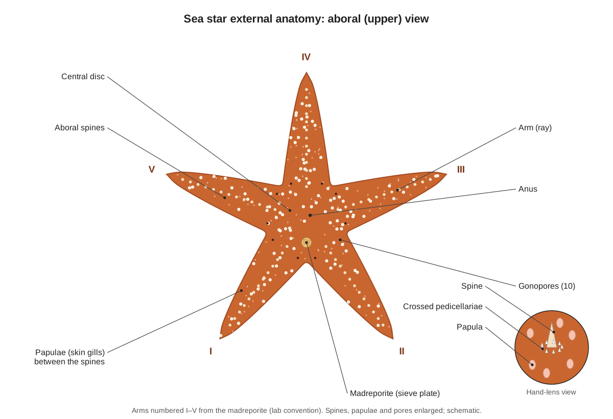
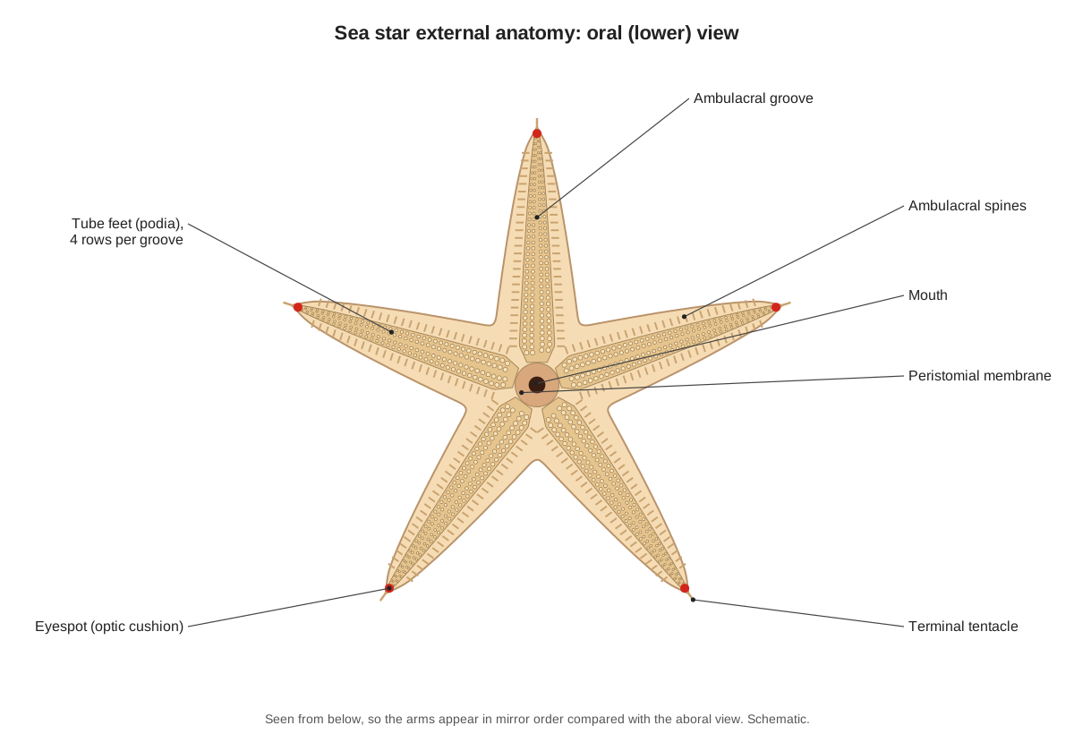
Start with the specimen whole. On the rough aboral side, find the central disc, the five arms and the light-coloured madreporite between two arms. With a hand lens, look for the blunt spines, the tiny white pedicellariae around them and the soft papulae. On the pale oral side, find the mouth, the five ambulacral grooves and their four rows of tube feet, and the red eyespot at each arm tip.
How to dissect a starfish: step-by-step instructions
1. Orient the specimen (aboral side up)
- Rinse the preserved sea star in water and place it in the dissecting tray with the rough, darker aboral (upper) side facing up.
- Find the central disc and count the five arms (rays).
- Find the madreporite: a small, smooth, light-coloured button off-centre on the disc, between two arms.
- Turn the tray so the madreporite is nearest to you. The arm to the left of it is arm I, the arm to the right is arm II, and arms III, IV and V follow counter-clockwise.
- Look near the centre of the disc for the tiny anus (it is hard to see).
Find: Central disc, Arms, Madreporite, Anus, Aboral spines
2. Spines, pedicellariae and papulae (hand lens)
- Use a hand lens or dissecting microscope on the aboral surface of one arm.
- Find the short, blunt aboral spines. Each is ringed by tiny white pincers: the crossed pedicellariae.
- Look for larger straight pedicellariae scattered between the spines.
- Find the papulae: soft, translucent, finger-like bumps between the spines. In preserved specimens they are often shrunken.
- Record a sketch of a spine with its wreath of pedicellariae.
Find: Aboral spines, Crossed pedicellariae, Straight pedicellariae, Papulae
3. Oral surface: mouth, grooves and tube feet
- Turn the sea star over so the pale oral surface faces up.
- Find the mouth at the centre, surrounded by the soft peristomial membrane.
- Follow an ambulacral groove from the mouth to the arm tip.
- Count the rows of tube feet in one groove (Asterias has four rows) and look at the sucker-like tips with the hand lens.
- Find the movable ambulacral spines along the edges of the groove.
Find: Mouth, Peristomial membrane, Ambulacral groove, Tube feet, Ambulacral spines
4. Arm tip: eyespot and terminal tentacle
- Look at the oral side of an arm tip with the hand lens.
- Find the red or orange eyespot (optic cushion) and the small unsuckered terminal tentacle at the very end of the groove.
Find: Eyespot, Terminal tentacle
5. Cut off an arm tip and view the cross-section
- With scissors, cut across arm III about 2 cm from its tip.
- Look at the cut face of the removed piece under the hand lens.
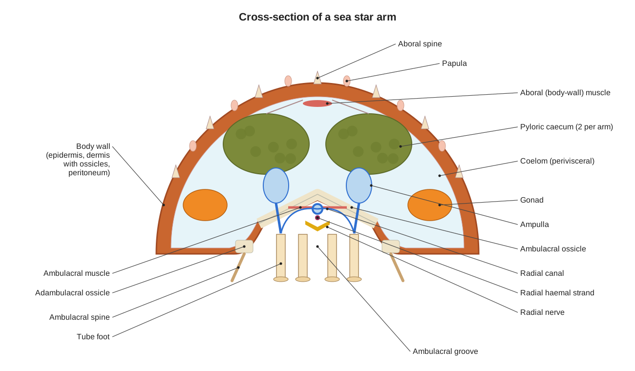
- Identify the body wall with its ossicles, the two pyloric caeca at the top, the V-shaped ambulacral ossicles forming the groove roof, the ampullae on either side, and the tube feet below.
- Note the open space around the organs: the perivisceral coelom.
Caution: Scissor tips point away from you; cut only through the body wall, which is hard and gritty.
Find: Pyloric caeca, Ambulacral ossicles, Ampullae, Tube feet, Radial canals, Radial nerves, Perivisceral coelom
6. Remove the aboral wall of one arm
- Return the sea star to aboral side up.
- Starting at the cut end of arm III, make two cuts along both sides of the arm towards the disc, keeping the scissor tips just under the body wall.
- Lift the flap of body wall with forceps and free it from the underlying organs by gently cutting the mesenteries.
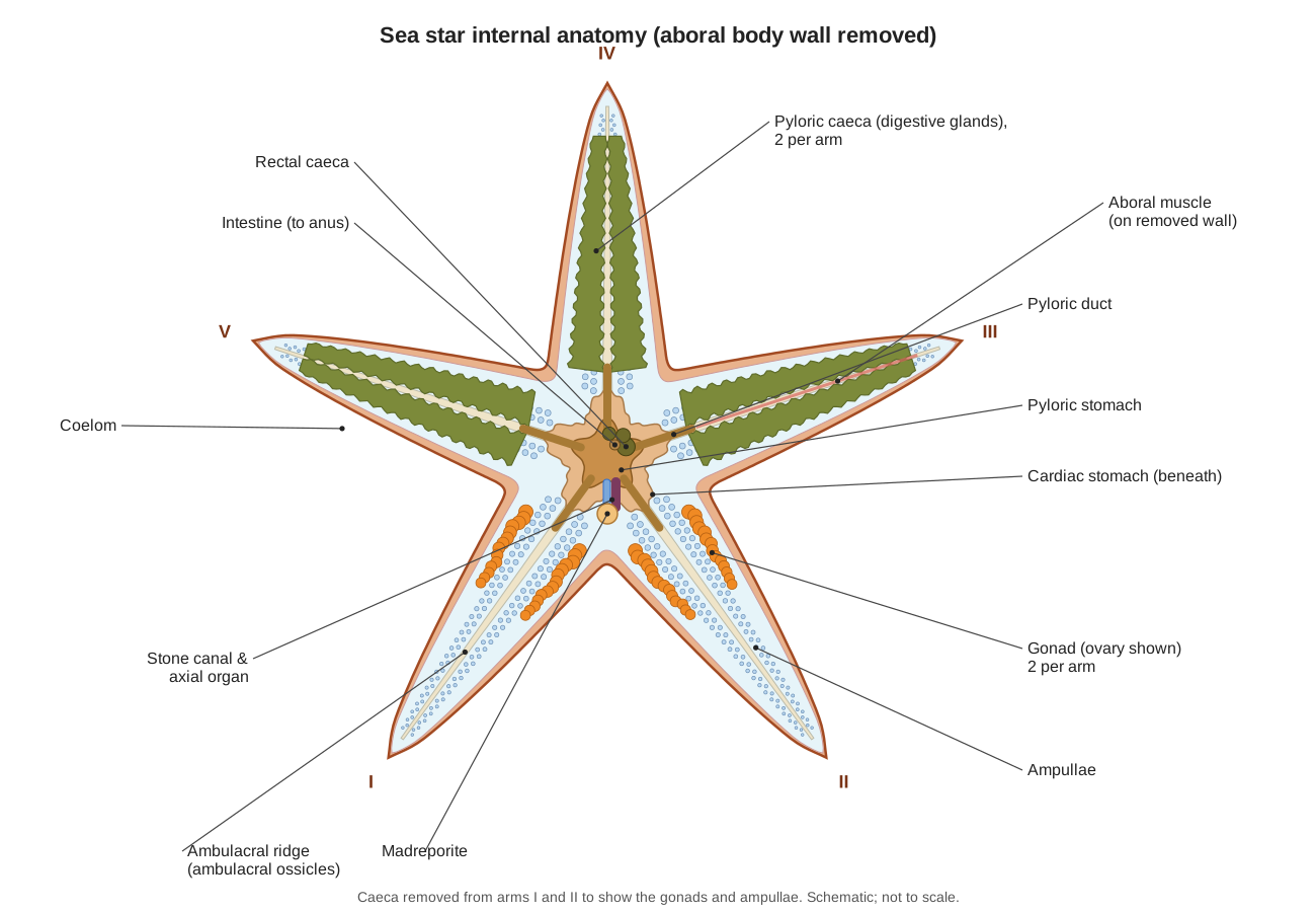
- The first organs you see are the two green-brown, feathery pyloric caeca filling the arm.
- Look on the inside of the removed flap for the aboral muscle band and the skeletal mesh.
Caution: Keep the scissor tips angled up against the body wall so you do not cut the caeca.
Find: Pyloric caeca, Pyloric ducts, Aboral longitudinal muscle, Dermal ossicles
7. Open the disc around the madreporite
- Extend your cuts around the edge of the disc and across the bases of the other arms. Do not cut between the madreporite and the edge of the disc.
- Carefully lift the aboral disc wall. Look for the small lobed rectal caeca and the tiny intestine attached to its underside.
- Cut around the madreporite so it stays attached to the stone canal, and remove the rest of the disc wall.
- Find the flat, star-shaped pyloric stomach in the centre and follow a pyloric duct into each arm.
Find: Rectal caeca, Intestine, Pyloric stomach, Pyloric ducts, Madreporite
8. Lift the caeca to find the gonads
- Gently lift the pyloric caeca of the opened arm with a probe.
- Find the two gonads at the base of the arm, one on each side, close to the disc.
- Note their colour: ovaries are usually orange and grainy; testes are creamy white. Switch the model between Female and Male to compare.
- Trace a gonoduct towards the aboral surface, where it opens at a tiny gonopore.
Caution: Gonad size depends on the season: in specimens collected after spawning they may be very small.
Find: Ovaries, Testes, Gonoducts, Gonopores
9. The two stomachs
- Remove the pyloric caeca from the opened arm (cut at the pyloric duct) and set them aside.
- Find the large, thin-walled, folded cardiac stomach under the pyloric stomach.
- Find the gastric ligaments that tie the cardiac stomach to the floor of each arm.
- Think about how this stomach can be pushed out through the mouth to digest a mussel.
Find: Cardiac stomach, Pyloric stomach, Gastric ligaments, Mouth
10. Water vascular system in the arm
- With the caeca removed, look at the floor of the opened arm.
- Find the ambulacral ridge (the ambulacral ossicles) running down the middle.
- Find the rows of small, clear, bead-like ampullae on both sides of the ridge.
- Gently squeeze an ampulla with forceps and watch the oral side: the matching tube foot may extend.
Find: Ampullae, Ambulacral ossicles, Tube feet, Lateral canals, Radial canals
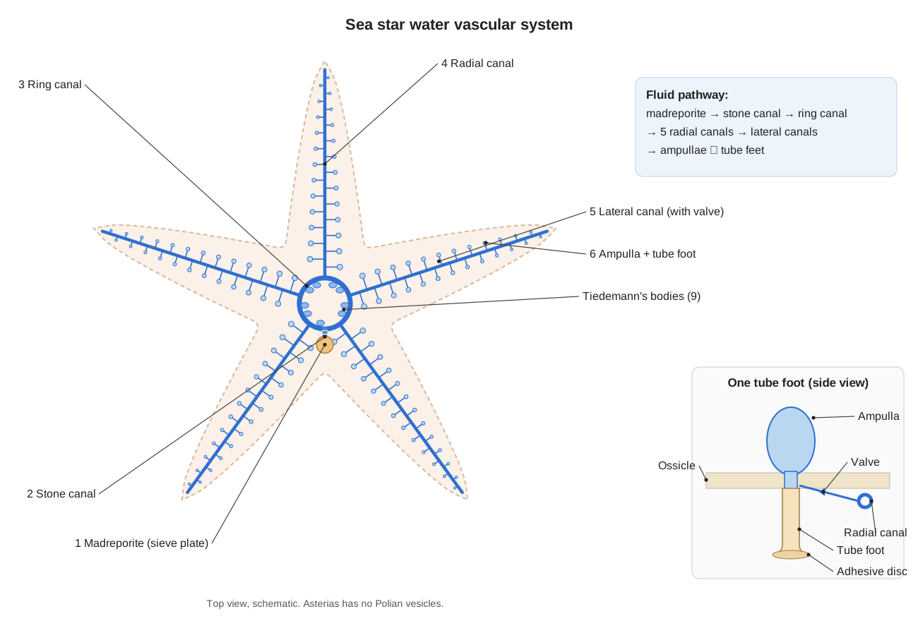
11. Stone canal, ring canal and Tiedemann's bodies
- Carefully remove the stomachs by cutting the oesophagus just above the mouth.
- Follow the stiff stone canal from the underside of the madreporite down to the mouth area.
- Find the hard white mouth frame. The thin ring canal lies against its inner edge.
- Look for small, pale Tiedemann's bodies on the inner edge of the ring canal (nine in Asterias). Asterias has no Polian vesicles.
- Next to the stone canal, look for the darker, spongy axial organ.
Find: Stone canal, Ring canal, Tiedemann's bodies, Mouth frame, Axial organ, Madreporite
12. Nervous system and clean-up
- Turn the specimen over. In the groove of the opened arm, push the tube feet aside to see the radial nerve along the middle of the groove roof.
- Follow it to the nerve ring around the mouth.
- Dispose of the specimen as your teacher directs, clean and dry your tools, and wash your hands.
Find: Radial nerves, Nerve ring, Hyponeural system, Eyespot
Starfish dissection labeled: what you should see
With the aboral wall removed, your dissected starfish should look like the labeled diagram below. Arms that still have their pyloric caeca look green-brown and feathery; in an arm where you lifted the caeca you should see the gonads at the base and two rows of ampullae along the floor.
Male vs female starfish
You cannot sex a sea star from the outside. Inside, each arm has two gonads at its base. Ovaries are usually orange and grainy (full of eggs); testes are creamy white and smoother. Gonad size depends on the season: just before spawning in spring they can fill much of the arm, and after spawning they may be very small. Use the Female/Male switch in the 3D viewer to compare.
Starfish dissection lab data sheet
Fill in the right-hand column from your specimen. Expected values are shown.
| Observation | Expected result (answer key) |
|---|---|
| Number of arms | 5 |
| Diameter (tip to opposite interradius), cm | your measurement |
| Rows of tube feet per groove | 4 |
| Interradius containing the madreporite | between arms I and II |
| Pyloric caeca per arm | 2 |
| Gonads per arm / total | 2 / 10 |
| Gonad colour and sex | orange = female, cream = male |
| Tiedemann's bodies counted | up to 9 (often hard to see) |
Starfish dissection worksheet questions and answer key
What type of symmetry does an adult sea star have, and what symmetry does its larva have?
Show answer
Adults are pentaradially symmetrical (five parts around a central axis). The larvae (bipinnaria and brachiolaria) are bilaterally symmetrical.
What is the function of the madreporite?
Show answer
It is the sieve-like opening of the water vascular system. It connects seawater with the stone canal and helps keep the system's fluid volume balanced.
List, in order, the parts of the water vascular system from the madreporite to a tube foot.
Show answer
Madreporite → stone canal → ring canal → radial canal → lateral canal (with a one-way valve) → ampulla and tube foot.
How does a tube foot extend and grip?
Show answer
The ampulla contracts and pushes fluid into the tube foot, which extends. The tip sticks to the surface with an adhesive secretion and later releases it with a de-adhesive secretion (the duo-gland system). Muscles in the tube foot wall shorten it again.
Name the two stomachs and give the job of each.
Show answer
The cardiac stomach (lower, larger) can be everted through the mouth to begin digesting prey outside the body. The pyloric stomach (upper, smaller) receives the partly digested food and passes it to the pyloric caeca.
What are the pyloric caeca and how many are there?
Show answer
They are digestive glands that make enzymes, absorb food and store nutrients. There are ten: two in each arm.
Where are the gonads and how can you tell a male from a female?
Show answer
Two gonads lie at the base of each arm (ten in all), under the pyloric caeca. Males and females look the same outside; inside, ovaries are usually orange and testes creamy white.
How does a sea star breathe and get rid of waste?
Show answer
Gas exchange happens across the papulae (skin gills) and the tube feet. There are no kidneys; ammonia diffuses out through the same thin surfaces, and coelomocytes carry particles out through the papulae.
What are pedicellariae and what do they do?
Show answer
Tiny pincers on the body surface (crossed ones around the spines and straight ones scattered between them) that keep the skin free of settling organisms and debris.
What did you see in the cross-section of the arm, from top to bottom?
Show answer
Aboral body wall with spines, two pyloric caeca (often with gonads near the base of the arm), the coelom, ampullae either side of the V-shaped ambulacral ossicles, the radial canal and radial nerve in the groove, and the tube feet.
Why is the name 'sea star' preferred to 'starfish'?
Show answer
Because these animals are echinoderms, not fish. They have no backbone, fins, gills or scales.
What part of the sea star is needed for a whole new animal to regenerate?
Show answer
In Asterias, a piece of the central disc must be present. A lost arm is regrown by the remaining body over several months.
Virtual starfish dissection (alternative to a real specimen)
Students who opt out of dissecting a real animal can complete the same lab here. Work through the 12 steps in the viewer, use the "Find these structures" chips to locate each part, and answer the worksheet questions above. The model can also be used to preview the lab before class or to review the anatomy afterwards.
Frequently asked questions
What sea star is used for school dissections?
Usually the Forbes sea star, Asterias forbesi, from the Atlantic coast of North America, supplied preserved. Its anatomy matches the common sea star Asterias rubens shown in the 3D model.
Which organs fill the arms of a dissected starfish?
The two green-brown pyloric caeca (digestive glands) in each arm. Under them, near the base of the arm, are the two gonads, and along the floor of the arm run the rows of ampullae.
How do you tell if a dissected starfish is male or female?
Look at the gonads at the base of the arms. Ovaries are usually orange and grainy; testes are creamy white. There is no external difference.
Why should you not cut through the madreporite?
The madreporite is attached to the stone canal underneath. Cutting around it, not through it, keeps the madreporite, stone canal and ring canal connected so you can trace the water vascular system.
Can I do a starfish dissection online?
Yes. The 3D model on this page is a free virtual starfish dissection. Each step fades the body wall and hides organ systems in the same order as a real dissection, and you can click any structure to read about it.
How long does a starfish dissection take?
About 45–60 minutes for external observation, opening one arm and the disc, and identifying the main organs.
References
- Fox, R. S. Asterias forbesi (sea star): Invertebrate Anatomy OnLine, including dissection procedure. Lander University.
- Ruppert, E. E., Fox, R. S. & Barnes, R. D. (2004). Invertebrate Zoology: A Functional Evolutionary Approach, 7th ed., ch. 28 Echinodermata. Brooks/Cole.
- Visible Body. Sea star lab activity (student worksheet).
- Algrain, M. et al. (2022). In the footsteps of sea stars: deciphering the catalogue of proteins involved in underwater temporary adhesion. Open Biology 12: 220103.
- Forbes sea star (Asterias forbesi). Wikipedia.
- Common starfish (Asterias rubens). Wikipedia.
More interactive anatomy models
- Starfish anatomyWater vascular system, tube feet, two stomachs and pyloric caeca of a sea star.
- Human anatomy1,000+ bones, muscles, organs, vessels and nerves from real anatomical data.
- Octopus anatomyThree hearts, a ring-shaped brain, eight arms and a beak, in 3D.
- Squid anatomyPen, ten arms, giant axons, ink sac and three hearts of the longfin squid.
- Squid dissectionStep-by-step guided squid dissection with every structure labeled.
- Fish anatomyExternal and internal anatomy of a bony fish: fins, gills, swim bladder, heart and brain.