

Ant anatomy at a glance
An ant has three main body parts: the head, the mesosoma (the middle part that carries all six legs) and the gaster (the swollen rear part). Between the mesosoma and the gaster is a narrow waist made of one or two small segments, the petiole and, in some ants, the postpetiole. The head carries two elbowed antennae, two compound eyes and strong jaws (mandibles).
This page uses the Florida carpenter ant, Camponotus floridanus, as its reference ant. Workers are 5.5–10 mm long, with a rusty orange-brown head and mesosoma, a black gaster and long golden hairs (UF/IFAS EDIS IN1075). Carpenter ants belong to the subfamily Formicinae: they have one waist node, no working sting and a round nozzle at the gaster tip, the acidopore, through which they spray formic acid. Where fire ants, army ants, leafcutters and other ants differ, the text and tables say so.
Directions: anterior is towards the head, posterior towards the gaster tip, dorsal is the back and ventral the belly. Like every insect, an ant has an exoskeleton of hard plates joined by soft membranes, six jointed legs, an open circulatory system, tracheae for breathing and a ventral nerve cord.
| Body part | Scientific name | What it carries |
|---|---|---|
| Head | Head capsule | Brain, compound eyes, ocelli (queens and males), antennae, mandibles, mouthparts |
| Middle part | Mesosoma (alitrunk) | Three pairs of legs, wings in queens and males, leg and flight muscles |
| Waist | Petiole (+ postpetiole in fire ants) | Narrow flexible joint; oesophagus, aorta and nerve cord pass through it |
| Rear part | Gaster | Crop, midgut, hindgut, heart, ovaries, venom apparatus, acidopore or sting |
Ant body parts diagram: external anatomy
The external anatomy of an ant includes the head with its eyes, antennae and mandibles; the mesosoma with the pronotum, mesonotum and propodeum on top; the petiole; the gaster; and six legs, each with a coxa, trochanter, femur, tibia and five-segmented tarsus ending in two claws. The diagram labels every part a typical "label the ant" worksheet asks for.
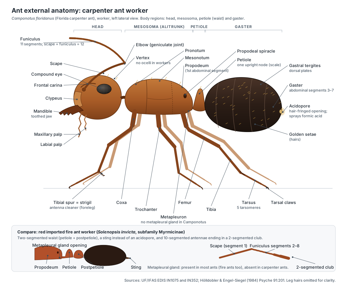
The mesosoma is worth a closer look. In simple diagrams it is called the thorax, but it is really the three thoracic segments plus the first abdominal segment, the propodeum, fused to them. That is why the waist is the second abdominal segment and the gaster begins with the third. A carpenter ant has an evenly rounded (convex) mesosoma in side view, which is a quick way to recognise the genus (Texas A&M E-412).
- Pronotum: Covers the prothorax, which carries the front legs, and anchors neck and foreleg muscles.
- Mesonotum: Covers the mesothorax, which carries the middle legs. In queens and males it is greatly enlarged to hold the flight muscles of the forewings.
- Propodeum: Forms the joint with the petiole and carries the propodeal spiracle. In most ants (but not carpenter ants) the opening of the metapleural gland lies just below it.
- Propodeal spiracle: Lets air into the tracheae (air tubes). It is one of the spiracles lab diagrams most often ask you to label.
- Petiole: Lets the gaster swing forward under the body, for example to aim the acidopore or sting at an enemy.
- Gaster: Holds the crop, midgut, hindgut, heart, ovaries and the venom (formic acid) apparatus.
- Acidopore: The opening of the poison gland. Formicine ants, including carpenter ants, have no working sting. They bite and then spray formic acid from the acidopore into the wound or at an attacker.
Ant head: eyes, antennae and mouthparts
An ant's head carries two compound eyes, two elbowed antennae and a set of mouthparts: mandibles, maxillae with palps, a labium with palps, and a short tongue called the glossa. Queens and males also have three small simple eyes (ocelli) on top of the head; carpenter ant workers do not.
Each antenna has a long first segment, the scape, and a chain of small segments, the funiculus. The joint between them makes the antenna bend like an elbow (geniculate). A carpenter ant worker's antenna has 12 segments and no club; a fire ant worker's has 10 segments ending in a two-segmented club (UF/IFAS EDIS IN1075, IN352). Ants smell, taste and touch with their antennae, which is why they constantly clean them with the strigil on the front legs.
The mandibles are the ant's tools. Carpenter ants use them to excavate galleries in soft or rotting wood, but they do not eat wood (Texas A&M E-412). Ants swallow mainly liquids. The maxillae, labium and glossa lap up nectar, honeydew and prey juices, while solid particles are trapped in the infrabuccal pocket inside the mouth (Richter & Economo, 2023).
- Compound eyes: Detect movement, light and landmarks used in navigation. Carpenter ant workers see reasonably well; many soil ants have tiny eyes.
- Antennal scape: Holds the rest of the antenna out in front of the head. The joint at its tip is what makes ant antennae elbowed (geniculate).
- Antennal funiculus: Carries thousands of sensilla (sense hairs and pegs) for smell, taste, touch, humidity and temperature. Ants recognise nestmates, trails and food with it.
- Clypeus: Covers the base of the mouth and is the attachment point for the muscles of the sucking pump (cibarium) inside.
- Frontal carinae: Partly shield the antennal sockets and guide the scape when the antenna is folded back.
- Mandibles: The ant's hands and tools: they dig, cut wood, carry brood and food, fight and grip. They do not grind food for swallowing; ants swallow liquids.
- Maxillae: Together with the labium they form the maxillolabial complex, which laps up liquids and pushes food particles into the mouth.
- Labium: Carries the glossa and the labial palps; the salivary (labial) gland duct opens on it.
- Glossa: Laps up liquids such as nectar, honeydew and water. Liquids lapped by the glossa pass straight into the mouth (Richter & Economo, 2023).
Ant legs and wings
All six legs attach to the mesosoma. Ant legs have the standard insect segments, plus two special features: a comb-like spur on each front tibia, the strigil, which with a notch on the first tarsal segment forms an antenna cleaner; and paired claws with a sticky pad (arolium) that let ants climb glass. Only queens and males have wings, and only for the mating flight. Ant forewings are longer than the hindwings (Texas A&M E-412). A queen breaks her wings off after mating, leaving small wing scars on the mesosoma.
- Strigil: The ant pulls an antenna through the gap between the spur and the notch to comb off dirt, so its smell receptors keep working.
- Tibial spurs: Grip the ground and help clean the legs and body.
- Tarsal claws & arolium: The claws hook onto rough surfaces; the pad sticks to smooth ones, so ants can walk up glass and upside down.
- Forewings: Used once, for the mating flight. Queens break their wings off afterwards and leave small wing scars.
Ant internal anatomy diagram
Inside an ant, the gut runs from the mouth through the narrow waist to the gaster, where the crop, proventriculus, midgut, Malpighian tubules, hindgut and rectum lie. The brain sits in the head, a ventral nerve cord runs along the belly, a tube-shaped heart lies along the top of the gaster, and air tubes (tracheae) reach every tissue. Venom glands and the ovaries fill the rear of the gaster.
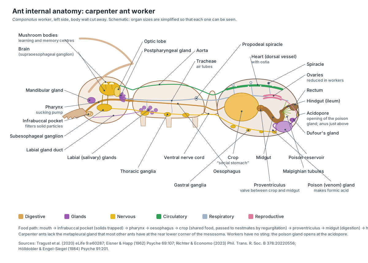
Ant digestive system
Food passes through the mouthparts, the infrabuccal pocket (a filter for solid particles), the pharynx and the thin oesophagus into the crop. The crop is the colony's "social stomach": workers store liquid food there and regurgitate it to nestmates (Eisner & Happ, 1962). The proventriculus is a valve and pump that keeps the crop sealed and lets small amounts into the midgut, where food is digested. Waste is removed by the Malpighian tubules and leaves through the hindgut and rectum.
- Infrabuccal pocket: A filter: food particles and dirt collect here, are pressed into a pellet and spat out. Only liquid and tiny particles reach the gut.
- Crop: Holds food that the ant shares with nestmates by regurgitation (trophallaxis). Food here is not yet digested.
- Proventriculus: Keeps the crop sealed so stored food stays shareable, and pumps small amounts into the midgut when the ant itself needs food.
- Midgut: Secretes digestive enzymes and absorbs nutrients into the hemolymph (insect blood).
- Malpighian tubules: The insect kidney: they draw out nitrogen waste and salts and empty them into the hindgut.
- Rectum: Reabsorbs water and stores faeces until the ant leaves the nest or drops them in a refuse chamber.
Heart, breathing and nervous system
Ants have an open circulation. The heart along the top of the gaster pumps hemolymph forward through the aorta into the head, and the blood then bathes the organs. Oxygen does not travel in the blood: it enters through spiracles on the sides of the body and flows through the tracheae. The nervous system has a brain with large mushroom bodies (for learning and memory), optic and antennal lobes, a subesophageal ganglion and a ventral nerve cord with ganglia in the mesosoma and gaster.
Venom: acidopore or stinger
About 70% of ant species have a stinger (Dejean et al., 2026). Carpenter ants and other formicines do not. Their poison gland makes a strongly acidic secretion, mostly formic acid, stored in a reservoir and sprayed from the acidopore. The Dufour's gland beside it makes oily signal chemicals. Fire ants inject venom through a sting at the gaster tip; their venom is about 95% alkaloids, which cause the white pustule a day after a sting (UF/IFAS EDIS IN352).
- Poison gland: In carpenter ants it makes a strong formic acid solution, sprayed from the acidopore. In stinging ants it makes the venom injected by the sting.
- Poison reservoir: Holds the venom until the ant sprays it through the acidopore.
- Dufour's gland: Makes oily hydrocarbons used in alarm signals, trail marking or defence; in fire ants it is the source of the trail pheromone.
- Brain: Processes smell, sight and touch and controls behaviour.
- Mushroom bodies: Learning and memory: they combine smell and vision, for example to remember landmarks on the way home.
- Heart: Pumps hemolymph forward into the aorta. Insects have an open circulation: the blood then bathes the organs.
Ant castes: worker, queen and male
An ant colony has three kinds of adult: wingless female workers, one or more egg-laying queens, and short-lived winged males. Use the caste switch above the 3D model to compare them. Workers and queens develop from fertilised eggs; males from unfertilised eggs.
| Feature | Worker | Queen | Male |
|---|---|---|---|
| Sex | Female (usually sterile) | Female, fertile | Male |
| Wings | Never | Until after mating, then shed | Yes, until death |
| Ocelli | Absent in carpenter ants | Present | Present, largest |
| Eyes | Medium | Larger | Largest, most facets |
| Mesosoma | Narrow, no flight muscles | Large, with flight muscles | Large, with flight muscles |
| Gaster | Medium, acidopore | Largest; big ovaries, spermatheca | Slender; testes, genital capsule |
| Antenna (Camponotus) | 12 segments | 12 segments | 13 segments |
| Lifespan | Weeks to months | Years | A few weeks; dies after mating |
Carpenter ant workers vary in size: small minors and larger majors with big heads full of mandible muscle. The first workers a new queen raises (minims) are small and uniform (UF/IFAS EDIS IN1075). Fire ant colonies have minor, median and major workers, all sterile females, and mature colonies may hold up to 240,000 workers (UF/IFAS EDIS IN352).
Life cycle of an ant diagram
Ants go through complete metamorphosis: egg → larva → pupa → adult. Larvae are legless grubs fed by the workers. Carpenter ant larvae pass through four instars and spin a silk cocoon before pupating; newly formed adults cannot free themselves and are helped out by nestmates (UF/IFAS EDIS IN1075). Fire ant pupae are naked, without a cocoon.
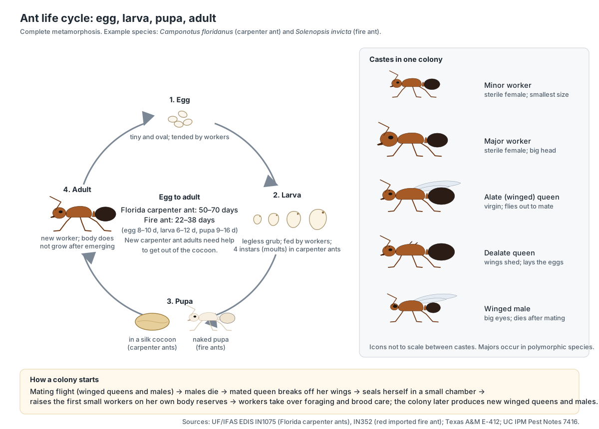
| Species | Egg | Larva | Pupa | Egg to adult |
|---|---|---|---|---|
| Florida carpenter ant (C. floridanus) | — | 4 instars | In silk cocoon | 50–70 days |
| Red imported fire ant (S. invicta) | 8–10 days | 6–12 days | 9–16 days, naked | 22–38 days |
A colony starts with a mating flight. Winged queens and males leave the nest, often after rain; after mating the males die, and each queen breaks off her wings, seals herself in a small chamber and raises her first workers (UC IPM 7416; Texas A&M E-412). Carpenter ant colonies take two to five years before they produce winged reproductives (UF/IFAS EDIS IN1075).
Ant colony diagram: inside an ant hill
An underground ant nest is a set of flat, horizontal chambers joined by vertical shafts. The chambers hold the queen, eggs, larvae, pupae, workers and stored food; waste is carried to refuse areas. Depth ranges from a few centimetres to more than 4 metres depending on the species (Tschinkel, 2011). The mound or crater at the surface is soil carried up during digging.
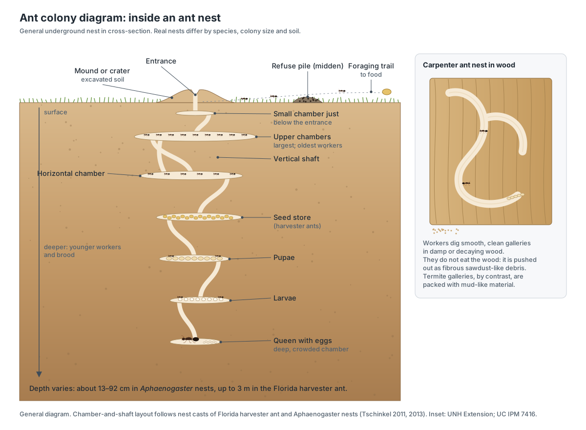
Nest casts of the Florida harvester ant show nests up to about 3 m deep, with ants sorted by age and brood by stage at different depths, and colonies that move and rebuild a near-identical nest about once a year (Tschinkel, 2013). Fire ant mounds are seldom more than 46 cm across (UF/IFAS EDIS IN352). Carpenter ants nest in wood instead: they excavate clean, smooth galleries in moist or decaying wood and often keep satellite nests away from the parent colony (UC IPM 7416).
Carpenter, fire, army, leafcutter and other ants compared
All ants share the same body plan, but the waist, sting, antennae and worker castes differ between groups. Common names such as "black ant", "red ant" and "sugar ant" are used for many different species, so check the waist and the tip of the gaster rather than the colour.
| Ant | Waist | Sting? | Notable anatomy |
|---|---|---|---|
| Carpenter ants (Camponotus) | 1 node | No; acidopore | Evenly rounded mesosoma, 12-segment antennae, no metapleural gland, major and minor workers |
| Red imported fire ant (Solenopsis invicta) | 2 nodes (petiole + postpetiole) | Yes | 10-segment antennae with 2-segment club, 4-toothed mandibles, workers 2.4–6 mm, alkaloid venom |
| Army ants (Eciton, Dorylus) | 1–2 nodes | Yes in Eciton | No permanent nest: workers form a living bivouac around the queen and brood; colonies of about 600,000 (Eciton) to 20 million (Dorylus) (Dejean et al., 2025, 2026) |
| Texas leaf-cutting ant (Atta texana) | 2 nodes | Reduced | Workers 1/16–1/2 inch; large workers guard, medium ones cut leaves, small ones tend the fungus garden; colonies of 2 million or more (Texas A&M Forest Service) |
| Trap-jaw ants (Odontomachus) | 1 node | Yes | Mandibles open 180° and snap shut in about 0.13 ms (Dejean et al., 2026) |
| 'Black ants' and 'sugar ants' | Varies | Varies | Names for many small household ants; several are formicines with an acidopore, like carpenter ants |
Leaf-cutting ants do not eat the leaves they carry. They take them underground to grow a fungus that is their main food, and each new queen carries a pellet of fungus with her to start a new garden (Texas A&M Forest Service). Army ants hunt in raids and relocate often; their bivouacs are made of workers clinging to each other by their legs, with chambers for the queen, eggs, larvae and food (Dejean et al., 2025).
Ant vs termite: how to tell them apart
Ants have a narrow, pinched waist, elbowed antennae and, if winged, forewings longer than the hindwings. Termites have a broad waist with no pinch, straight bead-like antennae and four wings of about equal length (Texas A&M E-412). Winged carpenter ants and termites can swarm at the same time of year, and control methods differ, so identification matters.
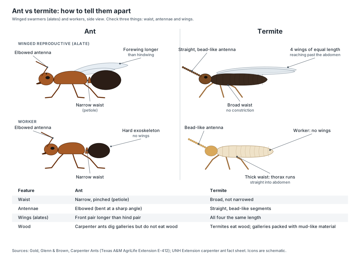
| Feature | Ant | Termite |
|---|---|---|
| Waist | Narrow petiole (1–2 nodes) | Broad, no constriction |
| Antennae | Elbowed (geniculate) | Straight, bead-like |
| Wings (alates) | Forewings longer than hindwings | All four about equal |
| Development | Complete metamorphosis (egg, larva, pupa, adult) | Incomplete metamorphosis (egg, nymph, adult) |
| Wood | Carpenter ants excavate it but do not eat it | Eat wood; galleries packed with mud-like material |
| Order | Hymenoptera (with bees and wasps) | Blattodea (with cockroaches) |
Formic acid: weapon, disinfectant and gut filter
Carpenter ants and other Formicinae lost the working sting of their wasp ancestors. Instead, the poison gland makes a strongly acidic secretion whose main component is formic acid, stored in a reservoir and sprayed through the acidopore at the tip of the gaster. Tragust and colleagues showed that Florida carpenter ants (Camponotus floridanus) also curl the gaster forward, take up a drop of this acid from the acidopore and swallow it. This 'acidopore grooming' keeps the crop at a median pH of about 2, even after the ants drink less acidic food. (Tragust et al. (2020): formicine ants swallow their acidic poison, eLife.)
The acid crop kills many bacteria that arrive with food, which matters in a colony where workers share every meal. It is also selective: some bacteria of the family Acetobacteraceae, which normally live in formicine guts, tolerate the acid and pass on to the midgut. The acidopore is the outlet of the poison gland alone; the anus opens just above it, under the last tergite. The same small nozzle therefore serves both defence and hygiene. (Tragust et al. (2020): formicine ants swallow their acidic poison, eLife.)
Brains that differ by caste
Ant brains are tiny but organised like those of other insects: two optic lobes for vision, two antennal lobes for smell, a central complex for orientation and two mushroom bodies for learning and memory. Ellendula and colleagues measured these regions in three castes of two Australian ants. Female castes had larger brains with pronounced mushroom bodies, matching their wider range of tasks. Males had the smallest brains overall but the largest optic lobes and central complexes, because they find mates in flight. Workers that navigate using visual memories had the largest mushroom bodies of all. (Ellendula et al. (2026): caste- and sex-specific brain investment in ants, iScience.)
The same study notes that males have the largest compound eyes, with more facets, and the largest ocelli. In carpenter ants, winged females and males have more facets and bigger eyes than workers of similar head size. That is why the 3D model's caste switch changes the eyes as well as the wings: a worker carpenter ant has no ocelli at all, while queens and males have three. (Ellendula et al. (2026): caste- and sex-specific brain investment in ants, iScience.)
The metapleural gland: an ant-only antibiotic gland that carpenter ants lack
Almost every ant has a pair of metapleural glands at the back of the mesosoma, just above the hind legs. Their secretions are antimicrobial, and the gland is often called one of the defining features of ants. The opening is a small slit or pore on each side. Carpenter ants are a notable exception. Hölldobler and Engel-Siegel found no metapleural gland in any Camponotus or Polyrhachis species they sectioned, nor in weaver ants (Oecophylla); of 27 Camponotus species checked from the outside, only C. gigas showed an opening. (Hölldobler & Engel-Siegel (1984): on the metapleural gland of ants, Psyche 91.)
For a student this has two practical uses. First, if you are labelling a fire ant, harvester ant or leafcutter, look for the gland opening low on the propodeum. Second, if you are keying out a large black or bicoloured ant with a single waist node, an evenly rounded mesosoma, a hair-fringed acidopore and no metapleural opening, you almost certainly have a carpenter ant. Our 3D model is a carpenter ant, so it has no metapleural gland; the external diagram shows it in the fire ant inset instead. (Hölldobler & Engel-Siegel (1984): on the metapleural gland of ants, Psyche 91.)
What an ant nest looks like underground
Most underground ant nests are built from the same two units: flat, horizontal chambers and the more or less vertical shafts that connect them. Tschinkel filled abandoned nests with plaster, wax or molten aluminium to make casts. The nests of three Aphaenogaster species were simple, small and vertical, with horizontal chambers joined by vertical shafts; shafts going down tended to start at chamber edges. Across species, nests can be a few centimetres deep or more than 4 metres. (Tschinkel (2011): nest architecture of three Aphaenogaster ants, J. Insect Science.)
The Florida harvester ant (Pogonomyrmex badius) digs species-typical nests up to about 3 metres deep, with a characteristic vertical pattern of chamber size and spacing and a sorting of ants by age and of brood by stage at different depths. Colonies move about once a year and build a near-replica of their old nest. These measurements are what our ant colony diagram is based on; it is a general picture, and the details differ from species to species. Carpenter ants instead excavate galleries in soft or rotting wood, and Texas leaf-cutting ants build enormous nests of fungus-garden chambers in deep sandy soil. (Tschinkel (2013): Florida harvester ant nest architecture, PLoS ONE.)
Label the ant: worksheet with answers
Use the 3D model and the diagrams to answer these questions.
- Name the three main body parts of an ant. Answer: head, mesosoma (thorax) and gaster, with the petiole as the waist.
- What are the two parts of an elbowed antenna? Answer: the scape and the funiculus.
- How many waist nodes does a carpenter ant have? A fire ant? Answer: one; two (petiole and postpetiole).
- Which organ is called the social stomach, and why? Answer: the crop, because workers store food in it and share it by regurgitation.
- What does the proventriculus do? Answer: it seals the crop and controls how much food passes into the midgut.
- How does a carpenter ant defend itself without a sting? Answer: it bites and sprays formic acid from the acidopore.
- Which castes have wings and ocelli? Answer: queens (before shedding the wings) and males.
- Give two ways to tell a winged ant from a winged termite. Answer: narrow waist and elbowed antennae (also unequal wings).
Compare with the honeybee anatomy page: bees and ants are both Hymenoptera and share the narrow waist, the antenna cleaner and the hooked wings.
Every structure in the 3D ant model
The labeled list below matches the model (worker, queen and male combined).
Exoskeleton & body regions
- Head (Head capsule)
- Body part 1: a hard, rounded capsule that carries the eyes, antennae and mouthparts. Protects the brain and the huge mandible muscles, and carries the main sense organs. In carpenter ant majors the head is very large, because most of it is filled with muscle.
- Mesosoma (Alitrunk; 'thorax' in simple diagrams)
- Body part 2: the middle section that carries all six legs. Holds the leg muscles and, in winged queens and males, the flight muscles. It is the three thoracic segments plus the first abdominal segment (propodeum) fused to them.
- Pronotum
- The collar-like top plate of the first thoracic segment. Covers the prothorax, which carries the front legs, and anchors neck and foreleg muscles.
- Mesonotum
- The top plate of the second thoracic segment. Covers the mesothorax, which carries the middle legs. In queens and males it is greatly enlarged to hold the flight muscles of the forewings.
- Propodeum
- The last part of the mesosoma: abdominal segment 1, fused to the thorax. Forms the joint with the petiole and carries the propodeal spiracle. In most ants (but not carpenter ants) the opening of the metapleural gland lies just below it.
- Propodeal spiracle
- A breathing hole on the side of the propodeum, the spiracle of abdominal segment 1. Lets air into the tracheae (air tubes). It is one of the spiracles lab diagrams most often ask you to label.
- Petiole (Waist node, abdominal segment 2)
- The narrow waist segment with a raised node or scale between the mesosoma and gaster. Lets the gaster swing forward under the body, for example to aim the acidopore or sting at an enemy.
- Gaster (The swollen part of the abdomen)
- Body part 3: the large, rounded rear section behind the waist. Holds the crop, midgut, hindgut, heart, ovaries and the venom (formic acid) apparatus.
- Acidopore
- A small round nozzle at the tip of the gaster, fringed with a circle of hairs. The opening of the poison gland. Formicine ants, including carpenter ants, have no working sting. They bite and then spray formic acid from the acidopore into the wound or at an attacker.
Head, eyes & mouthparts
- Compound eyes
- Two oval eyes made of many small facets (ommatidia), one on each side of the head. Detect movement, light and landmarks used in navigation. Carpenter ant workers see reasonably well; many soil ants have tiny eyes.
- Ocelli (Simple eyes)
- Three tiny simple eyes in a triangle on top of the head of queens and males. Help winged ants hold a level flight path and read the polarised light of the sky.
- Antennal scape (First antennal segment)
- The long first segment of each antenna, which forms the shaft before the 'elbow'. Holds the rest of the antenna out in front of the head. The joint at its tip is what makes ant antennae elbowed (geniculate).
- Antennal funiculus (Flagellum with pedicel)
- The chain of small segments beyond the elbow that does most of the smelling and touching. Carries thousands of sensilla (sense hairs and pegs) for smell, taste, touch, humidity and temperature. Ants recognise nestmates, trails and food with it.
- Clypeus
- The plate on the front of the face just above the mandibles. Covers the base of the mouth and is the attachment point for the muscles of the sucking pump (cibarium) inside.
- Frontal carinae
- Two raised ridges between the antennal sockets. Partly shield the antennal sockets and guide the scape when the antenna is folded back.
- Labrum (Upper lip)
- A small flap hanging below the clypeus, usually hidden behind the closed mandibles. Closes the mouth cavity from the front and helps hold food against the other mouthparts.
- Mandibles (Jaws)
- A pair of strong, toothed jaws that open sideways. The ant's hands and tools: they dig, cut wood, carry brood and food, fight and grip. They do not grind food for swallowing; ants swallow liquids.
- Maxillae
- The second pair of jaws, behind the mandibles, with a soft lobe (galea) at each tip. Together with the labium they form the maxillolabial complex, which laps up liquids and pushes food particles into the mouth.
- Maxillary palps
- A pair of jointed feelers on the maxillae. Taste and touch food before it is eaten.
- Labium (Lower lip)
- The fused lower lip that forms the floor of the mouth. Carries the glossa and the labial palps; the salivary (labial) gland duct opens on it.
- Labial palps
- A pair of short jointed feelers on the labium. Taste and handle food.
- Glossa (Tongue)
- A short, soft, hairy tongue at the tip of the labium. Laps up liquids such as nectar, honeydew and water. Liquids lapped by the glossa pass straight into the mouth (Richter & Economo, 2023).
Legs & wings
- Coxae
- The first, thick leg segment that joins each leg to the mesosoma. Swing the legs forward and back.
- Trochanters
- Small segments between the coxa and the femur. Act as a hinge that lets the femur lift and lower.
- Femora (Thighs)
- The long, strong third leg segment. Holds the muscles that move the tibia.
- Tibiae
- The long, slender leg segment after the femur. Gives the long stride of fast-running ants such as carpenter ants.
- Tibial spurs
- Small, stiff spurs at the tip of the tibiae of the middle and hind legs. Grip the ground and help clean the legs and body.
- Strigil (Antenna cleaner)
- A comb-like spur on the front tibia that meets a notch on the first tarsal segment. The ant pulls an antenna through the gap between the spur and the notch to comb off dirt, so its smell receptors keep working.
- Tarsi (Feet)
- The jointed 'foot' made of five small segments (tarsomeres). Spread the ant's weight and carry the claws and adhesive pad.
- Tarsal claws & arolium
- A pair of hooked claws with a soft adhesive pad (arolium) between them. The claws hook onto rough surfaces; the pad sticks to smooth ones, so ants can walk up glass and upside down.
- Forewings
- The larger front pair of wings of winged queens and males (alates). Used once, for the mating flight. Queens break their wings off afterwards and leave small wing scars.
- Hindwings
- The smaller back pair of wings of alates. Hook onto the forewing so both wings on each side beat as one surface.
Muscles
- Mandible closer muscles (Mandibular adductor muscles)
- Two huge muscles that fill most of the head and close the mandibles. Power biting, cutting and carrying. The big heads of major workers are mostly these muscles.
- Flight muscles
- Large blocks of muscle that fill the mesosoma of winged queens and males. Beat the wings during the mating flight. After a queen sheds her wings, she breaks these muscles down and uses them as food for her first brood.
Digestive
- Pharynx (Sucking pump (cibarium + pharynx))
- A muscular pump behind the mouth that sucks liquid food in. Draws liquids into the oesophagus. Ants mostly eat liquids; solid particles are filtered out first.
- Infrabuccal pocket (Infrabuccal pouch)
- A small pouch in the floor of the mouth that traps solid particles. A filter: food particles and dirt collect here, are pressed into a pellet and spat out. Only liquid and tiny particles reach the gut.
- Oesophagus (Esophagus)
- A thin tube that carries food from the head, through the mesosoma and waist, to the crop. Carries liquid food. It has to be thin to fit through the narrow waist.
- Crop (Social stomach)
- A thin-walled, stretchy sac at the front of the gaster that stores liquid food. Holds food that the ant shares with nestmates by regurgitation (trophallaxis). Food here is not yet digested.
- Proventriculus
- A muscular valve and pump between the crop and the midgut. Keeps the crop sealed so stored food stays shareable, and pumps small amounts into the midgut when the ant itself needs food.
- Midgut (Ventriculus, stomach)
- The 'true stomach', where food is digested and absorbed. Secretes digestive enzymes and absorbs nutrients into the hemolymph (insect blood).
- Malpighian tubules
- Fine, blind-ended tubes that remove wastes from the hemolymph. The insect kidney: they draw out nitrogen waste and salts and empty them into the hindgut.
- Hindgut (Ileum)
- A short tube that leads from the midgut to the rectum. Absorbs water and salts and carries waste to the rectum.
- Rectum
- The last, slightly swollen part of the gut, with rectal pads. Reabsorbs water and stores faeces until the ant leaves the nest or drops them in a refuse chamber.
Glands & defence
- Mandibular glands
- A pair of sac-like glands that open at the base of the mandibles. Produce alarm and defence chemicals that are released when the ant bites or is disturbed.
- Postpharyngeal glands
- Finger-like glands opening into the pharynx, found only in ants. Store and mix the colony's hydrocarbon smell, which ants use to recognise nestmates.
- Labial (salivary) glands
- Branched glands in the mesosoma whose duct opens on the labium. Make saliva. In carpenter ants the larvae, not adults, produce silk for their cocoons from their own labial glands.
- Poison gland (Venom gland)
- A pair of fine secretory filaments that make the ant's venom. In carpenter ants it makes a strong formic acid solution, sprayed from the acidopore. In stinging ants it makes the venom injected by the sting.
- Poison reservoir (Venom sac)
- A large, thin-walled sac that stores formic acid. Holds the venom until the ant sprays it through the acidopore.
- Dufour's gland
- A small sac-like gland beside the poison reservoir. Makes oily hydrocarbons used in alarm signals, trail marking or defence; in fire ants it is the source of the trail pheromone.
Heart & breathing
- Heart (Dorsal vessel (abdominal part))
- A long muscular tube along the top of the gaster with small valved openings (ostia). Pumps hemolymph forward into the aorta. Insects have an open circulation: the blood then bathes the organs.
- Aorta
- The front, narrow part of the dorsal vessel. Carries hemolymph forward and releases it around the brain.
- Tracheae (Tracheal system, air sacs)
- Branching air tubes that carry oxygen straight to the cells. Insects breathe without lungs: air flows in through the spiracles and along the tracheae to every tissue.
- Spiracles
- Small breathing holes along the sides of the body. Let air in and out of the tracheae. They can close to save water.
- Fat body
- Loose white tissue that lines the gaster. Stores fat and protein and makes many blood proteins; it works like an insect liver.
Nervous
- Brain (Supraoesophageal ganglion)
- The main nerve centre above the oesophagus in the head. Processes smell, sight and touch and controls behaviour.
- Mushroom bodies (Corpora pedunculata)
- A pair of cup-shaped centres on top of the brain. Learning and memory: they combine smell and vision, for example to remember landmarks on the way home.
- Optic lobes
- Side lobes of the brain that receive nerves from the compound eyes. Process images from the compound eyes.
- Antennal lobes
- Round lobes at the front of the brain that receive the antennal nerves. First processing centre for smells, including trail and nestmate pheromones.
- Subesophageal ganglion
- A nerve centre below the oesophagus, joined to the brain by a ring of nerves. Controls the mandibles and other mouthparts.
- Thoracic ganglia
- Nerve centres on the floor of the mesosoma. Control the legs (and wings in alates).
- Ventral nerve cord (With gastral ganglia)
- A double nerve cord along the belly with small ganglia in the gaster. Links the brain to the gaster and controls the gut, heart and acid spraying.
Reproductive
- Ovaries
- Egg-producing organs made of tubes called ovarioles: tiny in workers, huge in queens. In the queen they make all the colony's eggs. Carpenter ant workers have a few short ovarioles and may lay unfertilised eggs that become males or are fed to larvae.
- Spermatheca
- A small round sac in the queen that stores sperm from her mating flight. Keeps sperm alive for years. The queen fertilises each egg as it is laid: fertilised eggs become females (workers or queens), unfertilised eggs become males.
- Testes
- The male's sperm-producing organs. Make sperm before the mating flight; in many ants the testes have shrunk by the time the male flies, with sperm stored in the seminal vesicles.
- Male genitalia (Genital capsule)
- A hard capsule of claspers at the tip of the male's gaster. Grips the queen and transfers sperm during mating.
Frequently asked questions
What are the 3 main body parts of an ant?
An ant has a head, a mesosoma and a gaster. The head carries the eyes, elbowed antennae and mandibles. The mesosoma (often called the thorax) carries all six legs. The gaster is the swollen rear part. Between the mesosoma and gaster is the narrow waist, made of one segment (the petiole) in carpenter ants and two (petiole and postpetiole) in fire ants.
What is the petiole of an ant?
The petiole is the narrow waist segment between the mesosoma and the gaster. It usually has a raised hump called a node. It is really the second abdominal segment, and it lets the ant bend its gaster forward to sting or spray. Carpenter ants have one waist segment; fire ants and other Myrmicinae have two.
Do ants have a heart?
Yes. An ant has a tube-shaped heart (the dorsal vessel) running along the top of its gaster. It pumps hemolymph forward through the aorta into the head. Ants have an open circulation, and they breathe through spiracles and tracheae rather than through their blood.
Do ants have two stomachs?
In a sense, yes. The crop is a storage sac often called the 'social stomach', because ants regurgitate its contents to feed nestmates. Behind it, a valve called the proventriculus lets small amounts into the midgut, the 'personal stomach', where food is digested.
Do carpenter ants sting?
No. Carpenter ants belong to the Formicinae, which have no working sting. They bite with their mandibles and spray formic acid from a round, hair-fringed opening at the tip of the gaster called the acidopore. Fire ants, harvester ants and many others do sting.
How many legs and antennae does an ant have?
Six legs, all attached to the mesosoma, and two elbowed antennae. Each leg has a coxa, trochanter, femur, tibia and five-part tarsus with two claws. A carpenter ant worker's antenna has 12 segments; a fire ant worker's has 10 with a two-segmented club.
Do ants have eyes?
Most ants have two compound eyes. Queens and males also have three small simple eyes (ocelli) on top of the head, which carpenter ant workers lack. Some army ant and soil-dwelling workers are nearly or fully blind and rely on smell.
How can you tell an ant from a termite?
Ants have a narrow pinched waist, elbowed antennae and, when winged, forewings longer than the hindwings. Termites have a broad waist, straight bead-like antennae and four wings of about equal length (Texas A&M AgriLife E-412).
How long is the ant life cycle?
Ants go through complete metamorphosis: egg, larva, pupa and adult. A Florida carpenter ant takes about 50–70 days from egg to adult; a red imported fire ant takes 22–38 days (UF/IFAS EDIS IN1075 and IN352). Worker lifespans are weeks to months; queens can live for many years.
References
- Koehler, P. G., Pereira, R. M. & Scheffrahn, R. H. Florida Carpenter Ants. UF/IFAS EDIS SP486C/IN1075.
- Collins, L. & Scheffrahn, R. H. Red Imported Fire Ant, Solenopsis invicta Buren. UF/IFAS EDIS EENY-195/IN352.
- Gold, R. E., Glenn, G. J. & Brown, W. Carpenter Ants. Texas A&M AgriLife Extension E-412.
- Klotz, S. A., Hansen, L. D. & Rust, M. K. Carpenter Ants. UC IPM Pest Notes, UC ANR Publication 7416.
- Tragust, S. et al. (2020). Formicine ants swallow their highly acidic poison for gut microbial selection and control. eLife 9, e60287.
- Eisner, T. & Happ, G. M. (1962). The infrabuccal pocket of a formicine ant: a social filtration device. Psyche 69, 107–116.
- Richter, A. & Economo, E. P. (2023). The feeding apparatus of ants: an overview of structure and function. Phil. Trans. R. Soc. B 378, 20220556.
- Hölldobler, B. & Wilson, E. O. (1990). The Ants. Cambridge, MA: Belknap Press of Harvard University Press.
- Hölldobler, B. & Maschwitz, U. (1965). Der Hochzeitsschwarm der Rossameise Camponotus herculeanus L. Zeitschrift für vergleichende Physiologie 50, 551–568.
- Lanan, M. C. et al. (2016). A bacterial filter protects and structures the gut microbiome of an insect. The ISME Journal 10, 1866–1876.
- Hölldobler, B. & Engel-Siegel, H. (1984). On the metapleural gland of ants. Psyche 91, 201–224.
- Ellendula, S. et al. (2026). Caste- and sex-specific differential investment in brain regions of Australian ants. iScience.
- Dejean, A. et al. (2025). Foraging by predatory ants: a review. Insect Science.
- Dejean, A. et al. (2026). The predatory behavior of ants: an impressive panoply of morphological adaptations. Insect Science.
- Tschinkel, W. R. (2011). The nest architecture of three species of north Florida Aphaenogaster ants. Journal of Insect Science 11, 105.
- Tschinkel, W. R. (2013). Florida harvester ant nest architecture, nest relocation and soil carbon dioxide gradients. PLoS ONE 8, e59911.
- Texas A&M Forest Service. Texas leaf-cutting ant (Atta texana).
- Vergara-Martínez, M. F. et al. (2025). Age, caste, and social context shape ovarian morphology in red harvester ants. npj Aging.
More interactive anatomy models
- Insect body partsHead, thorax and abdomen: the three body parts every insect shares.
- Grasshopper anatomyExternal and internal anatomy of the grasshopper, with mouthparts.
- Grasshopper dissectionGuided grasshopper dissection: crop, gastric caeca, heart and tracheae.
- Butterfly anatomyScaled wings, proboscis, compound eyes and internal organs of a butterfly.
- Honeybee anatomyWorker bee anatomy: pollen baskets, honey stomach, sting and wax glands.
- Human anatomy1,000+ bones, muscles, organs, vessels and nerves from real anatomical data.
How ants share food: the crop, the proventriculus and trophallaxis
An ant worker has two 'stomachs' in a working sense. The crop at the front of the gaster is a stretchy storage bag. Food in it is not digested and can be brought back up and passed mouth-to-mouth to a nestmate, a behaviour called trophallaxis. Behind the crop, the proventriculus works as a pump with an intake valve on the crop side and an outlet valve on the midgut side. In formicine and most dolichoderine ants the intake is a narrow cross-shaped cleft that stays almost closed, so the crop can hold far more liquid than the ant itself needs. Eisner and Happ described the crop as a 'social stomach' because, taken together, the crops of all workers form a food store for the whole colony. (Eisner & Happ (1962): the infrabuccal pocket of a formicine ant, Psyche 69.)
This design depends on liquid food. Ants first strain what they eat. Solid particles are caught at the mouth and packed into the infrabuccal pocket, a pouch in the floor of the mouth; only fluid and tiny particles reach the gut. Liquids licked up by the glossa pass the filter directly. The pellets from the pocket are usually spat out onto refuse piles; in some ants workers feed them to larvae, which, unlike adults, can eat solid food. When you read an ant internal anatomy diagram, follow the food in this order: mouthparts → infrabuccal pocket → pharynx → oesophagus (through the narrow waist) → crop → proventriculus → midgut → hindgut and rectum. (Richter & Economo (2023): the feeding apparatus of ants, Phil. Trans. R. Soc. B.)