

Crayfish dissection at a glance
A crayfish dissection has three parts. First you study the outside and remove the appendages in order. Then you take off the carapace to expose the gills, heart, gonads, stomach and digestive gland. Finally you clear the head to find the green glands and brain, and open the abdomen to see the muscles and nerve cord. Most classes need two or three lab periods.
Any preserved cambarid crayfish works. This guide is written for the red swamp crayfish, Procambarus clarkii, and notes where other common lab species, such as Faxonius (formerly Orconectes) and Cambarus, look different. The internal organs are the same in all of them. For the full anatomy, including how each system works, see the crayfish anatomy page.
What tools are necessary for a crayfish dissection?
You need fine dissecting scissors, fine forceps, a blunt probe, pins, a dissecting tray and a hand lens. A scalpel is optional: the shell is cut more safely with scissors. Gloves and goggles are essential, and an index card with tape lets you mount the appendages.
Materials
- Preserved crayfish, about 8–12 cm long (one per pair of students)
- Dissecting tray with a wax or rubber base
- Fine-pointed dissecting scissors (the main cutting tool)
- Fine forceps and a blunt probe or dissecting needle
- Scalpel (optional; scissors are safer on the shell)
- Dissecting pins
- Hand lens or dissecting microscope, plus a slide and cover slip for a gill filament
- An index card and clear tape, or a small dish, for laying out the appendages in order
- Nitrile gloves, safety goggles, a lab apron and paper towels
Safety
- Wear gloves and goggles. Preserved specimens are stored in fluids that irritate skin and eyes; rinse the crayfish under running water before you start and work in a ventilated room.
- Point scissor tips upward and keep them just under the shell, so you cut the exoskeleton and not the organs below it.
- The tips of the rostrum, claws and telson are sharp. Handle the crayfish by the carapace.
- Never eat or drink in the lab. Dispose of the specimen and tissue as your teacher directs, and wash your hands afterwards.
Crayfish dissection labeled diagrams
These diagrams show what you should find at each stage: the internal organs from above once the carapace is off, the underside of a male and a female, and all the appendages laid out in order.
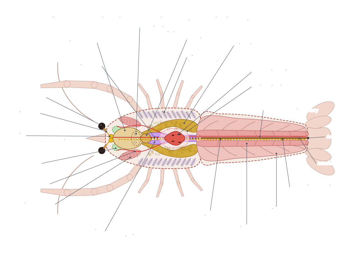
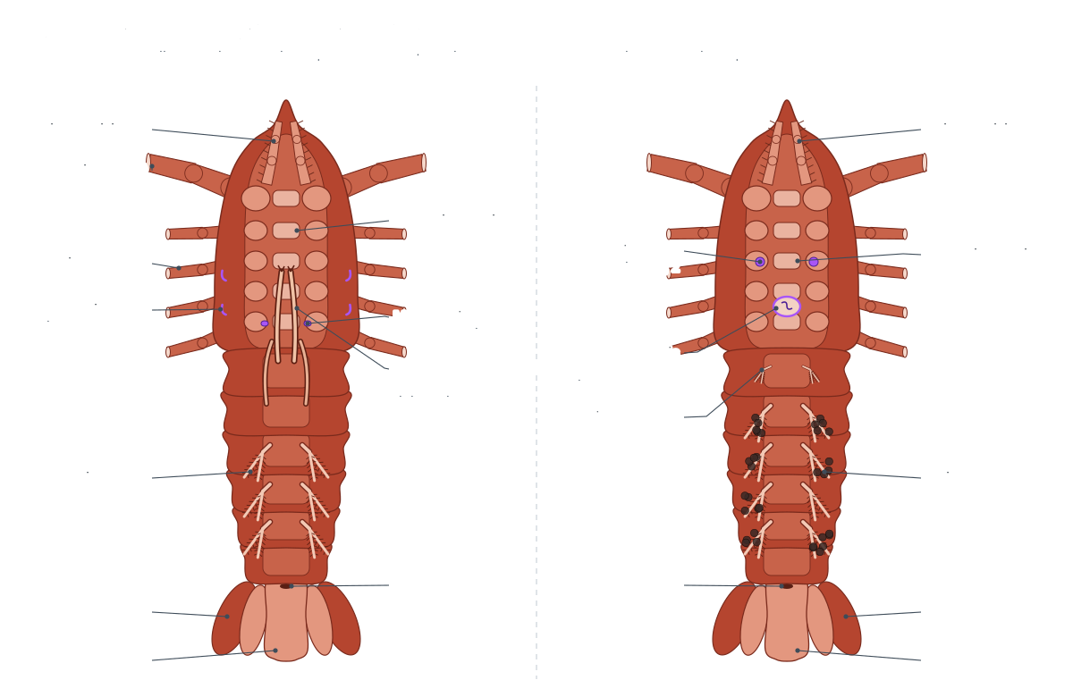
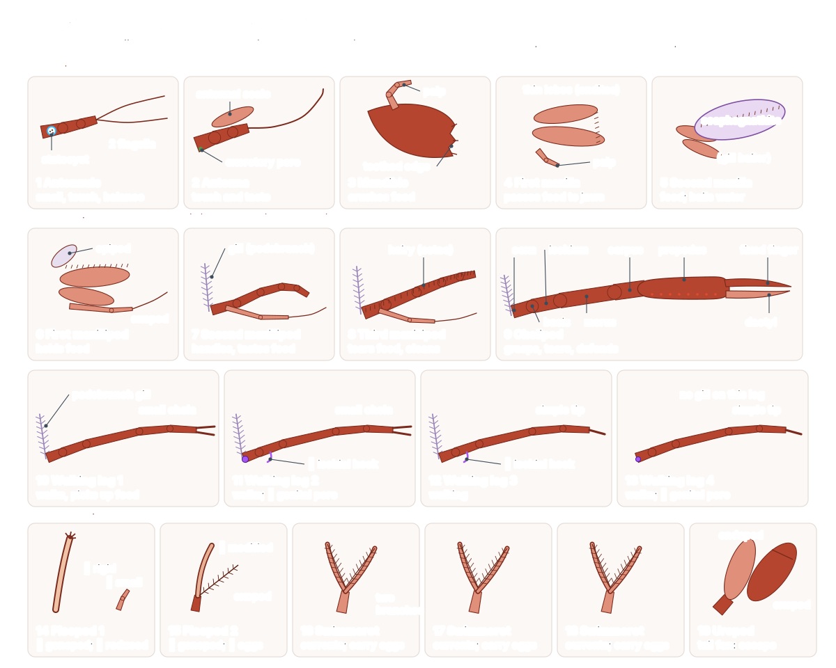
External observation before you cut
Spend the first part of the lab on the outside of the crayfish. You will answer most worksheet questions from this stage, and it is the only time the specimen is intact.
- Measure and draw: record the length from rostrum tip to telson tip, and draw the dorsal view larger than life, labelling the carapace, rostrum, cervical groove, eyes, antennules, antennae, chelipeds, walking legs, abdominal segments, telson and uropods.
- Count: abdominal segments (6), pairs of walking legs (4, plus the chelipeds), and branches on the antennules (2).
- Feel: the hard carapace against the flexible joints between abdominal segments. The difference is the reason the abdomen can curl for the tail-flip.
- Look closely: with a hand lens, examine the surface of a compound eye for the many tiny facets, each the lens of one ommatidium.
- Turn it over: find the third maxillipeds covering the mouth, the sternites between the leg bases, the swimmerets, and the anus on the underside of the telson. Then sex the crayfish (step 2).
How to dissect a crayfish: the procedure
The order below follows common high-school and college lab manuals: outside first, then appendages, then the organs from the top down in the order you meet them once the carapace is off. Each numbered step matches a step in the 3D viewer above.
1. External anatomy: dorsal view
- Place the crayfish in the tray with its back (dorsal side) up and the rostrum pointing away from you.
- Find the two body regions: the cephalothorax, covered by the carapace, and the six-segmented abdomen. Count the abdominal segments.
- Locate the rostrum, the cervical groove across the carapace, and the compound eyes on their movable eyestalks.
- Find the short, two-branched antennules and the long antennae. Note the flat antennal scale at the base of each antenna.
- At the rear, find the telson in the middle of the tail fan and a uropod on each side of it.
Identify: Carapace, Rostrum, Cervical groove, Compound eyes, Eyestalks, Antennules, Antennae, Antennal scale, Abdomen, Telson, Uropods, Branchiostegite.
2. Ventral view: legs, swimmerets and the sex of your crayfish
- Turn the crayfish on its back. Count the pairs of legs: one pair of chelipeds and four pairs of walking legs.
- Look at the swimmerets under the abdomen. If the first two pairs are hard and point forward between the last walking legs, they are gonopods and your crayfish is male.
- Male: find the genital pores on the inner side of the bases of the fourth (last) walking legs, and the small hooks on the walking legs.
- Female: find the genital pores on the bases of the second walking legs, and the round annulus ventralis between the bases of the third and fourth walking legs. Use the Female/Male switch on the model to compare.
- Find the thoracic sternites between the leg bases and the anus on the underside of the telson.
Identify: Chelipeds, First walking legs, Second walking legs, Third walking legs, Fourth walking legs, Swimmerets, Gonopods, Male genital pores, Ischial hooks, Female genital pores, Annulus ventralis, First pleopods (female), Thoracic sternites, Anus.
3. Remove and identify the appendages
- Work on one side only, so the other side stays intact for comparison.
- Remove each appendage by gripping it with forceps right at its base and pulling gently while twisting, so the whole appendage comes off, including the gill attached to it.
- Start at the front with the antennule and antenna and work backward: mouthparts (next step), cheliped, the four walking legs, the swimmerets and the uropod.
- Tape the appendages to an index card in order and label each one with its name and function. Note which are biramous (two-branched).
- Look at the base of a walking leg you removed: the feathery structure attached to it is a podobranch (gill).
Caution: Pull steadily. Jerking tears the appendage at the joint and leaves the base and gill behind.
Identify: Antennules, Antennae, Chelipeds, First walking legs, Second walking legs, Third walking legs, Fourth walking legs, Swimmerets, Uropods, Podobranchs.
4. Mouthparts: from the outside in
- With the crayfish on its back, find the large, hairy third maxillipeds covering the mouth region. Remove one.
- Underneath, remove in order the second maxilliped, the first maxilliped, the second maxilla and the first maxilla. Use a hand lens: they are thin and fragile.
- On the second maxilla, find the flat scaphognathite (gill bailer). It sits in the front of the gill chamber and beats to pump water over the gills.
- The last, hard mouthparts beside the mouth are the mandibles. Note their toothed edges and small palp.
Identify: Third maxillipeds, Second maxillipeds, First maxillipeds, Second maxillae, First maxillae, Scaphognathite, Mandibles, Mouth.
5. Remove the carapace and examine the gills
- Turn the crayfish dorsal side up. Insert the tip of the scissors under the rear edge of the carapace at one side.
- Cut forward along the side, just above where the legs join the body, up to the eye. Repeat on the other side.
- Lift the carapace from behind with forceps. Use the probe to free the thin membrane under it, especially over the heart, then remove it. (Some labs instead cut along the dorsal midline and pin the two halves back.)
- The feathery gills now lie exposed on each side in the branchial chamber. Find the podobranchs on the leg bases, the arthrobranchs on the joint membranes, and the pleurobranchs on the body wall.
- Cut off one gill, float it in water on a slide, and look at its filaments under the hand lens or microscope.
Caution: Keep the scissors shallow. The heart lies right under the carapace behind the cervical groove.
Identify: Podobranchs, Arthrobranchs, Pleurobranchs, Gill filaments, Scaphognathite, Branchiocardiac canals.
6. The heart and circulation
- Find the small, pale heart in the midline just behind where the cervical groove was. It sits in the pericardial sinus.
- With a hand lens, look for the slit-like ostia on its surface.
- Look for the fine arteries leaving the heart: the ophthalmic artery running forward, the antennal arteries forward and outward, and the dorsal abdominal artery running back into the abdomen.
- Note that no veins enter the heart. Blood returns through sinuses and the gills to the pericardial sinus: an open circulatory system.
Identify: Heart, Ostia, Pericardial sinus, Ophthalmic artery, Antennal arteries, Hepatic arteries, Dorsal abdominal artery, Sternal artery.
7. Reproductive organs
- Carefully remove the heart with forceps. The gonad lies just beneath it.
- Female: the ovary is a three-lobed mass, often orange or dark, with visible round eggs. Trace an oviduct down to the base of the second walking leg.
- Male: the testis is a smaller, white, three-lobed organ. Trace the coiled white sperm duct down to the base of the fourth walking leg.
- Compare with a lab partner who has the other sex, or use the Female/Male switch.
Identify: Ovary, Oviducts, Female genital pores, Testes, Sperm ducts, Male genital pores.
8. Stomach, gastric mill and digestive gland
- Find the large, thin-walled cardiac stomach in the head, behind the eyes. Note the mandibular muscles on each side of it.
- Find the large, soft, yellow-green digestive gland (hepatopancreas) filling both sides of the cephalothorax behind the stomach.
- Find the pyloric stomach behind the cardiac stomach and trace the thin intestine back into the abdomen.
- Cut the stomach out, open it, and rinse it. Find the three hard teeth of the gastric mill. Look at what your crayfish was eating.
- If your crayfish was close to molting, you may find two white, disc-shaped gastroliths in the stomach wall.
Identify: Cardiac stomach, Gastric mill, Pyloric stomach, Gastroliths, Digestive gland, Intestine, Oesophagus, Mandibular muscles.
9. Green glands (excretion)
- Remove the stomach and digestive gland. Look in the floor of the head, just behind the bases of the antennae.
- Find the two small, round green glands. They may look beige or brown rather than green in preserved specimens.
- Look for the thin bladder beside each one. On the outside, the excretory pore is on the underside of the antenna's first segment.
Identify: Green glands, Bladder, Excretory pores.
10. Brain and ventral nerve cord
- Remove the remaining tissue in the head. Find the small, white brain (supraoesophageal ganglion) between the eyes, just behind the rostrum.
- Trace the optic nerves to the eyes, and the circumoesophageal connectives that run down around the oesophagus.
- Remove the gills and the remaining organs from the thorax. Find the ventral nerve cord on the floor of the body, with a ganglion in each segment.
- Find the statocysts in the bases of the antennules; you may see sand grains inside.
Identify: Brain, Optic nerves, Circumoesophageal connectives, Suboesophageal ganglion, Ventral nerve cord, Statocysts, Sternal sinus.
11. Abdomen: muscles, intestine and nerve cord
- Cut through the tergites along the dorsal midline of the abdomen, from the front to the telson, and peel the shell back.
- Find the dorsal abdominal artery and the thin extensor muscles under the shell, then the intestine running back to the anus.
- Below it, the large, white, twisted flexor muscles fill the abdomen. These power the tail-flip.
- Remove the flexor muscles to see the ventral nerve cord and its six abdominal ganglia on the floor of the abdomen.
Identify: Abdominal extensor muscles, Abdominal flexor muscles, Intestine, Dorsal abdominal artery, Abdominal ganglia, Ventral nerve cord, Anus.
Removing the carapace: the key step
To remove the carapace, cut forward along each side from the rear edge to the eyes, just above where the legs join the body, then lift the shell off from behind while separating it from the membrane underneath with a probe. Done well, it leaves the heart in place and the gills fully exposed.
The heart is the organ most often lost at this stage, because it lies directly under the areola (the narrow strip of carapace behind the cervical groove) and the thin membrane around it is attached to the shell. Lift slowly and watch the midline. Some lab manuals, including Biology Junction's, instead make a single dorsal cut from the back of the thorax forward to the rostrum and pin the two halves out to the sides. That method keeps the branchiostegites attached so you can see how they enclose the gill chamber, but it is easier to cut through the heart.
Once the carapace is off, you are looking at the organs in layers. On the surface in the midline is the heart. In front of it, filling the head, is the large cardiac stomach, with a mandibular muscle on each side. Along both sides lie the feathery gills, and between the gills and the midline you can see the edges of the yellow-green digestive gland. The gonad is hidden under the heart.
Gills, heart, stomach, green glands and brain: what to look for
Each internal organ has a feature that proves you have found the right thing. Use these checks before you label your drawing.
- Gills: feathery plumes in rows, many attached to the leg bases. Pull a walking leg off at its base and a gill (podobranch) comes with it. Float one in water and its filaments spread out.
- Heart: small, pale and roughly diamond-shaped, in the dorsal midline of the thorax. Look for the slit-like ostia with a hand lens. There are no veins attached to it.
- Stomach: a thin-walled sac in the head. Cut it open and rinse it: the hard teeth of the gastric mill are inside its back end, and you may find the last meal.
- Digestive gland: soft, crumbly, yellow-green or brown tissue that fills most of the cephalothorax. It is by far the largest organ.
- Green glands: two small, round bodies in the floor of the head behind the antennae. They are often beige or brown in preserved crayfish, so find them by position, not colour.
- Brain: a tiny, white, two-lobed mass between the eyes, just behind the rostrum, with nerves running out to the eyes and antennae and two connectives looping down around the oesophagus.
- Ventral nerve cord: a white cord on the floor of the body with a swelling (ganglion) in each segment, best seen in the abdomen after the flexor muscles are removed.
Male vs female: what changes in the dissection
Only two steps differ between the sexes: sexing the specimen from below (step 2) and finding the gonads and their ducts (step 7). Everything else is the same.
- Female: small, soft first swimmerets; genital pores on the bases of the second walking legs; a round annulus ventralis between the bases of the third and fourth walking legs. Inside, a large three-lobed ovary under the heart, often orange or dark and packed with eggs, and short, wide oviducts.
- Male: two pairs of hard, forward-pointing gonopods; genital pores on the bases of the fourth walking legs; in the red swamp crayfish, small hooks on the ischium of the second and third walking legs. Inside, a smaller white three-lobed testis and long, coiled white sperm ducts.
If your crayfish is a female carrying eggs, you will see them glued to the swimmerets as dark beads. Count them and record it: females fan the eggs with the swimmerets to keep them supplied with oxygen. Male cambarids switch between a breeding form with hard, sharp gonopods and a non-breeding form with softer ones, so gonopods can look different between specimens.
Preparing a preserved crayfish
Rinse the specimen under running water for a minute to remove excess preservative, and blot it dry so it does not slide in the tray. Check it before you start: a crayfish with a soft, wrinkled shell had recently molted, and one missing a claw may show a small regenerating limb bud. Both are worth recording. Measure the body length from the tip of the rostrum to the end of the telson. Adult red swamp crayfish are 5.5 to 12 cm long. (USGS Nonindigenous Aquatic Species: Procambarus clarkii (red swamp crayfish) fact sheet.)
Plan the work over more than one session. A standard school lab spends the first session on external anatomy and drawing, the second on the thorax (carapace, gills, heart, stomach), and the third on the remaining organs and the abdomen. Between sessions, wrap the crayfish in a damp paper towel and seal it in a labelled bag so the tissues do not dry out. (Biology Junction: Crayfish dissection lab.)
Making an appendage card that earns full marks
Remove appendages from one side only and keep the other side intact for comparison. Grip each one with forceps as close to the body as possible and pull with a slight twist. If you pull from the middle of a leg, it breaks at a joint and you lose the coxa and its attached gill. Work from front to back so you do not mix up the mouthparts, which are small and similar-looking.
Tape the appendages to an index card in order and number them 1 to 19. For each, write its name, the body region (head, thorax or abdomen), whether it is biramous or uniramous, and one function. Add a note next to walking leg 2 or walking leg 4 about the genital pore if you can see it, and next to the first swimmerets about the sex of your specimen. This one card answers most of the external anatomy questions on a typical worksheet.
Common mistakes when opening the cephalothorax
The most common mistake is cutting too deep. The heart lies directly under the carapace, behind the cervical groove, and the membrane that holds it is attached to the inside of the shell. Keep the scissor points angled up against the shell and lift the carapace slowly, freeing the membrane with a probe as you go. If the heart comes away with the carapace, look for it on the inside of the shell before you discard it.
The second mistake is pulling out the digestive gland while looking for the gonads. The gonad lies between the heart and the digestive gland, so remove the heart first and look straight down. In a female the ovary is often large, orange or dark and full of eggs; in a male the white testis is much smaller and easy to miss. If you cannot tell, go back to the swimmerets: hard, forward-pointing first pairs mean male. (Huxley, T. H. (1880). The Crayfish: An Introduction to the Study of Zoology. Internet Archive.)
Where lab crayfish come from, and why you should never release one
Most preserved crayfish in North American classrooms are cambarids such as the red swamp crayfish, which is farmed on a large scale in the southern United States, especially Louisiana. Using a farmed or fishery-caught animal means no wild population is harvested just for teaching. Ask your supplier for the species name and record it in your lab report, because details such as gonopod shape and rostrum shape differ between species. (USGS Nonindigenous Aquatic Species: Procambarus clarkii (red swamp crayfish) fact sheet.)
If your class uses live crayfish for behaviour observations, never release them into a pond or stream afterwards. The red swamp crayfish has spread far beyond its native Gulf coastal plain and is an invasive species in many U.S. states and other countries, where it outcompetes native crayfish, reduces aquatic plant cover and eats the eggs and young of amphibians and fish. The USGS fact sheet lists aquaculture, the aquarium trade, bait use and release after classroom or lab use among the ways it has spread. (USGS Nonindigenous Aquatic Species: Procambarus clarkii (red swamp crayfish) fact sheet.)
Crayfish dissection lab questions and answer key
Answer these during or after the dissection. Click a question to reveal the answer.
What are the two main body regions of a crayfish, and what covers the first one?
The cephalothorax (head and thorax fused) and the abdomen. The cephalothorax is covered by the carapace.
How many pairs of appendages does a crayfish have? Which are on the head, the thorax and the abdomen?
19 pairs. Head: antennules, antennae, mandibles, first and second maxillae (5). Thorax: three pairs of maxillipeds, the chelipeds and four pairs of walking legs (8). Abdomen: five pairs of swimmerets and the uropods (6).
How did you tell the sex of your crayfish?
By the first two pairs of swimmerets. In males they are hard, forward-pointing gonopods; in females they are small and soft. Males have genital pores on the bases of the fourth walking legs; females have them on the second walking legs and have an annulus ventralis between the third and fourth walking legs.
What is the function of the swimmerets in a female?
She attaches her eggs to them and fans them to keep the eggs supplied with oxygen; the young cling to them after hatching.
Which structures form the tail fan, and what is it used for?
The telson and the two uropods. Spread out, they act as a paddle when the abdomen flexes, shooting the crayfish backward to escape.
Where are the gills, and what protects them?
In the branchial chambers on each side of the thorax. They are covered by the branchiostegite, the side flap of the carapace. Many are attached to the bases of the legs.
How is water moved over the gills?
The scaphognathite (gill bailer) on each second maxilla beats rapidly, pumping water forward out of the gill chamber. Fresh water is drawn in under the edges of the carapace. Leg movements also help.
Is the crayfish circulatory system open or closed? What evidence did you see?
Open. The heart has ostia (holes) through which blood enters directly from the pericardial sinus, and no veins lead back to it. Blood leaves the arteries and flows through sinuses before returning via the gills.
What is the function of the gastric mill?
Its three hard teeth grind food inside the cardiac stomach, doing the job of chewing.
What does the digestive gland (hepatopancreas) do, and which human organs does it resemble?
It produces digestive enzymes, absorbs digested food and stores reserves. It combines functions of our liver and pancreas (and partly the small intestine).
Where are the green glands and what do they do?
In the floor of the head, behind the antennae. They are the excretory organs (like kidneys): they filter the blood and reabsorb salts, producing dilute urine that leaves through the pores at the base of the antennae.
Describe the crayfish nervous system. How does it differ from ours?
A brain (supraoesophageal ganglion) above the oesophagus, connected by a nerve ring to a ventral nerve cord with a ganglion in each segment. Our nerve cord is dorsal, hollow and protected by vertebrae; the crayfish's is ventral and solid.
What are statocysts and where are they found?
Balance organs in the bases of the antennules. Sand grains rest on sensory hairs and tell the crayfish which way is down. They are shed at each molt and refilled with new sand.
Which muscles make up most of the abdomen, and what movement do they produce?
The flexor muscles. They snap the abdomen forward under the body, producing the backward tail-flip escape. The smaller dorsal extensors straighten it again.
What is the exoskeleton made of, and how does a crayfish grow?
Chitin and protein hardened with calcium carbonate. It cannot stretch, so the crayfish grows by molting: it sheds the old shell, swells by taking up water, and hardens a new, larger one.
Worksheet answers: the questions students ask most
Short answers to the reasoning questions that appear on most crayfish dissection worksheets.
- Is the crayfish closer to an insect or a spider? To an insect. DNA studies place insects inside the crustacean branch of the arthropods (together they form the Pancrustacea), while spiders belong to a separate branch, the chelicerates. The crayfish is still a crustacean, not an insect: it has ten legs (insects have six, spiders eight) and two pairs of antennae (insects have one, spiders none).
- Does a crayfish have binocular vision? Only partly. Its compound eyes sit on separate movable stalks and see a wide field each, with little overlap in front; they are best at detecting movement.
- Why are the gills attached to the legs? When the crayfish walks, the legs move the gills and stir the water around them, so breathing increases when the animal is most active.
- Does the dorsal abdominal artery carry blood to or from the heart? Away from the heart. It is an artery; blood returns through the sinuses and gills, not through veins.
- Is the "vein" in a cooked crayfish tail a blood vessel? No. It is the intestine, which runs along the top of the abdomen to the anus.
- What symmetry do the brain and nerves show? Bilateral symmetry: the brain has two lobes and the nerve cord is paired, with nerves leaving each ganglion on both sides.
Virtual crayfish dissection
The 3D viewer above is a complete virtual crayfish dissection. Each step sets how much of the shell is shown, hides the organs you would already have removed, and lists the structures to find; click any one for its location and function. Students who opt out of a physical dissection can work through the same 11 steps, make the same drawings from the model, and answer the same lab questions. Teachers can use it before a real lab as a pre-lab, so students arrive knowing what each organ looks like and where it is.
A virtual lab cannot show texture: the crunch of the carapace, how soft the digestive gland is, or how the gill filaments spread in water. If you can, combine it with a short look at a real specimen or a single removed gill under a hand lens. For a full explanation of every system, see the crayfish anatomy guide.
Frequently asked questions
What tools are necessary for a crayfish dissection?
Fine dissecting scissors, fine forceps, a blunt probe, dissecting pins, a dissecting tray and a hand lens. A scalpel is optional. You also need gloves, goggles and an index card with tape for mounting the appendages.
How do you remove the carapace of a crayfish?
Slide the scissor tips under the rear edge of the carapace and cut forward along each side, just above the leg bases, to the eyes. Then lift the carapace from the back with forceps while freeing the thin membrane underneath with a probe, so you do not tear off the heart.
What is the first organ you see when you remove the carapace?
The heart, a small pale organ in the dorsal midline just behind the cervical groove. On each side you also see the feathery gills, and in front the large cardiac stomach.
Why are the green glands of my crayfish not green?
Preservatives change tissue colours, so the green glands of preserved crayfish often look beige, brown or grey. Find them by position: small round bodies in the floor of the head, just behind the bases of the antennae.
How can you tell if a crayfish is male or female?
Look under the abdomen. Males have two pairs of hard, forward-pointing gonopods (modified first and second swimmerets). Females have small, soft first swimmerets and a round annulus ventralis between the bases of the third and fourth walking legs.
Where do lab crayfish come from?
Most preserved crayfish sold to schools in North America are farmed or wild-caught cambarid crayfish. The red swamp crayfish is farmed on a large scale in Louisiana. Never release live crayfish from a classroom: species such as the red swamp crayfish are invasive outside their native range.
Is there a virtual crayfish dissection?
Yes. The 3D model on this page follows the same 11 steps as a real dissection. Each step removes the right layers and highlights the structures to find, so students who opt out of a physical dissection can complete the same lab and answer the same questions.
How long does a crayfish dissection take?
Usually two to three class periods: one for the external anatomy and appendages, one for the gills, heart, gonads and digestive organs, and part of a third for the green glands, brain and abdomen.
References
- Biology Junction. Crayfish dissection lab (procedure and questions).
- Huxley, T. H. (1880). The Crayfish: An Introduction to the Study of Zoology. Internet Archive full text.
- Nagy, R., Fusaro, A. & Conard, W. Procambarus clarkii: red swamp crayfish. USGS Nonindigenous Aquatic Species Database.
- Bauer, R. T. (1986). Phylogenetic trends in sperm transfer and storage complexity in decapod crustaceans. Journal of Crustacean Biology 6(3): 313–325.
- McGaw, I. J. (2005). The decapod crustacean circulatory system: a case that is neither open nor closed. Microscopy and Microanalysis 11(1): 18–36.
- Wine, J. J. (1984). The structural basis of an innate behavioural pattern. Journal of Experimental Biology 112(1): 283–319.
- Western Australian Museum. Why freshwater crayfish don't need milk for healthy bones (gastroliths).
- Missouri Department of Conservation. Crayfishes (field guide).
- Biology Junction. Grasshopper dissection lab.
More interactive anatomy models
- Crayfish anatomyExternal appendages and internal organs of the crayfish.
- Human anatomy1,000+ bones, muscles, organs, vessels and nerves from real anatomical data.
- Octopus anatomyThree hearts, a ring-shaped brain, eight arms and a beak, in 3D.
- Squid anatomyPen, eight arms, two tentacles, giant axons and three hearts of the longfin squid.
- Squid dissectionStep-by-step guided squid dissection with every structure labeled.
- Fish anatomyExternal and internal anatomy of a bony fish: fins, gills, swim bladder, heart and brain.