

Before you start: materials and safety
You need a preserved pigeon, a dissecting tray, fine and medium scissors, forceps, a probe, bone cutters for the breastbone, and protective equipment. Allow two lab periods: one for the external anatomy, feathers and flight muscles, and one for the body cavity and brain.
Materials
- Preserved rock pigeon (Columba livia) from a biological supply company
- Dissecting tray with wax or a cutting mat, and dissecting pins
- Scalpel, fine and medium scissors (one blunt-tipped blade), forceps and a blunt probe
- Bone cutters or strong scissors for the furcula, coracoids and ribs
- Hand lens or dissecting microscope for feathers and the gizzard lining
- Nitrile gloves, safety glasses and a lab coat or apron
- Wash bottle of water, paper towels and a labeled bag for storing the specimen
Safety
- Wear gloves and eye protection. Preserved specimens contain fixative: work in a ventilated room and rinse the bird before you start.
- Use only preserved specimens supplied for teaching. Fresh or found wild birds can carry diseases that spread to people and may be protected by law.
- Cut away from your hands and from your partner. Lift skin and body wall with forceps before cutting so the scissors do not pierce the organs or air sacs below.
- Bone cutters can snap bone fragments: keep your face clear and cut slowly.
- Never eat or drink in the lab, wash your hands when you finish, and dispose of tissue as your teacher directs.
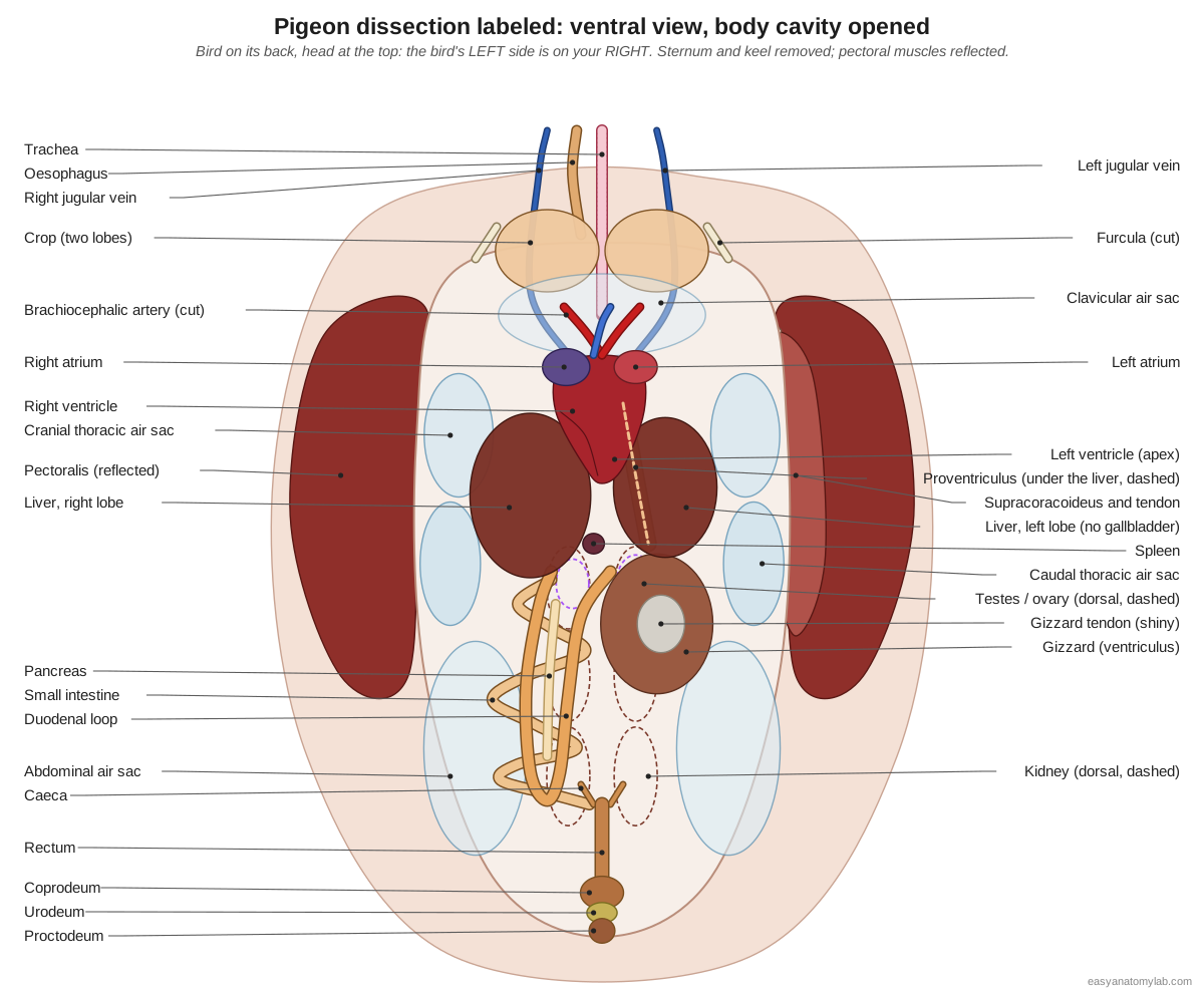
Pigeon dissection labeled diagram
This is what you should see after removing the sternum and keel and folding back the breast muscles: the crop at the base of the neck, the heart between the two liver lobes, the gizzard on the bird's left, the duodenal loop and coiled intestine on its right, and the cloaca at the back. Remember that with the bird on its back, its left side is on your right.
External anatomy of the pigeon: what to record first
Before you cut, record the external features. They are part of most pigeon lab worksheets and they help you orient the specimen later.
| Region | What to find | Note |
|---|---|---|
| Head | Bill (culmen and gonys), cere, nostrils, eyes, eyelids, nictitating membrane, ear opening | The ear opening is hidden by feathers behind the eye |
| Wing | Brachium, antebrachium and manus; wrist, elbow; patagium; primaries, secondaries, tertials, coverts, alula | Spread the wing to see each feather group |
| Tail | Rectrices, upper- and undertail coverts, uropygial (preen) gland | The rock pigeon's preen gland is small; some pigeon species lack it |
| Leg | Thigh, crus (drumstick), tarsus, four toes and claws | Anisodactyl: three toes forward, one back |
| Ventral | Keel (feel it), vent | Note the feather tracts and bare strips when you pluck |
How to dissect a pigeon: the procedure step by step
The order below follows the Weber State University rock dove guide and standard vertebrate zoology labs: external anatomy and feathers first, then the skin and crop, the flight muscles, the body cavity and air sacs, the heart, the respiratory tract, the digestive tract, the kidneys and gonads, the cloaca, and finally the brain.
1. External anatomy
- Lay the pigeon on its back, then on its side. Identify the head, neck, trunk, wings, tail and legs.
- On the head, find the bill, the soft white cere (operculum) at its base, the slit-like nostrils, the eyes with their eyelids and nictitating membrane, and the ear opening behind the eye (part the feathers).
- Find the vent under the tail and, on the back at the tail base, find the small preen (uropygial) gland. The rock pigeon's gland is small, and some other pigeon species lack it.
- Look at the scaly, bare feet. Count the toes and note which way each points (anisodactyl: three forward, one back).
Identify: Head, Bill (beak), Cere (operculum), Nostrils (external nares), Eye, Ear opening (external acoustic meatus), Vent (cloacal opening), Uropygial (preen) gland, Feet and scales (podotheca), Claws.
2. Feathers and the wing
- Spread one wing. Identify the primaries on the hand, the secondaries on the forearm, the tertials next to the body, the rows of coverts, and the alula at the wrist.
- Spread the tail and count the rectrices. Find the upper- and undertail coverts.
- Pluck a contour feather from the breast and a down feather from under it. With a hand lens, find the calamus, rachis, vanes, barbs and the fluffy afterfeather. Pull two barbs apart and stroke them together again: the hooklets re-zip the vane.
- Feel along the wing for the elbow and wrist; note the patagium, the skin fold in front of the elbow.
Identify: Primary flight feathers (primaries), Secondary flight feathers (secondaries), Tertials, Wing coverts, Alula (bastard wing), Tail feathers (rectrices), Upper- and undertail coverts, Contour feathers (body plumage), Down feathers.
3. Remove the breast skin: crop and flight muscles
- Pluck the feathers from the breast and neck (wet them first to stop them flying about). Note that feathers grow in tracts with bare strips between them.
- Pinch up the skin over the keel and make a midventral cut from the base of the neck back to just in front of the vent. Peel the thin skin to the sides.
- At the base of the neck, find the thin-walled, two-lobed crop just under the skin. Do not puncture it.
- Look at the large, dark red pectoralis on each side of the keel. Run a finger along the keel's edge.
Caution: Bird skin is very thin. Keep the scissor tips pointing up and cut only the skin.
Identify: Crop (ingluvies), Pectoralis (breast muscle), Keel (carina), Oesophagus (esophagus), Trachea (windpipe), Jugular veins.
4. Pectoralis and supracoracoideus
- Note the origin of the pectoralis (keel, sternum and furcula) and follow it to its insertion on the underside of the humerus. Move the wing to see what it does.
- Cut the pectoralis along its origin next to the keel and fold it outward. Underneath lies the smaller supracoracoideus.
- Follow the supracoracoideus forward. Its tendon passes up through the triosseal canal at the shoulder to the dorsal surface of the humerus. Pull it gently: the wing lifts.
Identify: Pectoralis (breast muscle), Supracoracoideus, Triosseal canal, Humerus, Coracoid, Furcula (wishbone), Keel (carina).
5. Open the body cavity: air sacs
- Cut through the supracoracoideus, the furcula and the sternum on each side of the keel, close to the ribs, and through the coracoids. Lift the sternum and keel away. Work slowly so you do not damage the organs below.
- Continue the opening back to the vent.
- The thin, transparent, cobweb-like membranes are the walls of the air sacs. Identify the clavicular sac at the thoracic inlet, the cranial and caudal thoracic sacs on the sides, and the large abdominal sacs at the back.
Caution: Air-sac walls are a single thin layer and tear at a touch; look before you probe.
Identify: Clavicular air sac, Cervical air sacs, Cranial thoracic air sacs, Caudal thoracic air sacs, Abdominal air sacs, Sternum (breastbone).
6. Heart and great vessels
- The large heart lies on the midline, ventral and cranial, in its pericardial sac between the two liver lobes. Open the pericardium.
- Identify the right and left atria at the base and the ventricles. The thick-walled left ventricle forms the apex; the thinner right ventricle wraps its right side.
- Find the aorta leaving the left ventricle and note that its arch curves to the bird's right. Find the two brachiocephalic arteries and their common carotid and subclavian branches.
- Find the pulmonary arteries leaving the right ventricle, the two cranial venae cavae and the caudal vena cava entering the right atrium, and the jugular veins in the neck (the right one is larger).
- Where each common carotid artery meets the jugular vein at the base of the neck, look for the small, oval, dark red thyroid gland.
Identify: Right atrium, Left atrium, Right ventricle, Left ventricle, Right aortic arch and dorsal aorta, Brachiocephalic arteries, Pulmonary arteries, Venae cavae, Jugular veins, Thyroid glands.
7. Trachea, syrinx and lungs
- Follow the trachea down the neck. Feel its complete cartilage rings.
- Move the heart aside gently and trace the trachea into the thorax to its fork into the two bronchi. The swelling at the fork is the syrinx.
- Find the bright pink lungs pressed into the ribs on the dorsal wall of the thorax. Note how small they are compared with the air sacs, and that they do not collapse.
- Open the mouth: find the glottis on a mound behind the tongue and the slit (choana) in the roof of the mouth.
Identify: Trachea (windpipe), Larynx and glottis, Syrinx, Primary bronchi, Lungs, Tongue.
8. Digestive tract
- Trace the oesophagus from the mouth down the neck to the crop and on through the thorax.
- On the left side of the abdomen, find the spindle-shaped proventriculus and behind it the large, round gizzard.
- Find the dark two-lobed liver. Look for a gallbladder: pigeons do not have one.
- Follow the duodenal loop from the gizzard and find the pale pancreas held inside it. Find the small round spleen between the proventriculus and the duodenum.
- Unravel the coiled small intestine. Where it joins the short rectum, look for the two tiny caeca.
Identify: Oesophagus (esophagus), Crop (ingluvies), Proventriculus (glandular stomach), Gizzard (ventriculus), Liver, Duodenum (duodenal loop), Pancreas, Spleen, Small intestine (jejunum and ileum), Caeca, Rectum (colon).
9. Open the gizzard and crop
- Cut the gizzard open along its edge. Note the thickness of its muscular wall and the shiny tendon on its sides.
- Scrape the yellowish, horny koilin lining and look at the grit and seed fragments inside.
- Open the crop and the proventriculus and compare their walls: thin and stretchy, thick and glandular.
Identify: Gizzard (ventriculus), Proventriculus (glandular stomach), Crop (ingluvies).
10. Kidneys and reproductive organs
- Move the intestines aside (or remove the digestive tract above the rectum). The dark kidneys lie in hollows of the synsacrum; each has three lobes.
- Trace a ureter from each kidney back to the cloaca. Note that there is no urinary bladder.
- Male: find the two bean-shaped testes at the front of the kidneys (the left is often larger) and the zigzag ductus deferens running back to the cloaca.
- Female: find the single left ovary with its yolk follicles and the coiled left oviduct with its funnel (infundibulum). Use the Female/Male switch to compare.
- At the front of each kidney, close to the gonad, find the small yellow-orange adrenal gland.
Identify: Kidneys, Ureters, Testes, Ductus deferens and seminal vesicle, Ovary (left), Oviduct (left), Adrenal glands.
11. The cloaca
- Cut along the cloaca and identify its three chambers.
- The coprodeum is continuous with the rectum. The urodeum, in the middle, receives the ureters and the ductus deferens or oviduct. The proctodeum opens through the vent.
- In a young bird, look on the dorsal wall of the proctodeum for the round bursa of Fabricius. It shrinks with age and may be hard to find in an adult.
Identify: Rectum (colon), Coprodeum, Urodeum, Proctodeum, Vent (cloacal opening), Bursa of Fabricius.
12. The brain
- Remove the skin from the top of the head. Cut away the roof of the skull in small pieces with fine scissors or bone cutters.
- Identify the two smooth cerebral hemispheres, the folded cerebellum behind them, the large optic lobes on the lower sides, and the medulla oblongata leading into the spinal cord.
- Lift the front of the brain to see the optic nerves running to the eyes.
Caution: The skull is thin. Small, shallow cuts protect the brain.
Identify: Cerebral hemispheres, Cerebellum, Optic lobes (optic tectum), Medulla oblongata, Spinal cord, Eye.
13. Review the skeleton
- If your class keeps the specimen, or with a prepared pigeon skeleton, identify the skull, neck vertebrae, notarium and synsacrum, pygostyle, ribs with uncinate processes, and the keeled sternum.
- Identify the furcula, coracoid and scapula, the wing bones from humerus to digits, and the leg bones from femur to toes.
Identify: Cervical vertebrae (C3–C12), Thoracic vertebrae (notarium region), Synsacrum, Pygostyle, Ribs (vertebral and sternal segments), Uncinate processes, Keel (carina), Furcula (wishbone), Coracoid, Scapula (shoulder blade), Humerus, Ulna, Carpometacarpus, Femur (thigh bone), Tibiotarsus, Tarsometatarsus.
Pigeon flight muscles: pectoralis and supracoracoideus
The pigeon's two flight muscles are the most striking part of the dissection. The pectoralis is the large, dark red outer muscle on each side of the keel; it pulls the wing down. The supracoracoideus lies underneath, against the keel, and lifts the wing through a tendon that runs over the shoulder through the triosseal canal.
Measure them if your class has a balance. In pigeons studied by Tobalske and Biewener (2008), one pectoralis weighed on average about 53 g and one supracoracoideus about 10 g. Compare the colour with chicken breast meat: a pigeon's breast is dark because it is built for sustained flight.
Pigeon crop, gizzard and digestive tract: what to look for
Three digestive observations are specific to pigeons. The crop is a large, thin-walled sac with two lateral lobes at the entrance to the chest. There is no gallbladder on the liver. And the caeca, which are large in chickens, are only tiny buds where the small intestine meets the rectum.
| Organ | In the pigeon | Compare with the chicken |
|---|---|---|
| Crop | Two-lobed, thin-walled; makes crop milk in breeding birds of both sexes | Single crop; no crop milk |
| Gizzard | Very thick muscle, koilin lining, grit and seeds inside | Similar |
| Gallbladder | Absent | Present |
| Caeca | Tiny, vestigial | Two long caeca |
| Breast muscle | Dark red | Pale |
When you open the gizzard, rinse the contents into a dish and look at them with a hand lens: you should find small stones and seed fragments. The tough, yellowish lining you can peel off is the koilin, a hard layer secreted by glands in the gizzard wall.
Pigeon heart: atria and ventricles
The pigeon heart lies on the midline in the front of the body cavity, between the two lobes of the liver, inside its pericardial sac. Its base, towards the head, carries the two thin-walled atria; the ventricles form the pointed apex.
To label the atria correctly, remember the bird is on its back: the right atrium is on your left. The aortic arch leaves the left ventricle and curves to the bird's right, the opposite of the human aorta. The pulmonary trunk leaves the right ventricle beside the aortic root and divides into left and right pulmonary arteries. Cutting across the ventricles shows the thick muscular wall of the left ventricle and the thinner crescent-shaped right ventricle.
Pigeon lungs, kidneys and brain
The lungs, kidneys and brain are the deepest organs and are easiest to find last. The bright pink lungs are pressed into the ribs on the roof of the chest and do not collapse when the body cavity is opened, unlike mammal lungs. The dark red-brown kidneys lie in hollows of the synsacrum behind the lungs; each has three lobes. Their ureters run back to the urodeum, and there is no bladder.
To expose the brain, cut away the roof of the skull in small pieces. The brain has two smooth cerebral hemispheres with no surface folds, a ridged cerebellum behind them, and two large optic lobes on the lower sides, a sign of how much a pigeon depends on its eyes.
Male vs female pigeon dissection
Only step 10 differs between the sexes. Use the Female/Male switch on the model to compare.
- Male: two bean-shaped, cream-coloured testes at the front of the kidneys (the left is often larger) and a white, zigzag ductus deferens on each side running back to the urodeum, ending in a small seminal vesicle.
- Female: a single left ovary that looks like a small bunch of grapes of yolk follicles, and a long, coiled left oviduct opening into the body cavity through a funnel-shaped infundibulum. The right ovary and oviduct do not develop.
Common mistakes in a pigeon dissection
- Cutting too deep through the breast skin and opening the crop, which spills seeds over the dissection.
- Labeling left and right from your point of view instead of the bird's.
- Tearing the air sacs before looking at them. They are the first thing you see when the sternum comes off.
- Searching for a gallbladder or a urinary bladder. Pigeons have neither.
- Mistaking the dark gizzard for the liver: the gizzard is round, hard and has a shiny tendon on its side.
- Missing the caeca: in pigeons they are tiny buds at the ileum–rectum junction.
- Destroying the brain with deep cuts. Remove the thin skull roof in small pieces.
Where do lab pigeons come from? Ethics and alternatives
Pigeons for dissection should come from a biological supply company as preserved teaching specimens, or from an institution's approved source, never from the street or from found birds. Fresh, feral and wild birds can carry diseases that spread to people, and many wild species are legally protected. Your teacher should be able to tell you how your specimen was obtained.
Policies on dissection vary between countries and schools. In 2011, India's University Grants Commission issued guidelines to phase out animal dissection in zoology and life-science courses, replacing it with field visits and digital alternatives; under them, undergraduates no longer dissect animals themselves. Many schools elsewhere let students choose an alternative. The 3D model on this page follows every step of the procedure, so a student who opts out can still identify every structure and answer the same lab questions. (Down To Earth (2011): UGC guidelines on phasing out animal dissection.)
Reading the opened body cavity
When the sternum and keel are lifted away, the first thing you see is not an organ but the air sacs: thin, shining membranes that fill the spaces between organs. The Weber State guide describes them as the many mesentery-like membranes in the body cavity. Look at them before you probe, because they tear at a touch and the view of how they wrap the heart and gut is lost. (Cavitt, Weber State University: external and internal anatomy of the rock dove (PDF).)
Orientation is the most common source of mistakes. With the pigeon on its back and its head away from you, the bird's left is on your right. The gizzard and proventriculus lie on the bird's left, the duodenal loop and most of the intestine on its right, and the aortic arch curves to the bird's right. Keep a sketch with the bird's left and right marked, and label it as you go. (Cavitt, Weber State University: external and internal anatomy of the rock dove (PDF).)
Male vs female pigeon: what changes in the dissection
In a preserved specimen, the reliable way to sex a pigeon is to look at the gonads. A male has two bean-shaped testes at the front of the kidneys, the left often larger, and a zigzag ductus deferens that widens into a seminal vesicle near the cloaca. A female has a single left ovary with yolk follicles and a coiled left oviduct opening into the body cavity through a funnel-shaped ostium. (Cavitt, Weber State University: external and internal anatomy of the rock dove (PDF).)
Gonad size depends on season and breeding condition: testes swell in breeding males and the ovary of a laying female carries large yellow follicles, while non-breeding birds may have small, pale gonads that are easy to overlook. A pigeon clutch is two eggs, incubated by both parents for 17 to 19 days, and both parents later produce crop milk, so a breeding bird of either sex may have a thickened crop lining. (Wikipedia: rock dove (Columba livia).)
Pigeon dissection lab questions and answer key
Answer these during or after the dissection. Click a question to reveal the answer.
Why is the pigeon's keel so large?
It anchors the two flight muscles, the pectoralis and supracoracoideus. A large keel gives a large attachment area for the powerful muscles a strong flier needs. Flightless birds such as ostriches and kiwi have no keel.
Which muscle raises the wing, and how can a muscle on the breast lift it?
The supracoracoideus. Its tendon runs up through the triosseal canal at the shoulder, which works as a pulley, and inserts on the dorsal surface of the humerus, so contraction pulls the wing up and twists the humerus.
Which muscle powers the downstroke, and where does it insert?
The pectoralis. It runs from the keel, sternum and furcula to the underside (deltopectoral crest) of the humerus.
What is the function of the crop? What is unusual about the pigeon crop?
The crop stores and softens food. In breeding pigeons of both sexes, the crop lining also produces crop milk, a fat- and protein-rich secretion fed to the squabs, under the control of prolactin.
How do the proventriculus and gizzard differ?
The proventriculus is the glandular stomach: it secretes acid and pepsinogen. The gizzard (ventriculus) is the muscular stomach: thick muscle and a hard koilin lining grind food with grit.
What did you find inside the gizzard, and why?
Usually grit (small stones) and seed fragments. Birds have no teeth; the gizzard uses grit to grind hard seeds.
Did your pigeon have a gallbladder?
No. Pigeons have no gallbladder; bile passes from the liver to the duodenum through two bile ducts.
Where is the pancreas, and why is it easy to find?
It lies inside the U-shaped duodenal loop, held between its two limbs.
Why do birds have no urinary bladder?
They excrete nitrogen as uric acid, a paste that needs little water, and send it straight to the cloaca. Not carrying stored urine saves weight for flight.
Name the three chambers of the cloaca and what each receives.
Coprodeum (faeces from the rectum), urodeum (ureters and the ductus deferens or oviduct) and proctodeum (opens to the outside through the vent).
Which ovary is functional in a female pigeon?
Only the left ovary and left oviduct develop in most birds, including pigeons. The right ones remain rudimentary.
On which side of the body does the aortic arch run in a bird, and in a mammal?
Birds keep the right aortic arch; mammals keep the left.
Describe the path of air through a bird's respiratory system.
On inspiration, fresh air passes down the trachea and bronchi mostly to the posterior (caudal thoracic and abdominal) air sacs. On expiration it is pushed from them through the lungs' parabronchi, where gas exchange happens. Used air moves to the anterior air sacs and leaves on the next expiration. Air crosses the lungs in one direction.
Why are the bird's lungs so small compared with a mammal's?
Bird lungs do not act as bellows. The air sacs do the pumping, and the rigid lungs, with their dense network of parabronchi and air capillaries, only exchange gases, which they do very efficiently.
Which part of the pigeon brain is unusually large, and what does that tell you?
The optic lobes. Vision is the pigeon's dominant sense.
How is the bird skeleton adapted for flight?
Air-filled (pneumatic) bones such as the humerus, a fused, rigid trunk (notarium, synsacrum, fused pelvis), a keeled sternum, a furcula, ribs braced by uncinate processes, fused hand bones (carpometacarpus) and a pygostyle for the tail feathers.
Virtual pigeon dissection
The 3D viewer above is a complete virtual pigeon dissection. Each of the 13 steps sets the feathers and skin, hides the layers you would have cut away, and lists the structures to find; click any one for its location and function. Students who opt out of a physical dissection can work through the same steps and answer the same lab questions. For the full anatomy of each organ system, see bird anatomy, and for every bone, the bird skeleton page.
Frequently asked questions
Where do you make the first cut in a pigeon dissection?
After plucking the breast, pinch up the skin over the keel and cut along the ventral midline from the base of the neck back to just in front of the vent. Cut only the thin skin and fold it to the sides; the crop lies directly under the skin at the base of the neck.
How do you find the supracoracoideus?
Cut the pectoralis along its origin beside the keel and fold it outward. The smaller, paler supracoracoideus lies underneath, against the keel. Follow it forward to its tendon, which runs up through the triosseal canal at the shoulder to the top of the humerus.
Does a pigeon have a gallbladder?
No. Pigeons have no gallbladder; bile flows from the two-lobed liver to the duodenum through two bile ducts.
What are the thin membranes inside the body cavity?
They are the walls of the air sacs. Birds have nine air sacs in total (four pairs and the single clavicular sac); they look like glistening, cobweb-like films and tear very easily.
How can you tell a male from a female pigeon in a dissection?
Look in front of the kidneys. A male has two bean-shaped testes and a zigzag ductus deferens on each side; a female has a single left ovary, often with yellow yolk follicles, and a coiled left oviduct.
What is the coprodeum?
The first and largest chamber of the cloaca, continuous with the rectum, which receives faeces. Behind it are the urodeum, which receives the ureters and reproductive ducts, and the proctodeum, which opens at the vent.
Is there a virtual pigeon dissection?
Yes. The 3D model on this page follows the same 13 steps as the lab. Each step removes the right layers and lists the structures to find, so students who opt out of a physical dissection can complete the same work and answer the same questions.
Why is the pigeon's breast muscle so dark?
Pigeons are strong, sustained fliers. Their breast muscle fibres are rich in myoglobin and mitochondria for aerobic work, which makes the muscle dark red, unlike the pale breast of a chicken, which flies only in short bursts.
References
- Cavitt, J. Weber State University. External and internal anatomy of the rock dove (pigeon dissection guide, ornithology lab).
- Savalli, U. M. BIO 370 Vertebrate Zoology: rock pigeon dissection and skeleton identification exercises.
- Crespo, R. et al. (2018). Galliformes and Columbiformes. In Pathology of Wildlife and Zoo Animals. Elsevier.
- Mahdy, M. A. A. & Mohammed, E. S. I. (2024). Oesophagus and crop of young and adult domestic pigeons. BMC Veterinary Research 20, 428.
- Khoder, D. A. & Hameed, B. K. (2025). Liver and biliary system of the pigeon and the gull. Iraqi Journal of Veterinary Sciences 39(4).
- Hussein, A. A., Ibrahim, R. S. & Ali, H. K. (2017). Morphological study of the air sacs in domestic pigeons. Diyala Agricultural Sciences Journal 9.
- Tobalske, B. W. & Biewener, A. A. (2008). Contractile properties of the pigeon supracoracoideus. Journal of Experimental Biology 211, 170–179.
- Iniyah, K., Kumaravel, A. & Gnanadevi, R. (2022). Axial skeleton of homing pigeon (Columba livia). Biological Forum 14(3), 167–170.
- Down To Earth (2011). UGC guidelines on phasing out animal dissection spark controversy.
- Wikipedia: Rock dove (Columba livia): size, plumage, clutch and incubation.
More interactive anatomy models
- Bird skeletonHollow bones, keeled sternum, wishbone and fused wing bones of a bird.
- Bird anatomyBird digestive system, air sacs, heart and feather anatomy in 3D.
- Chicken anatomyCrop, gizzard, reproductive tract, skeleton and external parts of the chicken.
- Human anatomy1,000+ bones, muscles, organs, vessels and nerves from real anatomical data.
- Octopus anatomyThree hearts, a ring-shaped brain, eight arms and a beak, in 3D.
- Squid anatomyPen, eight arms, two tentacles, giant axons and three hearts of the longfin squid.