

Do snakes have bones? The snake skeleton at a glance
Yes. Snakes are vertebrates with a complete bony skeleton: a skull, a backbone of a few hundred vertebrae and a pair of ribs on almost every vertebra of the trunk. What a snake lacks are limbs, a shoulder (pectoral) girdle and a breastbone (sternum). Boas and pythons, including the ball python shown here, also keep a tiny pelvis and thigh bone on each side of the vent, tipped with a visible claw called a spur.
The ball python is a good reference skeleton because pythons keep several bones that most snakes have lost: teeth on the premaxilla, a supraorbital bone above each eye, a coronoid bone in the lower jaw and the vestigial hind limb. A colubrid such as a corn snake has the same plan without those extras. Its skeleton is built from three parts:
| Part | Ball python | Notes |
|---|---|---|
| Skull | About 20 named bones per side, plus teeth | Kinetic: jaw and palate bones move independently |
| Vertebrae | Roughly 240: ~200 trunk, 4 cloacal, ~34 caudal | Across snakes, 115–546 vertebrae |
| Ribs | One pair on almost every trunk vertebra (~400 ribs) | Rib ends free; no sternum |
| Shoulder girdle and forelimbs | Absent | Absent in all living snakes |
| Pelvis and hind limbs | Pelvic rod, tiny femur and spur on each side | Lost in colubrids, elapids and vipers |
Snake skeleton labeled: diagram
A labeled snake skeleton shows the skull at the front, then the atlas and axis, the long series of rib-bearing trunk vertebrae, the cloacal vertebrae at the vent and the rib-less tail vertebrae. In boas and pythons, a small pelvis and femur sit beside the vent, outside the vertebral column.
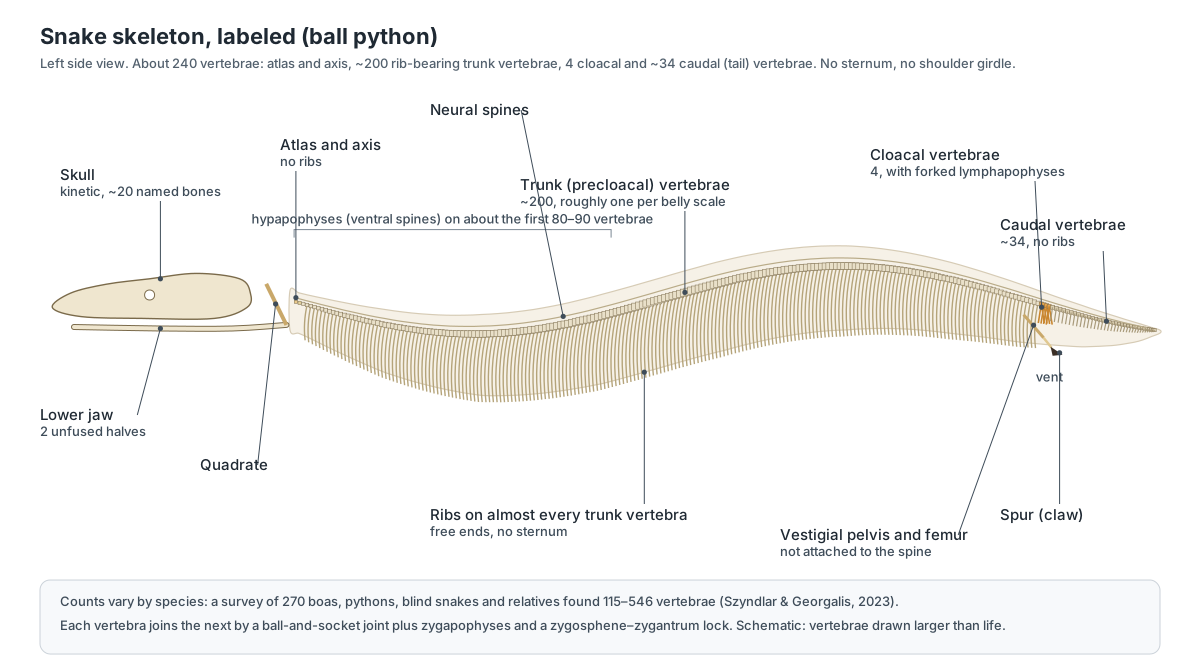
Snake skeleton anatomy: every bone and its function
This is the text version of the 3D skeleton: every labeled bone and bone group, grouped by region.
Body outline
- Body outline (skin)
- The skin of the ball python, shown faded so the skeleton inside is visible. Use the slider to fade it in and out and see how far the bones reach inside the body.
Skull roof & braincase
- Premaxilla
- The single small bone at the tip of the snout. Forms the tip of the snout and, in pythons, carries a few small teeth.
- Nasal bones
- Paired thin bones roofing the nasal cavity. Cover the nasal capsules; the frontal–nasal joint flexes slightly during swallowing.
- Septomaxillae
- Small paired bones inside the snout. Roof the vomeronasal (Jacobson's) organ and support the nasal capsule.
- Vomers
- Paired small bones on the roof of the mouth at the front of the palate. Form the floor of the vomeronasal organ, whose ducts open on the palate beside them.
- Prefrontals
- Paired bones that form the front wall of each eye socket. Support the maxilla through a movable joint that lets the maxilla rotate.
- Frontals
- Paired roofing bones between the eyes. Roof and enclose the front of the brain; in snakes they wrap down around it like a tube.
- Supraorbitals
- Small bones over each eye found in pythons. Complete the rim of the eye socket.
- Postorbitals (postfrontals)
- Slender bones forming the back rim of each eye socket. Close the back of the orbit and anchor jaw muscles.
- Parietal
- The large single bone roofing the back of the braincase. Protects the brain and anchors the jaw-closing muscles.
- Braincase (basicranium) (Basioccipital, exoccipitals, prootics, basisphenoid, otooccipitals)
- The solid box of fused bones that encloses the brain and inner ears. Protects the brain and inner ear; it is the rigid core that the movable jaws hang from.
- Occipital condyle
- The knob at the back of the skull that articulates with the atlas. Forms the joint between skull and neck.
- Stapes (columella)
- The only middle-ear bone: a thin rod from the inner ear to the quadrate. Carries vibrations from the jaw and quadrate to the inner ear.
Jaws, palate & suspensorium
- Supratemporals (Tabular (older usage))
- Flat rods on the back of the skull from which the quadrates hang. Carry the quadrate out behind the skull, adding length to the jaw and widening the gape.
- Quadrates
- Long, mobile bones that link the supratemporal to the lower jaw. Swing outward to spread the jaw joints apart; their lower end forms the jaw joint with the compound bone.
- Maxillae
- The main toothed bones of the upper jaw, one on each side. Carry the upper teeth and can rotate on the prefrontal to pull prey in.
- Palatines
- Toothed bones forming the front of the inner tooth row on the roof of the mouth. With the pterygoids, ratchet prey back into the throat ('pterygoid walk').
- Pterygoids
- Long rods running back from the palatines to the quadrates; toothed at the front. Moved forward and back by muscles to walk the palate over the prey.
- Ectopterygoids
- Short struts linking the back of each maxilla to the pterygoid. Transmit movement between the palate and the maxilla.
- Dentaries
- The toothed front bones of each lower jaw half. Carry the lower teeth.
- Compound bones (Fused articular, surangular and prearticular)
- The long rear bone of each lower jaw half, made of several fused bones. Forms the jaw joint with the quadrate and anchors the jaw muscles.
- Splenials and angulars
- Two small splint bones on the inner side of each lower jaw half. Brace the flexible joint between the front and back of the lower jaw.
- Coronoids
- A small bone on the top edge of each lower jaw half. Anchors jaw-closing muscles.
- Intermandibular ligament (Mandibular symphysis (unfused))
- The elastic ligament that loosely joins the tips of the two lower jaws. Lets the two jaw halves spread apart and move independently during swallowing.
- Hyoid (Hyoid cornua)
- A pair of thin cartilage rods in the floor of the mouth and throat. Anchors the tongue muscles.
Teeth
- Premaxillary teeth
- A few small teeth at the tip of the snout. Help hold prey at the front of the mouth.
- Maxillary teeth
- The outer row of long, backward-curving upper teeth. Strike and grip prey.
- Palatine and pterygoid teeth
- The inner rows of upper teeth on the roof of the mouth. Grip and pull prey back during swallowing.
- Dentary teeth
- The lower row of backward-curving teeth. Grip prey from below.
Vertebral column
- Atlas (First cervical vertebra)
- The first vertebra: a ring that cups the occipital condyle. Lets the head nod on the neck.
- Axis
- The second vertebra, joined to the atlas. Allows the head to turn.
- Trunk (precloacal) vertebrae (Body vertebrae, 'cervical' + 'thoracic')
- The long series of nearly identical rib-bearing vertebrae from neck to cloaca. Support the body and allow side-to-side and up-and-down bending.
- Condyles and cotyles (Ball-and-socket joints)
- The ball on the back of each vertebral body and the socket it fits into. Form flexible ball-and-socket joints between vertebrae.
- Neural spines
- The blade-like spines on top of each neural arch. Anchor the epaxial muscles and tendons.
- Zygapophyses (Prezygapophyses and postzygapophyses)
- Flat joint surfaces on each side of the arch that overlap the next vertebra. Guide bending and limit twisting.
- Zygosphene and zygantrum
- A wedge on the front of each arch (zygosphene) that fits into a socket (zygantrum) on the vertebra in front. Lock neighbouring vertebrae against twisting while still allowing bending.
- Hypapophyses
- Ventral spines under the vertebral bodies of the front part of the trunk. Anchor neck and trunk muscles used in striking and swallowing.
- Cloacal vertebrae (Lymphapophyses)
- A few vertebrae at the level of the cloaca with forked rib-like processes. Support the cloaca and the base of the tail.
- Caudal vertebrae (Tail vertebrae, pleurapophyses)
- The rib-less tail vertebrae, which get smaller towards the tip. Support the tail and the hemipenes; their side processes (pleurapophyses) anchor tail muscles.
Ribs
- Ribs
- A pair of long, curved ribs on almost every trunk vertebra. Protect the organs, move air in and out (there is no diaphragm) and let the body expand around prey.
Pelvic vestige
- Pelvic bones (vestigial pelvis) (Pelvic rudiment; ilium or pubis)
- A tiny rod-like hip bone on each side of the cloaca. Anchors the femur and spur and the muscles that move it.
- Femur vestiges
- A tiny thigh bone on each side, the last remnant of the hind leg. Carries the spur and lets the snake move it.
- Spurs (Cloacal (pelvic) spurs)
- The horny claw at the tip of each femur, visible beside the vent. Used by males to stimulate females in courtship and in combat.
Snake skull and head skeleton
A snake skull has a solid, box-like braincase surrounded by slender, loosely jointed bones of the snout, palate and jaws. This "kinetic" design lets the jaws move apart and forward independently, so a snake can swallow prey wider than its own head.
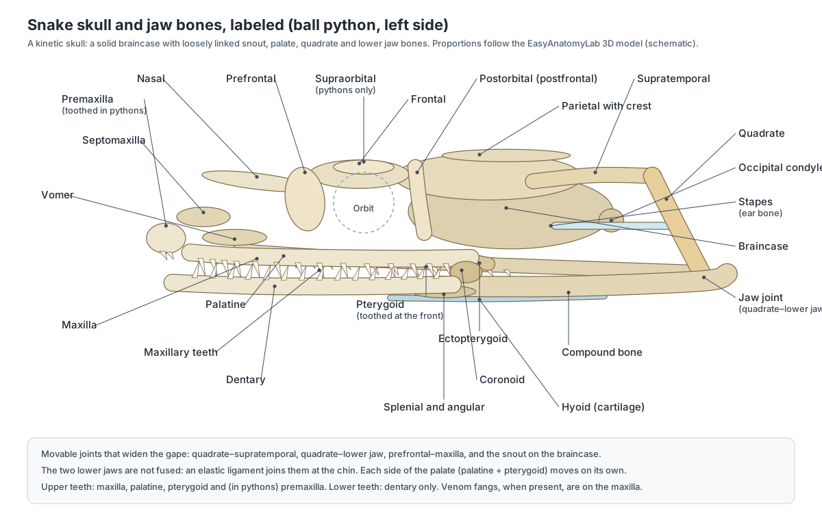
Skull roof, snout and braincase
- Premaxilla: The single small bone at the tip of the snout. Forms the tip of the snout and, in pythons, carries a few small teeth.
- Nasal bones: Paired thin bones roofing the nasal cavity. Cover the nasal capsules; the frontal–nasal joint flexes slightly during swallowing.
- Septomaxillae: Small paired bones inside the snout. Roof the vomeronasal (Jacobson's) organ and support the nasal capsule.
- Vomers: Paired small bones on the roof of the mouth at the front of the palate. Form the floor of the vomeronasal organ, whose ducts open on the palate beside them.
- Prefrontals: Paired bones that form the front wall of each eye socket. Support the maxilla through a movable joint that lets the maxilla rotate.
- Frontals: Paired roofing bones between the eyes. Roof and enclose the front of the brain; in snakes they wrap down around it like a tube.
- Supraorbitals: Small bones over each eye found in pythons. Complete the rim of the eye socket.
- Postorbitals (postfrontals): Slender bones forming the back rim of each eye socket. Close the back of the orbit and anchor jaw muscles.
- Parietal: The large single bone roofing the back of the braincase. Protects the brain and anchors the jaw-closing muscles.
- Braincase (basicranium): The solid box of fused bones that encloses the brain and inner ears. Protects the brain and inner ear; it is the rigid core that the movable jaws hang from.
- Occipital condyle: The knob at the back of the skull that articulates with the atlas. Forms the joint between skull and neck.
Snake jaw anatomy: suspensorium, palate and lower jaw
- Supratemporals: Flat rods on the back of the skull from which the quadrates hang. Carry the quadrate out behind the skull, adding length to the jaw and widening the gape.
- Quadrates: Long, mobile bones that link the supratemporal to the lower jaw. Swing outward to spread the jaw joints apart; their lower end forms the jaw joint with the compound bone.
- Maxillae: The main toothed bones of the upper jaw, one on each side. Carry the upper teeth and can rotate on the prefrontal to pull prey in.
- Palatines: Toothed bones forming the front of the inner tooth row on the roof of the mouth. With the pterygoids, ratchet prey back into the throat ('pterygoid walk').
- Pterygoids: Long rods running back from the palatines to the quadrates; toothed at the front. Moved forward and back by muscles to walk the palate over the prey.
- Ectopterygoids: Short struts linking the back of each maxilla to the pterygoid. Transmit movement between the palate and the maxilla.
- Dentaries: The toothed front bones of each lower jaw half. Carry the lower teeth.
- Compound bones: The long rear bone of each lower jaw half, made of several fused bones. Forms the jaw joint with the quadrate and anchors the jaw muscles.
- Splenials and angulars: Two small splint bones on the inner side of each lower jaw half. Brace the flexible joint between the front and back of the lower jaw.
- Coronoids: A small bone on the top edge of each lower jaw half. Anchors jaw-closing muscles.
- Intermandibular ligament: The elastic ligament that loosely joins the tips of the two lower jaws. Lets the two jaw halves spread apart and move independently during swallowing.
- Hyoid: A pair of thin cartilage rods in the floor of the mouth and throat. Anchors the tongue muscles.
Snake jaw: how snakes swallow without dislocating
Snakes do not dislocate their jaws. Four features let the mouth open wide: the long, swinging quadrate bones; the supratemporals that carry the quadrates out behind the skull; the elastic ligament joining the two lower jaws at the chin; and a palate whose left and right sides (palatine and pterygoid) move separately.
| Joint | How mobile | What it does |
|---|---|---|
| Quadrate – lower jaw | Very mobile | The jaw joint; opens the mouth far behind the braincase |
| Quadrate – supratemporal | Very mobile | Lets the jaw joints swing outward |
| Prefrontal – maxilla | Mobile | Lets the maxilla rotate to pull prey in (and erect the fang in vipers) |
| Frontal – nasal (snout) | Slight | Tilts the snout a little |
| Chin (intermandibular ligament) | Stretchy | Lets the two lower jaws spread apart |
| Supratemporal – parietal | Essentially immobile | Anchors the jaw suspension to the skull |
Snake teeth and fangs
Snakes have four rows of upper teeth (the maxillae and the palatine–pterygoid rows) and two lower rows on the dentaries. Pythons also have a few teeth on the premaxilla. All are thin, sharp and curved backward, and they are replaced throughout life.
- Premaxillary teeth: A few small teeth at the tip of the snout. Help hold prey at the front of the mouth.
- Maxillary teeth: The outer row of long, backward-curving upper teeth. Strike and grip prey.
- Palatine and pterygoid teeth: The inner rows of upper teeth on the roof of the mouth. Grip and pull prey back during swallowing.
- Dentary teeth: The lower row of backward-curving teeth. Grip prey from below.
In venomous snakes the fangs are modified maxillary teeth. Vipers have long hinged fangs on a short, rotating maxilla; cobras and other elapids have short fixed front fangs; rear-fanged colubrids have grooved fangs at the back of the maxilla. For the soft-tissue anatomy of the mouth, see the snake head and mouth anatomy section.
Snake vertebrae: how many and what they look like
Snakes have between about 115 and 546 vertebrae depending on the species, and many have more than 300. A ball python has roughly 240. Each trunk vertebra has a body (centrum) with a socket in front and a ball behind, a neural arch with a spine, paired zygapophyses, and an extra wedge-and-socket joint, the zygosphene and zygantrum, which stops the column twisting.
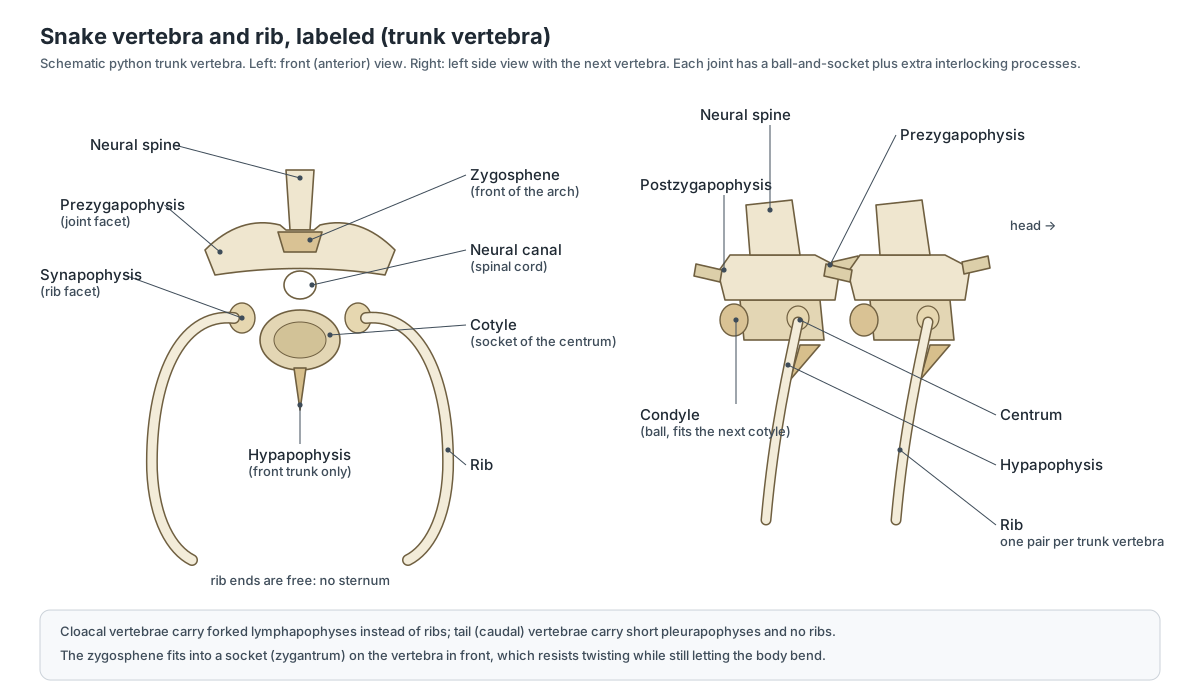
- Atlas: The first vertebra: a ring that cups the occipital condyle. Lets the head nod on the neck.
- Axis: The second vertebra, joined to the atlas. Allows the head to turn.
- Trunk (precloacal) vertebrae: The long series of nearly identical rib-bearing vertebrae from neck to cloaca. Support the body and allow side-to-side and up-and-down bending.
- Condyles and cotyles: The ball on the back of each vertebral body and the socket it fits into. Form flexible ball-and-socket joints between vertebrae.
- Neural spines: The blade-like spines on top of each neural arch. Anchor the epaxial muscles and tendons.
- Zygapophyses: Flat joint surfaces on each side of the arch that overlap the next vertebra. Guide bending and limit twisting.
- Zygosphene and zygantrum: A wedge on the front of each arch (zygosphene) that fits into a socket (zygantrum) on the vertebra in front. Lock neighbouring vertebrae against twisting while still allowing bending.
- Hypapophyses: Ventral spines under the vertebral bodies of the front part of the trunk. Anchor neck and trunk muscles used in striking and swallowing.
- Cloacal vertebrae: A few vertebrae at the level of the cloaca with forked rib-like processes. Support the cloaca and the base of the tail.
- Caudal vertebrae: The rib-less tail vertebrae, which get smaller towards the tip. Support the tail and the hemipenes; their side processes (pleurapophyses) anchor tail muscles.
Snake ribs and the missing sternum
Almost every trunk vertebra carries a pair of long, curved ribs that wrap around the body cavity. Their lower ends are free, because snakes have no sternum. Free rib ends let the body stretch around a large meal, and the ribs do the work of breathing in the absence of a diaphragm.
- Ribs: A pair of long, curved ribs on almost every trunk vertebra. Protect the organs, move air in and out (there is no diaphragm) and let the body expand around prey.
Ribs also have special jobs in some snakes. Cobras and hognose snakes spread their long front ribs to flatten the neck into a hood. In rectilinear crawling, the costocutaneous muscles pull the belly skin forward and back over ribs that hardly move.
Snake skeleton legs: vestigial pelvis and spurs
Living snakes have no working legs, but boas and pythons keep a remnant of the hind limb: a rod-like pelvic bone embedded in the muscles beside the cloaca, a tiny femur, and a horny spur that sticks out beside the vent. These bones are not joined to the vertebral column.
- Pelvic bones (vestigial pelvis): A tiny rod-like hip bone on each side of the cloaca. Anchors the femur and spur and the muscles that move it.
- Femur vestiges: A tiny thigh bone on each side, the last remnant of the hind leg. Carries the spur and lets the snake move it.
- Spurs: The horny claw at the tip of each femur, visible beside the vent. Used by males to stimulate females in courtship and in combat.
Fossils show how snakes lost their legs. Cretaceous snakes such as Najash and Pachyrhachis had a pelvis and small but complete hind limbs, and analysis of living snakes suggests that their common ancestor still had all three pelvic bones and rudimentary hind limbs. No snake, living or fossil, is known to keep a shoulder girdle.
Legless lizard vs snake skeleton
A legless lizard looks like a snake but differs in its skeleton and body plan. The quickest guide is proportions: a snake has a long body and a short tail, while a legless lizard has a short body and a very long tail.
| Feature | Snake | Legless lizard (e.g. glass lizard, slow worm) |
|---|---|---|
| Body vs tail | Long body, short tail | Short body, very long tail |
| Skull | Highly kinetic | Less mobile |
| Lower jaw | Two halves joined by a ligament | Halves firmly joined at the chin |
| Girdles | No shoulder girdle; pelvis only in basal snakes | Often small remnants of the girdles |
| Eyelids and ears | No eyelids, no ear opening | Eyelids and external ear openings |
| Belly scales | One row of wide ventral scales | No broad belly scales |
| Lungs | One main lung | Two lungs of roughly equal size |
Rattlesnake skeleton and the rattle
A rattlesnake skeleton is a typical viper skeleton: a short, rotating maxilla carrying a long hinged fang on each side, with reserve fangs behind it, a wide braincase, and a rib-bearing trunk. The rattle itself is not bone. It is a stack of hollow, interlocking keratin segments sitting on the last few tail vertebrae, which are fused together. A new segment is added at each shed, but old segments break off, so the rattle does not tell you the snake's age.
Titanoboa skeleton: the largest snake
Titanoboa cerrejonensis lived in what is now Colombia about 58–60 million years ago. It is known mainly from vertebrae, found in the Cerrejón coal mine, which were compared with the vertebrae of living boas to estimate a length of about 13 metres (42 feet) and a mass of over a tonne. That is roughly ten times the length of an adult ball python. Online images of "giant snake skeletons" on beaches or in deserts are hoaxes or misidentified whale and fish skeletons.
Sea snake skeleton
Sea snakes have the same kind of skeleton as cobras, their elapid relatives, but their body is flattened from side to side and the tail is a vertical paddle used for swimming. Their belly scales are so small that they are hard to tell from the other body scales, because they no longer need them to grip the ground.
Counting the bones in a snake
A snake skeleton is mostly backbone. The vertebrae are divided into an atlas–axis complex, trunk vertebrae (the front ones are sometimes called 'cervical'), cloacal vertebrae and caudal vertebrae. Across 270 species of boas, pythons, blind snakes and their relatives, total counts range from 115 to 546 vertebrae, and pythons are among the snakes with very high counts. Cloacal vertebrae, usually three to seven and most often four, bear forked processes called lymphapophyses, while caudal vertebrae bear unforked pleurapophyses. (Szyndlar & Georgalis (2023): atlas of snake vertebral morphology.)
Each new vertebra forms from a block of embryonic tissue called a somite. In corn snake embryos, a 'segmentation clock' runs much faster relative to the growth of the body than it does in mice or chickens, producing a new somite about every 100 minutes, so more than 300 segments form before the clock stops. Humans have only 33 vertebrae and mice stop at about 65 segments. (ScienceDaily: how the snake got its vertebrae (Gomez et al., 2008).)
For a ball python, a quick estimate comes from its scales: the species has fewer than about 210 ventral scales and fewer than about 38 subcaudals, and each belly scale corresponds roughly to one trunk vertebra. That gives around 200 trunk vertebrae with a pair of ribs on almost every one, plus a few dozen tail vertebrae: roughly 240 vertebrae and close to 400 ribs. (Schleip & O'Shea (2010): annotated checklist of pythons.)
Why the snake skull is called kinetic
The snake braincase is solidly built, but the bones around it are loosely joined so the mouth can open around prey larger than the head. The highly mobile joints are between the quadrate and the lower jaw, between the quadrate and the supratemporal, and between the prefrontal and the maxilla; the frontal–nasal joint and the lower jaw add a little more. The quadrate is usually long and hangs from the skull by the supratemporal. The two halves of the lower jaw are joined at the chin only by an elastic ligament. (Snake skeleton (overview).)
This is why 'snakes dislocate their jaws' is a myth. During swallowing a snake moves one side of its upper jaw and palate forward, grips with the backward-curving teeth, then moves the other side, so the head 'walks' over the prey. Most teeth are on the dentary, maxilla, palatine and pterygoid; pythons also have teeth on the premaxilla. Venom fangs, when present, are always on the maxilla. (Snake skeleton (overview).)
Embryo studies of 96 snakes from eight species showed that front fangs develop at the back of the upper jaw, as rear fangs do, and are later shifted forward by growth. The authors argued that a rear tooth-forming region became developmentally separate from the other teeth, letting it evolve together with the venom gland. (Naturalis: evolution of snake fangs (Vonk et al., 2008).)
Do snakes have legs? The pelvic vestige and spurs
No living snake has any trace of the front limbs or shoulder girdle. Boas and pythons, however, keep a tiny pelvic bone and a femur-like bone on each side, ending in an external claw, the spur, beside the cloaca. Leptotyphlopid blind snakes keep an ilium, pubis, ischium and a rudimentary femur. (Snake skeleton (overview).)
Palci and colleagues reported the first ossified pelvic vestiges in an anomalepidid blind snake and reviewed the pelvis across snakes. They concluded that the common ancestor of living snakes probably still had all three pelvic bones and rudimentary hind limbs, and that pelvic bones were lost, and sometimes regained, several times. The single rod in many basal snakes such as pythons is, in their view, the pubis, and the cloacal spur and femur are likely used in mating and courtship rather than being useless leftovers. (Palci et al. (2020): pelvis and hindlimb reduction in snakes.)
Legless lizard vs snake skeleton
Several groups of lizards, such as glass lizards and slow worms, have lost their legs and look like snakes. You can tell them apart from the outside: legless lizards have eyelids, external ear openings, a notched rather than forked tongue, no broad belly scales, two lungs of roughly equal size and a very long tail, whereas snakes have a long body and a short tail. (Legless lizard (overview).)
In the skeleton the same pattern shows up as body-to-tail proportions: a legless lizard has many more vertebrae behind the cloaca than in front of it compared with a snake of the same length, and its skull is far less mobile. Pelvic remnants alone do not separate the two groups, because the pelvic girdle is retained in limbless lizards and in the most basal snakes. (Legless lizard (overview).)
Titanoboa, rattles and sea snakes
Titanoboa cerrejonensis is known from fossil vertebrae of several individuals found in the Cerrejón coal mine in Colombia, about 58–60 million years old. Because every snake has a fixed number of segments and vertebra size scales with body length, its length could be estimated from vertebrae alone: about 13 metres (42 feet), with a mass of over a tonne. (National Geographic: Titanoboa.)
A rattlesnake's rattle is not bone. It is a set of hollow, interlocked keratin segments formed from modified scales at the tail tip, with a new segment added at each shed. Because end segments break off, the number of segments does not show the snake's age. Shaker muscles in the tail vibrate the segments, firing about 50 times per second. (Rattlesnake (overview).)
Sea snakes show how the same skeleton can be reshaped for water. The yellow-bellied sea snake has a body flattened from side to side and a paddle-shaped tail for swimming, valved nostrils, and belly scales so reduced that they are hard to tell from the other body scales. It also takes up oxygen through its skin, which extends its dives. (Yellow-bellied sea snake (overview).)
Snake skeleton worksheet: test yourself
Use the 3D model and diagrams to answer these. Answers are in brackets.
- Which bone carries the jaw joint and hangs from the supratemporal? (The quadrate.)
- What joins the two lower jaws at the chin? (An elastic intermandibular ligament.)
- Name the four bones that carry upper teeth in a python. (Premaxilla, maxilla, palatine, pterygoid.)
- Which bone in the lower jaw carries teeth? (The dentary.)
- Which snakes keep a vestigial pelvis and spurs? (Boas and pythons, among others.)
- What extra joint locks snake vertebrae against twisting? (The zygosphene–zygantrum joint.)
- What do cloacal vertebrae carry instead of ribs? (Forked lymphapophyses.)
- Why can a snake's body expand around a big meal? (Its ribs have free ends: there is no sternum.)
For the organs that fill the rib cage, see the snake anatomy page.
Frequently asked questions
Do snakes have bones?
Yes. Snakes are vertebrates with a full bony skeleton: a skull, a backbone of a few hundred vertebrae, and ribs. What they lack are limbs, shoulder bones and a breastbone. Boas and pythons also keep a tiny pelvis and thigh bone on each side of the vent.
How many bones does a snake have?
Most of a snake's bones are vertebrae and ribs. Different snakes have from about 115 to over 500 vertebrae, and most trunk vertebrae carry a pair of ribs, so a large snake can have well over 1,000 bones. A ball python has roughly 240 vertebrae and close to 400 ribs.
How many vertebrae does a snake have?
It depends on the species. A survey of 270 species of boas, pythons, blind snakes and their relatives found from 115 to 546 vertebrae (Szyndlar & Georgalis, 2023), and many snakes have more than 300. Humans have 33. A ball python has roughly 200 trunk vertebrae, about one per belly scale, plus a few dozen in its short tail.
Do snakes have legs?
No living snake has working legs, but boas and pythons keep a tiny pelvic bone and femur on each side of the vent, tipped with a visible claw-like spur. Fossil snakes such as Najash had small but complete hind legs. No living snake has any trace of front legs or a shoulder girdle.
Do snakes have a sternum?
No. Snakes have no sternum (breastbone) and no shoulder girdle. Their rib ends are free, which lets the body expand around a large meal and lets the ribs move freely during breathing and crawling.
Can snakes dislocate their jaws?
No. Snakes swallow large prey because their jaw bones are loosely connected, not because the jaws come apart. The two lower jaw halves are joined only by an elastic ligament, the quadrates swing outward, and the palate bones move independently on each side.
What is the difference between a snake and a legless lizard skeleton?
Many legless lizards keep small remnants of the limb girdles, have a long tail with many caudal vertebrae, and have a less mobile skull whose lower jaw halves are firmly joined at the chin. Snakes have a long body and short tail, a very kinetic skull, loosely joined lower jaws and no trace of a shoulder girdle. Outside the skeleton, legless lizards have eyelids and ear openings and lack broad belly scales.
How big was Titanoboa?
Titanoboa cerrejonensis, known from vertebrae found in a coal mine in Colombia and about 58–60 million years old, is estimated to have been about 13 m (42 ft) long and to have weighed over a tonne. Its length was calculated from the size of its vertebrae compared with living boas.
Is a rattlesnake's rattle made of bone?
No. The rattle is made of hollow, interlocking segments of keratin, the same material as scales. A new segment is added at each shed, but end segments break off, so the number of segments does not show the snake's age. The rattle sits on a knob formed by the last few tail vertebrae, which are fused together.
References
- Szyndlar, Z. & Georgalis, G. L. (2023). An illustrated atlas of the vertebral morphology of extant non-caenophidian snakes, with special emphasis on the cloacal and caudal portions of the column. Vertebrate Zoology 73, 717–886.
- Palci, A., Hutchinson, M. N., Caldwell, M. W., Smith, K. T. & Lee, M. S. Y. (2020). The homologies and evolutionary reduction of the pelvis and hindlimbs in snakes. Zool. J. Linn. Soc. 188, 630–652.
- Snake skeleton (overview of skull kinesis, dentition, vertebrae and limb vestiges).
- Schleip, W. D. & O'Shea, M. (2010). Annotated checklist of the recent and extinct pythons (Serpentes, Pythonidae). ZooKeys 66, 29–79.
- Howard Hughes Medical Institute / ScienceDaily (2008). How the snake got its vertebrae (Gomez et al., Nature 454, 335–339).
- Christensen, C. B. et al. (2012). Hearing with an atympanic ear: good vibration and poor sound-pressure detection in the royal python. J. Exp. Biol. 215, 331–342.
- National Geographic. Titanoboa: thirteen metres, one tonne, largest snake ever (Head et al., 2009, Nature 457, 715–717).
- Naturalis Biodiversity Center. Evolution of snake fangs (Vonk et al., 2008, Nature 454, 630–633).
- Rattlesnake (overview of the rattle, fangs and pit organs).
- Legless lizard (overview of differences from snakes).
- Yellow-bellied sea snake (overview of marine adaptations).
- Animal Diversity Web. Python regius (ball python).
More interactive anatomy models
- Snake anatomyElongated organs, one working lung, a flexible jaw and the snake heart.
- Turtle skeletonHow ribs and spine fuse into the shell: carapace, plastron and girdles.
- Lizard anatomySkull, scales, three-chambered heart and tail autotomy of a lizard.
- Human anatomy1,000+ bones, muscles, organs, vessels and nerves from real anatomical data.
- Octopus anatomyThree hearts, a ring-shaped brain, eight arms and a beak, in 3D.
- Squid anatomyPen, eight arms, two tentacles, giant axons and three hearts of the longfin squid.