

Dog anatomy at a glance
A dog is a four-legged carnivoran mammal that walks on its toes. Its body has a head with a long nasal cavity and powerful jaws, a flexible neck of seven vertebrae, a deep chest that holds the heart and lungs, an abdomen with a simple stomach and a short intestine, and four limbs. The forelimbs are attached to the trunk by muscles only, because the clavicle is reduced to a tiny remnant. The hind limbs are joined to the spine through the pelvis and drive the dog forward.
This page describes a medium mesaticephalic dog (a Labrador or Beagle type, about 57 cm at the shoulder). Dogs vary more in size and shape than almost any other domestic animal, from Chihuahuas to Great Danes, but almost every breed has the same organs and the same named bones. Where breeds differ, for example in skull shape, leg length, ear carriage or the number of tail vertebrae, the text says so.
Directions: cranial means towards the head and caudal towards the tail; rostral is used on the head for "towards the nose". Dorsal is the back, ventral the belly. On the legs, proximal is towards the body and distal towards the paw; the back of the front paw is palmar and of the hind paw plantar. Left and right are always the dog's own.
| Body system | Main organs in the model | Job |
|---|---|---|
| Skeletal | Skull, vertebral column, ribs, limb bones | Support, protection, levers for movement |
| Muscular | Superficial muscles of head, neck, trunk and limbs | Movement and posture |
| Digestive | Mouth, oesophagus, stomach, liver, pancreas, intestines | Digest food, absorb nutrients |
| Respiratory | Nose, larynx, trachea, lungs, diaphragm | Breathing and gas exchange |
| Cardiovascular | Heart (4 chambers), aorta, venae cavae, limb vessels | Pump and carry blood |
| Urinary | Kidneys, ureters, bladder, urethra | Filter blood, make and store urine |
| Reproductive | Testes, prostate, penis / ovaries, uterus, vagina | Make sperm or eggs; pregnancy |
| Nervous and senses | Brain, spinal cord, nerves, eye, ear, nose | Control, sensation and behaviour |
| Lymphatic and endocrine | Spleen, lymph nodes, thyroid and adrenal glands | Immunity and hormones |
Parts of a dog: external anatomy
The external parts of a dog, often called the "points of the dog", are the named regions of the body surface. Breeders, judges and vets use them to describe conformation. Click them on the model with the coat at full opacity.
| Point of the dog | Where it is | What lies under it |
|---|---|---|
| Muzzle and stop | Face in front of the eyes; the stop is the step up to the forehead | Nasal bones, maxilla, nasal cavity |
| Withers | Highest point of the back, behind the neck | Tops of the scapulae and long thoracic spinous processes |
| Back, loin, croup | Topline from withers to tail | Thoracic vertebrae, lumbar vertebrae, sacrum and pelvis |
| Chest and brisket | Rib cage; the brisket is its lowest point | Ribs, sternum, heart and lungs |
| Point of the shoulder | Front of the chest at the top of the foreleg | Greater tubercle of the humerus |
| Elbow | Back of the foreleg level with the chest floor | Olecranon of the ulna |
| Pastern | Between the wrist (carpus) and the paw | Metacarpal or metatarsal bones |
| Stifle | The knee, at the front of the hind leg | Femur, patella, tibia and cruciate ligaments |
| Hock | The ankle, pointing backwards | Tarsal bones; the point is the calcaneus |
| Tuck-up and flank | Belly line rising behind the ribs | Abdominal wall |
Other external structures are the nose leather (planum nasale), the whiskers (vibrissae), the ear flaps (pinnae), the paw pads and claws, the anus and the external genitalia. Whiskers are stiffer than ordinary hairs and rooted more deeply, with many touch-sensitive nerve endings at their base.
Dog internal anatomy: organs diagram
Inside a dog, the chest holds the heart and lungs, separated from the abdomen by the dome-shaped diaphragm. Just behind the diaphragm lie the large liver and the stomach, with the spleen along the stomach's left side. The small intestine fills the lower abdomen, the kidneys sit under the lumbar spine, and the bladder and reproductive organs lie at the entrance to the pelvis.
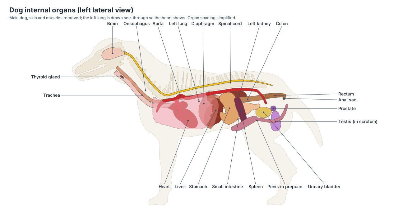
| Organ | Side of the body | Notes |
|---|---|---|
| Stomach | Mostly left | The pylorus crosses to the right |
| Spleen | Left | Along the greater curvature of the stomach |
| Liver | Both, more on the right | Six lobes against the diaphragm |
| Right kidney | Right, further forward | About T13–L2, in a hollow of the liver |
| Left kidney | Left, further back | About L1–L3; more mobile and easier to feel |
| Duodenum, pancreas right lobe, cecum | Right | The cecum is short and spiral (corkscrew-shaped) |
| Descending colon | Left | Leads to the rectum in the pelvis |
| Heart apex | Left | Points down and back towards the sternum |
Switch on Focus selected in the viewer and click an organ to see it on its own, with everything else ghosted.
Dog anatomy labeled: every part and its function
This is the text version of the 3D model: all 182 labeled structures, grouped by body system, with what each one does. Structures marked "males only" or "females only" appear when you switch sex in the viewer.
External
- Head
- The skull and its coverings: cranium, eyes, ears and the muzzle in front. Carries the brain, the main sense organs and the jaws.
- Muzzle (Snout, foreface)
- The face in front of the eyes: nose, lips (flews) and jaws. Houses the nose and the long nasal cavity used for smell, and the jaws for biting.
- Neck
- The region between the head and the shoulders, supported by seven cervical vertebrae. Moves and carries the head; the throat (larynx, trachea, oesophagus) and the jugular veins run through it.
- Withers
- The highest point of the back, just behind the neck, over the shoulder blades. Height at the withers is the standard way to measure a dog's height.
- Back
- The topline between the withers and the loin, over the thoracic vertebrae and ribs. Transmits the push of the hind legs forward to the forequarters.
- Loin
- The part of the back between the last rib and the pelvis. Flexes and extends the spine in the gallop; the epaxial muscles here are powerful.
- Croup (Rump)
- The rump: the region over the pelvis and sacrum, from the loin to the base of the tail. Its slope and length affect how the hind limbs drive the dog forward.
- Chest (Thorax, brisket)
- The thorax: the rib cage and its covering, from the forechest to the last rib. Protects the heart and lungs and gives room for breathing.
- Abdomen and flank (Belly)
- The belly and flanks between the rib cage and the hind legs. Encloses the stomach, intestines, liver, kidneys, bladder and reproductive organs.
- Shoulder and upper arm
- The point of the shoulder and the upper arm, over the scapula and humerus. The shoulder and arm muscles swing the forelimb and support the trunk.
- Elbow
- The joint between the upper arm and the forearm, at the back of the leg level with the chest floor. A hinge joint that flexes and extends the forelimb.
- Forearm (Antebrachium)
- The part of the front leg between the elbow and the wrist. Carries the muscles that move the carpus and toes.
- Carpus and front pastern (Wrist, metacarpus)
- The wrist (carpus) and the pastern (metacarpus) of the front leg. Absorbs shock; the pastern slopes slightly forward in a normal stance.
- Front paw (Forefoot, manus)
- The forefoot: four weight-bearing toes, pads and claws, plus the dewclaw on the inside of the leg. Supports the dog on its toes (a digitigrade stance), grips and digs.
- Thigh
- The upper part of the hind leg, over the femur, with the hamstring muscles behind. The large thigh muscles provide most of the push in walking, running and jumping.
- Stifle (knee) (Knee)
- The dog's knee: the joint between the thigh and the lower leg. A hinge-like joint stabilised inside by the cruciate ligaments and menisci.
- Lower leg (second thigh) (Crus, gaskin, second thigh)
- The hind leg between the stifle and the hock, over the tibia and fibula. Its muscles extend the hock and flex the toes.
- Hock (Tarsus, ankle)
- The ankle (tarsus) of the hind leg; the point of the hock is the heel bone. Acts as a lever: the calf muscles pull on the point of the hock to extend the leg.
- Rear pastern (Metatarsus)
- The part of the hind leg between the hock and the hind paw. Carries the weight to the toes.
- Hind paw (Hind foot, pes)
- The hind foot: usually four toes with pads and claws. Supports the weight and pushes off the ground.
- Tail
- The flexible continuation of the spine behind the croup. Balance when turning, and communication through position and wagging.
- Ear flap (pinna) (Auricle)
- The external ear flap, made of skin over a sheet of auricular cartilage. Collects sound and funnels it into the ear canal; moves to locate sounds.
- Nose (planum nasale) (Nose leather, rhinarium)
- The hairless, moist skin at the tip of the nose, around the two nostrils. Its moisture helps trap scent molecules; the comma-shaped nostrils let air in and out.
- Whiskers (vibrissae)
- Stiff tactile hairs on the muzzle, chin and above the eyes. Detect touch and air movement near the face.
- Digital pads
- The small round pad under each toe. Cushion the toe bones and joints with fatty tissue under thick, keratinised skin.
- Metacarpal and metatarsal pads (Main pad, palmar/plantar pad)
- The large, heart-shaped central pad of each front (metacarpal) and hind (metatarsal) paw. Takes much of the weight when the dog stands, walks and runs.
- Carpal pad (Stopper pad)
- A small pad high on the back of the front leg. Cushions the carpus and acts as a brake when the dog stops or comes down a slope.
- Claws (nails) (Nails)
- Hard keratin claws that cover the last bone of each toe. Grip and dig. Inside each claw is the quick: a blood vessel and nerve.
- Scrotum · males only
- The skin pouch that holds the testes. Keeps the testes cooler than the body core, which sperm production needs.
- Prepuce (sheath) (Sheath) · males only
- The tube of skin that covers the free part of the penis. Protects the glans penis. Urine leaves the male through the preputial opening.
- Vulva · females only
- The external genital opening of the female. Opening of the vestibule, the shared end of the urinary and reproductive tracts.
- Mammary glands and teats (Mammae) · females only
- Milk glands under the skin of the chest and belly, each with a teat. Produce milk for the puppies.
- Anus
- The opening of the digestive tract under the tail. Controlled by the anal sphincter muscles; releases faeces.
Skeletal
- Incisive bone (Premaxilla)
- The front bone of the upper jaw that holds the six upper incisors. Carries the upper incisor teeth and forms the front of the hard palate.
- Nasal bones
- A pair of long, narrow bones forming the bridge of the nose. Form the roof of the nasal cavity.
- Maxilla
- The main bone of the upper jaw and the side of the face. Holds the upper canine, premolars and molars and forms most of the hard palate.
- Lacrimal bone
- A small bone at the front (rostral) rim of the orbit. Contains the opening of the nasolacrimal canal, which drains tears into the nose.
- Frontal bone
- The forehead bone above and between the eyes. Encloses the frontal sinuses and the front of the braincase.
- Parietal bones
- A pair of curved plates forming the sides and roof of the braincase. Protect the cerebrum; the temporalis muscle attaches to them.
- Occipital bone
- The bone at the back and base of the skull. Its occipital condyles form the joint with the atlas; the spinal cord passes through the foramen magnum.
- Temporal bone
- The bone on the lower side of the braincase that holds the ear and the jaw joint. Forms the jaw joint (temporomandibular joint), the caudal part of the zygomatic arch and the bony ear.
- Zygomatic bone and arch (Cheekbone)
- The cheekbone, which forms the front of the wide zygomatic arch. Gives the masseter muscle a strong origin and protects the eye from below.
- Palatine bone
- A paired bone forming the back part of the hard palate. Separates the nasal cavity from the mouth at the back of the palate.
- Sphenoid bone
- The floor of the braincase in front of the occipital bone. Supports the pituitary gland and carries foramina for the optic and other cranial nerves.
- Mandible (Lower jaw)
- The lower jaw: two halves joined at the chin by a fibrous symphysis. Carries the lower teeth. The coronoid process anchors the temporalis, the masseteric fossa the masseter.
- Incisors
- Six small front teeth in each jaw (12 in all). Nibble, groom and pick up small objects.
- Canine teeth (Fangs)
- Four long, pointed teeth, one at each corner of the mouth. Grip and puncture.
- Premolars
- Sixteen blade-like cheek teeth, four in each half-jaw. Cut and shear food.
- Molars
- Ten broad back teeth: two in each upper half-jaw and three in each lower one. Crush and grind food.
- Carnassial teeth
- The large shearing pair of cheek teeth on each side: upper fourth premolar and lower first molar. Slide past each other like scissors to slice meat and tough food.
- Hyoid apparatus
- A chain of small bones that suspends the tongue and larynx from the skull. Anchors the tongue and larynx and moves them in swallowing.
- Atlas (C1) (First cervical vertebra)
- The first neck vertebra: a ring with broad, wing-like transverse processes. Its joint with the skull lets the head nod.
- Axis (C2) (Second cervical vertebra)
- The second and longest neck vertebra, with a tall, blade-like spinous process. Its dens (a peg) projects into the atlas, forming the pivot joint for turning the head.
- Cervical vertebrae (C3–C7)
- The remaining five neck vertebrae; dogs have seven cervical vertebrae in all. Support and move the neck and protect the spinal cord.
- Thoracic vertebrae (T1–T13)
- Thirteen vertebrae, each carrying a pair of ribs. Anchor the ribs; their tall spinous processes form the withers.
- Lumbar vertebrae (L1–L7)
- Seven large vertebrae of the loin with long transverse processes. Bear the strong back muscles and allow flexion in the gallop.
- Sacrum
- Three sacral vertebrae fused into one bone. Joins the spine to the pelvis at the sacroiliac joints.
- Caudal (tail) vertebrae (Coccygeal vertebrae)
- The vertebrae of the tail, usually about 20, becoming simple rods towards the tip. Support the tail and its muscles.
- Ribs
- Thirteen pairs of curved bones forming the rib cage. Protect the heart and lungs and move in breathing.
- Costal cartilages and costal arch
- Bars of cartilage that continue each rib towards the sternum. Make the rib cage flexible for breathing.
- Sternum (breastbone) (Sternebrae)
- The breastbone: eight sternebrae from the manubrium to the xiphoid process. Anchors the sternal ribs and the pectoral muscles.
- Scapula (Shoulder blade)
- The shoulder blade: a flat, triangular bone with a ridge (spine) on its lateral surface. Attaches the forelimb to the trunk through muscles only, and forms the shoulder joint.
- Clavicle (vestigial) (Collarbone remnant)
- A tiny remnant of a collarbone, not joined to any other bone. Has no supporting role; it marks the clavicular intersection of the brachiocephalicus.
- Humerus
- The upper arm bone, between the shoulder and the elbow. Transmits weight and carries the arm muscles.
- Radius
- The main weight-bearing bone of the forearm. Carries most of the weight to the carpus.
- Ulna
- The forearm bone behind the radius, forming the point of the elbow. The triceps pulls on the olecranon to extend the elbow.
- Carpal bones (Carpus)
- Seven small wrist bones in two rows. Let the wrist bend and absorb shock.
- Accessory carpal bone
- A carpal bone that projects backwards like a heel at the wrist. A lever for the flexor muscles of the carpus.
- Metacarpal bones (Metacarpals I–V)
- Five bones of the front pastern; I is short (the dewclaw), II–V are long. Support the paw at the correct angle.
- Phalanges of the front paw
- The toe bones: proximal, middle and distal phalanges in digits II–V; two in the dewclaw. The distal phalanx ends in the ungual process, which sits inside the claw.
- Sesamoid bones of the paws
- Small bones in tendons behind each knuckle joint of the paws. Reduce friction and act as pulleys for the flexor tendons.
- Ilium
- The largest part of the hip bone, with a wide wing that joins the sacrum. Transfers the push of the hind limb to the spine.
- Ischium
- The caudal part of the hip bone, ending in the ischial tuberosity (point of the buttock). Anchors the hamstring muscles.
- Pubis
- The ventral, cranial part of the hip bone, forming the front of the pelvic floor. Anchors the adductor and abdominal muscles.
- Femur (Thigh bone)
- The thigh bone: the longest bone of the hind limb. Carries weight and the large thigh muscles.
- Patella (kneecap) (Kneecap)
- The kneecap: a large sesamoid bone in the tendon of the quadriceps. Increases the leverage of the quadriceps when it straightens the stifle.
- Fabellae and popliteal sesamoid
- Small sesamoid bones behind the stifle. Act as pulleys for the calf muscle tendons.
- Tibia (Shin bone)
- The shin bone: the main bone of the lower hind leg. Carries the weight; the cruciate ligaments attach on its flat top (tibial plateau).
- Fibula
- A thin bone beside the tibia. Gives attachment to muscles and forms the lateral malleolus of the hock.
- Talus
- The ankle bone that forms the hinge of the hock with the tibia. Its pulley-shaped trochlea lets the hock flex and extend.
- Calcaneus (Heel bone)
- The heel bone, which forms the point of the hock. A lever for the calf muscles, which pull on it through the common calcanean (Achilles) tendon.
- Tarsal bones (Tarsus)
- The remaining five small hock bones: the central tarsal and tarsals I–IV. Form the lower joints of the hock and absorb shock.
- Metatarsal bones
- The long bones of the rear pastern: II–V, with I usually small or absent. Support the hind paw.
- Phalanges of the hind paw
- The toe bones of the hind paw, three in each of digits II–V. Their structure matches the front paw; the distal phalanx carries the claw.
- Os penis (Baculum, penile bone) · males only
- A bone inside the penis. Keeps the penis rigid for mating before full erection.
Muscular
- Temporalis
- The large jaw-closing muscle on the side and top of the skull. Closes the jaw powerfully.
- Masseter
- The thick cheek muscle of the jaw. Closes the jaw and grinds.
- Brachiocephalicus
- A long muscle from the head and neck to the arm. Pulls the forelimb forward, or bends the neck when the leg is planted.
- Sternocephalicus
- A strap muscle from the breastbone to the head. Pulls the head and neck down and to the side.
- Omotransversarius
- A strap muscle from the neck to the shoulder. Pulls the shoulder forward.
- Trapezius
- A thin triangular sheet over the neck and withers. Holds the scapula against the trunk and swings it.
- Deltoideus
- The muscle over the point of the shoulder. Flexes the shoulder joint.
- Triceps brachii
- The large muscle mass behind the upper arm. Extends (straightens) the elbow so the dog can bear weight.
- Latissimus dorsi
- A broad, fan-shaped muscle over the side of the chest. Pulls the forelimb backwards in the stride.
- Pectoral muscles (Superficial and deep pectorals)
- The chest muscles between the forelimbs. Hold the forelimbs against the body and pull them in and back.
- Extensors of the forearm
- The muscles on the front and outside of the forearm. Extend the carpus and toes.
- Flexors of the forearm
- The muscles on the back of the forearm. Flex the carpus and toes.
- External abdominal oblique
- The outer layer of the abdominal wall. Supports the abdomen and helps breathe out.
- Rectus abdominis
- A long strap muscle on each side of the belly midline. Supports the abdominal organs and flexes the back.
- Middle gluteal muscle (Gluteus medius)
- The largest gluteal muscle, over the wing of the ilium. Extends the hip and drives the hind limb.
- Superficial gluteal muscle (Gluteus superficialis)
- A smaller gluteal muscle behind the middle gluteal. Extends the hip and moves the leg outward.
- Tensor fasciae latae
- A triangular muscle at the front of the thigh. Tenses the thigh fascia, flexes the hip and helps extend the stifle.
- Sartorius
- A strap muscle on the cranial edge of the thigh. Flexes the hip and draws the leg forward.
- Biceps femoris
- The largest superficial muscle of the outer thigh. Extends the hip and hock; one of the hamstring muscles.
- Semitendinosus
- A hamstring muscle at the back of the thigh. Extends the hip and hock and flexes the stifle.
- Semimembranosus
- The innermost hamstring muscle. Extends the hip and adducts the limb.
- Gastrocnemius
- The calf muscle at the back of the lower hind leg. Extends the hock and pushes off the ground.
- Common calcanean tendon (Achilles tendon)
- The dog's Achilles tendon. Transmits the pull of the calf and hamstring muscles to the heel bone.
- Cranial tibial muscle (Tibialis cranialis)
- A muscle on the front of the lower hind leg. Flexes the hock.
Nervous
- Cerebrum
- The two cerebral hemispheres: the largest part of the brain. Senses, learning, behaviour and voluntary movement.
- Cerebellum
- The 'little brain' behind the cerebrum. Coordinates movement and balance.
- Brainstem
- The stalk that joins the brain to the spinal cord. Controls breathing, heart rate and consciousness; most cranial nerves arise here.
- Olfactory bulbs
- The front part of the brain that receives smell signals. First processing station for smell.
- Spinal cord
- The main nerve pathway inside the vertebral canal. Carries signals between the brain and the body.
- Brachial plexus
- The network of nerves to the forelimb. Supplies the forelimb muscles and skin.
- Sciatic nerve
- The largest nerve of the hind limb. Supplies the hamstrings and, through its tibial and common fibular branches, the lower leg and paw.
Sense organs
- Eye (Globe)
- The eyeball, about 2 cm across in a medium dog. Vision: dogs see well in dim light and detect movement.
- Third eyelid (Nictitating membrane)
- A fold of membrane at the inner corner of the eye. Protects the eye and spreads tears; its gland makes about a third or more of the tear film.
- Tapetum lucidum
- A reflective layer behind the retina. Reflects light back through the retina, improving vision in dim light; it causes eyeshine.
- Vertical ear canal
- The first, downward part of the L-shaped ear canal. Carries sound towards the eardrum.
- Horizontal ear canal
- The inner, horizontal part of the ear canal. Leads sound to the tympanic membrane.
- Tympanic membrane (Eardrum)
- The eardrum. Vibrates with sound and passes it to the middle-ear bones.
- Middle ear
- The air-filled cavity behind the eardrum. Contains the three ossicles that pass sound to the inner ear.
- Inner ear
- The cochlea and the balance organs (vestibular system). Hearing and balance.
- Vomeronasal organ (Jacobson's organ)
- A paired chemical-sense organ in the floor of the nasal cavity. Detects pheromones and other chemical signals.
Digestive
- Tongue
- The muscular organ on the floor of the mouth. Laps water, moves food, grooms and loses heat by panting.
- Salivary glands
- Glands that make saliva: parotid, mandibular, sublingual and zygomatic. Moisten food and the mouth.
- Oesophagus (Esophagus)
- The muscular tube from the throat to the stomach. Moves swallowed food to the stomach by peristalsis.
- Stomach
- A simple, J-shaped stomach on the left of the cranial abdomen. Stores and begins to digest food with acid and enzymes.
- Liver
- The largest gland, with six main lobes, against the diaphragm. Makes bile, processes nutrients, stores energy and removes toxins.
- Gallbladder
- A small sac that stores bile. Releases bile into the duodenum to digest fat.
- Pancreas
- A V-shaped gland with right and left lobes. Makes digestive enzymes and the hormones insulin and glucagon.
- Duodenum
- The first part of the small intestine. Receives bile and pancreatic juice; digestion begins here.
- Jejunum
- The long, coiled middle part of the small intestine. Digests food and absorbs nutrients.
- Ileum
- The short last part of the small intestine. Absorbs nutrients and passes the remains to the colon.
- Cecum
- A short blind pouch, coiled into a spiral (corkscrew) shape, at the start of the colon. A little microbial fermentation takes place in it; it is small in the dog.
- Colon
- The large intestine: ascending, transverse and descending parts. Absorbs water and salts and forms faeces.
- Rectum
- The last part of the large intestine. Stores faeces before defecation.
- Anal sacs (Anal glands)
- Two small scent glands beside the anus. Make a strong-smelling secretion that marks faeces for territory.
Respiratory
- Nasal cavity and conchae (Turbinates)
- The long air passage of the muzzle, filled with thin scroll-shaped bones (turbinates). Warms, filters and humidifies air; its caudal part carries the olfactory lining used for smell.
- Larynx (Voice box)
- The voice box at the top of the trachea. Protects the airway in swallowing (with the epiglottis) and produces barking.
- Trachea (Windpipe)
- The windpipe: a tube held open by C-shaped cartilage rings. Carries air between the larynx and the lungs.
- Main bronchi
- The two branches of the trachea, one to each lung. Carry air into the lungs, where they branch into smaller airways.
- Left lung
- The left lung, with a cranial lobe (cranial and caudal parts) and a caudal lobe. Gas exchange: oxygen in, carbon dioxide out.
- Right lung
- The right lung, with cranial, middle, caudal and accessory lobes. Gas exchange.
- Diaphragm
- The dome-shaped muscle that separates the chest from the abdomen. Contracts to flatten and draw air into the lungs; the main breathing muscle.
Cardiovascular
- Left ventricle
- The thick-walled pumping chamber that forms the apex of the heart. Pumps oxygen-rich blood into the aorta and around the body.
- Right ventricle
- The pumping chamber on the cranial and right side of the heart. Pumps oxygen-poor blood into the pulmonary trunk and lungs.
- Left atrium
- The receiving chamber for blood from the lungs. Receives oxygen-rich blood from the pulmonary veins and passes it to the left ventricle through the mitral valve.
- Right atrium
- The receiving chamber for blood from the body. Receives blood from the venae cavae and passes it to the right ventricle.
- Aorta
- The main artery leaving the left ventricle. Distributes oxygen-rich blood to the whole body.
- Brachiocephalic trunk
- The first and larger branch of the aortic arch, which supplies the head, neck and right forelimb. Gives off the left and right common carotid arteries and ends as the right subclavian artery.
- Left subclavian artery
- The second branch of the aortic arch, supplying the left forelimb and the left side of the neck and chest wall. Carries oxygen-rich blood to the left forelimb; it continues as the axillary artery.
- Pulmonary trunk and arteries
- The large artery from the right ventricle to the lungs. Carries oxygen-poor blood to the lungs.
- Cranial vena cava
- The large vein that returns blood from the head, neck and forelimbs. Returns oxygen-poor blood to the heart.
- Caudal vena cava
- The large vein that returns blood from the abdomen and hind limbs. Returns oxygen-poor blood to the heart.
- Common carotid arteries
- The two main arteries to the head. Supply the brain, face and tongue.
- External jugular veins
- Large superficial veins draining the head. Return blood from the head and face; a common blood-sampling site.
- Cephalic vein
- A superficial vein on the front of the forearm. Drains the front paw and forearm; the usual site for IV catheters and injections.
- Femoral artery
- The main artery of the hind limb. Supplies the hind limb. Vets check the pulse here.
- Lateral saphenous vein
- A superficial vein on the outside of the lower hind leg. Drains the hind paw; used for blood sampling and injections.
Lymphatic & endocrine
- Spleen
- A flat, tongue-shaped organ along the stomach. Stores blood, removes old red blood cells and helps immunity.
- Thyroid glands
- Two small endocrine glands on the trachea. Make thyroid hormones that control metabolism.
- Adrenal glands
- Two small endocrine glands near the kidneys. Make cortisol, aldosterone and adrenaline.
- Mandibular lymph nodes
- Lymph nodes under the angle of the jaw. Filter lymph from the head; they can be felt in a healthy dog.
- Superficial cervical lymph nodes (Prescapular lymph nodes)
- Lymph nodes in front of the shoulder. Filter lymph from the neck and forelimb.
- Popliteal lymph nodes
- Lymph nodes behind the stifle. Filter lymph from the lower hind limb.
Urinary
- Kidneys
- Two bean-shaped organs under the lumbar spine. Filter blood and make urine.
- Ureters
- Thin muscular tubes from each kidney to the bladder. Carry urine to the bladder by peristalsis.
- Urinary bladder
- A stretchy sac that stores urine. Stores urine until urination.
- Urethra
- The tube from the bladder to the outside. Carries urine out (and semen in males).
Reproductive
- Testes (Testicles) · males only
- The male gonads. Make sperm and testosterone.
- Epididymis · males only
- A coiled tube along each testis. Stores and matures sperm; it connects the testis to the ductus deferens.
- Ductus deferens (Vas deferens) · males only
- The tube that carries sperm from the epididymis to the urethra. Carries sperm during ejaculation.
- Prostate · males only
- A two-lobed gland around the urethra. Adds fluid to semen. It is the only accessory sex gland of male dogs.
- Penis · males only
- The male copulatory organ. Deposits semen and carries the urethra.
- Bulbus glandis · males only
- An expandable part of the penis at the base of the glans. Swells during mating and holds the dogs together in the 'tie'.
- Ovaries · females only
- The female gonads. Produce eggs and the hormones oestrogen and progesterone.
- Uterine horns · females only
- Two long tubes forming the main part of the uterus. Puppies develop in a row along both horns.
- Uterine body · females only
- The short common part of the uterus. Joins the horns to the cervix.
- Cervix · females only
- The muscular neck of the uterus. Stays closed except during heat and birth.
- Vagina · females only
- The long passage from the cervix to the vestibule. Receives the penis and forms the birth canal.
Dog muscle anatomy: the canine muscular system
The canine muscular system has several hundred named muscles. The ones you see first, just under the skin and the thin cutaneous trunci muscle, are the superficial muscles of the head, neck, shoulder, trunk and limbs shown below. In the forelimb, muscles such as the trapezius, rhomboids, serratus ventralis and pectorals form a muscular sling that attaches the leg to the body.
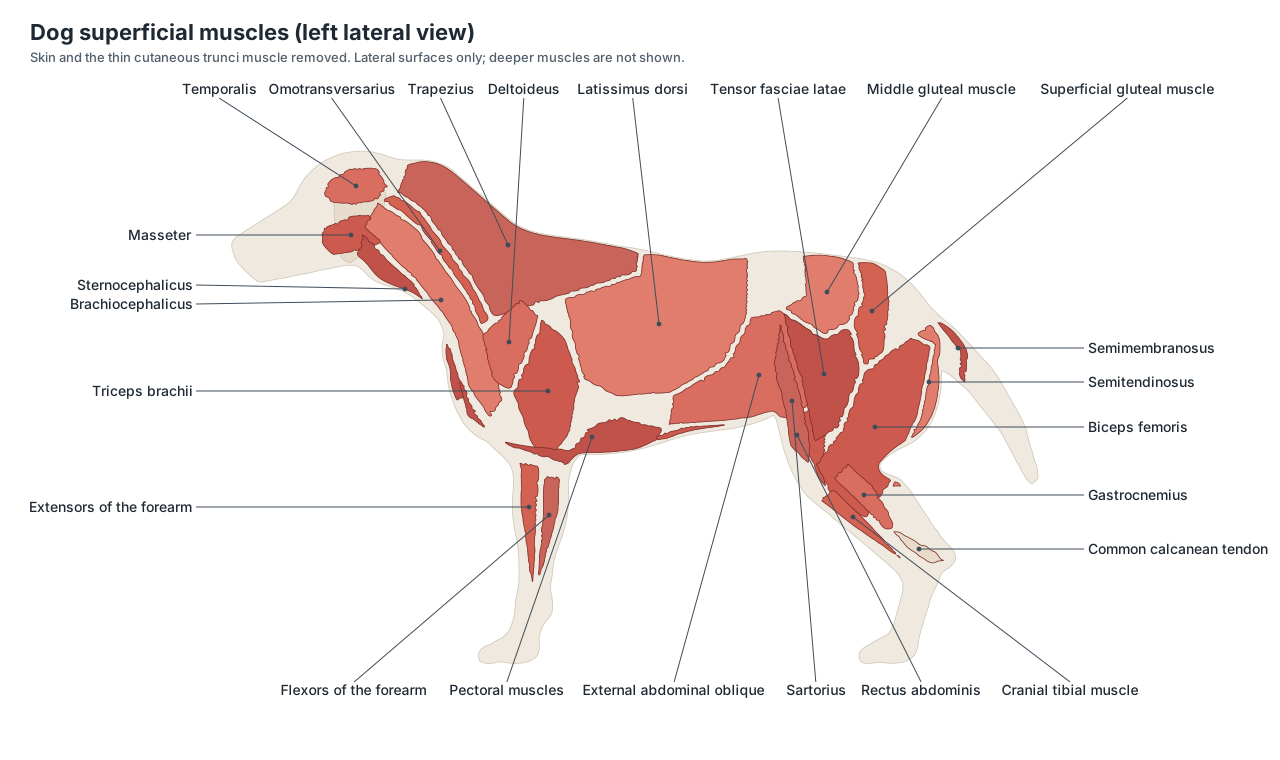
- Brachiocephalicus: A long muscle from the head and neck to the arm. Pulls the forelimb forward, or bends the neck when the leg is planted.
- Trapezius: A thin triangular sheet over the neck and withers. Holds the scapula against the trunk and swings it.
- Latissimus dorsi: A broad, fan-shaped muscle over the side of the chest. Pulls the forelimb backwards in the stride.
- Triceps brachii: The large muscle mass behind the upper arm. Extends (straightens) the elbow so the dog can bear weight.
- Middle gluteal muscle: The largest gluteal muscle, over the wing of the ilium. Extends the hip and drives the hind limb.
- Biceps femoris: The largest superficial muscle of the outer thigh. Extends the hip and hock; one of the hamstring muscles.
- Gastrocnemius: The calf muscle at the back of the lower hind leg. Extends the hock and pushes off the ground.
- Common calcanean tendon: The dog's Achilles tendon. Transmits the pull of the calf and hamstring muscles to the heel bone.
The brachiocephalicus is typical of carnivores: the remnant of the clavicle, a tendinous band called the clavicular intersection, splits it into a cleidocephalicus part running to the head and neck and a cleidobrachialis part running to the arm. In the hip, the superficial gluteal and the biceps femoris both attach to the sacrotuberous ligament, and the sciatic nerve runs dorsal and caudal to the hip joint deep to them. Vets avoid this area for injections into the back of the thigh.
Dog skeleton and bones
A dog's skeleton has roughly 320 bones; the exact number depends mainly on the length of the tail. The vertebral formula is C7, T13, L7, S3 (fused into the sacrum) and about 20 caudal vertebrae. There are 13 pairs of ribs, a breastbone of eight sternebrae, and limb bones that match ours in name but not in proportion: dogs stand on their toes, so the long bones of the paw (metacarpals and metatarsals) form part of the leg.
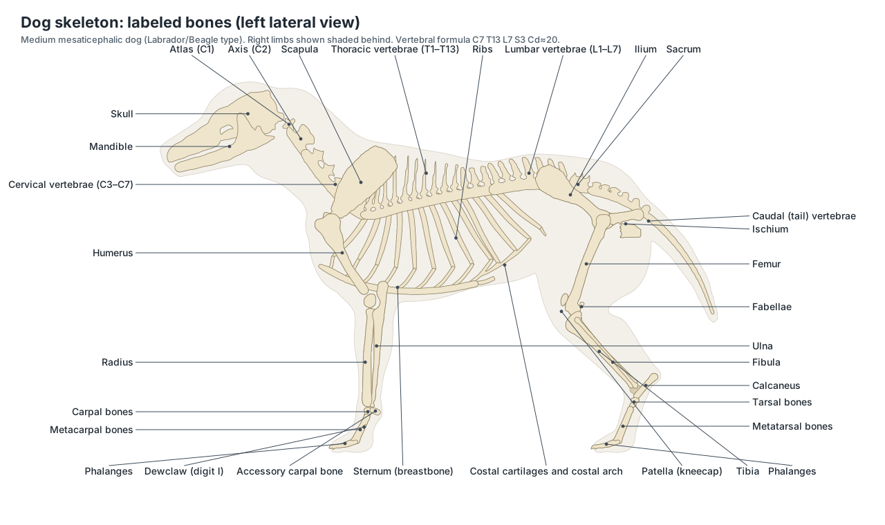
Every bone in the diagram can be clicked in the 3D model with the Skeletal system on. For bone-by-bone detail, the dental formula, skull bones and breed differences, see the dog skeleton guide.
Dog leg anatomy: front leg vs rear leg
A dog's front leg runs from the shoulder blade through the upper arm, elbow, forearm, wrist (carpus) and pastern to the paw. The rear leg runs from the hip through the thigh, knee (stifle), lower leg, ankle (hock) and rear pastern to the hind paw. The front legs carry more of the body weight when the dog stands; the hind legs provide most of the forward push.
| Front leg (forelimb) | Rear leg (hind limb) | |
|---|---|---|
| Attachment to the body | Muscles only (no clavicle joint) | Pelvis, joined to the sacrum |
| Upper bone | Humerus | Femur |
| First big joint | Shoulder | Hip |
| Middle joint | Elbow (points backwards) | Stifle / knee (points forwards) |
| Lower leg bones | Radius and ulna | Tibia and fibula |
| Ankle or wrist | Carpus: 7 carpal bones | Hock (tarsus): 7 tarsal bones |
| Pastern | 5 metacarpals | 4 metatarsals (I usually vestigial) |
| Toes | 4 + dewclaw | Usually 4 (rear dewclaws in some breeds) |
| Pads | Digital, metacarpal and carpal pads | Digital and metatarsal pads |
The dog's knee is the stifle, and its "backwards knee" lower down the hind leg is really the hock, the ankle. Dogs and humans share the same joints; the dog simply stands with its heel off the ground.
Dog paw anatomy: pads, nails and the quick
A dog's paw has four weight-bearing toes, each with a digital pad and a claw, a large heart-shaped metacarpal (front) or metatarsal (hind) pad behind them, and on the front leg a carpal pad higher up. The dewclaw is digit I, on the inner side of the leg; it has its own claw but does not touch the ground.
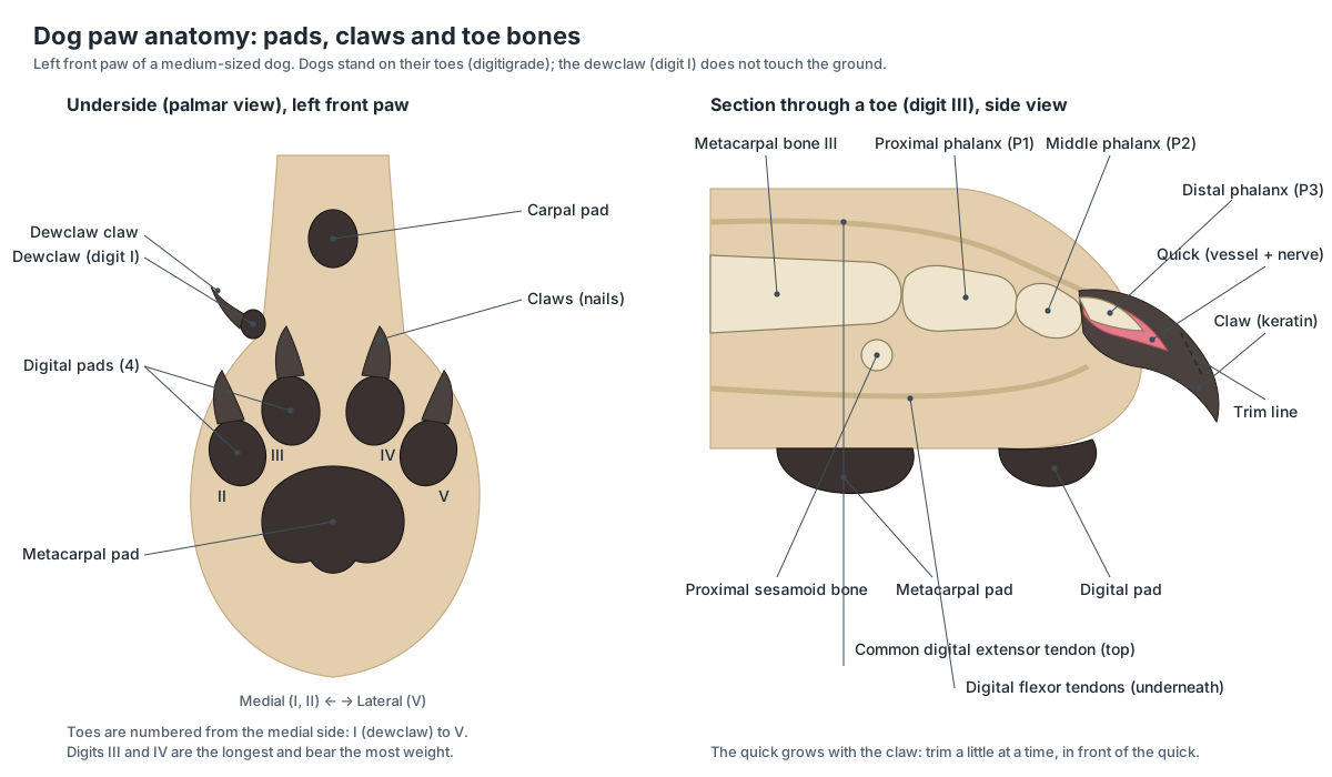
Each claw grows continuously around the last toe bone, the distal phalanx. Inside is the quick, a living core of blood vessels and nerves that grows as the claw grows. Cutting into it hurts and makes the claw bleed, sometimes heavily. When claws are trimmed regularly, the quick stays short; once a claw has grown long, it can only be trimmed back a little at a time. Most dogs need a trim every three to four weeks.
Pads are thick, keratinised skin over cushions of fat. They grip, absorb shock and protect the bones and joints of the paw. The carpal pad acts as a brake when a dog stops or comes down a slope.
Dog head anatomy: skull, mouth and teeth
The dog's head is built around the skull, which is divided into the braincase (cranium) and the face. Wide zygomatic arches give the jaw-closing masseter and temporalis muscles a large area to attach, and the jaw joint works mainly as a hinge for a strong scissor bite. Adult dogs have 42 teeth (2 × I 3/3, C 1/1, P 4/4, M 2/3); the carnassial pair, upper fourth premolar and lower first molar, slices food like scissors.
The tongue sits on the floor of the mouth, anchored to the hyoid bones. Saliva comes from four pairs of salivary glands: parotid, mandibular, sublingual and zygomatic. See the labeled dog skull for the individual bones.
Dog nose anatomy: why dogs smell so well
A dog's sense of smell starts at the moist planum nasale and the two comma-shaped nostrils, but the real work happens deep in the nasal cavity. Thin, scroll-shaped bones called nasal conchae (turbinates) fill the muzzle. Their front part warms and cleans the air; the caudal ethmoidal conchae carry the olfactory epithelium, where scent molecules bind to receptor cells. Dogs also have a second, separate chemical sense: the paired vomeronasal organ, which lies between the nasal and oral cavities near the vomer bone. (Kokocińska-Kusiak, A. et al. (2021). Canine olfaction: physiology, behavior, and possibilities for practical applications. Animals 11, 2463 (PMC8388720).)
The same review notes that a much smaller share of the dog's olfactory receptor genes are inactive pseudogenes (about 20%) than in humans (more than 50%). Popular figures such as '220 million receptors' or 'a million times better' are estimates that vary between sources, so treat them as rough comparisons. The Merck Veterinary Manual's owner guide gives the 'about a million times stronger' figure. (Merck Veterinary Manual (owners): Description and physical characteristics of dogs.)
Dog eye anatomy: third eyelid, tapetum and colour vision
Each eye has an upper and lower eyelid plus a third eyelid, the nictitating membrane: a pale fold in the inner (medial) corner that sweeps across the eye and protects it. A gland at its base makes a large part of the tear film. The American College of Veterinary Ophthalmologists puts it at 33–66% of tears, which is why vets replace a prolapsed gland ('cherry eye') rather than remove it: removal predisposes the eye to dry eye. (American College of Veterinary Ophthalmologists: Cherry eye.)
Behind the retina, the tapetum lucidum reflects light back through the light-sensitive cells. It makes the eyes glow in a torch beam and helps dogs see in dim light. Dogs have two cone types, so their colour vision is dichromatic: they see blues and yellows well but confuse reds and greens. With eyes set at the sides of the head, their total field of view is about 250°, wider than a human's. (American Kennel Club: What do dogs see?.)
Dog ear anatomy diagram
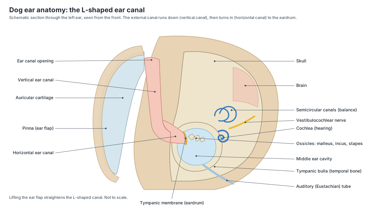
A dog's ear has three parts: the outer ear (the pinna and the L-shaped ear canal), the middle ear (eardrum and three ossicles inside the tympanic bulla) and the inner ear (cochlea and balance organs). Dogs move each pinna on its own to locate a sound, and hear high-pitched sounds far better than people.
Dog ear anatomy: vertical and horizontal ear canal
The dog's ear canal is L-shaped. From the opening at the base of the pinna, the vertical canal runs down the side of the head; it then bends to become the horizontal canal, which runs inwards to the eardrum (tympanic membrane). Lifting the ear flap straightens the canal, which is why ear drops are given with the pinna held up. (VCA Animal Hospitals: Applying ear drops to dogs.)
Behind the eardrum, the middle ear sits in the tympanic bulla, a bony bubble on the underside of the skull. It holds the three ossicles and connects to the throat through the auditory tube. The inner ear contains the cochlea for hearing and the vestibular system for balance. The canal is much longer than a human's, and ear problems are common in dogs. (Merck Veterinary Manual (owners): Ear structure and function in dogs.)
In otitis externa the canal lining swells and narrows, and medication may not reach the horizontal canal. Breeds with heavy, floppy, hairy ears are more prone; VCA reports that allergy is involved in up to 43% of cases. (VCA Animal Hospitals: Ear infections in dogs (otitis externa).)
Dog heart anatomy and circulation
A dog's heart has four chambers, like ours: two atria that receive blood and two ventricles that pump it. It lies in the lower chest between the forelimbs, roughly level with the elbows, with its apex angled down, back and to the left. The mitral and aortic valves are on the left side; the tricuspid and pulmonary valves on the right. At rest it beats about 70–120 times a minute (Merck Veterinary Manual), faster in small dogs.
- Right atrium: The receiving chamber for blood from the body. Receives blood from the venae cavae and passes it to the right ventricle.
- Right ventricle: The pumping chamber on the cranial and right side of the heart. Pumps oxygen-poor blood into the pulmonary trunk and lungs.
- Left atrium: The receiving chamber for blood from the lungs. Receives oxygen-rich blood from the pulmonary veins and passes it to the left ventricle through the mitral valve.
- Left ventricle: The thick-walled pumping chamber that forms the apex of the heart. Pumps oxygen-rich blood into the aorta and around the body.
- Aorta: The main artery leaving the left ventricle. Distributes oxygen-rich blood to the whole body.
- Brachiocephalic trunk: The first and larger branch of the aortic arch, which supplies the head, neck and right forelimb. Gives off the left and right common carotid arteries and ends as the right subclavian artery.
- Left subclavian artery: The second branch of the aortic arch, supplying the left forelimb and the left side of the neck and chest wall. Carries oxygen-rich blood to the left forelimb; it continues as the axillary artery.
- Pulmonary trunk and arteries: The large artery from the right ventricle to the lungs. Carries oxygen-poor blood to the lungs.
Blood returns from the body in the cranial and caudal venae cavae to the right atrium, is pumped by the right ventricle to the lungs, returns oxygenated to the left atrium, and leaves the left ventricle through the aorta. The aortic arch of the dog has just two large branches: the brachiocephalic trunk, which gives off both common carotid arteries and the right subclavian artery, and the left subclavian artery. Mitral valve disease is the most common acquired heart disease in older small-breed dogs.
| Vessel | Where | Why vets use it |
|---|---|---|
| External jugular vein | Jugular groove, lower neck | Blood samples |
| Cephalic vein | Front of the forearm | IV catheters and injections |
| Lateral saphenous vein | Outside of the hind leg above the hock | Blood samples |
| Femoral artery | Inside of the thigh | Feeling the pulse |
Dog throat, neck and lungs
Air enters through the nostrils, passes through the nasal cavity and pharynx, and reaches the larynx (voice box) in the throat. The trachea runs down the underside of the neck, held open by C-shaped cartilage rings, and divides above the heart into two main bronchi. The oesophagus runs beside it, on the left in the neck, and the thyroid glands lie on each side of the trachea just behind the larynx.
The lungs have lobes: the left lung has a cranial lobe (with cranial and caudal parts) and a caudal lobe; the right lung has cranial, middle, caudal and accessory lobes. The diaphragm, the main breathing muscle, bulges forward into the chest; when it contracts it flattens and draws air in. A resting dog takes about 18–34 breaths a minute.
Dog stomach and abdominal organs
Food travels from the mouth down the oesophagus, through the diaphragm into the stomach, then into the small intestine (duodenum, jejunum and ileum), the large intestine (cecum, colon and rectum) and out through the anus. The liver and pancreas pour bile and enzymes into the duodenum. The dog's gut is relatively short, as expected for a carnivore that also eats plant material.
- Stomach: A simple, J-shaped stomach on the left of the cranial abdomen. Stores and begins to digest food with acid and enzymes.
- Liver: The largest gland, with six main lobes, against the diaphragm. Makes bile, processes nutrients, stores energy and removes toxins.
- Gallbladder: A small sac that stores bile. Releases bile into the duodenum to digest fat.
- Pancreas: A V-shaped gland with right and left lobes. Makes digestive enzymes and the hormones insulin and glucagon.
- Jejunum: The long, coiled middle part of the small intestine. Digests food and absorbs nutrients.
- Colon: The large intestine: ascending, transverse and descending parts. Absorbs water and salts and forms faeces.
- Anal sacs: Two small scent glands beside the anus. Make a strong-smelling secretion that marks faeces for territory.
Dog stomach anatomy and bloat (GDV)
The dog has a simple, single-chambered stomach. It lies mostly on the left behind the liver, with the cardia where the oesophagus enters, a dome-shaped fundus, a body along the left flank, and the pyloric part that crosses to the right to join the duodenum. In gastric dilatation-volvulus (GDV, 'bloat'), the stomach fills with gas and can rotate on itself, cutting off its own blood supply. Risk is linked to a deep, narrow chest (a high thoracic height-to-width ratio); Great Danes, Weimaraners, St Bernards and setters are among the breeds most affected. (American College of Veterinary Surgeons: Gastric dilatation-volvulus.)
Dog urinary system
Two bean-shaped kidneys filter the blood under the lumbar spine. The right kidney lies further forward, at about T13–L2, tucked into a hollow of the liver; the left is further back and moves more. Urine runs down the ureters to the bladder in the caudal abdomen and leaves through the urethra, which in males runs the length of the penis and opens at the tip inside the prepuce.
Male vs female dog anatomy
Male and female dogs differ in their reproductive organs and in how they urinate. The male's penis lies in a sheath (prepuce) along the belly, and his testes hang in a scrotum between the thighs. The female's vulva sits below the anus, and she has two rows of mammary glands with teats.
| Male dog | Female dog | |
|---|---|---|
| Gonads | Testes in the scrotum | Ovaries behind the kidneys, in a fatty ovarian bursa |
| Ducts | Epididymis, ductus deferens, urethra | Uterine tubes, two long uterine horns, short uterine body |
| Glands | Prostate (the only accessory sex gland) | Mammary glands, usually 4–5 pairs |
| External | Prepuce on the belly, scrotum | Vulva below the anus |
| Special features | Os penis and bulbus glandis | Y-shaped uterus; puppies develop along both horns |
| Urethra | Long, through the penis; stones lodge more easily | Short, opening into the vestibule |
Switch the 3D viewer between Female and Male to see each set of organs in place.
Dog knee (stifle) anatomy and the cranial cruciate ligament
The stifle is the dog's knee: the joint between the femur, the tibia and the patella. Inside it, two cruciate ligaments cross between the femur and the tibia. The cranial cruciate ligament (CCL, the dog's equivalent of the human ACL) is one of the most important stabilisers of the joint, and its gradual degeneration and rupture is one of the most common reasons for hind-limb lameness in dogs. (American College of Veterinary Surgeons: Cranial cruciate ligament disease.)
Because the ligament stops the tibia sliding forward, a ruptured CCL lets the tibia shift ('cranial drawer') each time the dog bears weight. The menisci, cartilage pads between the bones, are often damaged as well. VCA describes CCL rupture as the most common knee injury in dogs. (VCA Animal Hospitals: Cruciate ligament rupture in dogs.)
Four sesamoid bones sit around the canine stifle: the patella in the quadriceps tendon, two fabellae in the heads of the gastrocnemius behind the femoral condyles (the lateral one is longer), and a small popliteal sesamoid behind the top of the tibia. (Mauragis, D. Small animal radiography: stifle joint and crus. Today's Veterinary Practice.)
Male and female dog reproductive anatomy
In the male, the testes sit in the scrotum and the epididymis on each testis connects it to the ductus deferens. The prostate is the only accessory sex gland of the male dog. The penis has two unusual features: a bone, the os penis, which keeps it rigid, and the bulbus glandis, which swells inside the female during mating and produces the 'tie'. (University of Illinois ANSC 207: Reproductive anatomy of the male dog.)
In the female, the paired ovaries lie behind the kidneys. The uterine horns join to form a short uterine body in front of the cervix, which connects the uterus to the vagina; the vagina opens at the vulva. (University of Illinois ANSC 207: Reproductive anatomy of the female dog.)
The bitch is monoestrous: she has one heat (oestrus) at a time with a long resting interval, roughly every 6–7 months, at any time of year. Puberty comes at about 6–24 months depending on breed size. (Merck Veterinary Manual: Features of the reproductive cycle (table).)
Most dogs have four to five pairs of mammary glands. Mammary tumours are common in intact females, which is one reason spaying is discussed early in life. (Merck Veterinary Manual: Mammary tumors in dogs.)
Dog nervous system: brain, spinal cord and nerves
The brain fills the cranial cavity: the large cerebrum in front, the cerebellum behind it for coordination, and the brainstem joining the spinal cord. The olfactory bulbs at the front are large. The spinal cord runs through the vertebral canal and, in a medium-sized dog, ends around the sixth to seventh lumbar vertebra, with the nerve roots of the cauda equina continuing beyond. The brachial plexus supplies the forelimb and the sciatic nerve the hind limb.
Because the cord lies directly above the intervertebral discs, a herniated disc can press on it and cause pain, weakness or paralysis. This is common in short-legged, long-backed breeds such as the Dachshund (see the Dachshund skeleton).
Dog tail anatomy
The tail is the continuation of the spine: about 20 caudal vertebrae, larger and with small processes near the base and simple rods towards the tip, moved by several groups of muscles. Dogs use the tail for balance when turning and for communication. Under the base of the tail lie the anus and, on each side of it at about the 4 and 8 o'clock positions, the anal sacs, whose strong-smelling secretion marks the faeces.
Dog anatomy ventral view
Seen from below (the ventral view), a dog shows the chest floor and sternum between the forelimbs, the belly with its two rows of teats in females or the prepuce in males, the inguinal region between the thighs, the scrotum or vulva, and the pads of all four paws. Inside, the ventral view is the classic dissection view: the liver lobes against the diaphragm, the stomach on the left, the coils of jejunum, and the bladder at the pelvic inlet. Rotate the 3D model and look up from below with the coat faded.
Lymph nodes and glands you can feel
Vets feel several lymph nodes at every check-up: the mandibular nodes under the angle of the jaw, the superficial cervical (prescapular) nodes in front of the shoulder, and the popliteal nodes behind the stifle. They are small and smooth in a healthy dog; enlargement of several at once is a common first sign of lymphoma. The spleen, thyroid and adrenal glands are not normally felt but are shown in the model.
How dog breeds differ anatomically
| Feature | Variation | Examples |
|---|---|---|
| Skull shape | Short-faced (brachycephalic), medium, long-faced (dolichocephalic) | Pug, Labrador, Greyhound |
| Leg length | Short, curved limbs from chondrodystrophy | Dachshund, Basset Hound, Corgi |
| Ear carriage | Drop, semi-erect or erect pinnae | Beagle, Collie, German Shepherd |
| Tail | Long, curled, naturally bobbed | Labrador, Husky, some Corgis |
| Chest | Deep and narrow vs barrel-shaped | Great Dane vs Bulldog |
| Rear dewclaws | Usually absent; present in some breeds | Great Pyrenees, Briard |
Dog anatomy worksheet: test yourself
- What is the dog's knee called, and which joint is the "backwards knee"?
- Name the four parts of the canine heart and the vessel that leaves the left ventricle.
- Which organ lies along the left side of the stomach?
- Why is a dog's ear canal described as L-shaped?
- What is the quick, and why should you not cut it?
- How many lobes does each lung have?
- Name two anatomical differences between male and female dogs.
Answers: 1 stifle; hock (ankle). 2 right and left atria and ventricles; aorta. 3 spleen. 4 vertical canal then horizontal canal. 5 the vessel and nerve in the claw; it bleeds and hurts. 6 left 2 (cranial, caudal), right 4 (cranial, middle, caudal, accessory). 7 for example os penis and prostate in males; uterus and mammary glands in females.
Frequently asked questions
How many bones does a dog have?
An adult dog has roughly 320 bones, but there is no single exact number: the count varies mainly with the length of the tail (about 20 caudal vertebrae in a long-tailed dog) and with whether rear dewclaws are present. The vertebral formula is C7, T13, L7, S3 (fused) and a variable number of caudal vertebrae.
What organs are on the left and right side of a dog?
On the left are most of the stomach, the spleen, the descending colon and the left kidney (further back than the right). On the right are the pylorus, duodenum, pancreas right lobe, cecum, much of the liver and the right kidney, which sits under the last rib in a hollow of the liver.
Where is a dog's heart?
The heart lies in the lower chest between the forelimbs, roughly at the level of the elbows, with its apex pointing down, back and to the left towards the sternum. The Merck Veterinary Manual gives a resting heart rate of about 70–120 beats per minute for adult dogs; small dogs have faster rates than large dogs.
What is a dog's knee called?
The dog's knee is called the stifle. It is the joint between the femur and the tibia at the front of the hind leg. The joint that points backwards lower down the hind leg is the hock, the dog's ankle.
Why are dog ear canals L-shaped?
A dog's ear canal runs down the side of the head (the vertical canal) and then turns inward (the horizontal canal) to the eardrum. This L shape traps moisture and debris, which is one reason ear infections are common, especially in breeds with floppy, hairy ears.
What is the quick in a dog's nail?
The quick is the blood vessel and nerve inside the claw. It grows as the claw grows. Cutting into it is painful and makes the claw bleed, so long claws are trimmed back a little at a time.
Do dogs have a third eyelid?
Yes. The third eyelid (nictitating membrane) is a pale fold at the inner corner of each eye. It protects the eye and spreads tears, and the gland at its base makes a large share of the tear film. 'Cherry eye' is a prolapse of that gland.
How many teeth does a dog have?
Adult dogs have 42 permanent teeth: 12 incisors, 4 canines, 16 premolars and 10 molars (dental formula 2 × I 3/3, C 1/1, P 4/4, M 2/3). Puppies have 28 deciduous teeth and no baby molars.
What is the difference between male and female dog anatomy?
Males have testes in a scrotum between the thighs, a penis with a bone (os penis) and a bulbus glandis inside a prepuce on the belly, and a prostate as their only accessory sex gland. Females have ovaries behind the kidneys, a Y-shaped uterus with long horns, a cervix, vagina and vulva below the anus, and usually 4–5 pairs of mammary glands.
References
- Merck Veterinary Manual (owners): Description and physical characteristics of dogs
- Merck Veterinary Manual (owners): Ear structure and function in dogs
- Merck Veterinary Manual (owners): Eye structure and function in dogs
- Merck Veterinary Manual (owners): Introduction to heart and blood vessel disorders in dogs
- Merck Veterinary Manual (owners): The gonads and genital tract of dogs
- Merck Veterinary Manual: Features of the reproductive cycle (table)
- Merck Veterinary Manual: Mammary tumors in dogs
- Merck Veterinary Manual: Dentition and dental nomenclature of animals
- VCA Animal Hospitals: Applying ear drops to dogs
- VCA Animal Hospitals: Ear infections in dogs (otitis externa)
- VCA Animal Hospitals: Cruciate ligament rupture in dogs
- VCA Animal Hospitals: Successful nail trims – training for happy handling
- VCA Animal Hospitals: Anal sac disease in dogs
- American College of Veterinary Surgeons: Cranial cruciate ligament disease
- American College of Veterinary Surgeons: Gastric dilatation-volvulus
- American College of Veterinary Ophthalmologists: Cherry eye
- American Kennel Club: What do dogs see?
- American Kennel Club: Why do dogs have whiskers?
- Kokocińska-Kusiak, A. et al. (2021). Canine olfaction: physiology, behavior, and possibilities for practical applications. Animals 11, 2463 (PMC8388720)
- Mauragis, D. Small animal radiography: stifle joint and crus. Today's Veterinary Practice
- University of Illinois ANSC 207: Reproductive anatomy of the male dog
- University of Illinois ANSC 207: Reproductive anatomy of the female dog
- IMAIOS vet-Anatomy: Lung lobes
- IMAIOS vet-Anatomy: Liver
- IMAIOS vet-Anatomy: Right kidney
- IMAIOS vet-Anatomy: Brachiocephalic muscle
- University of Minnesota veterinary anatomy, carnivore lab 5: lateral pelvic muscles of the dog
- WikiVet: Bones – Dog Anatomy
More interactive anatomy models
- Dog skeletonEvery major bone of the canine skeleton: skull, spine, limbs and paw.
- Human anatomy1,000+ bones, muscles, organs, vessels and nerves from real anatomical data.
- Octopus anatomyThree hearts, a ring-shaped brain, eight arms and a beak, in 3D.
- Squid anatomyPen, eight arms, two tentacles, giant axons and three hearts of the longfin squid.
- Squid dissectionStep-by-step guided squid dissection with every structure labeled.
- Fish anatomyExternal and internal anatomy of a bony fish: fins, gills, swim bladder, heart and brain.