

Earthworm dissection overview
In an earthworm dissection you open the worm along its back, pin the body wall aside and identify the gut, hearts, reproductive organs, nephridia and nervous system by counting segments from the mouth. The whole lab takes about 45–60 minutes. Almost everything that matters is in the first 20 segments, so most of your time will be spent with a hand lens on the front quarter of the worm.
Keep a segment count as you go. The peristomium around the mouth is segment 1. The brain is in segment 3, the hearts in 7–11, the seminal vesicles in 9–12, the female and male pores on 14 and 15, and the gizzard just behind the crop at about 17–19. If a structure is not where you expect it, recount: specimens differ by a segment or two, and contracted preserved worms make the front segments hard to separate.
Materials and safety
You need a worm, a wax-bottomed tray, fine scissors, forceps, a teasing needle and about twenty pins. Fine iris scissors make the dorsal cut much easier than a scalpel, because you can keep the lower blade just under the body wall.
Materials
- One preserved earthworm (Lumbricus terrestris, 15–25 cm), or a fresh worm anaesthetised under your teacher's supervision
- Dissecting tray with a wax base, or a cork or foam board
- Fine scissors (iris scissors are ideal), a scalpel or razor blade, fine forceps and a teasing (dissecting) needle
- About 15–20 dissecting pins
- Hand lens or dissecting microscope (needed for setae, pores, nephridia and the brain)
- Wash bottle of water (fresh specimens: weak ethanol, so the worm stays covered while you work)
- Nitrile gloves, safety glasses and a lab coat or apron
- Paper towels, a lab worksheet and coloured pencils for your labelled drawing
Safety
- Wear gloves and eye protection. Preserved worms are stored in fixative; rinse the worm in water and work in a ventilated room.
- Scissors and scalpels are sharp. Cut away from your fingers and lift the body wall with forceps before every cut.
- Keep the cut shallow. The body wall is only about a millimetre thick, and a deep cut destroys the gut and blood vessels under it.
- Never eat or drink in the lab. Wash your hands afterwards.
- Put tissue, pins and blades in the containers your teacher provides, not in the sink or bin.
Preserved earthworms, live worms and humane euthanasia
Most school labs use preserved earthworms bought from a biological supplier. They arrive already killed and fixed, so the question of how to kill an earthworm for dissection does not arise; you simply rinse off the preservative. Preserved worms are firmer and easier to pin, but their organs are paler and the blood vessels are harder to see than in a fresh worm.
University invertebrate labs often prefer fresh nightcrawlers, which are cheap from bait shops and show the beating dorsal vessel and the true colours of the organs. Lander University's lab exercise, for example, anaesthetises living worms by placing them in 5–7% non-denatured ethanol in tap water for 15–30 minutes before the class, and keeps the worm covered with the same solution while it is dissected. (Fox, Lander University: Lumbricus terrestris anatomy lab exercise.)
If a worm must be killed, it should be done humanely and only by, or under the direct supervision of, a teacher or researcher following their institution's animal-care rules. Published guidance on invertebrates generally recommends a two-step approach: first anaesthetise the animal so it cannot respond, then use a second step that makes death certain (for example, transfer into a stronger alcohol solution or fixative). Methods that cause obvious distress, such as dropping a conscious worm into concentrated alcohol, salt or boiling water, should not be used. Reviews point out that most invertebrate methods have not been properly tested in the laboratory, which is a good reason to follow your institution's current protocol rather than improvising. (Cooper (2011): anesthesia, analgesia and euthanasia of invertebrates (ILAR Journal).)
Earthworms do respond to harmful stimuli: touching the head triggers a fast escape withdrawal carried by the giant nerve fibres. Whatever your view on invertebrate pain, treating the animals carefully, using as few as necessary and choosing preserved or virtual specimens when they meet the learning goal is good laboratory practice. (Wikipedia: Earthworm (anatomy and physiology).)
Before you cut: external observation
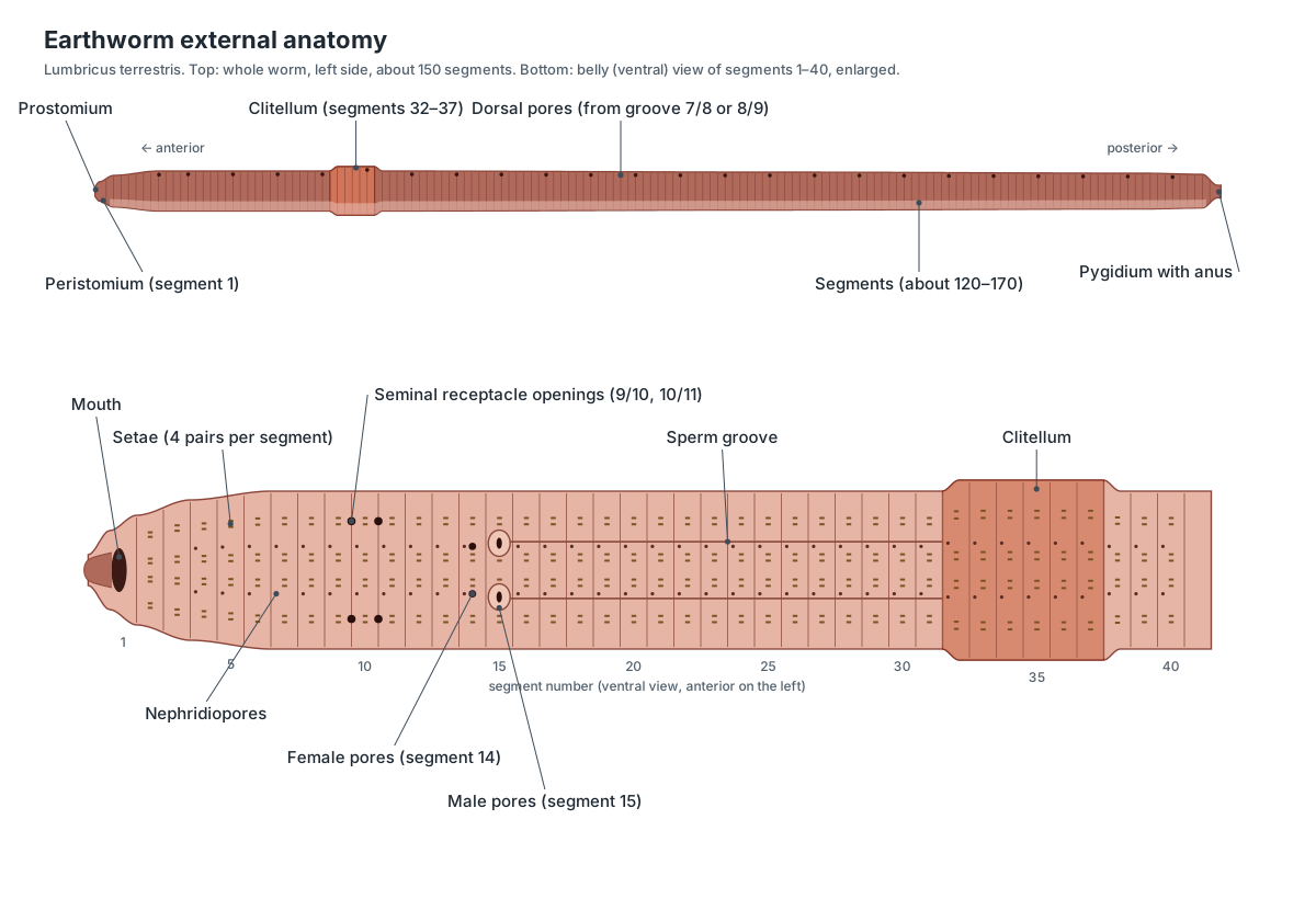
Before cutting, identify the anterior end, the dorsal and ventral sides, the clitellum, the setae and the genital pores. The clitellum is nearer the head, so the shorter part of the worm in front of it is the anterior end. The prostomium overhangs the mouth. The back is darker and rounded; the belly is paler and flatter.
Run a gloved finger along the belly from tail to head: the setae make it feel rough. Look at them with a hand lens. Then turn the worm belly up and count back from the mouth to segment 15 to find the male pores in their swollen lips, the female pores just in front on segment 14, and the seminal receptacle openings in the grooves between segments 9/10 and 10/11. Draw the front 40 segments and mark each feature before you start cutting, because the incision distorts the segments.
Dissected earthworm labeled diagram
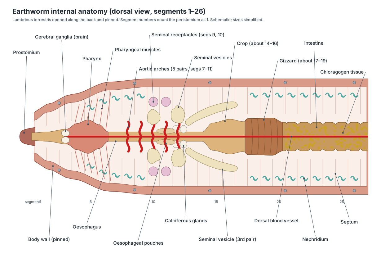
A dissected earthworm, opened along the back, shows the pharynx, hearts, oesophagus, seminal vesicles, seminal receptacles, crop, gizzard and intestine in a line from the head, with the dorsal blood vessel along the top of the gut. This is what the front 26 segments should look like after you pin the body wall open and tear the septa.
How to dissect an earthworm step by step
The order below follows university and high-school lab guides: outside first, then a dorsal incision, then the hearts, reproductive organs, gut, nephridia and finally the nervous system, which is only visible once the gut is moved. Each step matches a step in the 3D dissection guide above.
1. External anatomy: ends, sides and segments
- Rinse the worm and lay it in the tray. Find the anterior end: it is closer to the clitellum and more pointed.
- Find the dorsal side. It is darker and more rounded; the ventral side is paler and flatter. The dark dorsal blood vessel may show through the skin.
- Find the prostomium overhanging the mouth, the peristomium (segment 1) around the mouth, and the pygidium with the anus at the tail.
- Locate the clitellum and count the segments in front of it. In Lumbricus terrestris it begins at about segment 32 and covers about six segments. Look for the paired ridges (tubercula pubertatis) along its lower edges on segments 33–36.
- Run a finger along the ventral side from tail to head to feel the setae, then look at them with a hand lens: four pairs on each segment.
Identify: Prostomium, Peristomium, Mouth, Body segments, Clitellum, Tubercula pubertatis, Setae, Pygidium, Anus.
2. Ventral surface: genital and excretory pores
- Turn the worm ventral side up and examine segments 9 to 16 with a hand lens.
- On segment 15 find the male pores in their swollen, lip-like ridges. A faint sperm groove runs back from each one to the clitellum.
- On segment 14, just in front, look for the much smaller female pores.
- Look for the seminal receptacle openings in the grooves between segments 9/10 and 10/11, at the sides.
- With a microscope, look for nephridiopores near the front of most segments and the dorsal pores in the grooves on the back midline (from groove 7/8 or 8/9 backwards).
Identify: Male pores, Female pores, Seminal receptacle openings, Sperm grooves, Nephridiopores, Dorsal pores.
3. Dorsal incision and pinning
- Place the worm dorsal side up. Pin through the prostomium and through the body a few segments behind the clitellum, angling the pins away from the worm.
- Pinch up the skin with forceps a little behind the clitellum and slightly to one side of the dorsal midline, so you miss the dorsal blood vessel.
- Cut forwards with fine scissors, tips angled up, cutting only the body wall, all the way to the prostomium. To see the intestine as well, extend the cut 20–30 segments back behind the clitellum.
- Spread the body wall. Tear the septa (the thin walls between segments) with a teasing needle and pin the wall flat on each side, every five segments or so.
- Pins at 45° keep them out of your way. Add water so the organs float and separate.
Caution: Cut shallow and keep the scissor tips pointing up. Most failed earthworm dissections are caused by cutting too deep.
Identify: Septa, Coelom, Body segments, Dorsal blood vessel.
4. Hearts and blood vessels
- Find the dorsal blood vessel on top of the gut. Follow it forwards to the oesophagus region.
- In segments 7–11 find the five pairs of aortic arches ('hearts') curving down around the oesophagus. Move the seminal vesicles aside gently to see the last ones.
- Count the aortic arches and record the segments they are in.
- Lift the gut slightly to see the ventral blood vessel underneath it. Note the thin segmental vessels joining dorsal and ventral vessels behind the hearts.
Identify: Dorsal blood vessel, Aortic arches (hearts), Ventral blood vessel, Segmental vessels.
5. Reproductive organs (segments 9–15)
- Find the three pairs of large, cream-coloured seminal vesicles in segments 9 to 12. They arch over the oesophagus and are the biggest organs in this region.
- Below and in front of them, in segments 9 and 10, find the two pairs of small, round seminal receptacles near the body wall.
- With a microscope, look for the tiny testes and sperm funnels (segments 10 and 11) and the thin vas deferens running back to the male pore on segment 15.
- Look for the small ovaries hanging from the front septum of segment 13, and the short oviducts leading to the female pores on segment 14.
- Remember: every earthworm has both sets of organs. There is no male or female specimen.
Identify: Seminal vesicles, Seminal receptacles, Testes, Sperm funnels, Vas deferens, Ovaries, Oviducts, Ovisacs.
6. The alimentary canal
- Starting at the mouth, identify the buccal cavity (segments 1–3) and the muscular pharynx (segments 3–5) with fine pharyngeal muscles tying it to the body wall.
- Follow the narrow oesophagus back under the seminal vesicles (segments 6–13). Find the oesophageal pouches and the white calciferous glands at its rear end.
- Find the thin-walled crop (about segments 14–16) and the firm, muscular gizzard behind it (about 17–19). Squeeze each gently with forceps: the gizzard is hard, the crop soft.
- Trace the long intestine from the gizzard towards the tail. Note the yellow-brown chloragogen tissue covering it and the constrictions at each septum.
- Optional: slit the crop and gizzard to look for soil and sand grains, and the intestine to see the typhlosole on its dorsal wall.
Identify: Buccal cavity, Pharynx, Pharyngeal muscles, Oesophagus, Oesophageal pouches, Calciferous glands, Crop, Gizzard, Intestine, Chloragogen tissue, Rectum.
7. Nephridia
- Behind the reproductive region, push the intestine aside a little and look along the body wall of any segment.
- Find the pale, coiled nephridia, one on each side of each segment, attached to the septa.
- With a microscope, look for the small funnel (nephrostome) on the front face of a septum. Its tubule passes through the septum into the next segment.
- Remove one nephridium, mount it in water on a slide and look at the coiled tubule at low power.
Identify: Nephridia, Nephrostome, Septa, Nephridiopores.
8. Brain and ventral nerve cord
- Find the small, white, two-lobed cerebral ganglia (the brain) on top of the pharynx in segment 3. Do not cut the front of the worm until you have found them.
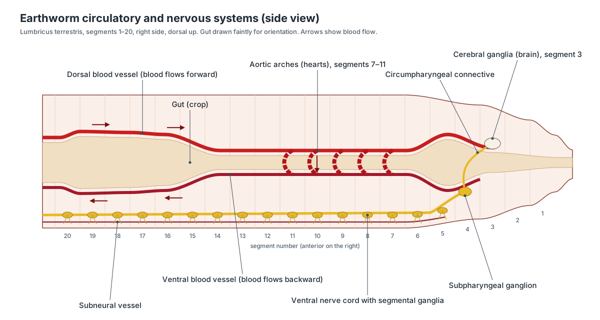
- Follow the circumpharyngeal connectives down each side of the pharynx to the subpharyngeal ganglion underneath.
- Cut through the oesophagus behind the pharynx and lift the gut back carefully, cutting septa as you go.
- On the floor of the body cavity find the white ventral nerve cord with a swelling (ganglion) in every segment, and the tiny segmental nerves branching off.
Caution: The brain is tiny and soft. Use a hand lens and a blunt probe rather than scissors near the front end.
Identify: Cerebral ganglia (brain), Circumpharyngeal connectives, Subpharyngeal ganglion, Ventral nerve cord, Segmental ganglia.
9. Cross-section through the intestine region
- Cut a thin slice across the worm behind the clitellum, or study a prepared cross-section slide under the microscope.
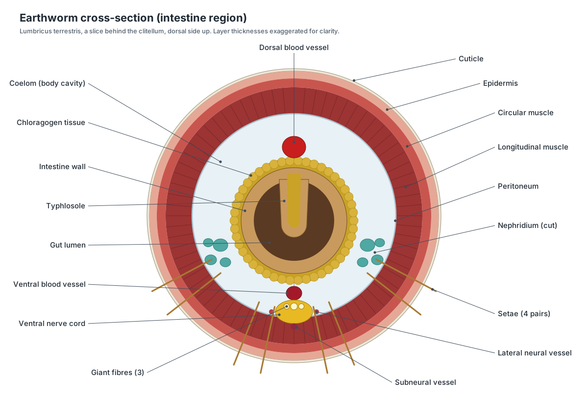
- From the outside in, identify the cuticle, epidermis, circular muscle and thick longitudinal muscle.
- Find the coelom, the intestine with its U-shaped space and the typhlosole hanging into it, and the chloragogen tissue around it.
- Find the dorsal vessel above the gut, the ventral vessel below it, and the nerve cord with its giant fibres and subneural vessel. Look for setae and nephridia in the body wall.
Identify: Cuticle, Epidermis, Circular muscle, Longitudinal muscle, Coelom, Typhlosole, Chloragogen tissue, Giant fibres, Subneural vessel, Lateral neural vessels.
10. Labelled drawing and clean-up
- Draw the opened anterior end (segments 1–20) and label every organ you found, with its segment number.
- Complete the lab questions below.
- Dispose of the worm, pins and blades as your teacher directs. Clean and dry the tools and the tray, then wash your hands.
Which segment is it in? Organ finder
Use segment numbers to find each organ in an earthworm dissection. Count from the mouth (segment 1). These are typical values for Lumbricus terrestris; expect a difference of one segment either way in some worms.
| Structure | Segment(s) | What it looks like |
|---|---|---|
| Cerebral ganglia (brain) | 3 | Top of the pharynx; tiny and white |
| Pharynx | 3–5 | Thick, muscular, with radial muscles to the body wall |
| Aortic arches (hearts) | 7–11 | Five pairs of red loops around the oesophagus |
| Seminal receptacles | 9, 10 | Small round sacs near the body wall |
| Seminal vesicles | 9, 11, 12 | Large cream lobes over the oesophagus |
| Testes, sperm funnels | 10, 11 | Under the vesicles, near the nerve cord (microscope) |
| Calciferous glands | 11–12 | White swellings at the end of the oesophagus |
| Ovaries | 13 | Front septum of segment 13, near the nerve cord |
| Female pores | 14 | Ventral surface, tiny |
| Male pores | 15 | Ventral surface, in swollen lips |
| Crop | About 14–16 | Soft, thin-walled swelling |
| Gizzard | About 17–19 | Hard, muscular swelling |
| Intestine | From about 20 | Long tube covered in yellow chloragogen |
| Clitellum | About 32–37 | Thick band on the outside |
If you are dissecting the Indian earthworm Pheretima, the numbers are different: its clitellum is on segments 14–16, the male pores on 18 and the gizzard in 8–9. See the comparison table on the earthworm anatomy page.
Dissecting the reproductive system of an earthworm
The reproductive organs are crowded into segments 9 to 15, just in front of where most students start looking. When you first open the front of the worm, the biggest things you see are the three pairs of creamy-white seminal vesicles in segments 9, 11 and 12, arching over the oesophagus. The rear pair is the largest and stretches back several segments; the front pair is the smallest. Seminal vesicles belong to the male system and store the worm's own sperm. (Fox, Lander University: Lumbricus terrestris anatomy lab exercise.)
Under and in front of them, in segments 9 and 10, are the two pairs of small, round seminal receptacles. They are smaller and more regular in shape than the vesicles and belong to the female system: they hold sperm received from a partner, and their ducts open in the grooves between segments 9/10 and 10/11. The testes, sperm funnels and ovaries are tiny. To see them, free the gut from the pharynx to the gizzard and lift it gently, then look with a dissecting microscope near the nerve cord in segments 10–13. The ovaries hang from the front septum of segment 13, and the oviducts lead to the female pores on segment 14. (Fox, Lander University: Lumbricus terrestris anatomy lab exercise.)
Earthworm brain and nervous system dissection
To find the earthworm brain, look at the very front of the opened worm. The two small, white cerebral ganglia sit on top of the junction between the buccal cavity and the pharynx in segment 3. If your incision went far enough forward to open the buccal cavity, you can see them without further cutting. Follow the circumpharyngeal connectives down each side of the pharynx with a fine probe. (Fox, Lander University: Lumbricus terrestris anatomy lab exercise.)
The ventral nerve cord is only visible once the gut is moved. Cut across the oesophagus behind the pharynx, lift the gut and fold it back, cutting the septa as you go. The cord is the white strand on the floor of the body cavity; with a hand lens you can see a ganglion (a slight swelling) in each segment and the segmental nerves leaving it. Biology Corner's high-school worksheet asks students to decide whether the brain attaches to the ventral nerve cord: it does, through the connectives and the subpharyngeal ganglion. (Biology Corner: earthworm dissection lab.)
Dissection of the alimentary canal
The alimentary canal is the easiest system to trace from end to end. Start at the mouth and follow the pharynx, then the narrow oesophagus hidden by the seminal vesicles and hearts. Behind the calciferous glands, the gut widens into the soft, thin-walled crop and then the firm gizzard, which feels hard when squeezed with forceps. Behind the gizzard the intestine runs to the anus, covered in yellow chloragogen tissue. (Fox, Lander University: Lumbricus terrestris anatomy lab exercise.)
Opening the gut tells you what each part does. Slit the crop and you usually find loose soil and plant fragments. The gizzard has a thick, iridescent cuticle lining that can be peeled off with forceps, and you may find small stones inside. In the intestine, look for a region where the typhlosole is smooth and unfolded on the dorsal wall, and note that the rectum at the end has no typhlosole at all. (Fox, Lander University: Lumbricus terrestris anatomy lab exercise.)
Nephridia and the cross-section
Nephridia are the small, pale, coiled tubules on each side of almost every segment. They are easiest to see behind the reproductive region, where nothing covers them. Each one starts with a funnel (nephrostome) on the front face of a septum and ends at a nephridiopore on the next segment. Remove one, mount it in a drop of water and look at the coiled tube at low power.
A cross-section shows how everything fits together. Cut a thin slice behind the clitellum with a new razor blade, or use a prepared slide. From the outside in you should see cuticle, epidermis, circular muscle, longitudinal muscle, the coelom, the intestine with the typhlosole hanging into it, and the chloragogen tissue around the intestine. The dorsal vessel is above the gut, the ventral vessel below it, and the nerve cord, with its three giant fibres, at the bottom.
Common mistakes and how to fix them
- Cutting too deep. The body wall is thin. If you see a brown smear, you have cut into the gut. Keep the scissor tips angled up and lift the wall with forceps ahead of every cut.
- Opening the wrong side. If you find the white nerve cord immediately under the cut, you have opened the belly. Note it, and use your partner's worm for the circulatory steps.
- Body wall will not lie flat. The septa are still attached. Tear each one with a teasing needle, working from the front, before you pin.
- Cannot find the hearts. They are under the seminal vesicles and septa of segments 7–11. Move the vesicles gently aside with a probe; do not pull them off.
- Mixing up crop and gizzard. Squeeze gently with forceps. The gizzard is hard and muscular; the crop in front of it is soft.
- Lost the brain. It is easily cut away with the front of the worm. Find it before cutting the pharynx, and stop the dorsal incision at the prostomium.
Earthworm dissection worksheet and answer key
Answer these during or after the dissection. They cover the usual pre-lab, lab and post-lab questions. Click a question to reveal the answer.
How can you tell the anterior end and the dorsal side of an earthworm?
The anterior end is nearer the clitellum and has the prostomium and mouth. The dorsal side is darker and more rounded, often with the dorsal blood vessel showing through; the ventral side is paler and flatter, with the setae easier to feel.
In which segments did you find the clitellum, and what does it do?
About segments 32–37 in Lumbricus terrestris (it can start at 31). It secretes mucus during mating and makes the cocoon that receives the eggs and sperm.
On which segments are the male and female pores?
Male pores on segment 15, female pores on segment 14 (in Lumbricus). In Pheretima, the male pores are on segment 18 and the single female pore on segment 14.
Why should the first incision be made on the dorsal side and slightly off the midline?
The nerve cord and most important structures lie ventrally, so opening the back keeps them intact. Cutting just to one side of the midline avoids cutting the dorsal blood vessel.
How many aortic arches did you count, and what is their function?
Five pairs (ten arches) in segments 7–11. They pump blood from the dorsal vessel down to the ventral vessel.
Is the earthworm's circulatory system open or closed? What is your evidence?
Closed. The blood stays inside vessels (dorsal, ventral and the aortic arches and their branches) and never empties into the body cavity.
Put these in order from mouth to anus: intestine, crop, pharynx, gizzard, oesophagus.
Pharynx → oesophagus → crop → gizzard → intestine (then rectum and anus).
Which feels harder, the crop or the gizzard? Why?
The gizzard. It has thick muscular walls and a tough cuticle lining because it grinds food against sand grains. The crop is thin-walled because it only stores food.
What is the typhlosole and why is it important?
A fold of the dorsal wall of the intestine that hangs into the gut. It increases the surface area for absorbing digested food.
Which organs are the large white structures that cover the oesophagus in segments 9–12?
The seminal vesicles. They store the worm's own sperm and are part of the male reproductive system.
Seminal vesicles and seminal receptacles both contain sperm. What is the difference?
Seminal vesicles (male system) store and mature the worm's own sperm. Seminal receptacles (female system) store sperm received from a partner during mating.
Where is the earthworm's brain, and how is it connected to the ventral nerve cord?
On top of the pharynx in segment 3. Two circumpharyngeal connectives run around the pharynx to the subpharyngeal ganglion, which begins the ventral nerve cord.
What organs remove nitrogen waste in an earthworm, and how many are there?
Nephridia. There is a pair in almost every segment, so a worm of 150 segments has nearly 300. Each opens to the outside through a nephridiopore.
How does an earthworm breathe, and why must it stay moist?
Gas exchange happens across the moist skin into a dense network of capillaries. Oxygen must dissolve in the mucus film first, so a dry worm cannot take in oxygen.
True or false: the earthworm is a hermaphrodite, so it fertilises its own eggs.
False. It is a hermaphrodite, but two worms mate and exchange sperm (cross-fertilisation). Eggs are fertilised in the cocoon by the partner's stored sperm.
Name the layers of the body wall from the outside in, as seen in a cross-section.
Cuticle, epidermis, circular muscle, longitudinal muscle, then the peritoneum lining the coelom.
True or false (answer key)
- The brain is connected to the ventral nerve cord. True (through the circumpharyngeal connectives).
- The dorsal side is lighter than the ventral side. False (the dorsal side is darker).
- The clitellum is closer to the anterior end. True.
- An earthworm has fourteen aortic arches. False (five pairs, ten in all).
- Seminal vesicles are part of the digestive system. False (they are male reproductive organs).
- The earthworm's circulatory system is open. False (it is closed).
- The gizzard has thicker walls than the crop. True.
Virtual earthworm dissection
The 3D viewer at the top of this page is a complete virtual earthworm dissection. Each of its ten steps sets the body wall, hides the systems you would have moved aside, and highlights the structures to find; click any of them to read where it is and what it does. Students who opt out of a physical dissection, or who missed the lab, can work through the same steps and answer the same worksheet. Teachers can use it as a pre-lab so students arrive knowing what to look for, which usually makes the real dissection faster and less wasteful.
For a full description of every organ system, the earthworm's movement and breathing, and the differences between Lumbricus and the Indian earthworm Pheretima, see the earthworm anatomy page.
What is the purpose of dissecting an earthworm?
An earthworm dissection lets students see, in one inexpensive animal, several ideas that are central to biology: a body built from repeated segments, a true coelom divided by septa, a complete one-way gut with specialised regions, a closed circulatory system with hearts, a ventral nerve cord, kidney-like nephridia, and a hermaphrodite reproductive system. Because these organs are arranged segment by segment, students also practise locating structures by counting, which is a useful habit for any later dissection.
It also connects anatomy to ecology. Earthworms move vast amounts of soil, drag leaves into their burrows and leave nutrient-rich castings. Charles Darwin devoted his last book to showing how worms shape the soil, and he studied their senses, feeding and gizzard as part of that work. A dissection shows the organs that make this possible. (Darwin (1881): The Formation of Vegetable Mould through the Action of Worms (Project Gutenberg).)
Frequently asked questions
Where do you cut to dissect an earthworm?
On the dorsal (back) side, slightly to one side of the midline. Start a little behind the clitellum and cut forwards through the body wall only, to the prostomium. Cutting off the midline avoids the dorsal blood vessel, and cutting the back keeps the ventral nerve cord intact.
How do you tell the dorsal side of an earthworm from the ventral side?
The dorsal side is darker and more rounded, and the dark dorsal blood vessel often shows through it. The ventral side is paler and flatter, and you can feel the setae more easily there. The genital pores are on the ventral side.
How many hearts will I find in a dissected earthworm?
Five pairs of aortic arches (ten 'hearts') in segments 7–11 of Lumbricus terrestris. They curve around the oesophagus from the dorsal vessel to the ventral vessel and may be partly hidden by the seminal vesicles.
How do you find the earthworm's brain?
Look at the very front of the opened worm, on top of the pharynx in segment 3. The brain is two tiny, white, pear-shaped lobes (cerebral ganglia). Use a hand lens and a blunt probe; it is easy to cut it off by accident.
How are earthworms killed for dissection?
School specimens are usually bought already preserved. When fresh worms are used, a teacher or researcher anaesthetises them first (for example in dilute ethanol) and then uses a second step that makes death certain, following their institution's animal-care protocol. Students should not try to kill worms themselves.
Why are preserved earthworms grey or pale?
The fixative stops decay but bleaches the natural colours and drains the blood, so organs look grey or cream and the vessels are harder to see. Fresh, anaesthetised worms show bright red vessels and a beating dorsal vessel.
Is there a virtual earthworm dissection?
Yes. The 3D model on this page follows the same ten steps as a real dissection. Each step removes the right layers and highlights the structures to find, so students who opt out of a physical dissection can complete the same lab and worksheet.
Do earthworms have a male and a female?
No. Every adult earthworm is a hermaphrodite, so every specimen has both testes and ovaries. Unlike a frog, shark or fetal pig dissection, you do not need to compare male and female worms.
References
- Fox, R. S. Lumbricus terrestris: invertebrate zoology laboratory exercise (after Ruppert, Fox & Barnes 2004). Lander University.
- Biology Corner. Earthworm dissection: procedure, structures and lab questions.
- Cooper, J. E. (2011). Anesthesia, analgesia, and euthanasia of invertebrates. ILAR Journal 52(2), 196–204.
- Darwin, C. (1881). The Formation of Vegetable Mould through the Action of Worms. Project Gutenberg edition.
- Kurth, J. A. & Kier, W. M. (2014). Scaling of the hydrostatic skeleton in the earthworm Lumbricus terrestris. Journal of Experimental Biology 217, 1860–1867.
- Earthworm: anatomy and physiology. Wikipedia.
- Lumbricus terrestris. Wikipedia.
- Pheretima: clitellum and genital pores. Wikipedia.
More interactive anatomy models
- Earthworm anatomySegments, clitellum, five aortic arches, crop and gizzard of the earthworm.
- Human anatomy1,000+ bones, muscles, organs, vessels and nerves from real anatomical data.
- Octopus anatomyThree hearts, a ring-shaped brain, eight arms and a beak, in 3D.
- Squid anatomyPen, eight arms, two tentacles, giant axons and three hearts of the longfin squid.
- Squid dissectionStep-by-step guided squid dissection with every structure labeled.
- Fish anatomyExternal and internal anatomy of a bony fish: fins, gills, swim bladder, heart and brain.