

Earthworm anatomy at a glance
The earthworm body is a series of similar segments with a gut running through the middle. On the outside you can see the prostomium, the mouth, the segments, the bristle-like setae, the swollen clitellum and the anus. Inside, a fluid-filled coelom separated by septa surrounds the gut, the blood vessels, the nerve cord and a pair of nephridia in almost every segment. The reproductive organs are crowded into segments 9 to 15.
- Phylum and class: Annelida (segmented worms), Clitellata, the oligochaetes.
- Reference species: Lumbricus terrestris, the nightcrawler or common earthworm, about 110–200 mm long with 120–170 segments.
- Body plan: bilateral symmetry, a true coelom, a complete gut from mouth to anus, and a closed circulatory system.
- Breathing: through the moist skin; there are no lungs or gills.
- Skeleton: none; the pressurised coelomic fluid acts as a hydrostatic skeleton.
- Reproduction: every adult is a hermaphrodite, but worms mate in pairs and swap sperm.
Lab manuals locate everything by segment number, counting the peristomium around the mouth as segment 1. Learn a few landmarks and the rest falls into place: brain in 3, hearts in 7–11, seminal receptacles in 9 and 10, female pores on 14, male pores on 15, gizzard just behind the crop at about 17–19 and the clitellum at about 32–37.
External anatomy of an earthworm
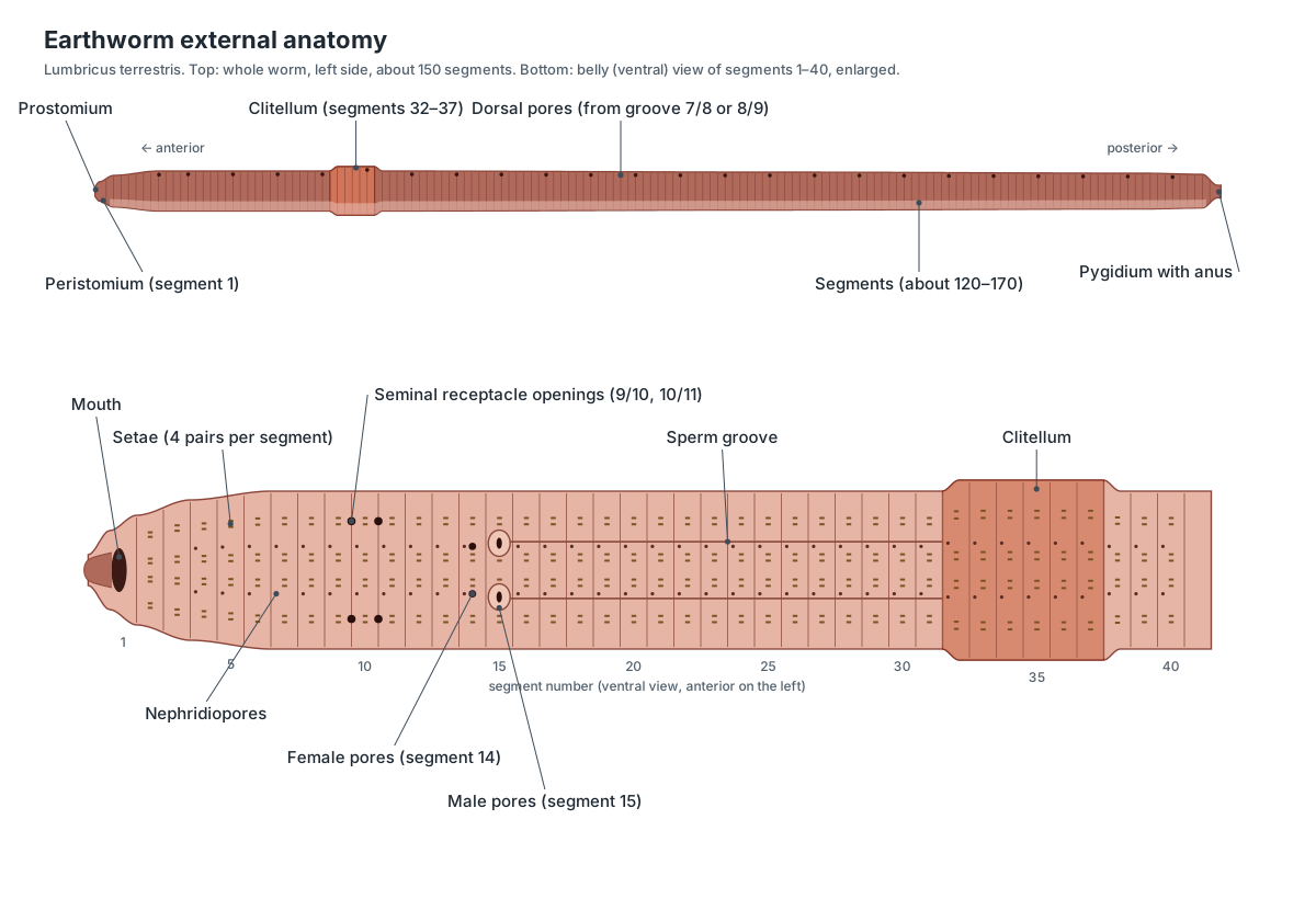
The external anatomy of an earthworm includes the prostomium, peristomium and mouth at the front, the segments and setae along the body, the clitellum, a set of small pores on the belly, and the pygidium with the anus at the tail. Tell the ends apart by the clitellum, which is much closer to the head. Tell the sides apart by colour and shape: the dorsal side is dark reddish-brown and rounded, with the dorsal blood vessel showing through as a dark line; the ventral side is paler and flatter.
| External feature | Where to find it (L. terrestris) | Function |
|---|---|---|
| Prostomium | Lobe over the mouth, in front of segment 1 | Probes soil; sensory |
| Peristomium and mouth | Segment 1, ventral | Takes in soil and leaf litter |
| Setae | 4 pairs on every segment except 1 | Grip the soil during crawling |
| Seminal receptacle openings | Grooves 9/10 and 10/11, at the sides | Receive a partner's sperm |
| Female pores | Segment 14, ventral | Release eggs |
| Male pores | Segment 15, on swollen lips | Release sperm |
| Sperm grooves | Segment 15 back to the clitellum | Carry sperm during mating |
| Clitellum | About segments 32–37 | Mucus for mating; makes the cocoon |
| Dorsal pores | Back midline, in every groove from 7/8 or 8/9 | Release coelomic fluid to keep skin moist |
| Nephridiopores | Most segments, at the sides | Release urine |
| Pygidium and anus | Tail tip | Releases castings |
Most of these pores are tiny. With a hand lens you can usually see the male pores, because they sit in conspicuous swellings, but the female pores, seminal receptacle openings, nephridiopores and dorsal pores normally need a dissecting microscope and a fine probe. Lumbricus has eight setae per segment, arranged in four pairs: two pairs on the belly (labelled a and b) and two on the sides (c and d). The tail of L. terrestris is flattened from top to bottom, which helps it brace itself in its burrow.
Earthworm internal anatomy
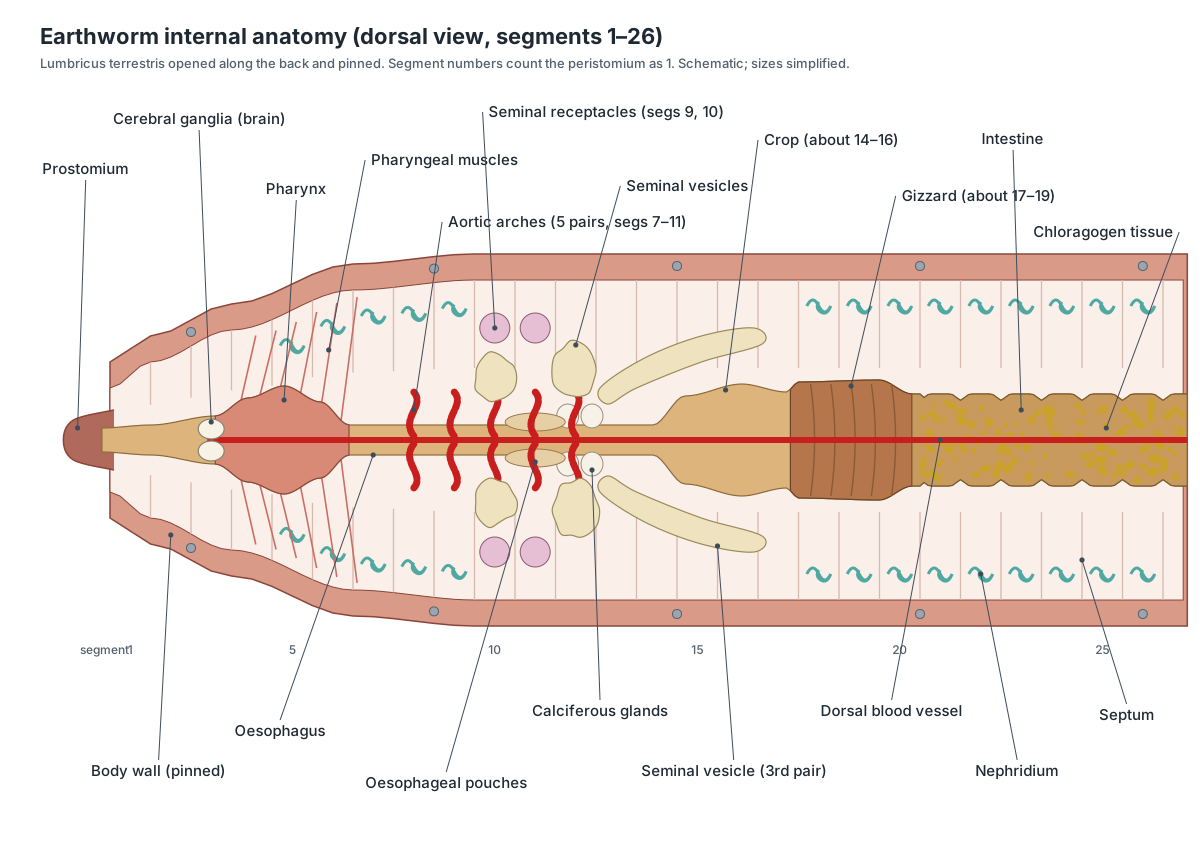
The internal anatomy of an earthworm is organised by segment: pharynx (3–5), oesophagus (6–13) with calciferous glands, crop (about 14–16), gizzard (about 17–19) and intestine (from about 20) form the gut, while five pairs of hearts (7–11), the brain (3), the reproductive organs (9–15) and a pair of nephridia in most segments surround it. The first thing you see when you open a worm along its back is the red dorsal blood vessel lying on the gut and the large, cream-coloured seminal vesicles in segments 9 to 12.
Earthworm parts and functions table
| Structure | Segment(s) | System | Function |
|---|---|---|---|
| Cerebral ganglia (brain) | 3 | Nervous | Process sensory information |
| Pharynx | 3–5 | Digestive | Sucks in food |
| Subpharyngeal ganglion | 4 | Nervous | Starts the ventral nerve cord |
| Oesophagus | 6–13 | Digestive | Carries food to the crop |
| Aortic arches (hearts) | 7–11 | Circulatory | Pump blood from dorsal to ventral vessel |
| Seminal receptacles | 9, 10 | Reproductive (female) | Store a partner's sperm |
| Seminal vesicles | 9, 11, 12 | Reproductive (male) | Store and mature own sperm |
| Testes and sperm funnels | 10, 11 | Reproductive (male) | Make and collect sperm |
| Calciferous glands | 11–12 | Digestive | Secrete calcium carbonate |
| Ovaries | 13 | Reproductive (female) | Make eggs |
| Crop | About 14–16 | Digestive | Stores food |
| Gizzard | About 17–19 | Digestive | Grinds food |
| Intestine with typhlosole | From about 20 | Digestive | Digests and absorbs food |
| Nephridia | Almost every segment | Excretory | Remove nitrogen waste |
| Ventral nerve cord | 4 to the tail | Nervous | Coordinates the segments |
The coelom is the space between the body wall and the gut. Thin septa divide it into one compartment per segment, in line with the grooves you can see on the outside. The gut, the main blood vessels and the nerve cord pierce every septum. That is why, in a dissection, you have to tear the septa before the body wall will lie flat.
Earthworm anatomy labeled: labeling answers
To label an earthworm diagram, start with landmarks you can count from: the mouth (segment 1), the clitellum and the male pores (segment 15). Typical worksheet labels and their answers are below. If your worksheet shows a dorsal dissection, use the internal diagram above; if it shows the outside, use the external diagram.
- Prostomium: the small lobe at the very front, over the mouth.
- Mouth: on the underside of segment 1.
- Cerebral ganglia (brain): two white lobes on top of the pharynx, segment 3.
- Pharynx: the thick, pear-shaped part of the gut in segments 3–5.
- Aortic arches (hearts): five pairs of red loops around the oesophagus, segments 7–11.
- Oesophagus: the thin tube under the hearts and seminal vesicles.
- Seminal vesicles: large cream-coloured lobes, segments 9–12.
- Seminal receptacles: small round sacs, segments 9 and 10.
- Crop: thin-walled swelling behind the oesophagus.
- Gizzard: firm, muscular swelling behind the crop.
- Intestine: the long tube to the anus, covered in yellow chloragogen tissue.
- Dorsal blood vessel: the red line along the top of the gut.
- Ventral nerve cord: the white cord on the floor of the body cavity.
- Nephridia: small coiled tubules at the sides of each segment.
- Clitellum: the thick band at about segments 32–37.
- Anus: at the tip of the tail.
In the 3D model, switch on All labels and set the skin slider to zero to check your answers. Turn off the reproductive system to see the hearts more clearly, or turn off the digestive system to reveal the nerve cord.
Earthworm digestive system
The earthworm's digestive system is a straight tube that runs from the mouth on segment 1 to the anus on the pygidium. In Lumbricus terrestris the order is: mouth, buccal cavity (segments 1–3), pharynx (3–5), oesophagus (6 to about 13) with its oesophageal pouches and calciferous glands, crop, gizzard, intestine and a short rectum. The crop sits in roughly segments 14–16 and the gizzard in roughly 17–19, although lab specimens vary by a segment or so. The buccal cavity, pharynx, oesophagus, crop and gizzard make up the foregut, which is lined with cuticle; the long intestine is the midgut. (Fox, Lander University: Lumbricus terrestris anatomy lab exercise.)
Each part has one main job. The muscular pharynx works as a suction pump, pulling soil and pieces of dead leaves into the mouth. The oesophagus carries food back. The thin-walled crop stores it. The gizzard, with thick muscle and a tough lining, grinds it against sand grains and tiny stones, since earthworms have no teeth or jaws. The intestine then secretes enzymes and absorbs the digested food. Its front part (the saccular region) is mainly secretory and has thin walls that tend to blister; further back the wall is thicker and mainly absorptive. (Fox, Lander University: Lumbricus terrestris anatomy lab exercise.)
The dorsal wall of the intestine folds down into the gut as the typhlosole, so the gut space is U-shaped in a cross-section. The fold greatly increases the absorptive surface. It runs along most of the intestine but is absent in the rectum. On the outside, the intestine is covered by yellow-brown chloragogen tissue. This layer works rather like a vertebrate liver: it stores glycogen and fat, breaks down amino acids and makes ammonia and urea for excretion. (Fox, Lander University: Lumbricus terrestris anatomy lab exercise.)
The calciferous glands are unusual. They take excess calcium from the blood and secrete it into the oesophagus as crystals of calcium carbonate, helping to regulate blood calcium and pH. Charles Darwin was struck by them; he wrote that nothing like them was known in any other animal, and suspected they served first for excretion and only secondarily for digestion. Darwin also argued that the gizzard, using swallowed stones like millstones, must do most of the grinding because worms have no jaws. (Darwin (1881): The Formation of Vegetable Mould through the Action of Worms (Project Gutenberg).)
Earthworm circulatory system
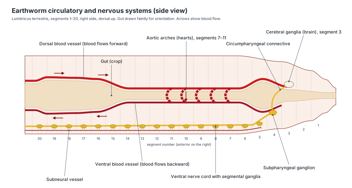
The earthworm has a closed circulatory system: blood stays inside vessels all the time. There are five main longitudinal vessels. The dorsal vessel runs along the top of the gut; the ventral vessel runs below the gut; the subneural vessel runs below the ventral nerve cord; and two small lateral neural vessels run beside the cord. In segments 7 to 11, five pairs of aortic arches loop around the oesophagus and act as hearts. (Wikipedia: Earthworm (anatomy and physiology).)
Blood moves forwards in the dorsal vessel, driven by waves of contraction, and valves prevent it flowing back. The ten 'hearts' then pump it downwards to the ventral vessel, which carries it backwards and sends branches to the body wall and organs of every segment. Blood returns to the dorsal vessel through paired segmental vessels. Because the dorsal vessel is also contractile, some books describe it as a heart too. The vessels supplying the gizzard's muscles and the skin are especially rich. (Fox, Lander University: Lumbricus terrestris anatomy lab exercise.)
A common exam answer says that an earthworm has five hearts. It is more accurate to say five pairs of aortic arches (ten in all) in Lumbricus. The number and position differ between earthworm families: Pheretima, the worm used in Indian textbooks, has four pairs of hearts. Earthworm blood is red because its oxygen-carrying pigment, a form of haemoglobin, is dissolved in the plasma rather than packed into red blood cells.
Earthworm nervous system and senses
The earthworm brain is a pair of small, white, pear-shaped cerebral ganglia that sit on top of the pharynx in segment 3. Two circumpharyngeal connectives run down either side of the pharynx and join the subpharyngeal ganglion below it, which serves the first few segments and starts the ventral nerve cord. The cord runs along the floor of the body to the tail, with a ganglion and segmental nerves in each segment. (Fox, Lander University: Lumbricus terrestris anatomy lab exercise.)
Three giant nerve fibres lie along the top of the ventral nerve cord: one median and two lateral. The median giant fibre carries signals from front to back, and the lateral ones from back to front. They conduct far faster than ordinary nerve fibres, which is why a worm touched on the head jerks back into its burrow in a fraction of a second. (Wikipedia: Earthworm (anatomy and physiology).)
Earthworms have no eyes or ears, but the skin is packed with sense cells for touch, taste (chemicals), vibration and light. Light-sensitive cells are single cells in the epidermis and are most abundant on the prostomium; the back is more sensitive to light than the belly. Darwin found that only the front end, where the cerebral ganglia lie, responds to light, and that touch is by far the best-developed sense. (Darwin (1881): The Formation of Vegetable Mould through the Action of Worms (Project Gutenberg).)
Earthworm excretory system: nephridia
Earthworms excrete with nephridia, tiny coiled tubules that work like the tubules of a vertebrate kidney. Almost every segment of Lumbricus has a pair of metanephridia. Each begins with a ciliated funnel, the nephrostome, that opens into the coelom of one segment. Its long, coiled tubule passes back through the septum and opens to the outside through a nephridiopore in the wall of the next segment. Useful substances are reabsorbed along the tubule, and urine containing ammonia and urea leaves the body. (Fox, Lander University: Lumbricus terrestris anatomy lab exercise.)
Pheretima is arranged differently. Instead of one pair per segment it has three kinds of nephridia: septal nephridia on the septa behind segment 15, integumentary nephridia attached to the body wall, and pharyngeal nephridia in tufts in segments 4, 5 and 6. This is one of the most commonly examined differences in Indian syllabuses. (Wikipedia: Earthworm (anatomy and physiology).)
Earthworm reproductive system: hermaphrodite, mating and cocoons
Every adult earthworm is a hermaphrodite, with complete male and female organs, all packed into a few segments in front of the clitellum (segments 9–15 in Lumbricus). The male system has two pairs of tiny testes in segments 10 and 11, enclosed in sperm reservoirs, three pairs of large seminal vesicles where the worm's own sperm matures, sperm funnels and the vasa deferentia that lead to the male pores on segment 15. The female system has a pair of ovaries in segment 13, egg funnels and oviducts that open at the female pores on segment 14, small ovisacs, and two pairs of seminal receptacles in segments 9 and 10 that store sperm received from a partner. (Fox, Lander University: Lumbricus terrestris anatomy lab exercise.)
Even though each worm has both sexes, Lumbricus terrestris must mate with another worm. Two worms lie head-to-tail with their undersides pressed together, held by mucus from the clitellum, and exchange sperm. L. terrestris mates at the surface while each partner keeps its tail anchored in its own burrow. Stored sperm can stay viable for months, so a worm can keep producing fertile cocoons long after one mating. (Wikipedia: Lumbricus terrestris.)
Later the clitellum secretes a tough band, the future cocoon, and nourishing albumen. The worm backs out of the band; as it slides forward over the head it collects the worm's eggs from the female pores and the partner's sperm from the seminal receptacles. Fertilisation happens inside the cocoon, which seals at both ends and is left in the soil. Development is direct: tiny worms hatch from the cocoon with no larval stage. (Wikipedia: Earthworm (anatomy and physiology).)
The clitellum is the easiest sign of maturity, because young worms do not have one. Its position, and how many segments it covers, is one of the main features used to identify earthworm species. Mature worms are often said to be ready to mate when the clitellum is swollen and orange-red. (Wikipedia: Clitellum.)
How earthworms move: peristalsis, setae and the hydrostatic skeleton
An earthworm has no bones. Its skeleton is the fluid in its coelom, held in a muscular body wall: a hydrostatic skeleton. Each segment's coelomic fluid is largely sealed off from the next by muscular septa. When the circular muscle of a segment contracts, the segment becomes long and thin; when the longitudinal muscle contracts, it becomes short and fat. Because the fluid cannot be squeezed into a smaller volume, one muscle layer stretches the other. (Kurth & Kier (2014): scaling of the hydrostatic skeleton in Lumbricus terrestris.)
Crawling uses waves of these two contractions that pass from the head towards the tail (peristalsis). Short, fat segments push their setae into the soil and act as anchors. Long, thin segments in front of them stretch forwards. As the wave moves back along the body, each part of the worm takes its turn as an anchor, and the worm moves forward without slipping. Mucus from the skin lubricates the burrow. (Quillin (1999): kinematics of peristaltic crawling in Lumbricus terrestris.)
The setae are what turn these shape changes into forward movement. Each seta sits in a sac and has its own protractor and retractor muscles, so it can be pushed out to grip or pulled in to let the segment slide. That is why the underside feels rough when you run a finger from tail to head and smooth the other way. Lumbricus has eight setae per segment, in four pairs. (Fox, Lander University: Lumbricus terrestris anatomy lab exercise.)
How earthworms breathe through their skin
Earthworms have no lungs, gills or other special breathing organs. Gas exchange takes place across the whole skin, which is thin and richly supplied with capillaries. Oxygen dissolves in the moist film of mucus on the skin, diffuses across the cuticle and epidermis into the blood and is carried around the body. Carbon dioxide leaves the same way. (Fox, Lander University: Lumbricus terrestris anatomy lab exercise.)
This only works while the skin is wet. The epidermis secretes mucus, and the dorsal pores release coelomic fluid that keeps the surface moist. A worm left on a dry surface cannot take in oxygen and soon dies. Breathing through the skin also limits how thick a worm can be, because oxygen has to diffuse inwards from the surface. (Wikipedia: Earthworm (anatomy and physiology).)
Earthworm cross-section
A cross-section of an earthworm shows a tube within a tube: the body wall on the outside, the coelom, and the intestine in the middle with the typhlosole hanging into it. From the outside in, the body wall is made of a thin cuticle, a single-celled epidermis, a thin layer of circular muscle, a thick layer of longitudinal muscle and the peritoneum lining the coelom. The dorsal blood vessel lies above the intestine and the ventral vessel below it, with the ventral nerve cord under that. Three giant fibres sit on top of the nerve cord, and the subneural vessel runs beneath it.
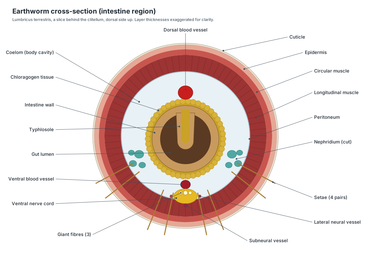
The 3D model includes the same section as an enlarged disc beside the worm. Click its layers to compare them with a prepared microscope slide. Remember that the layers in both diagrams are drawn thicker than they really are, and the typhlosole is not present in every slice: the rectum at the tail end has none.
Anatomy of earthworm class 11: Pheretima vs Lumbricus
Many Indian students search for 'anatomy of earthworm class 11' because older editions of the NCERT biology textbook described the Indian earthworm, Pheretima posthuma, often now placed in the genus Metaphire. The current rationalised NCERT Class XI chapter on structural organisation in animals covers only the frog, but the earthworm still appears in many state-board books and exam question banks. If your syllabus uses Pheretima, learn its segment numbers, because they are very different from the Lumbricus numbers in American and European lab manuals. (NCERT Biology Class XI, Chapter 7: Structural Organisation in Animals (2026–27 reprint, PDF).)
In Pheretima the clitellum covers segments 14–16, so the worm is divided into preclitellar, clitellar and postclitellar regions. There is a single female genital pore on segment 14 and a pair of male pores on segment 18, often with genital papillae nearby. In Lumbricus the clitellum is much further back (about 32–37) and the genital pores are in front of it, on segments 14 and 15. (Wikipedia: Pheretima.)
The gut is also arranged differently. In Pheretima the buccal cavity is in segments 1–3, the muscular pharynx about segment 4, the oesophagus 5–7 and the gizzard 8–9, followed by a stomach in segments 9–14 and the intestine from segment 15. A pair of short intestinal caeca arise on segment 26, and the typhlosole begins around there. Lumbricus has a crop in front of its gizzard and no separate stomach. (BYJU'S: earthworm digestive system (Pheretima).)
| Feature | Lumbricus terrestris (lab manuals in the US and Europe) | Pheretima (Metaphire) posthuma (Indian textbooks) |
|---|---|---|
| Number of segments | About 120–170 | About 100–120 |
| Clitellum | About segments 32–37, behind the genital pores | Segments 14–16, so the female pore is on the clitellum |
| Setae | 4 pairs per segment (lumbricine) | A ring of S-shaped setae on each segment (perichaetine) |
| Female pore(s) | A pair on segment 14 | A single pore on segment 14 |
| Male pores | A pair on segment 15 | A pair on segment 18 |
| Spermathecal (seminal receptacle) openings | 2 pairs: grooves 9/10 and 10/11 | 4 pairs: grooves 5/6, 6/7, 7/8 and 8/9 |
| Gizzard | About segments 17–19, behind the crop | Segments 8–9, in front of the stomach |
| Crop or stomach | Crop, about 14–16 | Stomach, 9–14 |
| Intestine | From about segment 20 | From segment 15; caeca on 26 |
| Hearts | 5 pairs, segments 7–11 | 4 pairs, segments 7, 9, 12 and 13 |
| Nephridia | 1 pair per segment (metanephridia) | Septal, integumentary and pharyngeal |
Pheretima values follow the older NCERT Class XI description and standard Indian zoology texts; check them against the textbook your exam uses.
All earthworm structures in the model
Every structure in the 3D model, grouped by organ system, with what it does. Use it as a revision list for an earthworm anatomy worksheet or a practical exam.
External
- Prostomium (Head lobe)
- A small fleshy lobe that overhangs the mouth at the very front of the worm. Probes and wedges into soil cracks, closes over the mouth, and carries many sensory cells, including light-sensitive cells.
- Peristomium (Segment 1, buccal segment)
- The first true segment, a ring that surrounds the mouth. Forms the lips around the mouth. It is the only segment of Lumbricus with no setae.
- Mouth
- The opening of the gut, on the underside of the front end. Takes in soil, dead leaves and other organic matter, which the muscular pharynx sucks in.
- Body segments (Metameres, annuli)
- The ring-like units that make up the body. Lumbricus terrestris has roughly 120 to 170 of them, often 135 to 150. Each segment is a fluid-filled compartment with its own muscles, nerves, setae and (in most) a pair of nephridia. This repetition (metamerism) lets one part of the body contract while another lengthens.
- Clitellum (Saddle, girdle)
- A thick, swollen, glandular band near the front of a mature worm. Secretes the mucus that holds two mating worms together and the tough cocoon into which eggs and sperm are released. It also secretes nourishing albumen into the cocoon.
- Tubercula pubertatis (Pubertal ridges)
- A pair of raised, glandular ridges along the lower edges of the clitellum. Press against the partner and add mucus during mating, helping the two worms hold together while they swap sperm.
- Setae (Chaetae, bristles)
- Tiny, curved, chitin bristles that stick out of the skin, four pairs on each segment. Grip the soil or burrow wall so that the stretched part of the body cannot slip backwards while the worm crawls.
- Dorsal pores (Coelomic pores)
- Tiny openings on the dorsal midline that lead from the coelom to the outside. Release coelomic fluid that keeps the skin moist for breathing and helps it slide through soil.
- Nephridiopores (Excretory pores)
- A pair of minute excretory openings on most segments. Release urine (mainly ammonia and urea in water) made by the nephridia.
- Male pores (Male genital pores, sperm duct openings)
- A pair of openings set in swollen, lip-like ridges on the underside. Release the worm's own sperm into a partner during mating.
- Female pores (Female genital pores, oviduct openings)
- A pair of tiny openings on the underside, one segment in front of the male pores. Release eggs from the oviducts into the cocoon as it slides forward over the head.
- Seminal receptacle openings (Spermathecal pores)
- Two pairs of tiny openings that lead into the seminal receptacles. Take in the partner's sperm during mating.
- Sperm grooves (Seminal grooves)
- A shallow groove on each side of the belly between the male pores and the clitellum. During mating, muscle contractions move sperm along these grooves, under the mucus tube, back towards the partner's seminal receptacles.
- Pygidium (Periproct, anal region)
- The tip of the tail that carries the anus. Surrounds the anus. New segments form just in front of it as a young worm develops.
Body wall & coelom
- Cuticle
- A thin, clear, non-living outer layer made of collagen fibres. Protects the epidermis. It is thin and permeable, so oxygen and carbon dioxide can pass through it.
- Epidermis
- A single layer of cells under the cuticle, full of mucus-secreting and sensory cells. Secretes the cuticle and the slimy mucus that keeps the skin moist, and senses touch, chemicals and light.
- Circular muscle
- A thin outer layer of muscle that runs around the body. When it contracts, a segment becomes long and thin, pushing the front of the worm forward.
- Longitudinal muscle
- A thick inner layer of muscle bands that run along the length of the body. When it contracts, a segment becomes short and fat, so its setae press into the soil as an anchor.
- Coelom (Body cavity, peritoneum)
- The fluid-filled body cavity between the body wall and the gut, lined by peritoneum. Its fluid acts as a hydrostatic skeleton and carries food, wastes and amoeboid cells (coelomocytes) that fight infection.
- Septa (Intersegmental septa)
- Thin, transparent walls that divide the coelom into one compartment per segment. Keep the coelomic fluid of each segment separate so segments can change shape independently.
Digestive
- Buccal cavity (Mouth cavity)
- A short, thin-walled chamber just inside the mouth. Receives food from the mouth and passes it to the pharynx.
- Pharynx
- A thick, muscular, pear-shaped part of the gut behind the mouth. Works as a suction pump that pulls soil and leaf fragments into the mouth. It also adds mucus.
- Pharyngeal muscles (Radial muscles of the pharynx)
- Many small muscle strands that run from the pharynx to the body wall. Contract to widen the pharynx, which creates the suction for feeding.
- Oesophagus (Esophagus)
- A narrow, thin-walled tube that carries food from the pharynx to the crop. Moves food back to the crop. Its rear part carries the oesophageal pouches and calciferous glands.
- Oesophageal pouches
- A pair of widened, finely striped pouches of the oesophagus. Part of the calcium-secreting region of the oesophagus.
- Calciferous glands (Oesophageal glands, Morren's glands)
- Two pairs of white swellings of the oesophagus that make calcium carbonate. Take excess calcium from the blood and secrete it into the gut as calcium carbonate crystals. This helps control blood calcium and pH.
- Crop
- A large, thin-walled, bulb-shaped chamber that stores food. Stores food temporarily before it is passed to the gizzard.
- Gizzard
- A hard, thick, very muscular chamber behind the crop. Grinds food against swallowed sand grains and tiny stones, because the worm has no teeth.
- Intestine (Stomach-intestine)
- The long, straight tube that runs from the gizzard almost to the anus. Secretes digestive enzymes and absorbs the digested food. Its front part is mainly secretory, the rest mainly absorptive.
- Typhlosole
- A large fold of the dorsal wall of the intestine that hangs down into the gut. Increases the surface area for absorbing digested food.
- Chloragogen tissue (Chlorogogen cells)
- A yellow-brown layer of cells covering the outside of the intestine. Works rather like a liver: it stores glycogen and fat, breaks down amino acids and makes ammonia and urea.
- Rectum (Hindgut)
- The short last part of the gut, with no typhlosole. Forms the castings (faeces) and passes them to the anus.
- Anus
- The opening at the tail end where castings leave the gut. Releases undigested soil and waste as castings.
Circulatory
- Dorsal blood vessel
- The main contractile vessel that runs along the top of the gut. Pumps blood forwards (towards the head) with waves of contraction. Valves stop the blood flowing back.
- Aortic arches (hearts) (Hearts, segmental hearts)
- Five pairs of thick, muscular vessels that loop around the oesophagus. Pump blood from the dorsal vessel down to the ventral vessel, keeping blood pressure up.
- Ventral blood vessel
- The large vessel that runs along the underside of the gut. Carries blood backwards and sends branches to the body wall and organs in every segment.
- Subneural vessel
- A thin vessel that runs below the ventral nerve cord. Drains blood from the nerve cord and body wall.
- Lateral neural vessels
- Two tiny vessels, one along each side of the ventral nerve cord. Are fed by the ventral vessel and supply the nerve cord with blood.
- Segmental vessels (Commissural and parietal vessels)
- Paired vessels in each segment that link the dorsal and ventral vessels. Supply and drain the body wall, gut and nephridia. Blood picks up oxygen in the capillaries of the skin.
Nervous
- Cerebral ganglia (brain) (Suprapharyngeal ganglia, brain)
- A pair of small, white, pear-shaped ganglia on top of the pharynx. Process information from the sensory cells of the head, especially light and touch, and help control behaviour.
- Circumpharyngeal connectives (Nerve ring, circumoesophageal connectives)
- Two nerve cords that run down either side of the pharynx. Connect the brain to the ventral nerve cord, forming a nerve ring around the gut.
- Subpharyngeal ganglion
- A ganglion below the pharynx at the front of the ventral nerve cord. Serves the first segments, including the peristomium, and starts the ventral nerve cord.
- Ventral nerve cord (Ventral nerve chain)
- A white, double nerve cord along the floor of the coelom. Carries signals along the body and coordinates the segments during crawling.
- Segmental ganglia (Ventral ganglia)
- A swelling of the nerve cord in each segment. Each ganglion controls the muscles and setae of its segment and passes on signals through segmental nerves.
- Giant fibres (Giant axons)
- Three very wide nerve fibres on top of the ventral nerve cord. Carry escape signals very fast. The median fibre carries signals backward, the two lateral fibres forward.
Excretory
- Nephridia (Metanephridia)
- Paired, coiled excretory tubules, one pair in almost every segment. Filter coelomic fluid, reabsorb useful substances and expel nitrogen waste and excess water. They work like the kidney tubules of vertebrates.
- Nephrostome (Nephridial funnel)
- The ciliated funnel at the start of each nephridium. Beating cilia draw coelomic fluid into the nephridium.
Reproductive
- Seminal receptacles (Spermathecae)
- Two pairs of small, round sacs that store a partner's sperm. Receive sperm during mating and release it into the cocoon later.
- Seminal vesicles
- Three pairs of large, cream-coloured, lobed sacs that arch over the oesophagus. Store and mature the worm's own sperm before mating.
- Testes
- Two pairs of tiny testes that make sperm. Produce immature sperm, which move into the seminal vesicles to mature.
- Sperm funnels (Seminal funnels)
- Folded, ciliated funnels that collect mature sperm. Lead sperm into the vasa efferentia and vas deferens.
- Vas deferens (Sperm duct, male duct)
- A thin duct on each side that carries sperm to the male pore. Carries sperm to the male pores during mating.
- Ovaries
- A pair of small ovaries that make eggs. Produce eggs, which are shed into the coelom of segment 13.
- Oviducts (Egg ducts, with egg funnels)
- Short ducts that carry eggs to the female pores. Collect eggs and lead them out of the body.
- Ovisacs (Egg sacs)
- Small pouches where ripe eggs are stored. Store eggs until they are laid into the cocoon.
Earthworm anatomy quiz
Test yourself. Click a question to see the answer.
Which segment is the peristomium?
Segment 1. It surrounds the mouth. The prostomium in front of it is not counted as a segment.
In which segments is the clitellum of Lumbricus terrestris?
About segments 32–37 (some keys say 31 or 32 to 37). In Pheretima it is segments 14–16.
How many setae does each segment of Lumbricus have?
Eight, in four pairs (two ventral pairs and two lateral pairs). Segment 1 has none.
On which segment are the male pores?
Segment 15. The female pores are one segment in front, on segment 14.
Name the five pairs of contractile vessels in segments 7–11.
The aortic arches, often called the hearts. They pump blood from the dorsal vessel to the ventral vessel.
Which organ grinds food, and how?
The gizzard. Its thick muscular wall squeezes food against swallowed sand grains.
What increases the surface area of the intestine?
The typhlosole, a fold of the dorsal intestinal wall that hangs into the gut.
Where is the brain?
On top of the pharynx in segment 3 (the cerebral ganglia).
Which organs excrete waste, and where do they open?
Nephridia, one pair in almost every segment. They open through nephridiopores.
Why do earthworms die if they dry out?
They breathe through their skin. Oxygen must dissolve in the moist mucus before it can diffuse into the blood.
Are earthworms self-fertilising?
No. They are hermaphrodites, but two worms exchange sperm (cross-fertilisation). Eggs are fertilised in the cocoon.
Which muscle layer makes a segment long and thin?
The circular muscle. The longitudinal muscle makes it short and fat.
Ready for the real thing? The earthworm dissection guide takes you through the same organs step by step, with a worksheet and answer key.
Frequently asked questions
How many hearts does an earthworm have?
Lumbricus terrestris has five pairs of aortic arches (ten 'hearts') in segments 7 to 11. They are thick, muscular vessels that pump blood from the dorsal vessel to the ventral vessel. The Indian earthworm Pheretima has four pairs, in segments 7, 9, 12 and 13.
Does an earthworm have a brain?
Yes. It has a small, two-lobed brain (the cerebral ganglia) above the pharynx in segment 3. It joins the ventral nerve cord through a nerve ring around the pharynx. Each segment also has its own ganglion, so the worm can still crawl without its brain.
How do earthworms breathe?
Through their skin. Earthworms have no lungs or gills. Oxygen dissolves in the moist mucus on the skin and diffuses into a dense network of capillaries; haemoglobin in the blood carries it around the body. That is why worms must stay moist and come to the surface when the soil is flooded.
Are earthworms male or female?
Both. Earthworms are hermaphrodites: every adult has testes and ovaries. They still mate with another worm and swap sperm (cross-fertilisation). Each worm later makes a cocoon with its clitellum, where its eggs are fertilised by the stored sperm.
What is the clitellum and what does it do?
The clitellum is the thick, swollen band near the front of a mature worm, around segments 32–37 in Lumbricus terrestris and 14–16 in Pheretima. It makes the mucus that holds mating worms together and the cocoon that protects the eggs.
How many segments does an earthworm have?
Lumbricus terrestris has about 120 to 170 segments, often 135 to 150. Pheretima has about 100 to 120. A worm hatches with its full number of segments and grows by enlarging them.
What are setae used for?
Setae are tiny bristles that anchor parts of the body in the soil while other parts stretch forward. Lumbricus has four pairs on every segment except the first; Pheretima has a ring of setae around each segment.
Why are earthworms important?
They mix and aerate soil, pull dead leaves underground and leave nutrient-rich castings. Charles Darwin spent decades studying how worms turn over the soil and bury objects on the surface.
References
- Fox, R. S. Lumbricus terrestris: invertebrate zoology laboratory exercise (after Ruppert, Fox & Barnes, Invertebrate Zoology, 7th ed., 2004). Lander University.
- Darwin, C. (1881). The Formation of Vegetable Mould through the Action of Worms, with Observations on their Habits. London: John Murray. Project Gutenberg edition.
- Kurth, J. A. & Kier, W. M. (2014). Scaling of the hydrostatic skeleton in the earthworm Lumbricus terrestris. Journal of Experimental Biology 217, 1860–1867.
- Quillin, K. J. (1999). Kinematic scaling of locomotion by hydrostatic animals: ontogeny of peristaltic crawling by the earthworm Lumbricus terrestris. Journal of Experimental Biology 202, 661–674.
- Lumbricus terrestris: description, behaviour and reproduction. Wikipedia.
- Earthworm: anatomy, circulatory, nervous, excretory and reproductive systems. Wikipedia.
- Clitellum. Wikipedia.
- Pheretima: clitellum and genital pores. Wikipedia.
- BYJU'S. Earthworm: alimentary canal of Pheretima with segment numbers.
- NCERT (2026–27 reprint). Biology Class XI, Chapter 7: Structural Organisation in Animals.
- Biology Corner. Earthworm dissection lab and questions.
More interactive anatomy models
- Earthworm dissectionGuided earthworm dissection with every internal organ labeled.
- Human anatomy1,000+ bones, muscles, organs, vessels and nerves from real anatomical data.
- Octopus anatomyThree hearts, a ring-shaped brain, eight arms and a beak, in 3D.
- Squid anatomyPen, eight arms, two tentacles, giant axons and three hearts of the longfin squid.
- Squid dissectionStep-by-step guided squid dissection with every structure labeled.
- Fish anatomyExternal and internal anatomy of a bony fish: fins, gills, swim bladder, heart and brain.