

The dog skeleton at a glance
A dog's skeleton has roughly 320 bones. It is divided into the axial skeleton (skull, hyoid, vertebral column, ribs and sternum), the appendicular skeleton (the bones of the four limbs, including the pelvis) and one visceral bone, the os penis of males. There is no single exact total because the number of tail vertebrae, and the presence of rear dewclaws, varies from dog to dog and breed to breed.
| Region | Bones | Count in a typical dog |
|---|---|---|
| Skull and jaw | Cranial and facial bones, mandible | Paired and unpaired bones fused at sutures, plus the mandible |
| Hyoid apparatus | Stylohyoid, epihyoid, ceratohyoid, thyrohyoid, basihyoid | 9 small bones (the tympanohyoids are cartilage) |
| Vertebral column | Cervical, thoracic, lumbar, sacral, caudal | C7 + T13 + L7 + S3 (fused) + Cd ≈ 20 |
| Rib cage | Ribs and sternebrae | 13 pairs of ribs, 8 sternebrae |
| Each forelimb | Scapula, humerus, radius, ulna, carpus, metacarpus, phalanges | 7 carpals, 5 metacarpals, 14 phalanges, sesamoids |
| Each hind limb | Hip bone, femur, patella, tibia, fibula, tarsus, metatarsus, phalanges | 7 tarsals, 4–5 metatarsals, 12 phalanges, sesamoids |
Directions: cranial is towards the head, caudal towards the tail, dorsal the back and ventral the belly. On a limb, proximal is towards the body and distal towards the paw; medial faces the midline and lateral faces outwards.
Dog skeleton labeled diagram
The labeled dog skeleton below shows every major bone from the left side, the standard view in veterinary anatomy books. Use it as a "label the dog skeleton" worksheet: cover the labels and name each bone, then check in the 3D model.
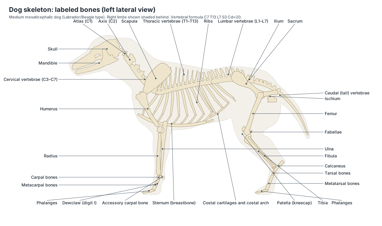
Axial skeleton of the dog
The axial skeleton is the central axis of the body: the skull, the hyoid apparatus that hangs the tongue and larynx from it, the vertebral column from the atlas to the tip of the tail, the 13 pairs of ribs and the sternum. It protects the brain, spinal cord, heart and lungs and anchors the muscles of the back, neck and breathing.
- Atlas (C1): The first neck vertebra: a ring with broad, wing-like transverse processes. Its joint with the skull lets the head nod.
- Axis (C2): The second and longest neck vertebra, with a tall, blade-like spinous process. Its dens (a peg) projects into the atlas, forming the pivot joint for turning the head.
- Cervical vertebrae (C3–C7): The remaining five neck vertebrae; dogs have seven cervical vertebrae in all. Support and move the neck and protect the spinal cord.
- Thoracic vertebrae (T1–T13): Thirteen vertebrae, each carrying a pair of ribs. Anchor the ribs; their tall spinous processes form the withers.
- Lumbar vertebrae (L1–L7): Seven large vertebrae of the loin with long transverse processes. Bear the strong back muscles and allow flexion in the gallop.
- Sacrum: Three sacral vertebrae fused into one bone. Joins the spine to the pelvis at the sacroiliac joints.
- Caudal (tail) vertebrae: The vertebrae of the tail, usually about 20, becoming simple rods towards the tip. Support the tail and its muscles.
- Ribs: Thirteen pairs of curved bones forming the rib cage. Protect the heart and lungs and move in breathing.
- Sternum (breastbone): The breastbone: eight sternebrae from the manubrium to the xiphoid process. Anchors the sternal ribs and the pectoral muscles.
Dog vertebral formula: C7 T13 L7 S3 Cd20
The dog's vertebral column has five regions. Dogs have seven cervical vertebrae (the first two are the atlas and axis), thirteen thoracic vertebrae, seven lumbar vertebrae and three sacral vertebrae fused into the sacrum. The number of caudal (tail) vertebrae varies widely between individuals and breeds. (WikiVet: Bones – Dog Anatomy.)
Comparative tables give the dog's formula as C7, T13 (range 12–14), L7 (sometimes 6), S3 and Cd 20–23. (Vetflow: Columna vertebralis (vertebral column) – vertebral formulas by species.)
The anticlinal vertebra, where the spinous processes switch from leaning backwards to leaning forwards, is a useful radiographic landmark. In a study of 100 dogs it was T11 in 75–89% of dogs depending on the definition used, and small breeds were more likely to have T10, so it should not be assumed to be T11 in every dog. (Baines, E. A. et al. (2009). Identification of the anticlinal vertebra in dogs. Veterinary Radiology & Ultrasound 50, 14–16.)
Ribs and sternum of the dog
Dogs have 13 pairs of ribs: 9 sternal (true) ribs whose cartilages reach the sternum, and 4 asternal ribs. The cartilages of the asternal ribs join the cartilage in front, forming the costal arch. (IMAIOS vet-Anatomy: Ribs.)
The sternum is made of eight sternebrae. The first is the manubrium and the last is the xiphoid process. In dogs and cats the sternebrae stay separate, joined by cartilage, rather than fusing into one bone. (IMAIOS vet-Anatomy: Sternebrae.)
Dog spine: vertebrae region by region
| Region | Number | Distinctive features |
|---|---|---|
| Cervical (neck) | 7 | Atlas with wide wings; long axis with a blade-like spinous process; C6 with a ventral lamina |
| Thoracic (chest) | 13 | Tall spinous processes forming the withers; each carries a pair of ribs; anticlinal vertebra usually T11 |
| Lumbar (loin) | 7 | Largest bodies; long transverse processes pointing cranioventrally |
| Sacral | 3, fused | Sacrum joins the pelvis at the sacroiliac joints |
| Caudal (tail) | About 20 (varies) | Arches and processes only on the first few; then simple rods |
Each typical vertebra has a body underneath, a vertebral arch around the spinal canal, a dorsal spinous process, paired transverse processes and articular processes that link it to its neighbours. Between the bodies lie the intervertebral discs. The spinal cord ends at about L6–L7 in a medium-sized dog; the nerve roots of the cauda equina continue through the sacrum.
Dog skull anatomy
The dog skull is made of the cranium, which encloses the brain, and the facial skeleton, which carries the nose, eyes and teeth. In adults the bones are joined at sutures, many of which close with age. The main bones visible from the side are listed below; the ethmoid, vomer, pterygoid and the deep parts of the sphenoid lie inside.
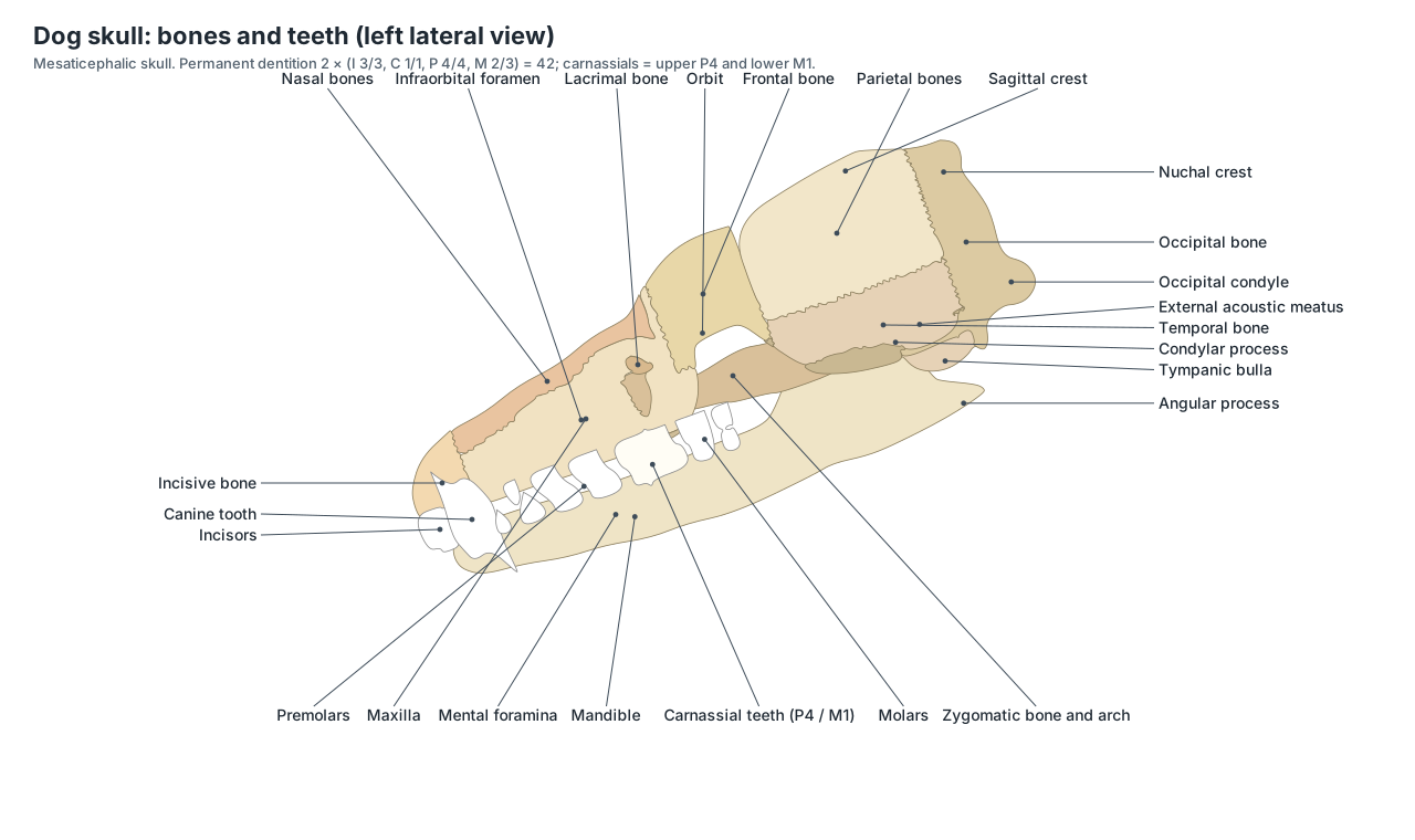
| Bone | Location | Key feature |
|---|---|---|
| Incisive | Tip of the upper jaw | Holds the six upper incisors |
| Nasal | Bridge of the nose | Roof of the nasal cavity |
| Maxilla | Side of the face | Holds the upper canine, premolars and molars; infraorbital foramen |
| Lacrimal | Front rim of the orbit | Opening of the tear duct |
| Frontal | Forehead | Frontal sinuses; zygomatic process at the top of the orbit |
| Parietal | Roof and side of the braincase | External sagittal crest along its midline |
| Occipital | Back and base of the skull | Occipital condyles, foramen magnum, nuchal crest |
| Temporal | Lower side of the braincase | Jaw joint, tympanic bulla, external acoustic meatus |
| Zygomatic | Below the orbit | Front of the zygomatic arch |
| Mandible | Lower jaw | Coronoid, condylar and angular processes; mental foramina |
Dogs have an incomplete bony orbit: the ring around the eye is closed behind only by the orbital ligament between the zygomatic process of the frontal bone and the zygomatic arch. The very wide zygomatic arches and the sagittal crest anchor the temporalis and masseter, the muscles that close the jaw.
Skull shapes: brachycephalic, mesaticephalic and dolichocephalic
Dog skulls are grouped by the relative length of the face. Brachycephalic breeds, such as the Pug and Bulldog, have a foreshortened facial skeleton; mesaticephalic (mesocephalic) breeds, such as the Labrador and Beagle, have a muzzle and cranium of medium proportions; dolichocephalic breeds, such as the Greyhound and Collie, have a long, narrow face. Packer and colleagues showed that brachycephaly is not an either/or trait: the risk of obstructive airway disease rises steadily as the muzzle gets shorter relative to the cranium. (Packer, R. M. A. et al. (2015). Impact of facial conformation on canine health: brachycephalic obstructive airway syndrome. PLOS ONE 10, e0137496.)
Dog teeth anatomy and dental formula
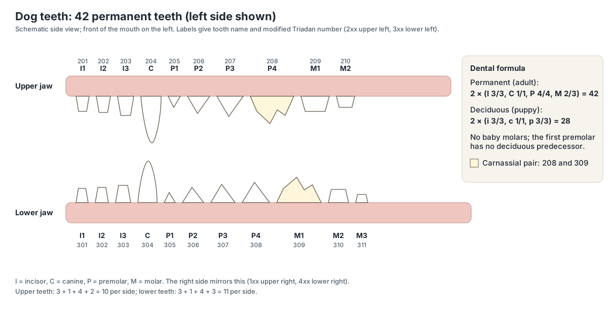
The permanent dental formula of the dog is 2 × (I 3/3, C 1/1, P 4/4, M 2/3) = 42: in each half of the upper jaw three incisors, one canine, four premolars and two molars, and in each half of the lower jaw the same plus a third molar. The upper fourth premolar and lower first molar are the large carnassial teeth.
Do dogs have a collarbone?
Not a functional one. In the dog the clavicle is rudimentary: a small plate about 1 cm long, not connected to the rest of the skeleton. It lies inside the brachiocephalicus muscle at the clavicular intersection, the tendinous line that divides that muscle into its cleidocephalicus and cleidobrachialis parts. Because no bone links the forelimb to the trunk, the shoulder is held by muscles alone, which lengthens the stride. (IMAIOS vet-Anatomy: Clavicle.)
WikiVet describes the canine clavicle as reduced to a fibrous area that may still be visible on radiographs of some dogs. (WikiVet: Bones – Dog Anatomy.)
Dog teeth and dental formula
Adult dogs have 42 permanent teeth: 12 incisors, 4 canines, 16 premolars and 10 molars. Vets name them with the modified Triadan system, which numbers each tooth by quadrant (1 = upper right, 2 = upper left, 3 = lower left, 4 = lower right) and position. (Merck Veterinary Manual: Dentition and dental nomenclature of animals.)
Puppies have 28 deciduous teeth. Baby teeth come in at about 3–5 weeks, permanent teeth start at about 4–5 months, and the adult set is complete at about 6–7 months. There are no deciduous molars. (Merck Veterinary Manual (owners): Dental development of dogs.)
Front leg bones of the dog
The front leg (thoracic limb) is built from the scapula, the humerus, the radius and ulna, seven carpal bones, five metacarpals and the phalanges of five digits. It is joined to the trunk only by muscles; there is no functional collarbone.
- Scapula: The shoulder blade: a flat, triangular bone with a ridge (spine) on its lateral surface. Attaches the forelimb to the trunk through muscles only, and forms the shoulder joint.
- Humerus: The upper arm bone, between the shoulder and the elbow. Transmits weight and carries the arm muscles.
- Radius: The main weight-bearing bone of the forearm. Carries most of the weight to the carpus.
- Ulna: The forearm bone behind the radius, forming the point of the elbow. The triceps pulls on the olecranon to extend the elbow.
- Carpal bones: Seven small wrist bones in two rows. Let the wrist bend and absorb shock.
- Accessory carpal bone: A carpal bone that projects backwards like a heel at the wrist. A lever for the flexor muscles of the carpus.
- Metacarpal bones: Five bones of the front pastern; I is short (the dewclaw), II–V are long. Support the paw at the correct angle.
- Phalanges of the front paw: The toe bones: proximal, middle and distal phalanges in digits II–V; two in the dewclaw. The distal phalanx ends in the ungual process, which sits inside the claw.
Hind leg bones and pelvis of the dog
The hind leg (pelvic limb) begins at the hip bone (os coxae), made of three fused bones: the ilium, ischium and pubis. The two hip bones and the sacrum form the pelvis. Below the hip joint come the femur, the patella, the tibia and fibula, seven tarsal bones, the metatarsals and the phalanges.
- Ilium: The largest part of the hip bone, with a wide wing that joins the sacrum. Transfers the push of the hind limb to the spine.
- Ischium: The caudal part of the hip bone, ending in the ischial tuberosity (point of the buttock). Anchors the hamstring muscles.
- Pubis: The ventral, cranial part of the hip bone, forming the front of the pelvic floor. Anchors the adductor and abdominal muscles.
- Femur: The thigh bone: the longest bone of the hind limb. Carries weight and the large thigh muscles.
- Patella (kneecap): The kneecap: a large sesamoid bone in the tendon of the quadriceps. Increases the leverage of the quadriceps when it straightens the stifle.
- Fabellae and popliteal sesamoid: Small sesamoid bones behind the stifle. Act as pulleys for the calf muscle tendons.
- Tibia: The shin bone: the main bone of the lower hind leg. Carries the weight; the cruciate ligaments attach on its flat top (tibial plateau).
- Fibula: A thin bone beside the tibia. Gives attachment to muscles and forms the lateral malleolus of the hock.
- Talus: The ankle bone that forms the hinge of the hock with the tibia. Its pulley-shaped trochlea lets the hock flex and extend.
- Calcaneus: The heel bone, which forms the point of the hock. A lever for the calf muscles, which pull on it through the common calcanean (Achilles) tendon.
- Tarsal bones: The remaining five small hock bones: the central tarsal and tarsals I–IV. Form the lower joints of the hock and absorb shock.
- Metatarsal bones: The long bones of the rear pastern: II–V, with I usually small or absent. Support the hind paw.
| Joint | Bones | Common problem |
|---|---|---|
| Shoulder | Scapula and humerus | Osteochondrosis in young large dogs |
| Elbow | Humerus, radius and ulna | Elbow dysplasia (e.g. ununited anconeal process) |
| Carpus | Radius, ulna, carpal and metacarpal bones | Hyperextension injuries |
| Hip | Femoral head and acetabulum | Hip dysplasia |
| Stifle (knee) | Femur, tibia, patella | Cranial cruciate ligament rupture; patellar luxation |
| Hock | Tibia, fibula, tarsal and metatarsal bones | Achilles (common calcanean) tendon injuries |
For the soft-tissue anatomy of the stifle, including the cruciate ligaments, see dog knee anatomy.
Carpal and tarsal bones
The dog's carpus (wrist) has seven bones. In carnivores the radial and intermediate carpal bones are fused into a single intermedioradial carpal bone; the proximal row also contains the ulnar and accessory carpal bones, and the distal row carpal bones I–IV. (IMAIOS vet-Anatomy: Carpal bones.)
The tarsus (hock) also has seven bones: the talus and calcaneus in the proximal row, the central tarsal bone, and tarsal bones I–IV in the distal row. (IMAIOS vet-Anatomy: Tarsal bones.)
Dog paw skeleton
Each toe of digits II–V has three phalanges: proximal, middle and distal. The distal phalanx ends in the ungual process, a hook of bone covered by the claw. The dewclaw (digit I) is shorter and has two phalanges. Behind each knuckle (the metacarpophalangeal or metatarsophalangeal joint) sits a pair of small proximal sesamoid bones, with a small dorsal sesamoid on the other side. Dogs walk on their toes (a digitigrade stance), so the metacarpals and metatarsals form the pastern.
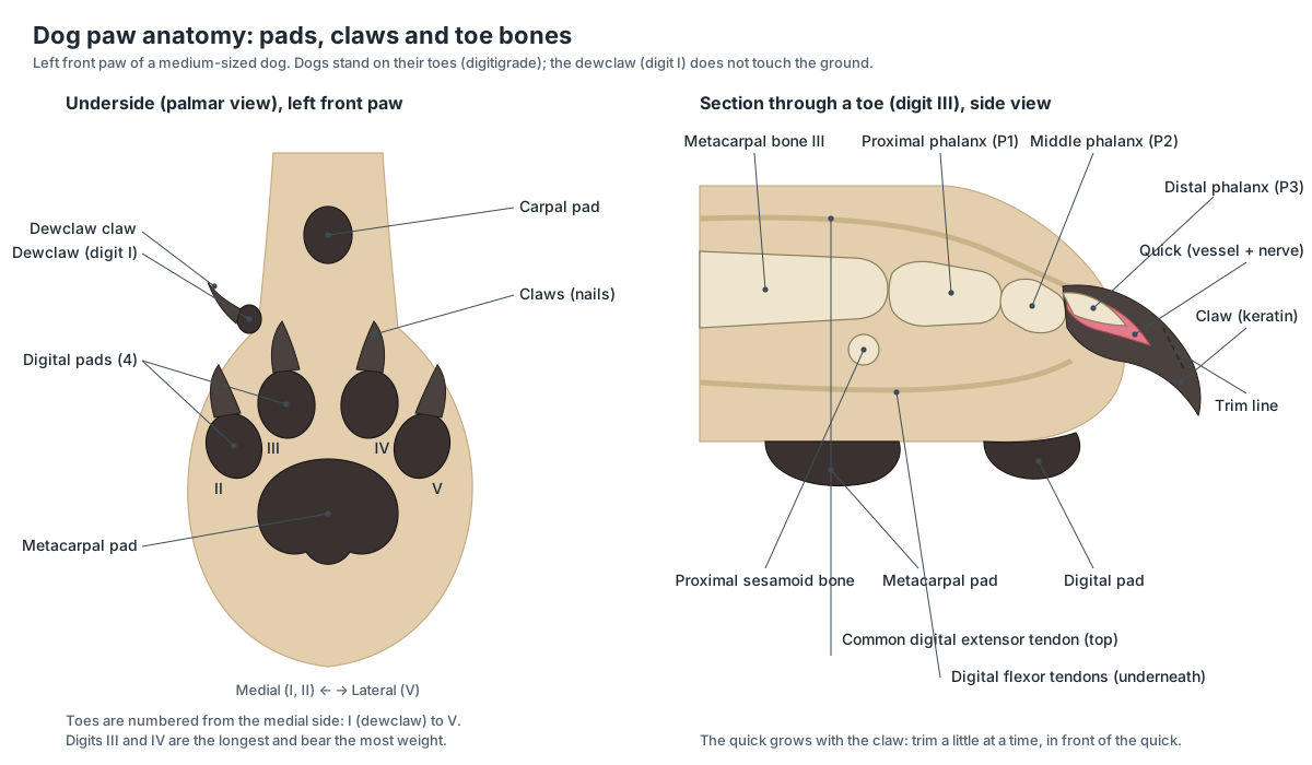
Sesamoid bones and the os penis
Sesamoid bones grow inside tendons where they run over a joint. The dog has the patella (kneecap), two fabellae behind the stifle, a popliteal sesamoid, the paired sesamoids behind each knuckle of the paws and small dorsal sesamoids. Males also have the os penis, a bone inside the penis with a groove for the urethra underneath; it keeps the penis rigid for mating.
- Patella (kneecap): The kneecap: a large sesamoid bone in the tendon of the quadriceps. Increases the leverage of the quadriceps when it straightens the stifle.
- Fabellae and popliteal sesamoid: Small sesamoid bones behind the stifle. Act as pulleys for the calf muscle tendons.
- Sesamoid bones of the paws: Small bones in tendons behind each knuckle joint of the paws. Reduce friction and act as pulleys for the flexor tendons.
- Os penis: A bone inside the penis. Keeps the penis rigid for mating before full erection.
Bones of the dog skeleton: complete list
Every bone and bone group in the 3D model, with its location and job.
Skeletal
- Incisive bone (Premaxilla)
- The front bone of the upper jaw that holds the six upper incisors. Carries the upper incisor teeth and forms the front of the hard palate.
- Nasal bones
- A pair of long, narrow bones forming the bridge of the nose. Form the roof of the nasal cavity.
- Maxilla
- The main bone of the upper jaw and the side of the face. Holds the upper canine, premolars and molars and forms most of the hard palate.
- Lacrimal bone
- A small bone at the front (rostral) rim of the orbit. Contains the opening of the nasolacrimal canal, which drains tears into the nose.
- Frontal bone
- The forehead bone above and between the eyes. Encloses the frontal sinuses and the front of the braincase.
- Parietal bones
- A pair of curved plates forming the sides and roof of the braincase. Protect the cerebrum; the temporalis muscle attaches to them.
- Occipital bone
- The bone at the back and base of the skull. Its occipital condyles form the joint with the atlas; the spinal cord passes through the foramen magnum.
- Temporal bone
- The bone on the lower side of the braincase that holds the ear and the jaw joint. Forms the jaw joint (temporomandibular joint), the caudal part of the zygomatic arch and the bony ear.
- Zygomatic bone and arch (Cheekbone)
- The cheekbone, which forms the front of the wide zygomatic arch. Gives the masseter muscle a strong origin and protects the eye from below.
- Palatine bone
- A paired bone forming the back part of the hard palate. Separates the nasal cavity from the mouth at the back of the palate.
- Sphenoid bone
- The floor of the braincase in front of the occipital bone. Supports the pituitary gland and carries foramina for the optic and other cranial nerves.
- Mandible (Lower jaw)
- The lower jaw: two halves joined at the chin by a fibrous symphysis. Carries the lower teeth. The coronoid process anchors the temporalis, the masseteric fossa the masseter.
- Incisors
- Six small front teeth in each jaw (12 in all). Nibble, groom and pick up small objects.
- Canine teeth (Fangs)
- Four long, pointed teeth, one at each corner of the mouth. Grip and puncture.
- Premolars
- Sixteen blade-like cheek teeth, four in each half-jaw. Cut and shear food.
- Molars
- Ten broad back teeth: two in each upper half-jaw and three in each lower one. Crush and grind food.
- Carnassial teeth
- The large shearing pair of cheek teeth on each side: upper fourth premolar and lower first molar. Slide past each other like scissors to slice meat and tough food.
- Hyoid apparatus
- A chain of small bones that suspends the tongue and larynx from the skull. Anchors the tongue and larynx and moves them in swallowing.
- Atlas (C1) (First cervical vertebra)
- The first neck vertebra: a ring with broad, wing-like transverse processes. Its joint with the skull lets the head nod.
- Axis (C2) (Second cervical vertebra)
- The second and longest neck vertebra, with a tall, blade-like spinous process. Its dens (a peg) projects into the atlas, forming the pivot joint for turning the head.
- Cervical vertebrae (C3–C7)
- The remaining five neck vertebrae; dogs have seven cervical vertebrae in all. Support and move the neck and protect the spinal cord.
- Thoracic vertebrae (T1–T13)
- Thirteen vertebrae, each carrying a pair of ribs. Anchor the ribs; their tall spinous processes form the withers.
- Lumbar vertebrae (L1–L7)
- Seven large vertebrae of the loin with long transverse processes. Bear the strong back muscles and allow flexion in the gallop.
- Sacrum
- Three sacral vertebrae fused into one bone. Joins the spine to the pelvis at the sacroiliac joints.
- Caudal (tail) vertebrae (Coccygeal vertebrae)
- The vertebrae of the tail, usually about 20, becoming simple rods towards the tip. Support the tail and its muscles.
- Ribs
- Thirteen pairs of curved bones forming the rib cage. Protect the heart and lungs and move in breathing.
- Costal cartilages and costal arch
- Bars of cartilage that continue each rib towards the sternum. Make the rib cage flexible for breathing.
- Sternum (breastbone) (Sternebrae)
- The breastbone: eight sternebrae from the manubrium to the xiphoid process. Anchors the sternal ribs and the pectoral muscles.
- Scapula (Shoulder blade)
- The shoulder blade: a flat, triangular bone with a ridge (spine) on its lateral surface. Attaches the forelimb to the trunk through muscles only, and forms the shoulder joint.
- Clavicle (vestigial) (Collarbone remnant)
- A tiny remnant of a collarbone, not joined to any other bone. Has no supporting role; it marks the clavicular intersection of the brachiocephalicus.
- Humerus
- The upper arm bone, between the shoulder and the elbow. Transmits weight and carries the arm muscles.
- Radius
- The main weight-bearing bone of the forearm. Carries most of the weight to the carpus.
- Ulna
- The forearm bone behind the radius, forming the point of the elbow. The triceps pulls on the olecranon to extend the elbow.
- Carpal bones (Carpus)
- Seven small wrist bones in two rows. Let the wrist bend and absorb shock.
- Accessory carpal bone
- A carpal bone that projects backwards like a heel at the wrist. A lever for the flexor muscles of the carpus.
- Metacarpal bones (Metacarpals I–V)
- Five bones of the front pastern; I is short (the dewclaw), II–V are long. Support the paw at the correct angle.
- Phalanges of the front paw
- The toe bones: proximal, middle and distal phalanges in digits II–V; two in the dewclaw. The distal phalanx ends in the ungual process, which sits inside the claw.
- Sesamoid bones of the paws
- Small bones in tendons behind each knuckle joint of the paws. Reduce friction and act as pulleys for the flexor tendons.
- Ilium
- The largest part of the hip bone, with a wide wing that joins the sacrum. Transfers the push of the hind limb to the spine.
- Ischium
- The caudal part of the hip bone, ending in the ischial tuberosity (point of the buttock). Anchors the hamstring muscles.
- Pubis
- The ventral, cranial part of the hip bone, forming the front of the pelvic floor. Anchors the adductor and abdominal muscles.
- Femur (Thigh bone)
- The thigh bone: the longest bone of the hind limb. Carries weight and the large thigh muscles.
- Patella (kneecap) (Kneecap)
- The kneecap: a large sesamoid bone in the tendon of the quadriceps. Increases the leverage of the quadriceps when it straightens the stifle.
- Fabellae and popliteal sesamoid
- Small sesamoid bones behind the stifle. Act as pulleys for the calf muscle tendons.
- Tibia (Shin bone)
- The shin bone: the main bone of the lower hind leg. Carries the weight; the cruciate ligaments attach on its flat top (tibial plateau).
- Fibula
- A thin bone beside the tibia. Gives attachment to muscles and forms the lateral malleolus of the hock.
- Talus
- The ankle bone that forms the hinge of the hock with the tibia. Its pulley-shaped trochlea lets the hock flex and extend.
- Calcaneus (Heel bone)
- The heel bone, which forms the point of the hock. A lever for the calf muscles, which pull on it through the common calcanean (Achilles) tendon.
- Tarsal bones (Tarsus)
- The remaining five small hock bones: the central tarsal and tarsals I–IV. Form the lower joints of the hock and absorb shock.
- Metatarsal bones
- The long bones of the rear pastern: II–V, with I usually small or absent. Support the hind paw.
- Phalanges of the hind paw
- The toe bones of the hind paw, three in each of digits II–V. Their structure matches the front paw; the distal phalanx carries the claw.
- Os penis (Baculum, penile bone) · males only
- A bone inside the penis. Keeps the penis rigid for mating before full erection.
Dachshund (sausage dog) skeleton and disc disease
A Dachshund has the same bones as any other dog, but its limb bones are short and curved because it is a chondrodystrophic breed: cartilage growth at the growth plates is altered. The same trait makes its intervertebral discs degenerate and calcify early. The American College of Veterinary Surgeons reports that chondrodystrophic breeds account for the vast majority of disc ruptures, with Dachshunds alone making up 45–70% of all cases; most ruptures are in the thoracolumbar spine. (American College of Veterinary Surgeons: Intervertebral disc disease.)
Pug, German Shepherd and Greyhound skeletons
| Breed type | Skeletal features | What it means |
|---|---|---|
| Pug, French Bulldog (brachycephalic) | Very short facial bones; crowded teeth; often malformed (hemi-)vertebrae in the tail and spine | Airway obstruction; screw tail |
| Labrador, Beagle (mesaticephalic) | Balanced skull; straight limbs | The reference on this page |
| Greyhound, Borzoi (dolichocephalic) | Long narrow skull; long limbs; deep chest | Sprinting; wide field of view |
| Dachshund, Basset (chondrodystrophic) | Short, curved limb bones; normal vertebra count | High risk of disc disease |
| German Shepherd | Sloping topline from the angled hind limbs | Prone to hip dysplasia and lumbosacral disease |
Dog skeleton vs human skeleton
| Feature | Dog | Human |
|---|---|---|
| Stance | Digitigrade (on the toes) | Plantigrade (on the soles) |
| Clavicle | Vestigial, not joined to other bones | Full collarbone joining shoulder to sternum |
| Vertebrae | C7 T13 L7 S3 Cd≈20 | C7 T12 L5 S5 (fused) Co4 |
| Ribs | 13 pairs | 12 pairs |
| Teeth | 42 permanent | 32 permanent |
| Penile bone | Os penis present | Absent |
| Forearm | Radius and ulna fixed, little rotation | Radius rotates around the ulna |
Dog skeleton worksheet
- Write the dog's vertebral formula.
- Which bone forms the point of the elbow, and which forms the point of the hock?
- Name the three bones that fuse to form each hip bone.
- How many pairs of ribs does a dog have, and how many are sternal?
- Which two teeth are the carnassials?
- Where are the fabellae?
Answers: 1 C7 T13 L7 S3 Cd≈20. 2 olecranon of the ulna; tuber of the calcaneus. 3 ilium, ischium, pubis. 4 13; 9 sternal and 4 asternal. 5 upper fourth premolar and lower first molar. 6 behind the femoral condyles, in the heads of the gastrocnemius.
See the organs, muscles, paw and ear on the dog anatomy page.
Frequently asked questions
How many bones are in a dog's skeleton?
About 320, but the number is not fixed. It varies mainly with the number of tail (caudal) vertebrae, usually about 20 in a long-tailed dog, and with whether rear dewclaws are present. Almost every dog has 7 cervical, 13 thoracic and 7 lumbar vertebrae, a sacrum of 3 fused vertebrae and 13 pairs of ribs.
What is the vertebral formula of a dog?
C7 T13 L7 S3 Cd20 (the caudal number varies, about 20–23 in a long tail). Occasional dogs have 12 or 14 thoracic or 6 lumbar vertebrae.
Do dogs have a collarbone?
Only a vestigial one. The canine clavicle is a tiny plate, about 1 cm long, buried in the brachiocephalicus muscle and not joined to any other bone. The forelimb is attached to the body by muscles alone.
What is the axial skeleton of a dog?
The axial skeleton is the skull, hyoid apparatus, vertebral column (cervical, thoracic, lumbar, sacral and caudal vertebrae), ribs and sternum. The appendicular skeleton is the limb bones and the pelvis; the os penis is a visceral bone.
How many toes and toe bones does a dog have?
Usually five digits on each front paw (four weight-bearing toes plus the dewclaw, digit I) and four on each hind paw. Digits II–V have three phalanges each; the dewclaw has two.
Does a dog have a bone in its penis?
Yes. The os penis lies in the glans penis with the urethra in a groove on its underside. It keeps the penis rigid for mating.
What are the fabellae in a dog?
Two small sesamoid bones behind the femoral condyles, in the two heads of the gastrocnemius muscle. With the patella and the popliteal sesamoid they make four sesamoid bones at the stifle.
Why does a Dachshund have a long back and short legs?
Dachshunds are chondrodystrophic: altered cartilage growth shortens and curves the limb bones. They have the same number of vertebrae as other dogs; the back only looks long. The same trait makes their discs degenerate early, so they have a high risk of intervertebral disc disease.
References
- WikiVet: Bones – Dog Anatomy
- Vetflow: Columna vertebralis (vertebral column) – vertebral formulas by species
- Baines, E. A. et al. (2009). Identification of the anticlinal vertebra in dogs. Veterinary Radiology & Ultrasound 50, 14–16
- IMAIOS vet-Anatomy: Ribs
- IMAIOS vet-Anatomy: Sternebrae
- IMAIOS vet-Anatomy: Clavicle
- IMAIOS vet-Anatomy: Carpal bones
- IMAIOS vet-Anatomy: Tarsal bones
- IMAIOS vet-Anatomy: Brachiocephalic muscle
- Mauragis, D. Small animal radiography: stifle joint and crus. Today's Veterinary Practice
- Merck Veterinary Manual: Dentition and dental nomenclature of animals
- Merck Veterinary Manual (owners): Dental development of dogs
- VCA Animal Hospitals: Teeth, teething and chewing in puppies
- American College of Veterinary Surgeons: Intervertebral disc disease
- American College of Veterinary Surgeons: Cranial cruciate ligament disease
- Packer, R. M. A. et al. (2015). Impact of facial conformation on canine health: brachycephalic obstructive airway syndrome. PLOS ONE 10, e0137496
- University of Illinois ANSC 207: Reproductive anatomy of the male dog
More interactive anatomy models
- Dog anatomyOrgans, muscles, paw, ear and eye of the domestic dog, in 3D.
- Human anatomy1,000+ bones, muscles, organs, vessels and nerves from real anatomical data.
- Octopus anatomyThree hearts, a ring-shaped brain, eight arms and a beak, in 3D.
- Squid anatomyPen, eight arms, two tentacles, giant axons and three hearts of the longfin squid.
- Squid dissectionStep-by-step guided squid dissection with every structure labeled.
- Fish anatomyExternal and internal anatomy of a bony fish: fins, gills, swim bladder, heart and brain.