

Anatomy of the crayfish at a glance
A crayfish is a freshwater decapod crustacean: an arthropod with a hard, jointed exoskeleton, two body regions and ten legs. Its body has a cephalothorax, the head and thorax fused under one shield called the carapace, and an abdomen of six segments ending in a tail fan. It carries 19 pairs of jointed appendages, breathes with gills hidden under the sides of the carapace, has an open circulatory system with a small dorsal heart, and excretes through a pair of green glands in its head.
This page describes the red swamp crayfish, Procambarus clarkii, family Cambaridae. Its native range runs along the Gulf coastal plain from the Florida panhandle into Mexico and up the Mississippi valley to southern Illinois, adults are 5.5 to 12 cm long, and it is farmed on a large scale in Louisiana (USGS). That makes it one of the easiest crayfish to obtain for teaching. Its anatomy is typical of the North American cambarid crayfishes, so almost everything here applies equally to Faxonius (formerly Orconectes) and Cambarus, the other genera often supplied to schools.
Where other crayfish differ, it is mostly in details used to identify species: the shape of the rostrum, the spines on the carapace, the width of the areola, the shape of the male gonopods and of the female annulus ventralis, and which walking legs carry hooks in males. The classic anatomy textbook, Huxley's The Crayfish (1880), describes the European Astacus; it has the same organs but no annulus ventralis, and its gill count of 18 per side may differ slightly from that of cambarids.
Directions used below: anterior is towards the rostrum, posterior towards the telson, dorsal is the back and ventral the belly. Left and right are the crayfish's own.
Crayfish external anatomy: labeled diagram
The external anatomy of a crayfish includes the carapace with its rostrum and cervical groove, two compound eyes on stalks, two pairs of antennae, the mouthparts, a pair of large chelipeds, four pairs of walking legs, the six-segmented abdomen with its swimmerets, and the tail fan made of the telson and two uropods.
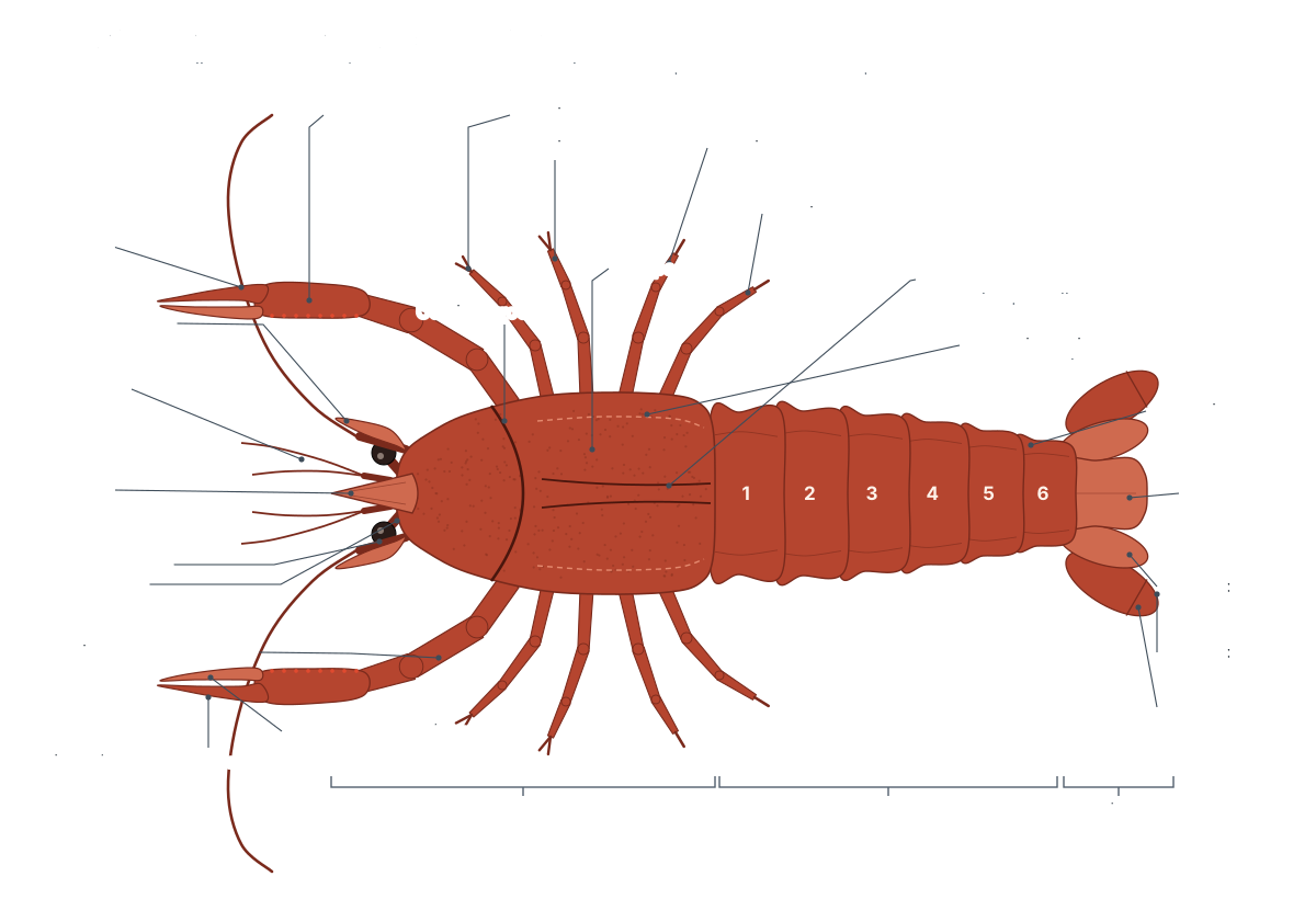
Seen from above, the carapace is divided by the cervical groove, which marks the boundary between the head in front and the thorax behind. Behind the groove, two branchiocardiac grooves outline a narrow strip called the areola, which lies directly over the heart. On each side, the carapace extends down as the branchiostegite, the gill cover. In the red swamp crayfish the rostrum is a triangular point without a central keel, and the spines on the sides of the carapace are small or absent.
The abdomen (often called the tail) is made of six hard rings. Each ring has a dorsal plate, the tergum, with side flaps called pleura, and a ventral plate, the sternum. Flexible joints between the rings let the abdomen curl under the body. The telson is the flat central piece at the end; it has no appendages, so it is not counted as a segment. The uropods on each side of it are the appendages of the sixth segment.
| Region | What covers it | Main external features |
|---|---|---|
| Head (cephalic region) | Front of the carapace, in front of the cervical groove | Rostrum, eyes on eyestalks, antennules, antennae, antennal scales, mouthparts |
| Thorax | Carapace behind the cervical groove, branchiostegites on the sides | Areola, three pairs of maxillipeds, chelipeds, four pairs of walking legs, sternites |
| Abdomen | Six tergites with pleura, six sternites | Swimmerets (pleopods), gonopods in males, uropods, telson, anus |
Crayfish appendages and their functions
A crayfish has 19 pairs of appendages: 5 on the head, 8 on the thorax and 6 on the abdomen. From front to back they are the antennules, antennae, mandibles, first and second maxillae, three pairs of maxillipeds, the chelipeds, four pairs of walking legs, five pairs of swimmerets and the uropods. All of them are built from the same basic two-branched limb.
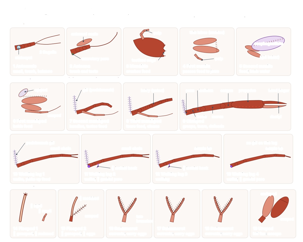
| Pair | Appendage | Region | Form | Function |
|---|---|---|---|---|
| 1 | Antennules | Head | Biramous | Smell, touch and balance (statocyst in the base) |
| 2 | Antennae | Head | Biramous (flagellum + antennal scale) | Touch and chemical sensing; green gland opens at the base |
| 3 | Mandibles | Head | Uniramous palp | Crush and cut food beside the mouth |
| 4 | First maxillae (maxillules) | Head | Leaf-like | Hold and pass food to the mandibles |
| 5 | Second maxillae | Head | With scaphognathite | Handle food; the gill bailer pumps water over the gills |
| 6 | First maxillipeds | Thorax | Biramous | Hold food and push it to the mouth |
| 7 | Second maxillipeds | Thorax | Biramous, with gill | Manipulate and taste food |
| 8 | Third maxillipeds | Thorax | Biramous, with gills | Hold and tear food; clean antennae and eyes |
| 9 | Chelipeds (first pereiopods) | Thorax | Uniramous, large chela | Capture and tear food, defence, fighting, holding the female |
| 10 | First walking legs | Thorax | Uniramous, small chela | Walking; picking up food |
| 11 | Second walking legs | Thorax | Uniramous, small chela | Walking; female genital pores on the coxa |
| 12 | Third walking legs | Thorax | Uniramous | Walking and support |
| 13 | Fourth walking legs | Thorax | Uniramous | Walking, cleaning; male genital pores on the coxa |
| 14 | First pleopods | Abdomen | Male: rigid tube; female: reduced | Male gonopod: transfers sperm |
| 15 | Second pleopods | Abdomen | Male: modified; female: biramous | Male: helps sperm transfer; female: carries eggs |
| 16–18 | Third to fifth pleopods (swimmerets) | Abdomen | Biramous | Create water currents; carry eggs and young in females |
| 19 | Uropods | Abdomen | Biramous, flat | Form the tail fan with the telson for the backward tail-flip |
Lab manuals sometimes number the legs differently. Zoologists call all five pairs of thoracic legs pereiopods, so the cheliped is pereiopod 1 and the last walking leg is pereiopod 5. Many school worksheets instead call the chelipeds the first pair of legs and the four pairs behind them the walking legs 1–4. This page uses walking legs 1–4 and gives the pereiopod number in brackets. Huxley counted the eyestalks as a twentieth pair of appendages; most modern texts do not.
Two features are easy to miss. The first two pairs of walking legs end in tiny pincers, while the last two end in a single point. And the scaphognathite, the flat blade on each second maxilla, sits out of sight in the front of the gill chamber, where it beats to keep water flowing over the gills.
Crayfish anatomy labeled: every part and its function
This is the text version of the 3D model: every labeled structure of the red swamp crayfish, grouped by body system, with what it does.
Exoskeleton & body regions
- Carapace (Dorsal shield of the cephalothorax)
- The single hard shield that covers the top and sides of the cephalothorax (the fused head and thorax). Protects the brain, stomach, heart, digestive gland and gonads, and gives the leg and mouthpart muscles a rigid place to attach.
- Rostrum
- The pointed beak at the front of the carapace, projecting forward between the eyes. Shields the eyes and the bases of the antennules from above.
- Cervical groove
- A curved groove across the carapace that marks the boundary between the head and the thorax. A landmark: the region in front of it is the head (cephalic region), the region behind it covers the thorax and gills.
- Branchiostegite (Gill cover, lateral carapace)
- The side flap of the carapace that hangs down over the gills on each side. Encloses the branchial (gill) chamber, protecting the delicate gills and guiding the water current that flows over them.
- Abdomen (Pleon, 'tail' (6 segments))
- The jointed rear part of the body, made of six movable segments, each covered by a dorsal tergite with side flaps (pleura). Holds the big flexor muscles that power the tail-flip escape, carries the swimmerets, and in females holds the eggs and young.
- Telson
- The flat, tongue-shaped last piece of the body, in the middle of the tail fan. Forms the centre of the tail fan, which acts like a paddle in the backward tail-flip.
- Thoracic sternites (Ventral plates of the cephalothorax)
- The row of narrow plates on the underside of the cephalothorax, between the bases of the legs. Floors the thorax, protects the ventral nerve cord, and carries the annulus ventralis in females.
Appendages
- Antennules (First antennae (pair 1))
- The short pair of feelers with two flagella each, at the front of the head below the eyes. Smell (chemoreception) with special setae on the flagella, touch, and balance: each base holds a statocyst.
- Antennae (Second antennae (pair 2))
- The long pair of whip-like feelers, often about as long as the body. Touch and chemical sensing over a wide area around the crayfish, especially in the dark.
- Antennal scale (Scaphocerite)
- A flat, pointed blade at the base of each antenna. Protects the base of the antenna and may help steer during swimming.
- Mandibles (Jaws (pair 3))
- A pair of hard, toothed jaws on each side of the mouth. Crush and cut food by moving side to side before it enters the mouth.
- First maxillae (Maxillules (pair 4))
- Small, thin, leaf-like mouthparts just behind the mandibles. Hold and pass food towards the mandibles.
- Second maxillae (Maxillae (pair 5))
- Thin, lobed mouthparts behind the first maxillae, each with a large flat blade (the gill bailer). Handle food, and drive the breathing current with the scaphognathite.
- First maxillipeds (Pair 6)
- The first and smallest of the three pairs of 'jaw-feet', flat and leaf-like. Hold food and push it towards the mouth.
- Second maxillipeds (Pair 7)
- The middle pair of maxillipeds, small and leg-like, with a gill at the base. Manipulate food and sense it by touch and taste.
- Third maxillipeds (Pair 8)
- The largest, outermost maxillipeds: hairy, leg-like appendages that cover the other mouthparts from below. Hold and tear food, and clean the antennae and eyes.
- Chelipeds (Claws, first pereiopods (pair 9))
- The first and largest pair of legs, each ending in a big pincer (chela). Capture and tear food, defence, fighting and display, and holding the female during mating.
- First walking legs (Second pereiopods (pair 10))
- The first pair of walking legs, each ending in a small pincer. Walking, and picking up and holding food with the small chela.
- Second walking legs (Third pereiopods (pair 11))
- The second pair of walking legs, also with small pincers. Walking and handling food; in females the oviducts open at their bases.
- Third walking legs (Fourth pereiopods (pair 12))
- The third pair of walking legs, ending in a simple pointed tip. Walking and support.
- Fourth walking legs (Fifth pereiopods (pair 13))
- The last pair of walking legs. Males have their genital pores on the bases of these legs. Walking, and cleaning the abdomen and swimmerets; in males the sperm ducts open at their bases.
- Swimmerets (Pleopods (pairs 14–18))
- Small, feathery, two-branched appendages under abdominal segments 1–5. Beat rhythmically to create water currents. In females they carry the eggs and newly hatched young.
- Uropods (Pair 19)
- The flat, two-branched appendages of the sixth abdominal segment that form the sides of the tail fan. Spread out with the telson to form the tail fan for the backward tail-flip escape and for steering.
Muscles
- Abdominal flexor muscles
- The thick, twisted mass of white muscle that fills most of the abdomen. Contract to snap the abdomen forward under the body. The tail fan then drives the crayfish rapidly backward: the tail-flip escape.
- Abdominal extensor muscles
- Two thin bands of muscle along the roof of the abdomen. Straighten (extend) the abdomen again after a tail-flip, re-cocking it for the next one.
- Mandibular muscles (Mandible adductors)
- A pair of strong muscles that run from each mandible up to the inside of the carapace. Close the mandibles to crush food.
Nervous
- Brain (Supraoesophageal (cerebral) ganglion)
- A small, pale, two-lobed mass of nerve tissue at the front of the head, above the oesophagus. Processes signals from the eyes, antennules and antennae and coordinates behaviour.
- Optic nerves
- The nerves that run from the brain out through each eyestalk to the eye. Carry visual signals from the compound eyes to the brain.
- Circumoesophageal connectives
- Two nerve cords that run from the brain down around each side of the oesophagus. Connect the brain to the ventral nerve cord.
- Suboesophageal ganglion
- A large fused ganglion below the oesophagus, at the front of the ventral nerve cord. Controls the mandibles, maxillae and maxillipeds.
- Ventral nerve cord
- A double nerve cord running along the floor of the body, with a ganglion in each segment. Carries signals between the brain and the body and controls the legs, swimmerets and tail locally.
- Abdominal ganglia
- Six swellings of the ventral nerve cord, one in each abdominal segment. Control the swimmerets, the abdominal muscles and the tail fan.
Sense organs
- Compound eyes
- Two dark eyes, each made of thousands of tiny units (ommatidia), on movable stalks. Detect light, shadows and movement in a wide field of view.
- Eyestalks
- Short, movable stalks that carry the compound eyes. Point the eyes and hold glands that release molt-inhibiting hormone.
- Statocysts (Balance organs)
- Small sacs in the base of each antennule containing sand grains resting on sensory hairs. Sense gravity and balance: when the body tilts, the grains press on different hairs.
Digestive
- Mouth
- The opening of the gut, on the underside of the head between the mandibles. Takes in food that the mouthparts have cut up and passed inward.
- Anus
- The opening at the end of the intestine, on the underside of the telson. Releases faeces.
- Oesophagus (Esophagus)
- A short, wide tube that runs straight up from the mouth to the stomach. Carries food from the mouth to the stomach.
- Cardiac stomach (Anterior chamber of the stomach)
- The large front chamber of the stomach, a thin-walled sac in the head. Stores food and grinds it with the gastric mill.
- Gastric mill
- Three hard, calcified teeth inside the back of the cardiac stomach. Grind food, doing the job of our teeth inside the stomach.
- Gastroliths ('Crab's eyes')
- Two lens-shaped stones of calcium carbonate in the wall of the cardiac stomach, present only around molting. Store calcium from the old shell so it can be used to harden the new one after the molt.
- Pyloric stomach (Posterior chamber of the stomach)
- The smaller rear chamber of the stomach, lined with fine bristles. Filters the ground food: fine particles go to the digestive gland, coarse material to the intestine.
- Digestive gland (Hepatopancreas, 'liver')
- The large, soft, yellow-green to brown gland that fills most of the cephalothorax. Makes digestive enzymes, absorbs digested food and stores food reserves. It does the jobs of our liver and pancreas.
- Intestine
- A thin, straight tube that runs from the stomach along the top of the abdomen to the anus. Absorbs some nutrients and water and carries faeces to the anus.
Circulatory (open)
- Heart
- A small, single-chambered, muscular heart on the dorsal side of the thorax. Pumps hemolymph (blood) out through seven arteries to the body.
- Ostia
- Slit-like openings with valves in the heart wall: three pairs in the crayfish. Let blood flow from the pericardial sinus into the heart and close when the heart contracts.
- Pericardial sinus
- The blood-filled space around the heart, enclosed by a thin membrane. Collects oxygenated blood returning from the gills before it enters the heart through the ostia.
- Ophthalmic artery (Anterior median artery, anterior aorta)
- A single artery running forward from the heart over the stomach to the head. Supplies the eyes, brain and front of the head.
- Antennal arteries (Anterolateral arteries)
- A pair of arteries that run forward and outward from the front of the heart. Supply the antennae, green glands, stomach and mouthpart muscles.
- Hepatic arteries
- A pair of arteries from the underside of the heart to the digestive gland. Supply the digestive gland (hepatopancreas).
- Dorsal abdominal artery (Superior abdominal artery, posterior aorta)
- An artery running from the back of the heart along the top of the abdomen. Supplies the abdominal muscles and the intestine.
- Sternal artery
- A large artery that runs straight down from the heart and passes through the ventral nerve cord. Feeds the ventral thoracic and ventral abdominal arteries, which supply the legs, mouthparts and nerve cord.
- Ventral thoracic and abdominal arteries (Subneural artery)
- An artery running along the floor of the body, just below the ventral nerve cord. Supplies the legs, mouthparts, swimmerets and nerve cord.
- Sternal sinus (Ventral sinus)
- A large blood space along the floor of the thorax, where blood collects after supplying the tissues. Returns oxygen-poor blood to the bases of the gills.
- Branchiocardiac canals
- Channels running up the inner wall of the gill chamber from the gills to the pericardial sinus. Carry oxygenated blood from the gills back to the heart.
Respiratory (gills)
- Scaphognathite (Gill bailer)
- A flat, oval paddle on the second maxilla that beats to pump water through the gill chamber. Bails water forward out of the gill chamber. This draws fresh water in under the edges of the carapace and over the gills.
- Podobranchs (Foot gills)
- Feathery gills attached to the bases (coxae) of the maxillipeds and legs. Gas exchange. Because they move with the legs, walking helps stir water over them.
- Arthrobranchs (Joint gills)
- Gills attached to the soft membrane between a leg's base and the body wall. Gas exchange.
- Pleurobranchs (Side gills)
- Gills attached directly to the side wall of the thorax above the legs. Gas exchange.
- Gill filaments
- The many fine threads on each gill's central axis that give the gills their feathery look. Provide a large, thin surface where oxygen passes from the water into the blood and carbon dioxide passes out.
Excretory (green glands)
- Green glands (Antennal glands)
- A pair of small, round, greenish glands in the front of the head; the crayfish's kidneys. Filter the blood, remove wastes and, above all, reabsorb salts so the crayfish can excrete a large volume of dilute urine.
- Bladder
- A thin-walled sac attached to each green gland that stores urine. Stores urine before it is released through the excretory pore.
- Excretory pores (Nephropores, renal openings)
- Tiny openings on a bump on the underside of the base of each antenna. Release urine from the bladder.
Reproductive
- Testes (Testis (three-lobed)) · males only
- A white, three-lobed organ under the heart, with two lobes in front and one behind. Produce sperm.
- Sperm ducts (Vasa deferentia) · males only
- A pair of long, coiled, white tubes from the testis down to the bases of the last walking legs. Carry sperm, packaged in spermatophores, to the male genital pores.
- Male genital pores (Male gonopores) · males only
- Small openings on the inner side of the base of each fourth walking leg. Release sperm into the grooved gonopods.
- Gonopods (Modified first and second pleopods (swimmerets)) · males only
- The first two pairs of swimmerets of a male, hard and tube-like and pointing forward between the last legs. Transfer sperm to the female's annulus ventralis during mating.
- Ischial hooks · males only
- Small hooks on the third segment (ischium) of the second and third walking legs of males. Help the male grip the female during mating.
- Ovary (Ovaries (three-lobed)) · females only
- An orange to dark, three-lobed organ under the heart, packed with round eggs when mature. Produce eggs.
- Oviducts · females only
- A pair of short, wide tubes from the ovary down to the bases of the second walking legs. Carry eggs to the female genital pores.
- Female genital pores (Female gonopores) · females only
- Round openings on the base of each second walking leg. Release eggs, which are fertilised as they pass the stored sperm and are glued to the swimmerets.
- Annulus ventralis (Seminal receptacle) · females only
- A small, raised, ring-like plate on the female's underside that stores sperm after mating. Receives and stores sperm from the male's gonopods until she lays her eggs.
- First pleopods (female) · females only
- The first pair of swimmerets of a female, small and soft. Reduced in females; the egg-carrying is done by swimmerets 2–5.
Crayfish internal anatomy
Inside the cephalothorax, the heart lies just under the carapace behind the cervical groove. The three-lobed gonad lies below it, the large digestive gland (hepatopancreas) fills both sides, the two-chambered stomach sits in the head, and the gills lie in a chamber on each side. The green glands and the brain are at the very front, and the ventral nerve cord runs along the floor of the body. The abdomen is filled mostly by muscle, with the intestine running along its top.
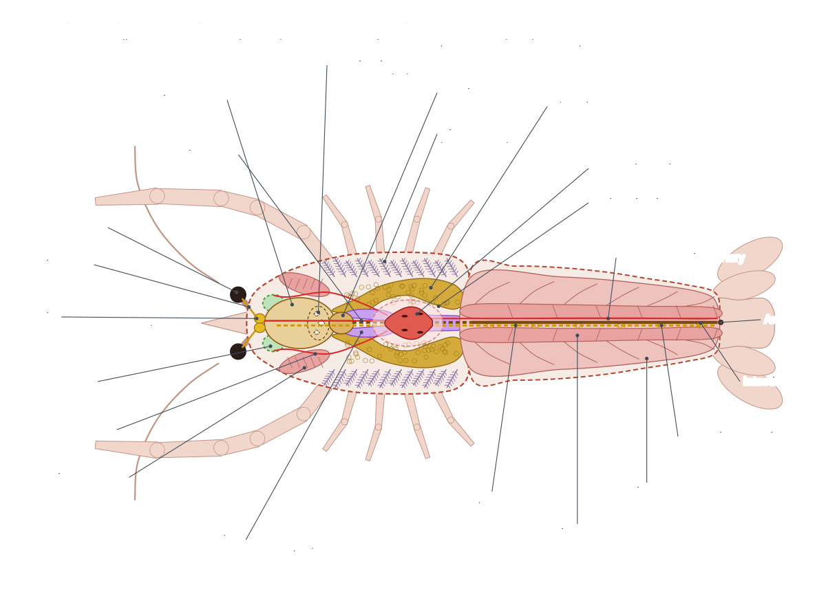
Viewed from the side, the organs are stacked in layers. From the top down in the thorax you find the heart in its pericardial sinus, the gonad, the intestine in the midline between the lobes of the digestive gland, and finally the sternal sinus and ventral nerve cord just above the sternites. The gills sit outside this stack, between the body wall and the branchiostegite. For a step-by-step guide to finding each organ in a real specimen, see our crayfish dissection guide.
Crayfish circulatory system: an open system
The crayfish has an open circulatory system. A single-chambered heart pumps colourless to pale blue hemolymph (blood) out through arteries. The blood then leaves the vessels and flows through spaces called sinuses, passes through the gills, and returns to the pericardial sinus around the heart. It enters the heart through three pairs of valved openings, the ostia, because no veins lead back to it.
- Heart: Pumps hemolymph (blood) out through seven arteries to the body.
- Ostia: Let blood flow from the pericardial sinus into the heart and close when the heart contracts.
- Pericardial sinus: Collects oxygenated blood returning from the gills before it enters the heart through the ostia.
- Ophthalmic artery: Supplies the eyes, brain and front of the head.
- Antennal arteries: Supply the antennae, green glands, stomach and mouthpart muscles.
- Hepatic arteries: Supply the digestive gland (hepatopancreas).
- Dorsal abdominal artery: Supplies the abdominal muscles and the intestine.
- Sternal artery: Feeds the ventral thoracic and ventral abdominal arteries, which supply the legs, mouthparts and nerve cord.
- Ventral thoracic and abdominal arteries: Supplies the legs, mouthparts, swimmerets and nerve cord.
- Sternal sinus: Returns oxygen-poor blood to the bases of the gills.
- Branchiocardiac canals: Carry oxygenated blood from the gills back to the heart.
Follow one loop: heart → arteries → tissues → sternal sinus on the floor of the thorax → afferent channels into the gills (oxygen picked up) → branchiocardiac canals up the sides → pericardial sinus → ostia → heart. Unlike a fish heart, the crayfish heart receives blood that has already been oxygenated in the gills. The oxygen is carried by hemocyanin, a copper-containing protein dissolved in the blood, which is why oxygenated crayfish blood can look faintly blue. Huxley described it as "a clear fluid, either colourless, or of a pale neutral tint or reddish hue".
Crayfish respiratory system: gills and the gill bailer
Crayfish breathe with feathery gills inside a branchial chamber on each side of the thorax, covered by the branchiostegite. Water is pulled in under the lower edge of the carapace near the legs, flows up and forward over the gills, and is pumped out at the front by the beating scaphognathite (gill bailer).
The gills are named by where they attach. Podobranchs are attached to the bases of the legs and maxillipeds, arthrobranchs to the joint membrane between leg and body, and pleurobranchs to the side wall of the thorax. Huxley counted 18 functional gills per side in Astacus: 6 podobranchs, 11 arthrobranchs and 1 pleurobranch, plus 2 rudimentary pleurobranchs. Because so many gills are fixed to the legs, pulling off a walking leg in a dissection usually brings a gill with it.
- Podobranchs: Gas exchange. Because they move with the legs, walking helps stir water over them.
- Arthrobranchs: Gas exchange.
- Pleurobranchs: Gas exchange.
- Gill filaments: Provide a large, thin surface where oxygen passes from the water into the blood and carbon dioxide passes out.
- Scaphognathite: Bails water forward out of the gill chamber. This draws fresh water in under the edges of the carapace and over the gills.
Each gill is a central axis covered in many fine filaments, which gives a large, thin surface for gas exchange. In fresh water the gills also take up salts from the water, helping the green glands keep the body fluids in balance.
Crayfish digestive system
Food passes from the mouthparts into the mouth, up a short oesophagus into the large cardiac stomach, where the three teeth of the gastric mill grind it. It is then sorted in the pyloric stomach: fine particles go into the digestive gland, where most digestion and absorption happen, and the rest passes along the intestine to the anus under the telson.
- Mandibles: Crush and cut food by moving side to side before it enters the mouth.
- Cardiac stomach: Stores food and grinds it with the gastric mill.
- Gastric mill: Grind food, doing the job of our teeth inside the stomach.
- Pyloric stomach: Filters the ground food: fine particles go to the digestive gland, coarse material to the intestine.
- Digestive gland: Makes digestive enzymes, absorbs digested food and stores food reserves. It does the jobs of our liver and pancreas.
- Intestine: Absorbs some nutrients and water and carries faeces to the anus.
- Gastroliths: Store calcium from the old shell so it can be used to harden the new one after the molt.
The stomach is lined with cuticle, like the outside of the body, so its lining and teeth are replaced at every molt. Huxley noted that the digestive gland, often called the liver, is made of "an immense number of short tubes, or caeca", and that its secretion is more like pancreatic juice than bile. Crayfish are omnivores and scavengers: in a dissection the stomach may hold plant fragments, detritus, insect parts or pieces of other animals.
Crayfish excretory system: green glands
The excretory organs of the crayfish are the two green glands, also called antennal glands, in the floor of the head. Each filters the blood, reabsorbs useful salts, stores urine in a thin bladder, and releases it through an excretory pore on the underside of the base of the antenna.
Because the crayfish lives in fresh water, water keeps flowing into its body. The main job of the green glands is therefore to get rid of that water as large amounts of dilute urine while holding on to salts. Most nitrogenous waste, in the form of ammonia, simply diffuses out across the gills.
Crayfish nervous system and sense organs
The crayfish nervous system has a small brain (the supraoesophageal ganglion) above the oesophagus, two circumoesophageal connectives that loop around the gut, and a ventral nerve cord with a ganglion in each segment. Huxley counted thirteen sets of ganglia: the brain, six thoracic ganglionic masses and six abdominal ganglia.
- Brain: Processes signals from the eyes, antennules and antennae and coordinates behaviour.
- Optic nerves: Carry visual signals from the compound eyes to the brain.
- Circumoesophageal connectives: Connect the brain to the ventral nerve cord.
- Suboesophageal ganglion: Controls the mandibles, maxillae and maxillipeds.
- Ventral nerve cord: Carries signals between the brain and the body and controls the legs, swimmerets and tail locally.
- Abdominal ganglia: Control the swimmerets, the abdominal muscles and the tail fan.
Compare this with our own nervous system: a crayfish's nerve cord is ventral, solid and paired, while ours is dorsal, hollow and single. This is a basic difference between arthropods and vertebrates.
The main sense organs are the compound eyes, each made of many units called ommatidia; the antennules and antennae, covered with touch and chemical sensors; and the statocysts, balance organs in the base of each antennule. Huxley described the statocyst hairs as embedded in "a gelatinous mass, which contains irregular particles of sand". The lining of the statocyst is shed at each molt and the crayfish has to put new grains of sand back in.
Crayfish muscles and the tail-flip escape
Most of the crayfish abdomen is muscle. The large flexor muscles on the underside snap the abdomen forward under the body, and the tail fan drives the animal rapidly backward. The thin extensor muscles along the top straighten the abdomen again. This is the tail-flip, the crayfish's emergency escape.
- Abdominal flexor muscles: Contract to snap the abdomen forward under the body. The tail fan then drives the crayfish rapidly backward: the tail-flip escape.
- Abdominal extensor muscles: Straighten (extend) the abdomen again after a tail-flip, re-cocking it for the next one.
- Mandibular muscles: Close the mandibles to crush food.
The tail-flip is controlled by giant nerve fibres in the ventral nerve cord, which connect quickly to the motor neurons of the fast flexors. Wine (1984) counted about 650 neurons in each abdominal ganglion and roughly 100,000 in the whole crayfish central nervous system, which is why the escape circuit has become a classic model in neuroscience.
Male vs female crayfish anatomy
You can tell a male crayfish from a female by turning it over. In males, the first two pairs of swimmerets are hard, forward-pointing gonopods lying between the bases of the last walking legs. In females, the first swimmerets are small and soft, and there is a round annulus ventralis between the bases of the third and fourth walking legs. The genital pores are also in different places.
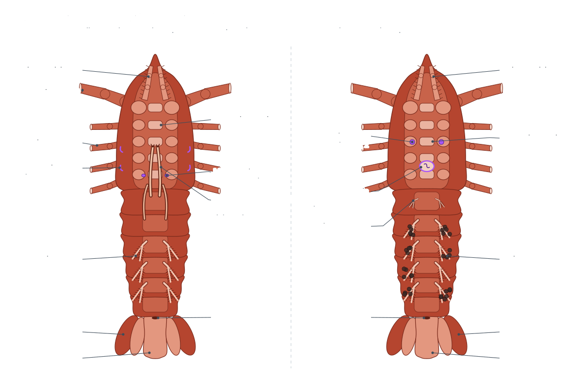
| Male crayfish | Female crayfish | |
|---|---|---|
| Gonads | White, three-lobed testis under the heart | Orange to dark, three-lobed ovary with visible eggs |
| Ducts | Long, coiled sperm ducts (vasa deferentia) | Short, wide oviducts |
| Genital pores | Bases of the fourth walking legs (5th pereiopods) | Bases of the second walking legs (3rd pereiopods) |
| First two pleopods | Rigid gonopods for sperm transfer | First pair reduced; second pair normal swimmerets |
| Sperm storage | — | Annulus ventralis between the 3rd and 4th walking legs |
| Other | Larger chelae; ischial hooks on walking legs 2 and 3 in P. clarkii | Wider abdomen; carries eggs and young on the swimmerets |
Huxley described both gonads as three-lobed ("a body of a trefoil form"), the testis having narrower and more elongated lobes than the ovary. Switch the 3D model between Female and Male to compare the gonads, ducts, pores and swimmerets.
Crayfish molting
Crayfish grow by molting (ecdysis): they shed the whole exoskeleton, swell by taking up water, and harden a new, larger shell. Young crayfish molt often; adults molt less frequently. Hormones from glands in the eyestalks hold molting back until conditions are right.
Before a molt, a freshwater crayfish moves calcium from its old shell into two gastroliths in the stomach wall, then uses it to harden the new shell. Red swamp crayfish grow quickly: the USGS notes that they can exceed 50 g within 3 to 5 months. A recently molted crayfish has a soft shell and is very vulnerable, which is why molting crayfish hide.
Comparative anatomy: crayfish vs grasshopper
Crayfish and grasshoppers are both arthropods, so they share a jointed exoskeleton, segmented body, ventral nerve cord, dorsal heart and open circulation. They differ mainly because the crayfish breathes water and the grasshopper breathes air.
| Feature | Crayfish (crustacean) | Grasshopper (insect) |
|---|---|---|
| Body regions | Cephalothorax and abdomen | Head, thorax and abdomen |
| Antennae | Two pairs (antennules and antennae) | One pair |
| Legs | Five pairs (chelipeds + four walking legs) | Three pairs on the thorax; hind legs for jumping |
| Wings | None | Two pairs: leathery forewings, membranous hind wings |
| Breathing | Gills in branchial chambers | Spiracles and air tubes (tracheae) |
| Excretion | Green glands in the head | Malpighian tubules that empty into the gut |
| Gut | Two-chambered stomach with gastric mill, digestive gland | Crop, gizzard and gastric caeca |
| Circulation | Open; blood carries oxygen on hemocyanin | Open; blood carries little oxygen, tracheae deliver it |
| Reproduction | Eggs carried on the female's swimmerets | Eggs laid in soil with an ovipositor |
Because insects deliver oxygen straight to their tissues through tracheae, their blood plays little part in breathing. In the crayfish, every drop of blood must pass through the gills, so the circulation is built around them. See the grasshopper dissection pages from Biology Junction for the insect side of this comparison.
Crayfish vs lobster anatomy
Crayfish and lobsters are close relatives with the same basic anatomy: a carapace, chelipeds, four pairs of walking legs, a six-segmented abdomen, swimmerets and a tail fan. The differences are mostly size, habitat and life history.
| Feature | Red swamp crayfish | American lobster |
|---|---|---|
| Habitat | Fresh water: swamps, ditches, ponds, streams | Sea floor, in burrows and crevices |
| Adult size | 5.5–12 cm long | Much larger: the biggest weigh up to 44 lb (about 20 kg) |
| Claws | Similar left and right, long and narrow | Unequal: a heavy crusher and a finer ripper claw |
| Development | Young hatch as small crayfish and cling to the mother | Free-swimming larvae molt four times before they look like adults |
| Calcium | Stores calcium in gastroliths before molting | Often eats its shed shell to replace lost calcium |
| Sperm storage | Annulus ventralis in females | Females have a thelycum leading to a median sperm receptacle (Bauer, 1986) |
Lobster figures come from NOAA Fisheries; crayfish figures from the USGS. A preserved lobster can be dissected with exactly the same procedure as a crayfish.
One limb plan, nineteen jobs: reading crayfish appendages
Every crayfish appendage is a variation on one basic limb. Huxley described it as a base, the protopodite, made of two joints (the coxopodite and basipodite), which carries an inner branch, the endopodite, and an outer branch, the exopodite. Where both branches are present the limb is called biramous (two-branched). The antennules, the antennae with their antennal scale, the maxillipeds, the swimmerets and the uropods all show both branches. In the walking legs and chelipeds the exopodite has been lost, and the endopodite forms the long jointed leg. (Huxley, T. H. (1880). The Crayfish: An Introduction to the Study of Zoology. Internet Archive.)
This is why lab worksheets ask you to lay the appendages out in order. Seen side by side, the same plan turns into feelers, jaws, food handlers, a water pump, pincers, legs, egg holders and paddles. Biologists call parts that share an origin but differ in form serially homologous. When you label an appendage card, add one more column: whether the appendage is biramous or uniramous (one-branched). It turns a memory exercise into evidence for how arthropod bodies evolved by modifying a repeated segment.
Crayfish molting and gastroliths
A crayfish grows by molting, because its hardened exoskeleton cannot stretch. Before a molt, calcium is moved out of the old shell. Freshwater holds little dissolved calcium, so many freshwater crayfish store part of it in a pair of gastroliths, lens-shaped stones that form in the wall of the cardiac stomach. After the molt the gastroliths are broken down and their calcium is used to harden the new exoskeleton. The Western Australian Museum notes that only freshwater crustaceans form gastroliths. (Western Australian Museum: Why freshwater crayfish don't need milk for healthy bones.)
Electron microscopy of the gastroliths of the redclaw crayfish, Cherax quadricarinatus, showed that they are made mostly of amorphous (non-crystalline) calcium carbonate, laid down as nanogranules along chitin–protein fibres. Amorphous mineral dissolves more easily than crystals, which suits a store that must be emptied quickly. Huxley knew the same stones as 'crabs' eyes'. In a dissection you will only find them in a crayfish that died shortly before or after a molt, so their absence is normal. (Luquet, G. et al. (2016). Calcium deposits in the crayfish, Cherax quadricarinatus: microstructure versus elemental distribution. Microscopy and Microanalysis 22: 22–38.)
Molting replaces every cuticle-lined surface: the outer shell, the lining of the stomach with its gastric mill, the hindgut, the internal skeleton (apodemes) and the lining of the statocysts. Huxley recorded that apodemes are shed along with the other cuticular structures at ecdysis (the act of shedding). A freshly molted crayfish is soft and hides until the new shell hardens, and many crayfish eat the shed skin, recovering more of its minerals. (Huxley, T. H. (1880). The Crayfish: An Introduction to the Study of Zoology. Internet Archive.)
Why a freshwater crayfish needs green glands
A crayfish's body fluids are saltier than the fresh water around it, so water constantly leaks in across the gills and other thin surfaces, and salts leak out. The green glands (antennal glands) solve the water problem. Huxley described each one as 'a discoidal body of a dull green colour' lying near the antennae, draining into a 'spacious sac', the bladder, which opens on the basal joint of the antenna. Fluid filtered from the blood is processed as it passes through the gland, salts are taken back, and the excess water leaves as dilute urine. (Huxley, T. H. (1880). The Crayfish: An Introduction to the Study of Zoology. Internet Archive.)
Compare this with a marine fish, which loses water to the sea and drinks to replace it. The crayfish has the opposite problem and the opposite solution: it makes a lot of urine and wins back salts, partly in the green gland and partly by active uptake across the gills. In decapods the antennal gland also has an unusually rich blood supply: corrosion casts showed complete capillary beds in it, one of only two places in the body where McGaw found them. (McGaw, I. J. (2005). The decapod crustacean circulatory system: a case that is neither open nor closed. Microscopy and Microanalysis 11: 18–36.)
The tail-flip: a reflex you can trace from nerve to muscle
The crayfish tail-flip is one of the best-understood behaviours in neuroscience. Wine's review describes six abdominal ganglia with about 650 neurons each, out of roughly 100,000 neurons in the whole central nervous system. A dedicated escape circuit of 90 pairs of motor neurons and 18 pairs of interneurons drives the fast flexor and extensor muscles of the abdomen. The lateral giant interneuron, a wide, fast-conducting nerve fibre in the cord, triggers the escape response; serotonin inhibits it and octopamine excites it. (Wine, J. J. (1984). The structural basis of an innate behavioural pattern. Journal of Experimental Biology 112: 283–319.)
In a dissection you can see the hardware. The thick, twisted flexor muscles fill the abdomen, while two thin extensor bands run under its roof. Fast flexors fire as powerful twitches for escape, whereas small superficial muscles hold posture. The ventral nerve cord, with a ganglion in each segment, lies under the flexors. Wine notes that an isolated abdomen can still run some of its motor programmes, although most are started by commands from the brain. (Wine, J. J. (1984). The structural basis of an innate behavioural pattern. Journal of Experimental Biology 112: 283–319.)
Open circulation, with qualifications
Textbooks call crayfish circulation open because blood leaves the arteries and flows through sinuses rather than returning in veins. The heart has no veins entering it: Huxley described six valved openings (three pairs of ostia) through which blood is drawn in from the surrounding pericardial sinus. From the heart, arteries run forward to the eyes, antennae and digestive gland, back along the abdomen, and down through the sternal artery to the ventral side. (Huxley, T. H. (1880). The Crayfish: An Introduction to the Study of Zoology. Internet Archive.)
Detailed corrosion casting in crabs showed that the arterial side branches far more finely than the word 'open' suggests. Seven arteries leave the single-chambered heart and divide into capillary-like vessels throughout the body, and the sinuses they drain into are organised, membrane-bound channels rather than one big cavity. McGaw concluded that the decapod system is best described as 'partially closed'. For a school exam, 'open' is still the expected answer; the research shows why it is a simplification. (McGaw, I. J. (2005). The decapod crustacean circulatory system: a case that is neither open nor closed. Microscopy and Microanalysis 11: 18–36.)
How crayfish mate: gonopods, the annulus ventralis and clones
Crayfish families differ in how sperm reaches the female. In cambarid crayfish, the family that includes Procambarus, Faxonius and Cambarus, the male's first pleopods are tube-like gonopods with sperm channels and terminal elements, and the second pair is also modified. The female has a median spermatheca, the annulus ventralis, on her ventral thorax. In European astacid crayfish, by contrast, the female has no spermatheca and the male attaches sperm packets to the outside of her body (Bauer, 1986). (Bauer, R. T. (1986). Phylogenetic trends in sperm transfer and storage complexity in decapod crustaceans. Journal of Crustacean Biology 6: 313–325.)
Male cambarids change form with the breeding season: they molt into a breeding form with hardened gonopods (often called Form I) and later back to a non-breeding form (Form II). The Missouri Department of Conservation notes that males typically have a different body form during the autumn and winter mating period. Females carry the fertilised eggs glued to the swimmerets, and the young stay attached to the mother until they have completed two molts. (Missouri Department of Conservation: Crayfishes field guide.)
Red swamp crayfish normally reproduce sexually, but genetic testing of 120 wild individuals in China found 15 genetically identical females in four clonal groups, evidence that P. clarkii can occasionally produce offspring without mating. The authors argued that this ability, combined with the species' fast growth, makes moving it to new waters especially risky. (Yue, G. H. et al. (2008). Discovery of four natural clones in a crayfish species Procambarus clarkii. International Journal of Biological Sciences 4: 279–282.)
Crayfish anatomy worksheet: test yourself
Use the 3D model or the diagrams above to answer these. Answers are on the dissection page.
- What two body regions make up a crayfish, and what is the carapace?
- Name the 19 pairs of appendages in order and give one function for each.
- Which appendage holds the statocyst, and what does it sense?
- Where are the gills, and which structure pumps water over them?
- Why is the crayfish circulatory system called open?
- What are the two chambers of the stomach and what happens in each?
- Which organ does the job of our kidneys?
- Give three differences between a male and a female crayfish.
Full lab questions with an answer key are in the crayfish dissection guide.
Frequently asked questions
What are the main parts of a crayfish?
A crayfish has two body regions: the cephalothorax (head and thorax fused under one shield, the carapace) and the abdomen (six segments ending in the telson). It has 19 pairs of jointed appendages: antennules, antennae, mandibles, two pairs of maxillae, three pairs of maxillipeds, chelipeds, four pairs of walking legs, five pairs of swimmerets and the uropods.
Does a crayfish have an open or closed circulatory system?
Open. The heart pumps hemolymph out through arteries, but the blood then leaves the vessels and flows through spaces called sinuses. It collects in the sternal sinus, passes through the gills and returns to the pericardial sinus, entering the heart through three pairs of ostia. Research on decapods shows the small arteries branch very finely, so some biologists call it 'partially closed'.
How does a crayfish breathe?
With feathery gills inside a chamber on each side of the cephalothorax, covered by the branchiostegite. A paddle on the second maxilla, the scaphognathite or gill bailer, beats several times a second to pull water in under the edge of the carapace, over the gills and out at the front.
What do the green glands do in a crayfish?
The green glands (antennal glands) are the crayfish's kidneys. They filter the blood and reabsorb salts so the crayfish, which lives in fresh water, can get rid of extra water as dilute urine. The urine leaves through excretory pores at the base of the antennae.
How can you tell a male crayfish from a female?
Turn it over. A male's first two pairs of swimmerets are hard, forward-pointing gonopods, and his genital pores are on the bases of the last walking legs. A female has small, soft first swimmerets, genital pores on the bases of the second walking legs, and a round annulus ventralis between the third and fourth walking legs.
Where is the crayfish's heart?
On the dorsal side of the thorax, just under the carapace and behind the cervical groove. It sits inside the pericardial sinus. In a dissection it is the first organ you see when you lift the carapace: a small, pale, roughly diamond-shaped organ in the midline.
Why does a crayfish have two stomachs?
It has one stomach with two chambers. The large cardiac stomach stores food and grinds it with three teeth (the gastric mill). The smaller pyloric stomach filters it: fine particles go to the digestive gland, coarse material to the intestine.
What is the function of the swimmerets?
Swimmerets (pleopods) beat to move water under the abdomen. In females they hold the eggs and newly hatched young, and keep them supplied with oxygen. In males the first two pairs are modified into gonopods for transferring sperm.
How does a crayfish escape so fast?
By a tail-flip. Giant nerve fibres in the ventral nerve cord fire the powerful abdominal flexor muscles, which snap the abdomen and tail fan forward under the body. This shoots the crayfish backward. The extensor muscles then straighten the abdomen for the next flip.
References
- Huxley, T. H. (1880). The Crayfish: An Introduction to the Study of Zoology. London: Kegan Paul. Internet Archive full text.
- Nagy, R., Fusaro, A. & Conard, W. Procambarus clarkii (Girard, 1852): red swamp crayfish. USGS Nonindigenous Aquatic Species Database.
- Bauer, R. T. (1986). Phylogenetic trends in sperm transfer and storage complexity in decapod crustaceans. Journal of Crustacean Biology 6(3): 313–325.
- McGaw, I. J. (2005). The decapod crustacean circulatory system: a case that is neither open nor closed. Microscopy and Microanalysis 11(1): 18–36.
- Wine, J. J. (1984). The structural basis of an innate behavioural pattern. Journal of Experimental Biology 112(1): 283–319.
- Luquet, G. et al. (2016). Calcium deposits in the crayfish, Cherax quadricarinatus: microstructure versus elemental distribution. Microscopy and Microanalysis 22(1): 22–38.
- Western Australian Museum. Why freshwater crayfish don't need milk for healthy bones (gastroliths).
- Yue, G. H. et al. (2008). Discovery of four natural clones in a crayfish species Procambarus clarkii. International Journal of Biological Sciences 4(5): 279–282.
- Missouri Department of Conservation. Crayfishes (field guide).
- Biology Junction. Crayfish dissection lab.
- Biology Junction. Grasshopper dissection lab.
- NOAA Fisheries. American lobster species profile.
More interactive anatomy models
- Crayfish dissectionGuided crayfish dissection: gills, green gland, stomach and heart.
- Human anatomy1,000+ bones, muscles, organs, vessels and nerves from real anatomical data.
- Octopus anatomyThree hearts, a ring-shaped brain, eight arms and a beak, in 3D.
- Squid anatomyPen, eight arms, two tentacles, giant axons and three hearts of the longfin squid.
- Squid dissectionStep-by-step guided squid dissection with every structure labeled.
- Fish anatomyExternal and internal anatomy of a bony fish: fins, gills, swim bladder, heart and brain.