

Before you start: materials and safety
You need a preserved fetal pig, a dissecting tray, simple tools and protective equipment. A double-injected specimen makes arteries and veins much easier to tell apart. Plan for about three to four hours of lab time.
Materials
- Preserved fetal pig, ideally double-injected (red latex in the arteries, blue in the veins)
- Dissecting tray (wax or a cutting mat) and string or rubber bands to tie the legs
- Sharp scissors (one blunt blade is best), scalpel, forceps, blunt probe and a dissecting needle
- Ruler or tape measure for the crown–rump length
- Nitrile gloves, safety glasses and a lab coat or apron
- Paper towels, a wash bottle of water and a labeled zip bag for storing the specimen between lab days
- Hand lens (for tongue papillae and the umbilical cord cross-section)
Safety
- Wear gloves and eye protection. Most specimens are stored in a preservative; rinse the pig under running water and work in a ventilated room.
- Cut away from your hands and your partner. Lift the body wall with forceps before cutting, and use scissors with the blunt blade inside, so you do not slice the organs.
- Scalpels are only needed for a few cuts (the angles of the jaw). Hand a scalpel over handle first, and never leave a blade loose in the tray.
- Do not eat or drink in the lab. Wash your hands when you finish, even if you wore gloves.
- Dispose of tissue, preservative and sharps exactly as your teacher directs.
Fetal pig dissection labeled diagram
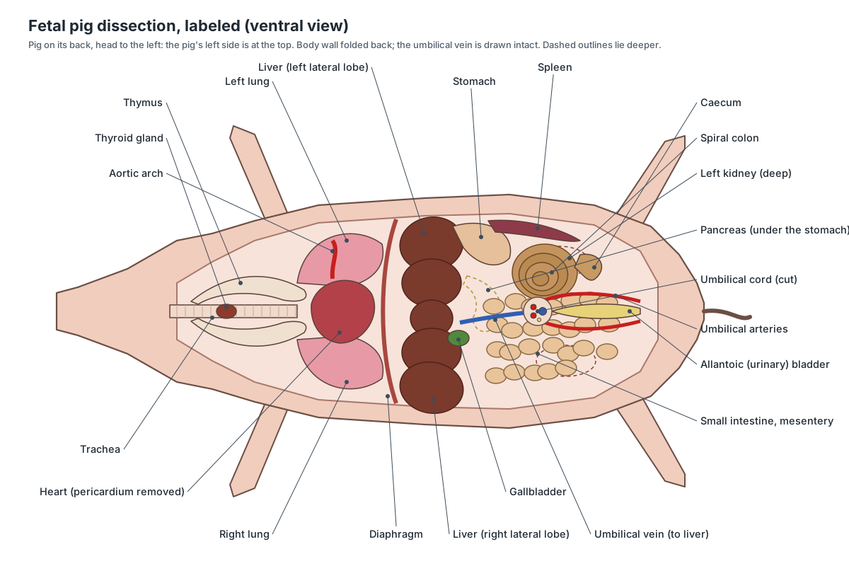
This is what you should see after opening the chest and abdomen: the thymus and thyroid in the neck, the heart between the lungs, the diaphragm, the dark lobed liver with the green gallbladder, the stomach and spleen on the pig's left, the coils of small intestine, the spiral colon and caecum, and the bladder between the two umbilical arteries.
External anatomy: sexing the pig and the umbilical cord
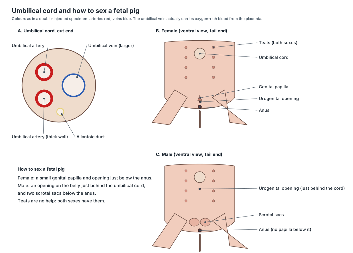
Start outside. A fetal pig has a rostral disk with two nostrils, usually closed eyelids, folded ear flaps, two rows of teats in both sexes, four toes on each foot (two hooves and two dewclaws) and the cut umbilical cord on its belly.
| Feature | Female | Male |
|---|---|---|
| Below the anus | Genital papilla with the urogenital opening | No papilla; scrotal sacs (may be small) |
| Behind the umbilical cord | Nothing | Urogenital (preputial) opening |
| Teats | Two rows | Two rows (not a sex difference) |
| Inside | Ovaries, uterine horns, uterine body, vagina, urogenital sinus | Testes, epididymis, ductus deferens, bulbourethral glands, penis |
How old is my fetal pig? Length–age table
Measure the crown–rump length, from the top of the head to the base of the tail (do not include the tail), and read the approximate age from this table. Pig gestation is about 114 days.
| Crown–rump length | Approximate age (days after mating) |
|---|---|
| 1.1 cm | 21 |
| 1.7 cm | 35 |
| 2.8 cm | 49 |
| 4 cm | 56 |
| 8 cm | 68 |
| 10 cm | 75 |
| 16 cm | 86 |
| 22 cm | 100 |
| 30 cm | about 114 (birth) |
Values to 56 days and 22–30 cm from the Biology Corner lab guide; 8–16 cm from the Wikipedia fetal pig article. Breed and litter size make real fetuses vary by several days.
Fetal pig dissection procedure, step by step
The order below follows common school and college lab guides. Each step matches a step of the 3D viewer, which sets the skin and hides the layers you would have moved aside.
1. External anatomy and age
- Rinse the pig and lay it on its side. Identify the head, neck, trunk and tail, and the four limbs.
- Find the rostral disk and nostrils, the eyelids (usually still closed), the ear flaps and the two rows of mammary papillae on the belly.
- Count the toes on one foot: two large hoofed toes and two small dewclaws.
- Find the cut umbilical cord. Use a hand lens to look at its cut end: two small thick-walled umbilical arteries, one larger umbilical vein and the small allantoic duct.
- Measure the crown–rump length (from the top of the head to the base of the tail, not including the tail) and use the table on this page to estimate the age.
Identify: Skin and body wall, Snout and rostral disk, Nostrils, Eyelids, Ears, Mammary papillae, Umbilical cord, Hooves (main digits), Dewclaws, Tail, Anus.
2. Determine the sex
- Turn the pig on its back. Look just below the anus: a small fleshy genital papilla with an opening means a female.
- In a male there is no papilla under the anus. Instead, find the urogenital opening on the belly just behind the umbilical cord, and the scrotal sacs as swellings below the anus.
- Remember that both sexes have teats, so the teats cannot tell you the sex. Use the Female/Male switch on the model to compare.
Identify: Genital papilla and urogenital opening, Urogenital opening (male), Scrotal sacs, Anus, Mammary papillae, Umbilical cord.
3. Mouth and oral cavity
- Open the mouth. With scissors or a scalpel, cut through the angle of the jaw on both sides so the lower jaw can be pulled down.
- Find the hard palate (ridged, bony) and the soft palate (smooth, behind it) on the roof of the mouth.
- Look at the tongue with a hand lens to see the papillae. Feel the edges of the jaws for teeth: the needle teeth may already have erupted.
- At the back of the throat, find the cone-shaped epiglottis and the glottis opening behind it, which leads to the larynx. Find the opening of the oesophagus dorsal to it.
Caution: The jaw cut needs firm pressure. Cut away from your fingers and keep the scissors' blunt blade inside the mouth.
Identify: Hard palate, Soft palate, Tongue, Tongue papillae, Teeth, Epiglottis, Larynx, Oesophagus.
4. Neck: salivary glands, thymus and thyroid
- Remove the skin from one side of the face and upper neck to expose the masseter muscle and the soft salivary glands near the angle of the jaw.
- On the ventral neck, cut through the skin along the midline from the chin to the chest and pull the muscles aside.
- Find the large, pale, lobed thymus on both sides of the trachea. It continues back over the heart.
- Find the small, dark thyroid gland on the ventral surface of the trachea near the entrance to the chest (between the neck lobes of the thymus), and the larynx at the front end of the trachea.
Identify: Masseter, Salivary glands, Thymus, Thyroid gland, Trachea, Larynx, Jugular veins, Common carotid arteries.
5. Opening the abdominal cavity
- Tie the legs apart with string under the tray. With scissors, cut through the skin and muscle along the midline from the base of the ribs back towards the hind legs.
- Cut around the umbilical cord to leave it on a flap, and make cuts sideways in front of the hind legs and along the lower edge of the ribs, so the walls fold back like doors.
- As you lift the umbilical flap, you will find the umbilical vein running forward from it to the liver. Cut this vein (tie a thread on each end so you can find it again) to fold the flap back.
- Rinse out any fluid. Note the shiny peritoneum lining the cavity and the diaphragm at its front.
Caution: Keep the scissor tips pointing up. The liver lies right under the body wall and is easily cut.
Identify: Abdominal wall muscles, Umbilical vein, Umbilical cord, Diaphragm, Liver.
6. Liver, stomach, spleen and pancreas
- Identify the large, dark, lobed liver just behind the diaphragm. Count its lobes.
- Lift the right side of the liver to find the green gallbladder and the thin bile duct running to the duodenum.
- Lift the liver towards the head to find the stomach on the pig's left. Find where the oesophagus enters it and the pyloric sphincter at its exit.
- Find the long, flat, dark spleen along the left side of the stomach.
- Lift the stomach and look along the first part of the duodenum for the pancreas: pale, granular, bumpy tissue in the loop of the duodenum and under the stomach.
Identify: Liver, Gallbladder, Bile duct, Stomach, Oesophagus, Pyloric sphincter, Spleen, Duodenum, Pancreas.
7. Intestines and mesentery
- Follow the duodenum into the coiled jejunum and ileum. Note the thin mesentery holding the coils and the blood vessels in it.
- Trace the red arteries in the mesentery back to the cranial mesenteric artery on the aorta, and find the hepatic portal vein carrying gut blood forward to the liver (yellow in a triple-injected pig).
- Follow the ileum to where it joins the large intestine. Find the blind-ended caecum at this junction.
- Find the spiral colon, a cone of tight coils on the left. Follow it to the rectum, which runs along the roof of the pelvis to the anus.
- Optional: cut open a short piece of intestine to see the dark green meconium (fetal faeces) inside.
Identify: Duodenum, Jejunum and ileum, Mesentery, Coeliac and cranial mesenteric arteries, Hepatic portal vein, Caecum, Spiral colon, Rectum, Anus.
8. Thoracic cavity: lungs and heart
- Extend the midline cut forward through the sternum (cut slightly to one side of it) to the neck, and cut along the front of the diaphragm on each side to open the chest.
- Find the lungs on either side of the heart. Count the lobes: four on the right (cranial, middle, caudal, accessory) and two on the left.
- Follow the trachea to where it divides into the bronchi. Look for the tracheal bronchus to the right cranial lobe, which leaves the trachea before it divides.
- Note the thymus lying over the front of the heart. Then gently remove the thin pericardium from the heart.
Caution: The sternum and ribs of a fetal pig are soft cartilage and bone. Cut beside the sternum, not through the heart under it.
Identify: Lungs, Trachea, Bronchi (with tracheal bronchus), Diaphragm, Thymus, Pericardium, Sternum, Ribs.
9. Heart and great vessels
- Identify the right and left atria at the top of the heart and the right and left ventricles below. The coronary vessels run in the groove between the ventricles.
- Find the pulmonary trunk crossing the front of the heart, and the aorta arching up behind it to the left.
- Find the short ductus arteriosus joining the pulmonary trunk to the aorta: a fetal shunt.
- Trace the branches of the aortic arch: the brachiocephalic trunk (to the common carotid arteries and right subclavian artery) and the left subclavian artery.
- Find the cranial vena cava (with the jugular veins draining into it) and the caudal vena cava entering the right atrium.
Identify: Right atrium, Left atrium, Right ventricle, Left ventricle, Coronary vessels, Pulmonary trunk, Aorta, Ductus arteriosus, Brachiocephalic trunk, Common carotid arteries, Subclavian arteries, Cranial vena cava, Caudal vena cava, Jugular veins.
10. Fetal circulation: the shunts
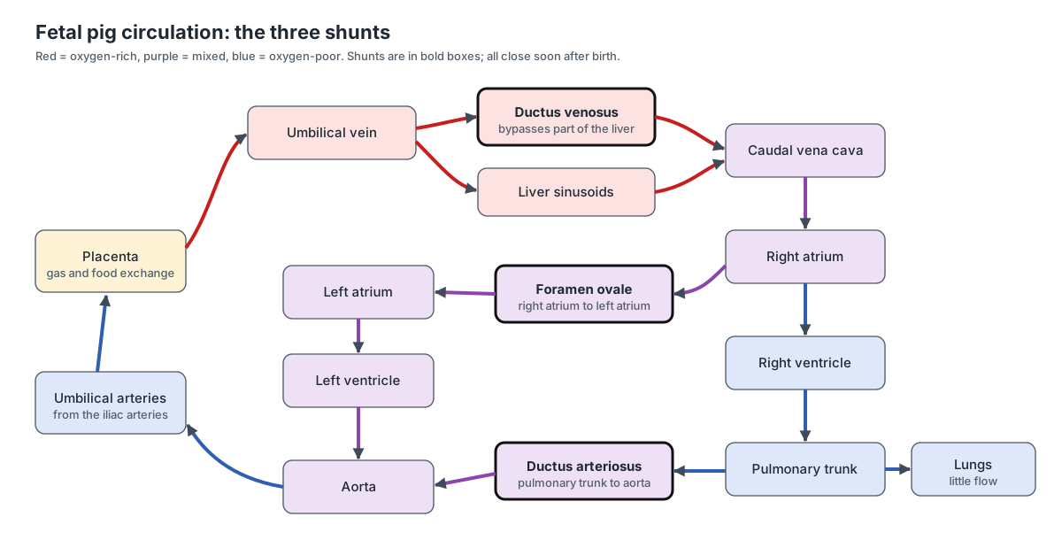
- Follow the umbilical vein from the cord to the liver. In the fetus its blood flows through the liver (partly through the ductus venosus) to the caudal vena cava and the right atrium.
- If you cut the heart open (optional), look in the wall between the atria for the foramen ovale, the flap-covered hole that lets blood skip the right ventricle.
- Trace the two umbilical arteries back from the cord along both sides of the bladder to the iliac arteries at the end of the aorta.
- Explain why the fetus needs these shunts: its lungs are not used, so blood is routed to the placenta instead.
Identify: Umbilical vein, Ductus venosus, Caudal vena cava, Foramen ovale, Ductus arteriosus, Umbilical arteries, Iliac arteries, Aorta.
11. Urinary system
- Push the intestines aside. Find the two bean-shaped kidneys against the dorsal body wall and the small adrenal glands beside their front ends.
- Find the renal artery and vein at each kidney, and the thin ureters running back to the bladder.
- Find the long allantoic (urinary) bladder between the two umbilical arteries, and follow it into the umbilical cord as the allantoic duct.
- Lift the bladder to find the urethra leaving it towards the pelvis.
Identify: Kidneys, Adrenal glands, Renal arteries and veins, Ureters, Urinary (allantoic) bladder, Allantoic duct, Umbilical arteries, Urethra.
12. Male reproductive system
- Switch the model to Male. Open a scrotal sac below the anus to find a testis with the coiled epididymis on it. If the testes are not there, look in the inguinal canal in the groin.
- Follow the ductus (vas) deferens forward through the inguinal canal, over the ureter, to the urethra.
- Find the paired bulbourethral glands beside the urethra at the back of the pelvis.
- Cut away the skin of the belly flap behind the umbilical cord to find the penis running forward to the urogenital opening.
Identify: Testes, Epididymis, Ductus (vas) deferens, Bulbourethral glands, Penis, Urethra, Scrotal sacs, Urogenital opening (male).
13. Female reproductive system
- Switch the model to Female. Find the small, bean-shaped ovaries just behind the kidneys, with the tiny coiled oviducts next to them.
- Follow each uterine horn back to where they join to form the uterine body, dorsal to the bladder.
- With scissors, cut through the middle of the pelvic bones to follow the vagina into the urogenital sinus, which opens at the genital papilla below the anus.
- Compare with a male pig at a neighbouring table.
Identify: Ovaries, Oviducts, Uterine horns, Uterine body, Vagina, Urogenital sinus, Genital papilla and urogenital opening, Urethra.
14. Optional: brain, spinal cord and skeleton
- With the pig on its belly, remove the skin from the top of the head. Carefully cut away the thin skull bones in small pieces to expose the brain.
- Identify the cerebrum, the cerebellum behind it and the brainstem joining the spinal cord.
- Feel or view the skeleton: skull with the rostral bone, vertebral column (7 cervical, 14–15 thoracic, 6–7 lumbar), ribs, sternum, and the bones of the limbs.
Caution: Fetal skull bones are thin and partly cartilage. Peel them off with forceps rather than cutting deeply.
Identify: Cerebrum, Cerebellum, Brainstem and medulla, Spinal cord, Skull, Rostral bone, Cervical vertebrae, Thoracic vertebrae, Lumbar vertebrae, Ribs, Pelvis, Femur.
Where is the pancreas in a fetal pig?
The pancreas is the bumpy, pale organ along the underside of the stomach. Its duct leads into the duodenum, and it makes insulin as well as digestive enzymes. (Biology Corner, Fetal pig dissection and lab guide.)
In practice, lift the liver towards the head and the stomach to the pig's left, then look along the first loop of the duodenum: the pancreas is a cream-coloured, granular, slightly lumpy tissue lying in that loop and running behind the stomach. Reflecting the small intestine to the pig's right also exposes it. Its enzymes reach the duodenum through the pancreatic duct, and its hormones (insulin and glucagon) leave in the blood. (Whitman College, Virtual Pig Dissection.)
Fetal pig umbilical cord and umbilical vessels
Cut the cord across and look at the end with a hand lens: you will see two umbilical arteries, one umbilical vein and the small allantoic duct. Inside the body, the umbilical vein runs forward to the liver; you have to cut it to fold back the belly flap. The two umbilical arteries run back on either side of the flattened urinary bladder. (Biology Corner, Fetal pig dissection and lab guide.)
Before birth, oxygen and nutrients diffuse from the mother's blood into the fetal blood of the placenta and are carried to the fetus in the umbilical vein. Wastes and carbon dioxide go back to the placenta in the umbilical arteries and diffuse into the mother's blood to be cleared. (Whitman College, Virtual Pig Dissection.)
Fetal circulation: the shunts
A fetus does not breathe, so its circulation bypasses the lungs. The foramen ovale, an opening between the right and left atria, lets blood pass straight to the left side of the heart, and the ductus arteriosus diverts blood from the pulmonary trunk into the aortic arch. Both close soon after birth. (Fetal pig, Wikipedia.)
A third shunt, the ductus venosus, carries part of the umbilical vein blood through the liver to the caudal vena cava. It is buried in liver tissue and is rarely seen in a student dissection. After birth the shunts become ligaments: the ligamentum arteriosum, the fossa ovalis, the ligamentum venosum and the round ligament of the liver.
Fetal ventricles have walls of about equal thickness. After birth the left ventricle thickens because it pumps blood around the long, high-resistance body circuit. (Whitman College, Virtual Pig Dissection.)
Why is the thymus so large in a fetal pig?
The thymus lies in the throat and over the heart. It is large in young pigs because it is critical in the development of the immune system's T cells; later in life it shrinks. (Whitman College, Virtual Pig Dissection.)
Male vs female: what changes in the dissection
Only steps 12 and 13 differ. Sex the pig first from the outside (step 2).
- Male: testes in or near the scrotal sacs (or still in the inguinal canal), the epididymis on each testis, the ductus deferens looping over the ureter to the urethra, paired bulbourethral glands at the back of the pelvis, and the penis under the belly skin running to the opening behind the cord.
- Female: small ovaries just behind the kidneys, tiny coiled oviducts, two long uterine horns joining into a short uterine body, the vagina dorsal to the urethra, and the urogenital sinus opening at the genital papilla.
Swap pigs with a neighbouring group so that everyone sees both sexes.
Where do fetal pigs for dissection come from?
Fetal pigs are a by-product of the meat-packing industry. They are not bred or killed for dissection: they are removed from the uterus of pregnant sows that have already been slaughtered for pork. Because they would otherwise be waste, they are cheap, and fetuses not sold for teaching are often used for fertiliser or discarded. (Fetal pig, Wikipedia.)
Biological supply companies preserve the fetuses and ship them to schools. Many are injected with coloured latex: red in the arteries and blue in the veins (double-injected), sometimes yellow in the hepatic portal system too. Older specimens were stored in formaldehyde, which is a carcinogen, so modern suppliers often use other preservatives; whatever is used, wear gloves and work in a ventilated room. (Fetal pig, Wikipedia.)
Why are fetal pigs used for dissection?
Pigs are mammals, like us, so they share hair, mammary glands, live birth and the same basic organ systems. A fetal pig also lets students see fetal circulation, which differs from the adult. Fetal pigs are easy to get at low cost, their soft tissues and partly cartilaginous skeleton are easy to cut, and at 25–35 cm they are large enough that the organs are easy to see. (Fetal pig, Wikipedia.)
How old is a fetal pig? Estimating age from length
Pig gestation lasts about 114 days. Lab guides give 112–115 days, and you may see the memory aid '3 months, 3 weeks and 3 days'. To estimate the age of your specimen, measure its crown–rump length (from the top of the head to the base of the tail, not including the tail) and compare it with the table on this page. (Biology Corner, Fetal pig dissection and lab guide.)
Growth is fast in the last third of pregnancy: a fetus is about 8 cm at 68 days, 10 cm at 75 days, 16 cm at 86 days, 22 cm at 100 days and about 30 cm at 114 days. Most lab specimens are therefore within the last few weeks before birth. (Fetal pig, Wikipedia.)
Fetal pig dissection worksheet: lab questions and answer key
Answer these during or after the dissection. Click a question to reveal the answer.
How did you determine the sex of your fetal pig?
By the urogenital opening. A female has a genital papilla with the urogenital opening just below (ventral to) the anus. A male has its urogenital opening on the belly just behind the umbilical cord, and scrotal sacs below the anus.
Why can't you use the teats (mammary papillae) to tell males from females?
Both male and female pigs have two rows of mammary papillae.
Your pig is 22 cm long (crown–rump). About how old is it, and how long is pig gestation?
About 100 days. Pig gestation is about 114 days (lab guides give 112–115 days), and a pig is about 30 cm long at birth.
Name the vessels in the umbilical cord and say what each carries.
Two umbilical arteries carry oxygen-poor blood and wastes from the fetus to the placenta. One umbilical vein carries oxygen- and nutrient-rich blood from the placenta to the fetus. The allantoic duct carries urine from the bladder to the allantois.
Which vessel did you have to cut to fold back the umbilical flap, and where did it go?
The umbilical vein, which runs from the umbilical cord forward to the liver.
Where is the pancreas, and what does it look like?
It lies under (dorsal to) the stomach and along the loop of the duodenum. It is pale, granular and bumpy. It makes digestive enzymes and the hormone insulin.
What is the function of the epiglottis?
It covers the glottis during swallowing so that food goes down the oesophagus and not into the trachea.
How can you tell the trachea from the oesophagus?
The trachea is ventral and has rings of cartilage that keep it open. The oesophagus lies dorsal to the trachea, has no rings and is flat when empty.
Why is the thymus so large in a fetal pig?
The thymus trains the T cells of the immune system early in life. It is large in the fetus and young pig and shrinks after puberty.
Why do the fetal lungs look solid and dark, and would a piece float?
The fetus has never breathed, so its lungs are not inflated with air. A piece of fetal lung sinks in water; lung tissue from a piglet that has breathed floats.
Name the three fetal shunts and what each bypasses.
Foramen ovale (between the atria: bypasses the right ventricle and lungs), ductus arteriosus (pulmonary trunk to aorta: bypasses the lungs) and ductus venosus (umbilical vein to caudal vena cava: bypasses part of the liver).
What happens to the fetal shunts after birth?
They close. The foramen ovale becomes the fossa ovalis, the ductus arteriosus the ligamentum arteriosum, the ductus venosus the ligamentum venosum, and the umbilical vein the round ligament of the liver.
What is the caecum, and where is it?
A blind-ended pouch at the junction of the small and large intestine, on the left side next to the spiral colon. It holds microbes that ferment plant fibre.
Which structure lies between the two umbilical arteries?
The urinary (allantoic) bladder.
Why does a vasectomy not affect urine flow?
It cuts the ductus (vas) deferens, which carries only sperm. Urine flows from the kidneys through the ureters to the bladder and out through the urethra, which is not cut.
Which part of the female pig matches the human Fallopian tube, and where do piglets develop?
The oviduct matches the Fallopian tube. Piglets develop along the two long uterine horns, not in a single uterine body as in humans.
How many toes does a pig have on each foot?
Four: two large weight-bearing toes (digits III and IV) and two small dewclaws (digits II and V).
Fetal pig quiz: identify the structure
Name the structure from its description. Answers are in brackets at the end of the list.
- A dead-end pouch where the small and large intestines meet.
- A green sac under the liver that stores bile.
- The muscle sheet between the chest and the abdomen.
- A small, dark reddish-brown gland on the ventral surface of the trachea, near the entrance to the chest.
- The vessel you cut to fold back the umbilical flap.
- The structure lying between the two umbilical arteries.
- The vessel joining the pulmonary trunk to the aorta.
- The thin membrane that holds the coils of the small intestine.
(1 caecum, 2 gallbladder, 3 diaphragm, 4 thyroid gland, 5 umbilical vein, 6 urinary bladder, 7 ductus arteriosus, 8 mesentery.)
Virtual fetal pig dissection and student choice
The National Association of Biology Teachers supports well-planned dissections but asks teachers to respect students' right to make informed decisions about taking part, and to provide meaningful alternatives where possible. It also notes that software-only approaches have limits. (NABT position statement: The use of animals in biology education (2019).)
Several comparative studies have found that students taught with non-animal methods do as well as those who dissect. The 3D model on this page follows the same 14 steps as the bench dissection, so a student who opts out can locate every structure and answer the same lab questions. (Fetal pig, Wikipedia.)
The poem 'On Learning to Dissect Fetal Pigs' is often searched alongside this lab; it is about the classroom experience, not a guide to the anatomy.
Learn the anatomy
For adult pig anatomy (the spiral colon length, liver lobes, skeleton, heart and xenotransplantation, and pig vs human differences), see our pig anatomy page.
Frequently asked questions
Where do fetal pigs for dissection come from?
They are a by-product of the pork industry. When pregnant sows are slaughtered for meat, the fetuses are removed from the uterus and sold to biological supply companies, which preserve them. The pigs are not bred or killed for dissection.
Are fetal pigs killed for dissection?
No. The fetuses die when the sow is slaughtered for pork; they would otherwise be discarded or used for fertiliser. Supply companies collect and preserve them afterwards.
How old are fetal pigs used in dissection?
Most are in the last few weeks of a gestation of about 114 days. Measure the crown–rump length: about 22 cm means roughly 100 days, and about 30 cm means the pig was close to birth.
How do you tell if a fetal pig is male or female?
Look below the anus. A female has a small genital papilla with the urogenital opening there. A male has no papilla; his urogenital opening is on the belly just behind the umbilical cord, and he has scrotal sacs below the anus. Both sexes have teats.
What are the vessels in the umbilical cord of a fetal pig?
Two umbilical arteries, which carry blood from the fetus to the placenta, and one larger umbilical vein, which brings oxygen-rich blood back. The small fourth opening is the allantoic duct, which drains the bladder.
Where is the pancreas in a fetal pig?
Under the stomach and along the first loop of the duodenum. It is pale, granular and bumpy. Lift the liver and stomach, then follow the duodenum from the pylorus to find it.
Why are fetal pigs used for dissection?
They are mammals with organs much like ours, they are cheap because they are a meat-industry by-product, they are large enough to see the organs easily, their soft tissue is easy to cut, and they show fetal circulation.
Is there a virtual fetal pig dissection?
Yes. The 3D model on this page follows the same 14 steps as the bench dissection: each step removes the right layers and highlights the structures to find. It works as an alternative for students who opt out of dissection.
References
- Biology Corner, Fetal pig dissection and lab guide
- Whitman College, Virtual Pig Dissection
- Fetal pig, Wikipedia
- NABT position statement: The use of animals in biology education (2019)
- Bazer et al. (2012), Uterine biology in pigs and sheep, Journal of Animal Science and Biotechnology
- Gonzalez, Moeser & Blikslager (2015), Porcine models of digestive disease, Translational Research
- Nykonenko, Vávra & Zonča (2017), Anatomic peculiarities of pig and human liver, Experimental and Clinical Transplantation
- IMAIOS vet-Anatomy: vertebral column (vertebral formulae of domestic mammals)
- Tracheal bronchus, Radiopaedia
- Pig, Wikipedia
More interactive anatomy models
- Pig anatomyOrgans, spiral colon and skeleton of the domestic pig.
- Human anatomy1,000+ bones, muscles, organs, vessels and nerves from real anatomical data.
- Octopus anatomyThree hearts, a ring-shaped brain, eight arms and a beak, in 3D.
- Squid anatomyPen, eight arms, two tentacles, giant axons and three hearts of the longfin squid.
- Squid dissectionStep-by-step guided squid dissection with every structure labeled.
- Fish anatomyExternal and internal anatomy of a bony fish: fins, gills, swim bladder, heart and brain.