

Anatomy of a snake at a glance
A snake is a legless reptile (suborder Serpentes) whose organs are long, thin and arranged one behind another inside a body that is mostly trunk. It has a highly mobile skull, hundreds of vertebrae with ribs, a three-chambered heart, usually one working lung, no urinary bladder, no external ears and no eyelids. All of its waste and reproductive products leave through a single opening, the vent.
This page uses the ball python (royal python, Python regius) as the reference snake. It is the most commonly kept pet snake, it has been studied with ultrasound, CT and physiology, and as a python it keeps several structures that most snakes have lost: a functional left lung, a vestigial pelvis with external spurs, a caecum, and heat-sensing labial pits. So it shows the full list of structures a lab manual asks for. A colubrid such as the corn snake has the same plan without those extras; the differences are flagged below.
Directions: cranial means towards the head, caudal towards the tail, dorsal the back and ventral the belly. Positions inside a snake are given as a percentage of snout–vent length (SVL), because the tail behind the vent varies a lot in length between species.
| Feature | Ball python | Most other snakes |
|---|---|---|
| Lungs | Right lung plus a smaller working left lung | One right lung; left tiny or absent |
| Heart | 3 chambers; strong pressure separation | 3 chambers |
| Hind-limb remnants | Pelvic rod, femur and spur | None in colubrids, elapids and vipers |
| Heat pits | Row of labial pits | None (colubrids, elapids); one loreal pit in pit vipers |
| Teeth | Solid teeth, including on the premaxilla; no fangs | Fangs in vipers, elapids and some colubrids |
| Reproduction | Lays 3–11 eggs and coils around them | Egg-laying or live-bearing, depending on species |
External anatomy of a snake
The external anatomy of a snake includes the head with its nostrils, eyes and lip scales, the long trunk covered in small dorsal scales and one row of wide ventral scales, the vent with its cloacal scale, and the tail with its subcaudal scales. Pythons and boas also have heat pits in the lips and a pair of spurs beside the vent.
- Head: Holds the brain, eyes, nostrils, heat-sensing pits, jaws and the forked tongue.
- Body (trunk) and dorsal scales: Protects the organs, limits water loss and anchors the muscles that bend the body and move the belly scales.
- Ventral scales (scutes): Grip the ground. Muscles pull them forward and back for straight-line (rectilinear) crawling.
- Subcaudal scales: Protect the tail and are counted to identify species.
- Vent and cloacal scale: Outlet for faeces, urates (urine), eggs or sperm. The hemipenes are everted through it.
- Pelvic (cloacal) spurs: Used in courtship and male combat; males rub or scratch the female with them.
- Tail: Holds the hemipenes (in males), the cloacal scent glands and the tail vertebrae; it is used for grip and balance.
Scales are not separate plates glued on: they are folds of skin covered in keratin. A snake sheds the outer layer of the whole skin, including the clear eye caps, usually in one piece. Before a shed the skin dulls and the eyes turn milky blue as fluid builds up under the old layer.
Snake internal anatomy: organs diagram
Inside a snake, the organs run in a line from head to tail: trachea and oesophagus, thyroid, heart, lung, liver, stomach, the gallbladder–pancreas–spleen triad, small intestine, gonads, kidneys and large intestine, ending at the cloaca. Long strands of fat bodies fill the back of the body cavity. There is no diaphragm, so a single body cavity (the coelom) holds everything from the heart to the cloaca.
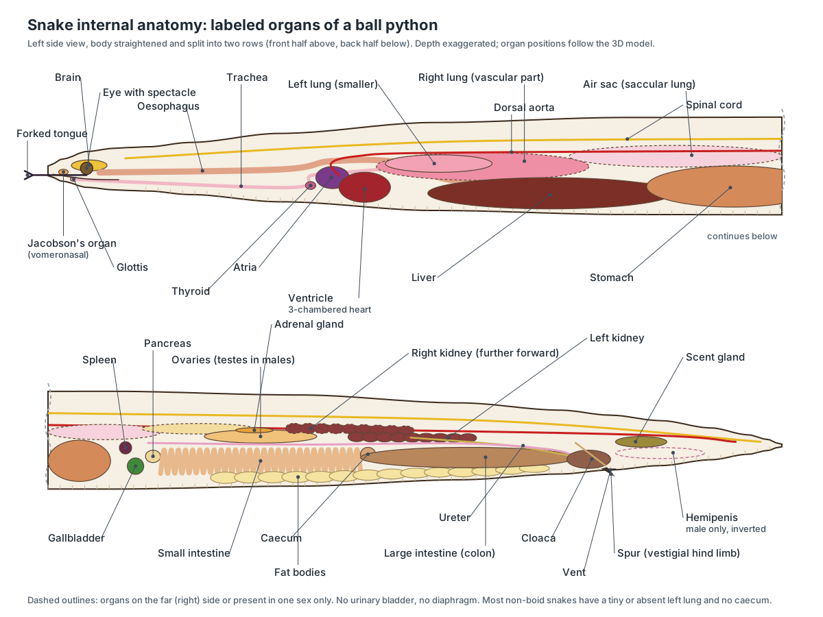
Paired organs are staggered to fit the narrow body: the right kidney, ovary or testis and adrenal gland lie in front of the left ones. Many single organs are long and thin: the liver is an elongated lobed strip, the lung can be a third of the body long, and the kidneys are chains of lobules.
Snake anatomy labeled: every part and its function
This is the text version of the 3D model: every labeled structure of the ball python, grouped by body system, with what it does.
External & scales
- Head (Cephalic region, head scales)
- A broad, slightly flattened head that is clearly wider than the slim neck in a ball python. Holds the brain, eyes, nostrils, heat-sensing pits, jaws and the forked tongue.
- Body (trunk) and dorsal scales (Precloacal body, skin)
- The long body from the neck to the vent, covered with small smooth overlapping scales. Protects the organs, limits water loss and anchors the muscles that bend the body and move the belly scales.
- Tail (Caudal region (postcloacal))
- The short, tapering part of the body behind the vent; about a tenth of the total length in a ball python. Holds the hemipenes (in males), the cloacal scent glands and the tail vertebrae; it is used for grip and balance.
- Ventral scales (scutes) (Belly scales, gastrosteges)
- One row of wide, overlapping belly plates, one roughly for each trunk vertebra. Grip the ground. Muscles pull them forward and back for straight-line (rectilinear) crawling.
- Subcaudal scales (Subcaudals, urosteges)
- The belly scales under the tail, behind the vent. Protect the tail and are counted to identify species.
- Vent and cloacal scale (Cloacal opening, anal plate)
- The transverse slit where the cloaca opens to the outside, covered by the cloacal (anal) scale. Outlet for faeces, urates (urine), eggs or sperm. The hemipenes are everted through it.
- Nostrils (External nares)
- Two small openings near the tip of the snout. Let air in and out for breathing and carry airborne odours to the nasal cavity.
- Pelvic (cloacal) spurs (Spurs, vestigial hind limbs)
- A small horny claw on each side of the vent: the visible tip of a vestigial hind limb. Used in courtship and male combat; males rub or scratch the female with them.
Sense organs
- Eyes (Eyeballs)
- Two eyes on the sides of the head, each permanently covered by a clear scale (the spectacle). Detect movement and light; vision works together with the heat pits.
- Spectacle (brille) (Brille, eye cap, ocular scale)
- A transparent scale that covers and protects each eye in place of eyelids. Protects the cornea and keeps the eye moist; it is shed with the skin.
- Labial heat pits (Pit organs, infrared receptors)
- A row of small pits in the lip scales that sense infrared (heat) from warm-blooded prey. Detect body heat, so the snake can strike accurately at a mouse in the dark.
- Vomeronasal organ (Jacobson's organ) (Jacobson's organ, VNO)
- Paired chemical-sensing chambers above the roof of the mouth that read the scent molecules collected by the tongue. Detects prey, predators and mates from scent trails; its nerves run to the accessory olfactory bulbs.
- Forked tongue (Bifid tongue)
- A long, slender tongue split into two tips, flicked in and out to sample scent. Collects odour particles from air and ground and carries them to the vomeronasal organ. It is not used for swallowing.
- Inner ear (Otic capsule, cochlear duct)
- The hearing and balance organ inside the braincase; snakes have no outer ear or eardrum. Senses vibration and head movement. Ground and skull vibrations reach it through the jaw, quadrate and stapes.
Brain & nerves
- Olfactory bulbs (Main and accessory olfactory bulbs, olfactory tracts)
- The front of the brain, which receives smell from the nose and the vomeronasal organ. Process odours; the accessory bulbs handle signals from Jacobson's organ.
- Cerebrum (cerebral hemispheres) (Telencephalon, forebrain)
- Paired hemispheres that integrate smell and other senses and control behaviour. Integrates sensory input and organises behaviour such as hunting and courtship.
- Optic tectum (Optic lobes, midbrain roof)
- Two rounded lobes on the roof of the midbrain that build a map of the surroundings. Combines vision with heat-pit (infrared) input and directs the strike.
- Cerebellum (Hindbrain roof)
- A small, thin plate of brain behind the optic tectum. Coordinates movement and balance.
- Medulla oblongata (Brainstem)
- The base of the brain, which continues into the spinal cord. Controls breathing and heart rate and relays signals between brain and body.
- Spinal cord (Medulla spinalis)
- The nerve cord that runs inside the neural arches of the whole vertebral column. Carries signals to and from the brain and coordinates the waves of muscle contraction that move the body.
Digestive
- Mouth (Oral opening, lips (labial scales))
- A wide mouth edged by rows of lip scales (labials), with a small notch at the front for the tongue. Grabs prey and swallows it whole; snakes do not chew.
- Teeth (Maxillary, palatine, pterygoid, dentary and premaxillary teeth)
- Many sharp, backward-curving teeth that grip prey; pythons have no fangs. Hold prey and ratchet it into the throat during swallowing. They do not chew.
- Tongue sheath (Lingual sheath)
- A pocket in the floor of the mouth into which the tongue retracts. Protects the tongue when it is pulled in; the tongue's retractor muscles lie alongside it.
- Oesophagus (Esophagus, gullet)
- A long, very stretchy tube that carries whole prey from the mouth to the stomach. Moves prey backward by waves of muscle contraction and by the snake 'walking' its body over the meal.
- Stomach
- A thick-walled, spindle-shaped, muscular and stretchable stomach. Stores and digests prey with acid and enzymes; even bone is dissolved.
- Liver
- A long, dark red, lobed organ, the largest gland in the body. Makes bile, stores energy and processes absorbed nutrients and toxins.
- Gallbladder (Gall bladder)
- A green, round sac of bile behind the stomach, well away from the liver. Stores bile and releases it into the intestine through several ducts that pass through the pancreas.
- Pancreas
- A small, firm, pale gland next to the gallbladder and spleen. Makes digestive enzymes and the hormones insulin and glucagon.
- Small intestine (Duodenum, jejunum, ileum)
- A narrow, gently folded tube with short transverse loops, not tightly coiled as in mammals. Completes digestion and absorbs nutrients.
- Caecum (Cecum)
- A small blind pouch where the small intestine joins the colon. Part of the large intestine; its exact role in snakes is not well studied.
- Large intestine (colon) (Colon)
- A wider tube that stores faeces and absorbs water. Absorbs water, including water from urine that flows back from the cloaca.
- Cloaca (Coprodeum, urodeum, proctodeum)
- The shared chamber at the end of the gut, urinary and reproductive tracts. Collects faeces, urates and eggs or sperm before they leave the body.
Respiratory
- Glottis (Laryngeal opening)
- The opening of the windpipe, far forward on the floor of the mouth. Lets air into the trachea. It can be pushed out to the side so the snake keeps breathing while swallowing large prey.
- Trachea (Windpipe)
- A long windpipe stiffened by incomplete cartilage rings, from the glottis to the lung. Carries air to and from the lungs.
- Right lung (Vascular (respiratory) lung)
- The main, long lung, with a spongy, blood-rich front part for gas exchange. Takes up oxygen and releases carbon dioxide.
- Left lung
- A smaller second lung that pythons and boas keep but most snakes have reduced to a stub or lost. Adds gas-exchange surface.
- Air sac (saccular lung) (Posterior air sac, avascular lung)
- The thin-walled back part of the lung, which stores air but does little gas exchange. Holds most of the lung's air volume; it may act as a reserve during swallowing and inflates the body when the snake is threatened.
Heart & vessels
- Right atrium
- Heart chamber that receives oxygen-poor blood from the body. Fills from the sinus venosus and pumps blood into the ventricle.
- Left atrium
- Heart chamber that receives oxygen-rich blood from the lungs. Fills from the pulmonary veins and pumps blood into the ventricle.
- Ventricle (Cavum arteriosum, cavum venosum, cavum pulmonale)
- The single, muscular pumping chamber, divided by ridges into three linked sub-chambers. Pumps blood to the lungs and the body while keeping the two streams largely separate.
- Sinus venosus
- A thin-walled chamber on the back of the heart that collects returning body blood. Receives the venae cavae and jugular veins and empties into the right atrium.
- Left and right aortic arches (Systemic arches)
- Two aortas that leave the heart, curve dorsally around the oesophagus and trachea and join. Carry oxygen-rich blood to the head and body.
- Dorsal aorta (Abdominal aorta, caudal artery)
- The main artery running back under the vertebral column. Supplies the gut, kidneys, gonads, muscles and tail.
- Pulmonary artery
- Vessel carrying oxygen-poor blood from the ventricle to the lung. Sends blood to the lungs at low pressure, which protects their delicate surfaces.
- Posterior vena cava (Postcava, caudal vena cava)
- The large vein returning blood from the kidneys and liver to the heart. Returns oxygen-poor blood from the back two-thirds of the body.
- Jugular vein (Right jugular vein)
- The vein that drains the head and neck. Returns blood from the head to the sinus venosus.
- Spleen
- A small, dark, round organ of the immune and blood system. Filters blood and produces immune cells.
Urinary
- Kidneys
- Two long, lobed kidneys; the right one lies further forward than the left. Filter blood and excrete nitrogen mainly as uric acid (white urates), which saves water.
- Ureters
- Tubes that carry urine from each kidney to the cloaca. Drain urine to the cloaca. Snakes have no urinary bladder, so urine is not stored.
Reproductive
- Testes (Testicles) · males only
- Two elongated testes inside the body, the right one in front of the left. Produce sperm and testosterone.
- Vas deferens (Ductus deferens, sperm duct) · males only
- A coiled duct from each testis to the cloaca. Carries sperm to the cloaca, from where it runs along a groove on the everted hemipenis.
- Hemipenes (Hemipenis (singular), copulatory organs) · males only
- Two copulatory organs that lie inside-out (inverted) in the base of the tail. One hemipenis is everted through the vent during mating to transfer sperm.
- Ovaries · females only
- Two elongated ovaries holding developing follicles, the right in front of the left. Produce eggs (ova) and hormones.
- Oviducts (Infundibulum, uterus, shell gland) · females only
- Two long ducts that collect eggs from the ovaries and carry them to the cloaca. Site of fertilisation and of shell formation; in live-bearing snakes, where embryos develop.
Glands & fat bodies
- Thyroid gland
- A single, round endocrine gland just in front of the heart. Makes thyroid hormones that control metabolism and the shedding cycle.
- Adrenal glands
- Small, elongated glands next to the gonads. Make stress hormones (corticosterone, adrenaline).
- Fat bodies (Coelomic (abdominal) fat bodies)
- Long, lumpy strands of yellow fat in the back part of the body cavity. Store energy for long fasts, egg production and the breeding season.
- Cloacal scent glands (Musk glands)
- Paired glands in the tail base that release a strong-smelling musk. Defence (a smelly discharge when handled) and chemical signalling.
Muscles
- Epaxial muscles (Spinalis, semispinalis, longissimus, iliocostalis)
- Long chains of back muscles above and beside the vertebral column. Bend the body side to side and up and down, for lateral undulation, climbing and constriction.
- Costocutaneous muscles (Costocutaneous superior and inferior)
- Small muscles that link the ribs to the belly skin. Move the belly skin forward and back over the ribs for rectilinear (straight-line, 'caterpillar') crawling.
- Jaw muscles (Adductor mandibulae externus)
- The muscles behind the eye that close the jaws. Close the mouth and hold prey; other head muscles move the palate and jaws during swallowing.
Skeleton
- Skull (Cranium, braincase, palate)
- A light, highly mobile (kinetic) skull with a solid braincase and loosely joined jaw and palate bones. Protects the brain and lets the jaws spread to swallow prey larger than the head.
- Lower jaw (mandibles) (Dentary, compound bone)
- Two separate halves joined at the chin only by a stretchy ligament. Each half moves independently, so the snake can 'walk' its jaws over prey.
- Quadrate bones (Quadratum)
- Long, mobile bones that hang from the skull and carry the jaw joint. Swing outward and down to widen the gape; also pass vibrations to the inner ear through the stapes.
- Vertebral column (Backbone, spine)
- Hundreds of small vertebrae joined by ball-and-socket joints and extra interlocking processes. Supports the body and allows great flexibility while resisting twisting.
- Ribs
- A pair of curved ribs on almost every trunk vertebra; there is no breastbone (sternum). Protect the organs, power breathing and allow the body to stretch around large prey.
- Vestigial pelvis and femur (Pelvic rudiment)
- A tiny rod-like pelvic bone and a rudimentary femur buried in the muscles beside the cloaca. Anchors the spur; used in courtship and mating.
Snake head anatomy: mouth, jaw and tongue
A snake's head holds the brain inside a solid braincase, flanked by loosely linked jaw and palate bones that let the mouth stretch around large prey. The mouth has rows of backward-curving teeth, a forked tongue that lives in a sheath in the floor of the mouth, and a glottis (windpipe opening) placed far forward.
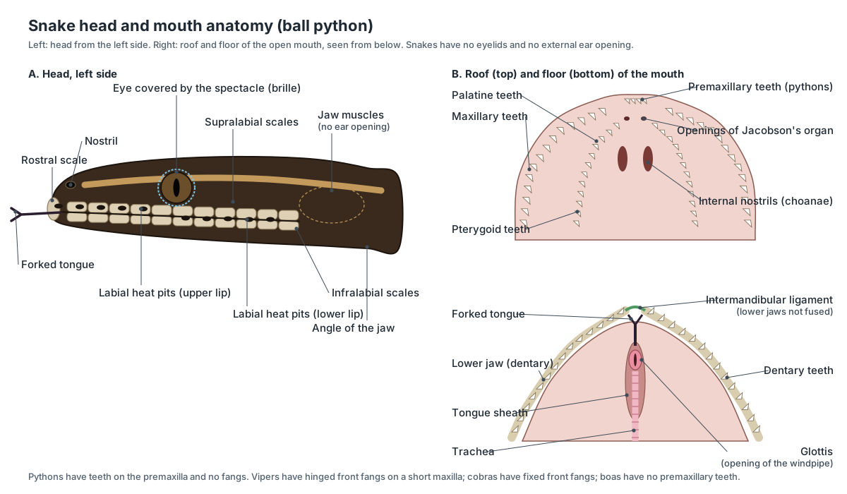
- Mouth: Grabs prey and swallows it whole; snakes do not chew.
- Teeth: Hold prey and ratchet it into the throat during swallowing. They do not chew.
- Forked tongue: Collects odour particles from air and ground and carries them to the vomeronasal organ. It is not used for swallowing.
- Tongue sheath: Protects the tongue when it is pulled in; the tongue's retractor muscles lie alongside it.
- Glottis: Lets air into the trachea. It can be pushed out to the side so the snake keeps breathing while swallowing large prey.
- Vomeronasal organ (Jacobson's organ): Detects prey, predators and mates from scent trails; its nerves run to the accessory olfactory bulbs.
- Labial heat pits: Detect body heat, so the snake can strike accurately at a mouse in the dark.
- Jaw muscles: Close the mouth and hold prey; other head muscles move the palate and jaws during swallowing.
Snake mouth anatomy explains how swallowing works. The two lower jaw halves are joined only by a stretchy ligament, and each side of the upper jaw and palate moves independently. The snake hooks the teeth of one side into the prey, pushes that side forward, then repeats with the other side. The bones of the jaw and skull are described one by one on the snake skeleton page.
Snake fangs and venom glands
Ball pythons have no fangs and no venom: they kill by constriction. Venomous snakes have enlarged fangs on the maxilla, connected by a duct to a venom gland, a modified salivary gland in the upper jaw behind the eye. Muscles around the gland squeeze venom through the duct when the snake bites.
| Fang type | Fang position | Examples |
|---|---|---|
| Aglyphous (no fangs) | All teeth solid | Pythons, boas, corn snakes |
| Opisthoglyphous (rear-fanged) | Grooved fangs at the back of the maxilla | Some colubrids; hognose snakes have enlarged rear teeth |
| Proteroglyphous | Fixed hollow fangs at the front | Cobras, mambas, sea snakes (elapids) |
| Solenoglyphous | Long hinged fangs that fold against the palate | Rattlesnakes and other vipers |
Embryo studies show that front fangs develop at the back of the upper jaw, like rear fangs, and are moved forward by growth. Rattlesnakes are born with working fangs and replace them every 6–10 weeks from a row of reserve fangs behind the functional pair.
Snake heart anatomy
A snake has a three-chambered heart: a right atrium, a left atrium and a single ventricle. It lies in the front third of the body, at about 23.5% of snout–vent length in Burmese pythons, and it can slide within the rib cage because there is no diaphragm.
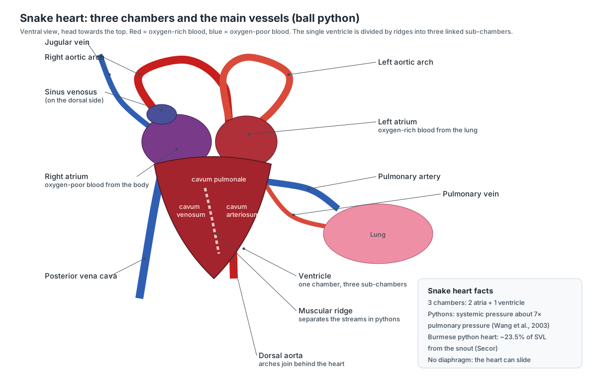
- Right atrium: Fills from the sinus venosus and pumps blood into the ventricle.
- Left atrium: Fills from the pulmonary veins and pumps blood into the ventricle.
- Ventricle: Pumps blood to the lungs and the body while keeping the two streams largely separate.
- Sinus venosus: Receives the venae cavae and jugular veins and empties into the right atrium.
- Left and right aortic arches: Carry oxygen-rich blood to the head and body.
- Dorsal aorta: Supplies the gut, kidneys, gonads, muscles and tail.
- Pulmonary artery: Sends blood to the lungs at low pressure, which protects their delicate surfaces.
- Posterior vena cava: Returns oxygen-poor blood from the back two-thirds of the body.
Heart position varies with lifestyle. Snakes that climb tend to have their heart closer to the head, which helps keep blood flowing to the brain when the body is vertical; some aquatic snakes have it nearer the middle of the body.
Snake lungs and breathing
Most snakes breathe with one long right lung; the left lung is small or missing. The ball python is an exception, with a smaller but working left lung. The back part of the lung is a thin air sac that stores air but does little gas exchange.
- Glottis: Lets air into the trachea. It can be pushed out to the side so the snake keeps breathing while swallowing large prey.
- Trachea: Carries air to and from the lungs.
- Right lung: Takes up oxygen and releases carbon dioxide.
- Left lung: Adds gas-exchange surface.
- Air sac (saccular lung): Holds most of the lung's air volume; it may act as a reserve during swallowing and inflates the body when the snake is threatened.
With no diaphragm, a snake breathes by moving its ribs. When it is constricting prey or has a large meal in its gut, it can shift which part of the rib cage does the work.
Snake digestive system
A snake swallows prey whole. Food passes down a very stretchy oesophagus into a muscular stomach, where it is broken down, bones included. It then moves through a short, gently looped small intestine and a wider colon to the cloaca. The liver, gallbladder and pancreas add bile and enzymes.
- Oesophagus: Moves prey backward by waves of muscle contraction and by the snake 'walking' its body over the meal.
- Stomach: Stores and digests prey with acid and enzymes; even bone is dissolved.
- Liver: Makes bile, stores energy and processes absorbed nutrients and toxins.
- Gallbladder: Stores bile and releases it into the intestine through several ducts that pass through the pancreas.
- Pancreas: Makes digestive enzymes and the hormones insulin and glucagon.
- Small intestine: Completes digestion and absorbs nutrients.
- Caecum: Part of the large intestine; its exact role in snakes is not well studied.
- Large intestine (colon): Absorbs water, including water from urine that flows back from the cloaca.
- Cloaca: Collects faeces, urates and eggs or sperm before they leave the body.
Pythons are famous for remodelling their gut. After a large meal the stomach and intestines grow and speed up, then shrink again during the fast that follows. Ball pythons may eat only every week or two as adults and can go months without food.
Snake kidneys and the cloaca
Snakes have two long, lobed kidneys in the back third of the body, the right one in front of the left. They have no urinary bladder. Urine goes straight to the cloaca as white uric acid paste (urates), which saves water.
- Kidneys: Filter blood and excrete nitrogen mainly as uric acid (white urates), which saves water.
- Ureters: Drain urine to the cloaca. Snakes have no urinary bladder, so urine is not stored.
- Cloaca: Collects faeces, urates and eggs or sperm before they leave the body.
- Cloacal scent glands: Defence (a smelly discharge when handled) and chemical signalling.
Snake reproductive anatomy: male vs female
Male snakes have two internal testes and two hemipenes, which lie inverted in the base of the tail. Female snakes have two ovaries and two oviducts. Both sexes have a cloaca, cloacal scent glands and, in pythons, spurs. Use the Female and Male buttons on the 3D model to compare them.
| Male snake anatomy | Female snake anatomy | |
|---|---|---|
| Gonads | Two elongated testes, right in front | Two ovaries with follicles, right in front |
| Ducts | Coiled vas deferens to the cloaca | Long oviducts that form the eggshell |
| Copulatory organs | Two hemipenes inverted in the tail base | None |
| Tail | Usually longer and thicker at the base | Usually shorter and tapering sooner |
| Spurs (pythons) | Larger; used in courtship | Smaller |
| Probe depth | About 8–10 subcaudal scales | About 2–4 subcaudal scales |
- Testes: Produce sperm and testosterone.
- Vas deferens: Carries sperm to the cloaca, from where it runs along a groove on the everted hemipenis.
- Hemipenes: One hemipenis is everted through the vent during mating to transfer sperm.
- Ovaries: Produce eggs (ova) and hormones.
- Oviducts: Site of fertilisation and of shell formation; in live-bearing snakes, where embryos develop.
- Fat bodies: Store energy for long fasts, egg production and the breeding season.
Snake eye, ear and brain anatomy
Snake eyes have no movable eyelids; a clear spectacle covers each eye. Snakes also have no ear opening or eardrum, but they do have inner ears, which pick up vibrations passed through the jaw and quadrate bone. Their brain is small and long, with large olfactory bulbs for smell.
- Eyes: Detect movement and light; vision works together with the heat pits.
- Spectacle (brille): Protects the cornea and keeps the eye moist; it is shed with the skin.
- Inner ear: Senses vibration and head movement. Ground and skull vibrations reach it through the jaw, quadrate and stapes.
- Olfactory bulbs: Process odours; the accessory bulbs handle signals from Jacobson's organ.
- Cerebrum (cerebral hemispheres): Integrates sensory input and organises behaviour such as hunting and courtship.
- Optic tectum: Combines vision with heat-pit (infrared) input and directs the strike.
- Cerebellum: Coordinates movement and balance.
- Medulla oblongata: Controls breathing and heart rate and relays signals between brain and body.
- Spinal cord: Carries signals to and from the brain and coordinates the waves of muscle contraction that move the body.
Snake muscle anatomy
Most of a snake's body weight is muscle. Long chains of epaxial muscles run along the back on either side of the spine and bend the body; small costocutaneous muscles link each rib to the belly skin and let the snake creep forward in a straight line; jaw muscles behind the eyes close the mouth.
- Epaxial muscles: Bend the body side to side and up and down, for lateral undulation, climbing and constriction.
- Costocutaneous muscles: Move the belly skin forward and back over the ribs for rectilinear (straight-line, 'caterpillar') crawling.
- Jaw muscles: Close the mouth and hold prey; other head muscles move the palate and jaws during swallowing.
Snakes move in several ways: lateral undulation (the familiar S-shaped slither), concertina movement in tunnels and on branches, sidewinding on loose sand, and rectilinear (caterpillar-like) crawling, which heavy pythons and boas often use. In rectilinear movement the ribs stay still and the skin slides over them.
Snake skeleton: do snakes have bones?
Yes. A snake has a full bony skeleton: a kinetic skull, a backbone of a few hundred vertebrae and a pair of ribs on almost every trunk vertebra. It has no sternum and no shoulder girdle; boas and pythons keep a tiny pelvis and thigh bone on each side of the vent.
- Skull: Protects the brain and lets the jaws spread to swallow prey larger than the head.
- Lower jaw (mandibles): Each half moves independently, so the snake can 'walk' its jaws over prey.
- Quadrate bones: Swing outward and down to widen the gape; also pass vibrations to the inner ear through the stapes.
- Vertebral column: Supports the body and allows great flexibility while resisting twisting.
- Ribs: Protect the organs, power breathing and allow the body to stretch around large prey.
- Vestigial pelvis and femur: Anchors the spur; used in courtship and mating.
See every skull bone, vertebra type and the vestigial hind limb in 3D on the snake skeleton page.
Ball python anatomy
The ball python is a stout constrictor from West and Central Africa. Adults average about 111 cm (males) to 116 cm (females) in snout–vent length, with a short tail and a broad head clearly wider than the neck. It has fewer than about 210 belly scales, a row of labial heat pits, spurs in both sexes, and a functional left lung. Its name comes from the way it rolls into a tight ball with its head hidden when threatened.
Corn snake anatomy
The corn snake (Pantherophis guttatus) is a North American rat snake in the family Colubridae and the most common colubrid kept as a pet. It is slimmer and longer-tailed than a ball python, reaching 61–182 cm in total length. Like other colubrids it has only a vestigial left lung, no pelvic remnants or spurs, no heat pits and no caecum. It is non-venomous, kills by constriction, and lays clutches of about 12–24 eggs. Its belly has a black-and-white checkerboard pattern.
Cobra anatomy
Cobras (mostly genus Naja) are elapids with fixed, hollow front fangs (proteroglyphous) connected to venom glands in the upper jaw. Their famous hood is made by spreading long ribs in the neck and stretching the skin between them. Indian cobra venom is mainly a postsynaptic neurotoxin with cardiotoxins, and hatchlings already have working venom glands. Like most snakes other than boids, cobras have one working lung.
Garter snake and hognose snake anatomy
Garter snakes (Thamnophis) are small to medium colubrids, about 46–130 cm long. They have a Duvernoy's gland whose secretions are only mildly toxic, and some populations resist the tetrodotoxin of the newts they eat, storing the toxin in their liver. Hognose snakes (Heterodon) have an upturned, keeled rostral scale for digging, two enlarged rear maxillary teeth on each side, and a defensive display in which they spread their front ribs to flatten the neck like a cobra and then roll over and play dead.
Sea snake anatomy
Sea snakes are elapids adapted to life in the ocean. The yellow-bellied sea snake has a body flattened from side to side, a paddle-shaped tail, nostrils with valves and a palatine seal to keep seawater out, greatly reduced belly scales and skin that takes up oxygen during dives. True sea snakes give birth to live young at sea. In aquatic snakes the air-sac part of the lung can reach all the way to the cloaca.
How to read a snake anatomy diagram
A snake has almost the same list of organs as a lizard, but they are stretched out and lined up one behind another. Vets and herpetologists therefore describe organ positions as a percentage of snout–vent length (SVL), the distance from the tip of the snout to the cloacal opening. In a study of Burmese pythons the heart sat on average 23.5% of SVL from the snout, and its relative position moved by up to about 6% of SVL as the snakes grew. Organs further back tended to shift with the heart, so the whole chain of organs moves together between individuals. (Secor (SICB abstract): heart position in snakes.)
When you study the 3D model or the labeled diagram above, read the body in four blocks. The neck and front third hold the trachea, oesophagus, thyroid and heart. The second block holds the lung and liver. The middle third has the stomach, then the small 'triad' of gallbladder, pancreas and spleen. The back third has the intestines, gonads, kidneys and fat bodies, ending at the cloaca. Paired organs are staggered, with the right one further forward. Remember that a dissected or X-rayed snake is usually stretched out straight, so a coiled live snake will not look like the diagram.
Why most snakes have one lung (and pythons have two)
Most snakes breathe with a single long right lung. The left lung is always reduced: it can be as long as 85% of the right lung or completely absent. It is relatively well developed in boas and pythons but only vestigial in colubrids such as corn snakes and garter snakes. The front part of the lung is the respiratory, blood-rich part; the back part is a thin-walled, largely non-respiratory air sac that holds most of the air. In terrestrial snakes it ends near the gallbladder, while in aquatic species it may reach the cloaca and is thicker and more muscular. (WikiVet: snake respiratory system.)
The windpipe is unusual too. Its cartilage rings are incomplete: the ventral part is rigid and the dorsal quarter is only membrane. In some snakes that membrane balloons into a tracheal lung, a pocket that can exchange gas, for example while a large meal compresses the main lung. Pythons have no tracheal lung, but the glottis is placed far forward on the floor of the mouth and can be pushed to the side, so the snake can keep breathing during the long process of swallowing large prey. (WikiVet: snake respiratory system.)
The python heart: three chambers that work like four
The snake heart has two atria and one ventricle. The right atrium receives oxygen-poor blood from the body and the left atrium receives oxygen-rich blood from the lungs. Inside the ventricle, muscular ridges divide the space into three linked sub-chambers, the cavum pulmonale, cavum venosum and cavum arteriosum, which keep the two streams largely apart. Because snakes have no diaphragm, the heart is not fixed in place and can slide within the rib cage, which may help when large prey passes down the oesophagus. (WikiVet: snake cardiovascular system.)
Pythons take this further. When Wang and colleagues measured blood pressure in young Burmese pythons, systemic arterial pressure was about seven times higher than pressure in the pulmonary artery. A well-developed muscular ridge separates the two halves of the ventricle during contraction, so the body gets high-pressure blood while the delicate lungs are protected. Functionally, the python ventricle behaves almost like the two ventricles of a mammal, even though anatomically it is one chamber. (J. Exp. Biol. summary of Wang et al. (2003): pythons keep the pressure up.)
Snake senses: tongue, heat pits, spectacles and 'ears'
The forked tongue is a smelling tool, not a taste organ. When a snake flicks its tongue, the two tips pick up odour particles and carry them to the vomeronasal (Jacobson's) organs, small chambers above the palate that open to the mouth through tiny holes. Each tip feeds its own side, so the brain can compare the left and right samples and follow a scent trail. Laser imaging of tongue flicks in air showed swirling vortices that pull odours towards each tip. (UConn Today: Schwenk on why snakes have forked tongues.)
Pythons and boas have a row of labial pits in their lip scales; pit vipers such as rattlesnakes have one larger loreal pit on each side between the eye and nostril. In both, the heat receptors are endings of the trigeminal nerve. The infrared signal is sent to the optic tectum of the brain, where it is overlaid on the visual map, so a snake effectively 'sees' a warm mouse even in darkness. Corn snakes, garter snakes and cobras have no heat pits. (Infrared sensing in snakes (overview).)
Snakes cannot blink. Each eye is covered by a transparent scale called the spectacle or brille, which has three layers like a cornea and even contains blood vessels and nerves in its middle layer. Only its outermost keratin layer is shed with the skin, which is why a retained 'eye cap' is a common problem after a poor shed in captive snakes. (Da Silva et al. (2014): morphology of the snake spectacle.)
There is no ear opening and no eardrum, but snakes do have an inner ear. Brainstem recordings in 11 royal (ball) pythons found the best sensitivity at low frequencies, about 80–160 Hz. Head vibrations caused by sound at threshold were at least as large as those caused by threshold-level ground vibration, so the authors concluded that pythons respond to sound by sensing the vibration of their skull rather than sound pressure, while keeping acute vibration sensitivity. (Christensen et al. (2012): hearing in the royal python.)
Digestion and excretion without a bladder
The snake oesophagus is very stretchy, with longitudinal folds, and it passes through the ring formed by the two aortic arches near the heart. The stomach is thick-walled, spindle-shaped and distensible. The liver is long and lobed, and there is a long gap between its back end and the gallbladder, which lies next to the duodenum. In most species the gallbladder, pancreas and spleen sit close together; in some snakes the pancreas and spleen are fused into a splenopancreas. A small caecum at the start of the colon is characteristic of boas and pythons, which is why the ball python model has one. (WikiVet: snake digestive system.)
The kidneys are paired, lobed and placed in the back part of the body cavity, the right one further forward than the left. Snakes have no urinary bladder. Ureters empty directly into the urodeum of the cloaca, and urine can flow back into the colon so that water is reabsorbed. Uric acid is the main nitrogen waste, which is why snake 'urine' is a white paste of urates. In males, a sexual segment at the end of the kidney tubules adds secretions to the semen. (WikiVet: snake urinary system.)
Ultrasound of 46 healthy boid snakes from four species, including ball pythons, found the liver, stomach, gallbladder, pancreas, intestines, kidneys, cloaca and scent glands in every snake, but the spleen in only 5 snakes and the hemipenes in only 10 of 21 males. Small organs such as the spleen are easy to miss even with modern imaging, and they are also the ones students most often fail to find in a dissection. (Banzato et al. (2012): ultrasonographic anatomy of boid snakes (Am. J. Vet. Res. 73: 634–645).)
Male and female snake anatomy
A male snake has two hemipenes, which lie inside-out in the base of the tail and are everted through the vent by filling with blood. Each hemipenis is linked to the testis on its own side, and only one is used per mating. Many are ornamented with spines. A missing hemipenis does not prove a snake is female: juveniles can have small organs, and females have scent-gland papillae that can be mistaken for them. (Hemipenis (overview).)
In ball pythons both sexes have pelvic spurs, but males' are usually larger and are used to stimulate the female during courtship. When sexing a ball python with a probe, the probe passes about 8–10 subcaudal scales in males (into a hemipenis pocket) but only 2–4 in females. Females are larger, with longer jaws, and they lay clutches of up to 11 eggs, coiling around them until they hatch after about two months. (Animal Diversity Web: Python regius.)
Snake anatomy worksheet: test yourself
Use the 3D model and diagrams to answer these. Answers follow each question in brackets.
- How many chambers does a snake heart have? (Three: two atria and one ventricle.)
- Which lung is the main lung in most snakes? (The right lung.)
- Name the organ that receives scent from the forked tongue. (The vomeronasal or Jacobson's organ.)
- Which kidney lies further forward? (The right kidney.)
- What organ do snakes lack that stores urine in mammals? (The urinary bladder.)
- Where are the hemipenes found? (Inverted in the base of the tail, behind the vent.)
- What covers a snake's eye instead of eyelids? (The spectacle, or brille.)
- Which three organs form the triad behind the stomach? (Gallbladder, pancreas and spleen.)
Frequently asked questions
What organs does a snake have?
Snakes have the same main organs as other reptiles: brain, eyes, heart, lungs, liver, stomach, intestines, kidneys, gonads and a cloaca. They are long and thin and arranged one behind another. Paired organs such as the kidneys and gonads are staggered, the right in front of the left, and most snakes have only one working lung.
Do snakes have one lung or two?
Most snakes have one long, working right lung and a tiny or missing left lung. Pythons and boas, including the ball python, are an exception: they keep a smaller but functional left lung. The back part of the right lung is a thin air sac that does little gas exchange.
How many chambers does a snake heart have?
Three: a right atrium, a left atrium and one ventricle. Ridges inside the ventricle divide it into three linked sub-chambers. In pythons they separate the blood streams so well that the body gets about seven times the pressure the lungs get.
Where is a snake's heart?
Roughly a quarter to a third of the way from the head to the vent, depending on the species. In the Burmese python it averages 23.5% of snout–vent length from the snout. Climbing snakes have hearts closer to the head, and some aquatic snakes have them nearer the middle.
Do snakes have ears?
Snakes have inner ears but no outer ear opening and no eardrum. The stapes connects the inner ear to the quadrate bone of the jaw, so snakes detect ground and skull vibrations very well. Ball pythons are most sensitive to low frequencies around 80–160 Hz.
Why do snakes flick their tongues?
To smell. The forked tongue picks up scent particles from the air and ground and delivers them to the vomeronasal (Jacobson's) organ in the roof of the mouth. Because the two tips sample two places at once, the snake can tell which side a scent trail is stronger on.
What is a hemipenis?
Male snakes have two hemipenes, which normally lie inside-out in pockets in the base of the tail. During mating one is everted through the vent by filling with blood and is pulled back by a retractor muscle. Females have no hemipenes, so their tail base is usually shorter and slimmer.
What is the cloaca in a snake?
The cloaca is the single chamber at the end of the body cavity into which the intestine, ureters and reproductive ducts all open. It leaves the body through the vent on the belly. Snakes have no urinary bladder; urine is passed as white, semi-solid urates.
Do snakes have eyelids?
No. Each eye is covered by a clear scale called the spectacle or brille, which is shed with the skin. Before a shed the spectacles turn cloudy blue. Legless lizards, which look like snakes, usually have movable eyelids.
How does a snake swallow prey bigger than its head?
It does not dislocate its jaw. The two halves of the lower jaw are joined only by an elastic ligament, the long quadrate bones swing outward, and the palate bones move independently. The snake ratchets each side forward in turn, its backward-curved teeth gripping the prey.
References
- WikiVet. Snake respiratory system.
- WikiVet. Snake digestive system.
- WikiVet. Snake urinary system.
- WikiVet. Snake cardiovascular system.
- Animal Diversity Web. Python regius (ball python).
- Schleip, W. D. & O'Shea, M. (2010). Annotated checklist of the recent and extinct pythons (Serpentes, Pythonidae). ZooKeys 66, 29–79.
- Secor, S. M. Heart position in snakes, ontogenetic shifts and correlation with other organs. SICB meeting abstract.
- Wang, T., Altimiras, J., Klein, W. & Axelsson, M. (2003). Ventricular haemodynamics in Python molurus: separation of pulmonary and systemic pressures. J. Exp. Biol. 206, 4241–4245 (summary: 'Pythons keep the pressure up').
- Christensen, C. B., Christensen-Dalsgaard, J., Brandt, C. & Madsen, P. T. (2012). Hearing with an atympanic ear: good vibration and poor sound-pressure detection in the royal python, Python regius. J. Exp. Biol. 215, 331–342.
- Da Silva, M. et al. (2014). Morphology of the snake spectacle. WSAVA Congress Proceedings (VIN).
- Banzato, T. et al. (2012). Ultrasonographic anatomy of the coelomic organs of boid snakes. Am. J. Vet. Res. 73, 634–645.
- UConn Today. Smelling in stereo: the real reason snakes have flicking, forked tongues (on the work of K. Schwenk).
- Infrared sensing in snakes (overview, with primary references including Gracheva et al., 2010, Nature 464, 1006–1011).
- Naturalis Biodiversity Center. Evolution of snake fangs (Vonk et al., 2008, Nature 454, 630–633).
- Palci, A., Hutchinson, M. N., Caldwell, M. W., Smith, K. T. & Lee, M. S. Y. (2020). The homologies and evolutionary reduction of the pelvis and hindlimbs in snakes. Zool. J. Linn. Soc. 188, 630–652.
More interactive anatomy models
- Snake skeletonHundreds of vertebrae and ribs, a kinetic skull and vestigial hip bones.
- Turtle skeletonHow ribs and spine fuse into the shell: carapace, plastron and girdles.
- Lizard anatomySkull, scales, three-chambered heart and tail autotomy of a lizard.
- Human anatomy1,000+ bones, muscles, organs, vessels and nerves from real anatomical data.
- Octopus anatomyThree hearts, a ring-shaped brain, eight arms and a beak, in 3D.
- Squid anatomyPen, eight arms, two tentacles, giant axons and three hearts of the longfin squid.