

The horse skeleton at a glance
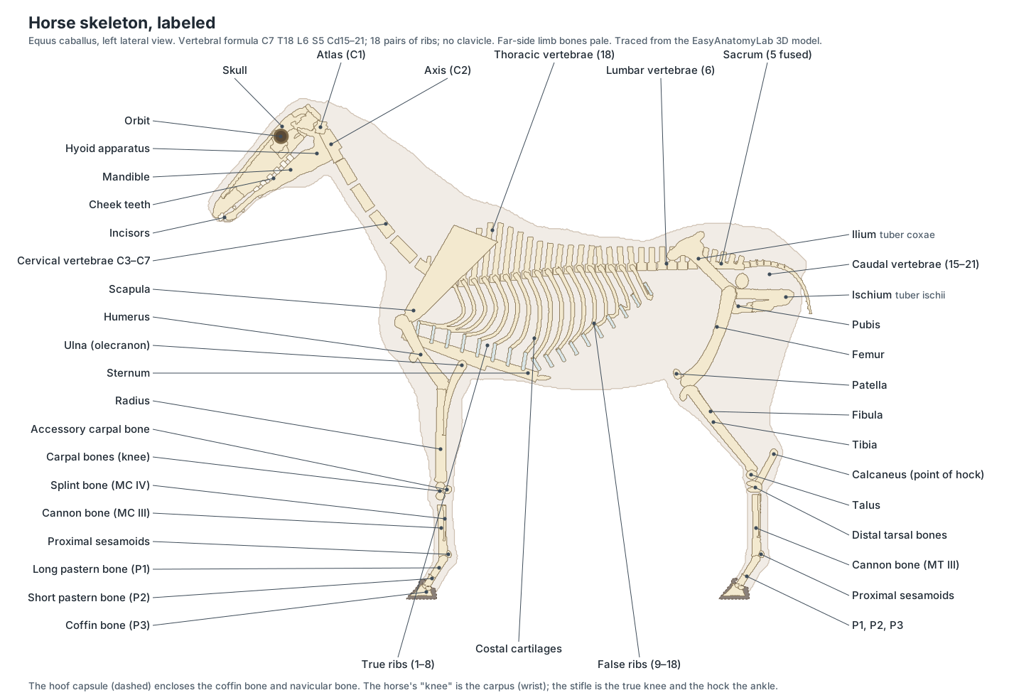
A horse skeleton has about 205 bones. It is divided into the axial skeleton (skull, hyoid, vertebral column, ribs and sternum) and the appendicular skeleton (the four limbs and the pelvis). Its most distinctive features are a very long skull with large grinding teeth, a stiff back with tall spines at the withers, no collarbone, and legs that end in a single toe.
| Feature | Horse | Human, for comparison |
|---|---|---|
| Total bones | About 205 (varies with tail length) | About 206 |
| Vertebrae | C7 T18 L6 S5 Cd15–21 (about 51–57) | C7 T12 L5 S5 Co4 |
| Ribs | 18 pairs (8 true, 10 false) | 12 pairs |
| Clavicle | None | Present |
| Digits per limb | One (digit III), plus splint bones from II and IV | Five |
| Stands on | The tip of the third digit (coffin bone) | The whole sole of the foot |
Horse skeleton labeled diagram
The diagram below names every major bone of the horse from the left side. The same bones can be clicked in the 3D model above, where you can also turn the skeleton to see the pelvis from above or the rib cage from below.
Horse skull anatomy and teeth
The horse skull is long because it must hold 24 large cheek teeth and long nasal passages; the braincase is small and lies behind the eyes. The face is made mainly of the incisive bones, nasal bones and maxillae, and the braincase of the frontal, parietal, temporal and occipital bones. A complete bony ring surrounds each orbit because the supraorbital process of the frontal bone reaches the zygomatic arch.
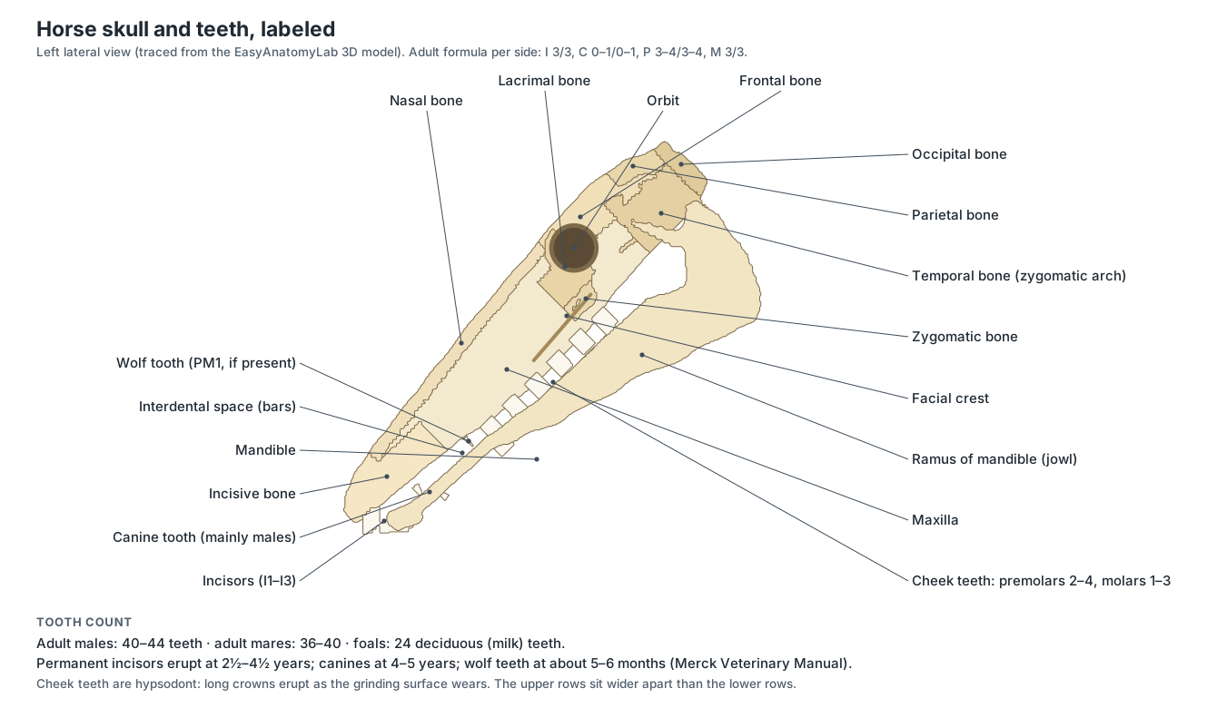
- Incisive bone: The paired front bones of the upper jaw; each carries three upper incisors, six in all. Holds the upper incisor teeth and frames the nasal opening (nasoincisive notch).
- Nasal bones: Two long, narrow bones forming the roof of the nasal cavity along the bridge of the nose. Roof the long nasal passages that warm and filter inhaled air.
- Maxilla: The large upper jaw bone on each side of the face, carrying the upper cheek teeth. Holds the six upper cheek teeth and encloses the maxillary sinuses; its facial crest anchors the masseter.
- Lacrimal bone: A small bone forming the front rim of the orbit. Houses the start of the nasolacrimal duct that drains tears to the nostril.
- Zygomatic bone: The bone below the orbit that forms the front of the zygomatic arch. Part of the cheek arch that braces the skull against chewing forces.
- Frontal bones: The paired bones of the forehead between and behind the eyes. Roof the front of the braincase; the supraorbital process forms a complete bony ring behind the eye.
- Parietal bones: Paired bones forming the roof of the braincase behind the frontal bones. Protect the cerebral hemispheres; the temporalis muscle attaches along the temporal lines.
- Temporal bones: The side walls of the braincase, which carry the jaw joint and the ear. Form the temporomandibular joint with the mandible and contain the middle and inner ear.
- Occipital bone: The bone at the back of the skull, around the foramen magnum. Its condyles articulate with the atlas; the nuchal crest anchors the nuchal ligament and neck muscles.
- Mandible: The lower jaw: two long halves joined at the chin, each with a tall ramus behind. Holds the lower incisors and cheek teeth; moved side to side by the masseter and pterygoid muscles to grind forage.
- Hyoid apparatus: A chain of small bones that suspends the tongue and larynx from the skull. Anchors the tongue, pharynx and larynx.
Horse teeth
- Incisors: Six upper and six lower chisel-shaped front teeth, arranged in an arc: central, intermediate and corner. Crop grass close to the ground.
- Canine teeth: Small pointed teeth in the gap between incisors and cheek teeth, usually present only in males. Used for fighting in stallions; no role in eating.
- Wolf teeth: Small, often missing teeth just in front of the upper cheek teeth. No function; they are often removed because they can interfere with the bit.
- Cheek teeth: Six large, square grinding teeth in each row, with long (hypsodont) crowns that keep erupting through life. Grind fibrous forage between their ridged enamel tables.
Horse spine: the vertebral column
The horse's spine usually has 7 cervical, 18 thoracic, 6 lumbar, 5 fused sacral and 15–21 caudal vertebrae. The neck vertebrae lie low in the neck in an S-shaped curve; the thoracic spines are tallest at the withers; the lumbar vertebrae have long, flat transverse processes; and the sacrum joins the pelvis at the sacroiliac joints.
| Region | Usual count | Key features |
|---|---|---|
| Cervical (C) | 7 | Atlas without a body; axis is the longest vertebra; C7 has no transverse foramen |
| Thoracic (T) | 18 (17–19) | Each carries a pair of ribs; spines longest at the withers, about T3–T6 |
| Lumbar (L) | 6 (sometimes 5 or 7) | Long flat transverse processes; the last ones often fuse with each other and the sacrum |
| Sacral (S) | 5, fused | Fusion usually complete by 4–5 years; forms the sacrum |
| Caudal (Cd) | 15–21 | Shrink to simple cylinders towards the tip of the tail |
- Atlas (C1): The first neck vertebra: a ring with broad wings and no body. Carries the head; the atlanto-occipital joint lets the horse nod.
- Axis (C2): The second neck vertebra, the longest vertebra in the body, with a blade-like spinous process. Its dens forms a pivot with the atlas so the head can rotate.
- Cervical vertebrae C3–C7: The remaining five neck vertebrae; long bodies that run low in the neck in an S-shaped curve. Support and move the neck; their transverse foramina carry the vertebral artery.
- Thoracic vertebrae (T1–T18): Eighteen vertebrae that carry the ribs; the tall spinous processes of the cranial ones form the withers. Anchor the ribs and the back muscles and support the rider.
- Lumbar vertebrae (L1–L6): Usually six vertebrae with long, flat transverse processes, in the loin. Form a stiff bridge that carries hindlimb thrust forward.
- Sacrum: Five sacral vertebrae fused into one triangular bone under the croup. Transmits hindlimb thrust from the pelvis to the spine through the sacroiliac joints.
- Caudal vertebrae: The vertebrae of the dock, usually 15–21, getting smaller and simpler towards the tip. Support and move the tail.
The spine is supported by strong ligaments: the nuchal ligament holds up the head and neck from the poll to the withers, and continues as the supraspinous ligament along the tips of the thoracic and lumbar spines.
Rib cage and sternum
A horse has 18 pairs of ribs. The first 8 pairs are true ribs whose costal cartilages join the sternum; the last 10 pairs are false ribs whose cartilages join the one in front to form the costal arch. The rib cage is narrow between the forelimbs and widens behind to make room for the large gut.
- True ribs (sternal ribs 1–8): The first eight pairs of ribs, whose costal cartilages join the sternum directly. Form a stiff front to the chest that protects the heart and carries the forelimb muscles.
- False ribs (asternal ribs 9–18): The last ten pairs of ribs, whose cartilages join the one in front to form the costal arch. Enclose the caudal lungs, liver, stomach and spleen; their movement aids breathing.
- Costal cartilages: The bars of cartilage that continue each rib to the sternum or to the costal arch. Give the rib cage flexibility for breathing.
- Sternum: A canoe-shaped breastbone of seven or eight segments (sternebrae) joined by cartilage, with a keel-like cartilage at the front. Anchors the true ribs and the pectoral muscles.
Horse front leg skeleton (thoracic limb)
The front legs carry more of the horse's weight than the hind legs at rest, yet have no bony joint with the trunk. From top to bottom it contains the scapula, humerus, radius and ulna, seven or eight carpal bones, the cannon bone (MC III) with two splint bones, two proximal sesamoids, the long and short pastern bones, the coffin bone and the navicular bone.
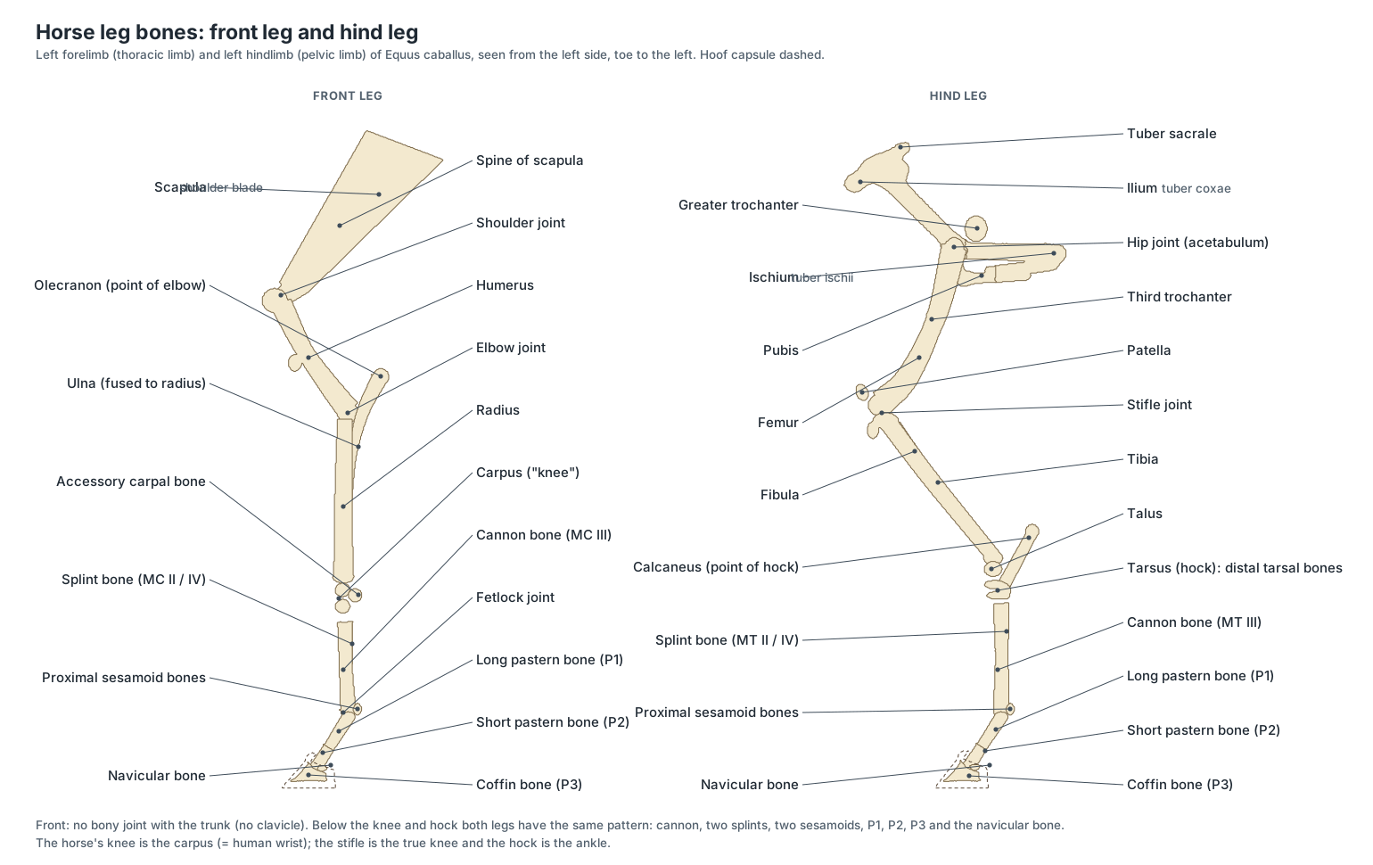
- Scapula: The long, triangular shoulder blade with a spine but no acromion, topped by a wide scapular cartilage. Attached to the trunk only by muscles: horses have no clavicle (collarbone).
- Humerus: The short, stout arm bone between the shoulder joint and the elbow. Lever for the shoulder and elbow muscles.
- Radius: The main weight-bearing bone of the forearm. Carries body weight from the elbow to the carpus.
- Ulna: A reduced bone fused to the back of the radius; only its top, the olecranon, is large. The olecranon is the lever arm for the triceps brachii, which extends the elbow.
- Carpal bones: Seven or eight small bones in two rows: radial, intermediate, ulnar and accessory above; (first), second, third and fourth below. Form the knee joint, which flexes the foreleg and locks straight under load.
- Accessory carpal bone: A disc-shaped bone projecting from the back of the knee. A lever for the flexor muscles of the carpus; it forms the wall of the carpal canal.
Horse hind leg skeleton (pelvic limb)
The hind leg is joined to the spine through the pelvis, so it can push the horse forward. It contains the hip bone (ilium, ischium and pubis), femur, patella, tibia and fibula, six tarsal bones in the hock, and the same cannon, splints, sesamoids and phalanges as the front leg.
- Ilium: The largest part of the pelvis: a wide wing with the point of the hip and the point of the croup. Joins the sacrum at the sacroiliac joint and anchors the gluteal and abdominal muscles.
- Ischium: The caudal part of the pelvis, ending in the point of the buttock. Origin of the hamstring muscles; forms the back of the pelvic floor.
- Pubis: The smallest part of the pelvis, forming the front of the pelvic floor. Anchors the prepubic tendon, which carries the weight of the abdominal muscles and viscera.
- Femur: The heavy thigh bone between the hip and the stifle, with a large third trochanter on its side. Lever for the powerful gluteal and thigh muscles; its trochlea guides the patella.
- Patella: A sesamoid bone at the front of the stifle, held by three patellar ligaments. Increases the leverage of the quadriceps; with the medial patellar ligament it locks the stifle.
- Tibia: The main bone of the gaskin, between the stifle and the hock. Carries body weight; its tuberosity receives the patellar ligaments.
- Fibula: A thin, incomplete bone on the lateral side of the tibia. Gives attachment to muscles; it carries no significant weight.
- Talus: The hock bone with a deep, spiral trochlea that meets the tibia. Forms the hinge of the tibiotarsal joint.
- Calcaneus: The long hock bone that projects up and back to form the point of the hock. The lever arm on which the gastrocnemius and superficial digital flexor pull to straighten the hock.
- Distal tarsal bones: The flat bones of the lower hock, in two rows below the talus and calcaneus. Form the low-motion intertarsal and tarsometatarsal joints that absorb shock.
Two features of the hind leg skeleton belong to the stay apparatus: the femur's enlarged medial trochlear ridge, over which the patella locks, and the long lever of the calcaneus, on which the gastrocnemius and superficial digital flexor pull.
Horse leg skeleton below the knee and hock
Below the carpus and tarsus, front and hind legs have the same bones. The single large cannon bone carries all the weight. Along its back edges lie the two splint bones, the remains of the side toes of three-toed ancestors. At the fetlock, two proximal sesamoid bones form a pulley for the flexor tendons; then come the long pastern bone (P1), the short pastern bone (P2) and the coffin bone (P3), with the small navicular bone behind the coffin joint.
- Cannon bones: The single large bone between the knee or hock and the fetlock, the main bone of the horse's one remaining toe. Carries all the weight of the limb; its distal condyle forms the fetlock joint.
- Splint bones: Two slender bones lying along the back edges of each cannon bone and ending about two-thirds of the way down. Help support the carpus and tarsus; they are the remains of digits II and IV of three-toed ancestors.
- Proximal sesamoid bones: A pair of small pyramid-shaped bones behind each fetlock joint. Form a groove for the flexor tendons and receive the suspensory ligament.
- Long pastern bone (P1): The first bone of the digit, between the fetlock and the pastern joint. Transfers load from the fetlock to the short pastern bone.
- Short pastern bone (P2): A short cube of bone between the pastern joint and the coffin joint, half inside the hoof. The superficial digital flexor tendon inserts on it.
- Coffin bone (P3): The crescent-shaped last bone of the digit, entirely inside the hoof and shaped like a small hoof. Suspended from the wall by the laminae; the common digital extensor and deep digital flexor tendons insert on it.
- Navicular bone: A small boat-shaped bone behind the coffin joint, over which the deep digital flexor tendon turns. Acts as a pulley that keeps the deep digital flexor tendon at a constant angle of insertion.
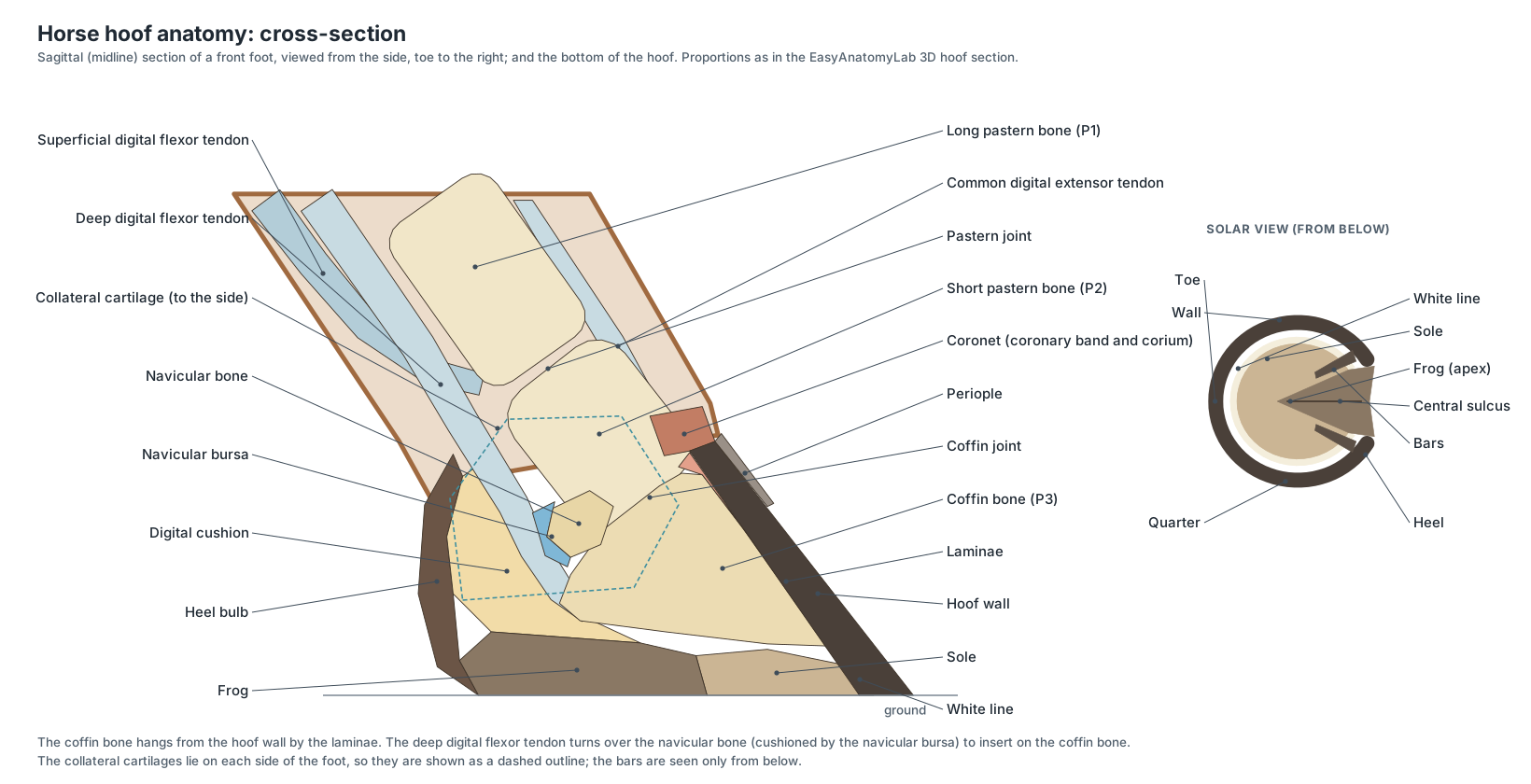
In the 3D model, turn on Hoof section (enlarged) to see these bones in a hoof cut down the middle. Inside the hoof, the coffin bone is suspended from the hoof wall by the laminae, and its dorsal surface lies roughly parallel to the front of the wall. For the soft tissues of the foot, the frog, sole and digital cushion, see the horse hoof anatomy section of the anatomy page.
Horse skeleton vs human skeleton
Horses and humans have the same bones in the same order, but the proportions are very different. The horse's hand and foot bones are long and fused into a single digit, so its "knee" is our wrist and its hock is our ankle; its upper arm and thigh are short and buried in muscle against the body.
| Human bone | Horse equivalent | Difference |
|---|---|---|
| Clavicle | None | The forelimb is attached by muscles only |
| Radius and ulna | Radius with a reduced ulna fused to it | No forearm rotation (pronation and supination) |
| Wrist (carpus) | Knee (carpus), 7–8 bones | Locks straight under load |
| Metacarpals I–V | Cannon bone (III) and two splint bones (II, IV) | Digits I and V absent |
| Finger phalanges | Long pastern, short pastern and coffin bone | One digit; the coffin bone sits in the hoof |
| Kneecap | Patella with three ligaments | Can lock the stifle |
| Fibula | Thin, incomplete fibula | Carries no weight |
| Ankle (tarsus), 7 bones | Hock, 6 bones | Moves almost only at one joint |
Every bone in the model
The full list of bones, bone groups and teeth in the 3D model, with what each one does.
Skeleton
- Incisive bone (Premaxilla)
- The paired front bones of the upper jaw; each carries three upper incisors, six in all. Holds the upper incisor teeth and frames the nasal opening (nasoincisive notch).
- Nasal bones
- Two long, narrow bones forming the roof of the nasal cavity along the bridge of the nose. Roof the long nasal passages that warm and filter inhaled air.
- Maxilla
- The large upper jaw bone on each side of the face, carrying the upper cheek teeth. Holds the six upper cheek teeth and encloses the maxillary sinuses; its facial crest anchors the masseter.
- Lacrimal bone
- A small bone forming the front rim of the orbit. Houses the start of the nasolacrimal duct that drains tears to the nostril.
- Zygomatic bone (Malar bone)
- The bone below the orbit that forms the front of the zygomatic arch. Part of the cheek arch that braces the skull against chewing forces.
- Frontal bones
- The paired bones of the forehead between and behind the eyes. Roof the front of the braincase; the supraorbital process forms a complete bony ring behind the eye.
- Parietal bones
- Paired bones forming the roof of the braincase behind the frontal bones. Protect the cerebral hemispheres; the temporalis muscle attaches along the temporal lines.
- Temporal bones
- The side walls of the braincase, which carry the jaw joint and the ear. Form the temporomandibular joint with the mandible and contain the middle and inner ear.
- Occipital bone
- The bone at the back of the skull, around the foramen magnum. Its condyles articulate with the atlas; the nuchal crest anchors the nuchal ligament and neck muscles.
- Mandible (Lower jaw)
- The lower jaw: two long halves joined at the chin, each with a tall ramus behind. Holds the lower incisors and cheek teeth; moved side to side by the masseter and pterygoid muscles to grind forage.
- Hyoid apparatus
- A chain of small bones that suspends the tongue and larynx from the skull. Anchors the tongue, pharynx and larynx.
- Atlas (C1)
- The first neck vertebra: a ring with broad wings and no body. Carries the head; the atlanto-occipital joint lets the horse nod.
- Axis (C2)
- The second neck vertebra, the longest vertebra in the body, with a blade-like spinous process. Its dens forms a pivot with the atlas so the head can rotate.
- Cervical vertebrae C3–C7
- The remaining five neck vertebrae; long bodies that run low in the neck in an S-shaped curve. Support and move the neck; their transverse foramina carry the vertebral artery.
- Thoracic vertebrae (T1–T18)
- Eighteen vertebrae that carry the ribs; the tall spinous processes of the cranial ones form the withers. Anchor the ribs and the back muscles and support the rider.
- Lumbar vertebrae (L1–L6)
- Usually six vertebrae with long, flat transverse processes, in the loin. Form a stiff bridge that carries hindlimb thrust forward.
- Sacrum
- Five sacral vertebrae fused into one triangular bone under the croup. Transmits hindlimb thrust from the pelvis to the spine through the sacroiliac joints.
- Caudal vertebrae (Coccygeal vertebrae, tail bones)
- The vertebrae of the dock, usually 15–21, getting smaller and simpler towards the tip. Support and move the tail.
- True ribs (sternal ribs 1–8)
- The first eight pairs of ribs, whose costal cartilages join the sternum directly. Form a stiff front to the chest that protects the heart and carries the forelimb muscles.
- False ribs (asternal ribs 9–18)
- The last ten pairs of ribs, whose cartilages join the one in front to form the costal arch. Enclose the caudal lungs, liver, stomach and spleen; their movement aids breathing.
- Costal cartilages
- The bars of cartilage that continue each rib to the sternum or to the costal arch. Give the rib cage flexibility for breathing.
- Sternum (Breastbone)
- A canoe-shaped breastbone of seven or eight segments (sternebrae) joined by cartilage, with a keel-like cartilage at the front. Anchors the true ribs and the pectoral muscles.
- Scapula (Shoulder blade)
- The long, triangular shoulder blade with a spine but no acromion, topped by a wide scapular cartilage. Attached to the trunk only by muscles: horses have no clavicle (collarbone).
- Humerus
- The short, stout arm bone between the shoulder joint and the elbow. Lever for the shoulder and elbow muscles.
- Radius
- The main weight-bearing bone of the forearm. Carries body weight from the elbow to the carpus.
- Ulna
- A reduced bone fused to the back of the radius; only its top, the olecranon, is large. The olecranon is the lever arm for the triceps brachii, which extends the elbow.
- Carpal bones (Carpus, 'knee')
- Seven or eight small bones in two rows: radial, intermediate, ulnar and accessory above; (first), second, third and fourth below. Form the knee joint, which flexes the foreleg and locks straight under load.
- Accessory carpal bone
- A disc-shaped bone projecting from the back of the knee. A lever for the flexor muscles of the carpus; it forms the wall of the carpal canal.
- Cannon bones (Third metacarpal (MC III) and third metatarsal (MT III))
- The single large bone between the knee or hock and the fetlock, the main bone of the horse's one remaining toe. Carries all the weight of the limb; its distal condyle forms the fetlock joint.
- Splint bones (MC/MT II (medial) and MC/MT IV (lateral))
- Two slender bones lying along the back edges of each cannon bone and ending about two-thirds of the way down. Help support the carpus and tarsus; they are the remains of digits II and IV of three-toed ancestors.
- Proximal sesamoid bones
- A pair of small pyramid-shaped bones behind each fetlock joint. Form a groove for the flexor tendons and receive the suspensory ligament.
- Long pastern bone (P1) (Proximal phalanx, first phalanx)
- The first bone of the digit, between the fetlock and the pastern joint. Transfers load from the fetlock to the short pastern bone.
- Short pastern bone (P2) (Middle phalanx, second phalanx)
- A short cube of bone between the pastern joint and the coffin joint, half inside the hoof. The superficial digital flexor tendon inserts on it.
- Coffin bone (P3) (Pedal bone, distal phalanx, third phalanx)
- The crescent-shaped last bone of the digit, entirely inside the hoof and shaped like a small hoof. Suspended from the wall by the laminae; the common digital extensor and deep digital flexor tendons insert on it.
- Navicular bone (Distal sesamoid bone)
- A small boat-shaped bone behind the coffin joint, over which the deep digital flexor tendon turns. Acts as a pulley that keeps the deep digital flexor tendon at a constant angle of insertion.
- Ilium
- The largest part of the pelvis: a wide wing with the point of the hip and the point of the croup. Joins the sacrum at the sacroiliac joint and anchors the gluteal and abdominal muscles.
- Ischium
- The caudal part of the pelvis, ending in the point of the buttock. Origin of the hamstring muscles; forms the back of the pelvic floor.
- Pubis
- The smallest part of the pelvis, forming the front of the pelvic floor. Anchors the prepubic tendon, which carries the weight of the abdominal muscles and viscera.
- Femur (Thigh bone)
- The heavy thigh bone between the hip and the stifle, with a large third trochanter on its side. Lever for the powerful gluteal and thigh muscles; its trochlea guides the patella.
- Patella (Kneecap)
- A sesamoid bone at the front of the stifle, held by three patellar ligaments. Increases the leverage of the quadriceps; with the medial patellar ligament it locks the stifle.
- Tibia
- The main bone of the gaskin, between the stifle and the hock. Carries body weight; its tuberosity receives the patellar ligaments.
- Fibula
- A thin, incomplete bone on the lateral side of the tibia. Gives attachment to muscles; it carries no significant weight.
- Talus (Tibial tarsal bone)
- The hock bone with a deep, spiral trochlea that meets the tibia. Forms the hinge of the tibiotarsal joint.
- Calcaneus (Fibular tarsal bone, point of the hock)
- The long hock bone that projects up and back to form the point of the hock. The lever arm on which the gastrocnemius and superficial digital flexor pull to straighten the hock.
- Distal tarsal bones (Central, third, fourth and fused first-second tarsal bones)
- The flat bones of the lower hock, in two rows below the talus and calcaneus. Form the low-motion intertarsal and tarsometatarsal joints that absorb shock.
Teeth
- Incisors
- Six upper and six lower chisel-shaped front teeth, arranged in an arc: central, intermediate and corner. Crop grass close to the ground.
- Canine teeth (Tushes) · males only
- Small pointed teeth in the gap between incisors and cheek teeth, usually present only in males. Used for fighting in stallions; no role in eating.
- Wolf teeth (First premolars)
- Small, often missing teeth just in front of the upper cheek teeth. No function; they are often removed because they can interfere with the bit.
- Cheek teeth (Premolars 2–4 and molars 1–3)
- Six large, square grinding teeth in each row, with long (hypsodont) crowns that keep erupting through life. Grind fibrous forage between their ridged enamel tables.
How variable is the horse's vertebral formula?
Textbooks give the horse's vertebral formula as C7, T18, L6, S5 and 15–21 caudal vertebrae, with the five sacral vertebrae usually fully fused by 4–5 years of age. Minor variations are well recognised. (Withers (2016), sacral and coccygeal vertebral lesions, BEVA Congress (IVIS).)
How common they are was measured by CT in 77 cadavers: the 'standard' 18 thoracic, 6 lumbar and 5 sacral vertebrae were found in 78% of semi-feral Konik horses, but only 53% of Warmbloods and 38% of Shetland ponies. Most specimens had 18 pairs of ribs, but some had 17 or 19, and one Konik and one Warmblood had 19 thoracic, 5 lumbar and 6 sacral vertebrae. Joints or fusion between the transverse processes of the last two lumbar vertebrae were common, especially in Shetland ponies (61%). In 18% of the specimens the total number of thoracic, lumbar and sacral vertebrae was 28 or 30 instead of 29. (Spoormakers et al. (2021), breed differences in the equine vertebral column, J Anat 239: 829–838.)
So when a label says '18 thoracic vertebrae', read it as 'usually 18'. The popular claim that every Arabian has 17 thoracic and 5 lumbar vertebrae is not supported by a breed-wide study; variation of that kind occurs in many breeds.
A forelimb without a collarbone
The horse's forelimb has no bony attachment to the trunk, in sharp contrast with the hindlimb, which is joined to the spine through the pelvis and sacroiliac joint. The forelimb is held on entirely by extrinsic muscles. The serratus ventralis is the main weight-bearing muscle: if the forelimbs are posts, it is a sling that suspends the trunk between their tops. The pectorals stop the legs from splaying, the deep pectoral pulls the leg back in the power stroke, and the brachiocephalicus pulls it forward. (University of Minnesota: Lab 1, forelimb osteology and extrinsic muscles.)
This arrangement makes the shoulder mobile, lengthening the stride, and lets the muscles absorb shock as the forefeet land. Because the equine scapula has no acromion, there is no bony prominence for the omotransversarius muscle, which instead merges with the brachiocephalicus. (University of Minnesota: Lab 1, forelimb osteology and extrinsic muscles.)
How the bones and ligaments let a horse stand all day
In the forelimb, the lacertus fibrosus links the biceps tendon to the extensor carpi radialis, creating a fibrous cable from the scapula to the cannon bone that supports the shoulder, elbow and carpus without muscle work. Below, the suspensory ligament (anatomically the interosseous tendon) and the distal sesamoidean ligaments form the suspensory apparatus that keeps the fetlock from sinking; the digital flexor tendons, anchored by their check ligaments, add passive support. (University of Minnesota: Lab 3, distal forelimb and suspensory apparatus.)
In the hindlimb, a ligamentous loop formed by the patella and its medial and middle ligaments hooks over the enlarged medial trochlear ridge of the femur and locks the stifle. The reciprocal apparatus then locks the hock as well: the fibularis tertius in front and the superficial digital flexor tendon behind force the hock to move with the stifle. (University of Minnesota: Lab 6, stifle and reciprocal apparatus.)
If the patellar loop stays hooked when the horse tries to move, the hind leg is held stiffly extended with the fetlock flexed until it releases with a jerk. This 'locked stifle' is most common in young, poorly muscled horses and ponies and in horses with straight hindlimbs. (Merck Veterinary Manual: Upward Fixation of the Patella in Horses (Durgam).)
Reading a horse's age from its teeth
Most horses have 24 deciduous teeth. Adult stallions have 40–44 teeth and mares 36–40, because canines appear at about 4–5 years mainly in males. Permanent incisors erupt between 2½ and 4½ years, the premolars between 2½ and 4 years, and the permanent molars at about 1, 2 and 4 years. Wolf teeth, when present, erupt at about 5–6 months, usually in the upper jaw. (Merck Veterinary Manual: Dental Development of Horses (Easley).)
Equine teeth are hypsodont: their long crowns erupt continuously, and in a horse eating forage the rate of eruption is matched by wear. As an incisor wears, the cup of its infundibulum disappears first and the deeper enamel spot remains for some years, followed by the dental star. These changes, together with the shape of the tables, are the basis of age estimation, which becomes less precise in older horses. (University of Minnesota: Lab 18, skull and teeth.)
Horse skeleton quiz
Use the labeled diagram or the 3D model to answer, then click a question to check.
How many cervical vertebrae does a horse have?
Seven, like almost all mammals.
What is the usual vertebral formula of the horse?
C7, T18, L6, S5 and Cd15–21.
How many pairs of ribs, and how many are true ribs?
18 pairs; the first 8 pairs are true (sternal) ribs.
Which bone forms the point of the hock?
The calcaneus (its calcanean tuber).
Which bone forms the point of the elbow?
The olecranon of the ulna.
Which bones are the remains of the side toes?
The splint bones: metacarpal or metatarsal II and IV.
What is another name for the navicular bone?
The distal sesamoid bone.
Which three bones fuse to form each half of the pelvis?
The ilium, ischium and pubis, meeting in the acetabulum.
Why does a horse have no collarbone, and what holds the forelimb on?
The forelimb has no bony joint with the trunk; muscles such as the serratus ventralis and pectorals sling the trunk between the limbs.
What is unusual about the femur of a horse?
It has a third trochanter, and an enlarged medial trochlear ridge over which the patella can lock.
For the muscles, organs and hoof soft tissues of the same horse, see the horse anatomy page.
Frequently asked questions
How many bones does a horse have?
About 205 in a typical adult horse. The exact number varies, mainly with the number of caudal (tail) vertebrae (15–21) and with occasional extra or missing thoracic and lumbar vertebrae.
Do horses have a collarbone?
No. Horses have no clavicle. The forelimb has no bony joint with the trunk at all; it is attached only by muscles such as the serratus ventralis and the pectorals, which sling the chest between the shoulder blades.
How many ribs does a horse have?
Usually 18 pairs: 8 pairs of true (sternal) ribs that join the sternum and 10 pairs of false (asternal) ribs whose cartilages form the costal arch. Some horses have 17 or 19 pairs, together with a matching change in the number of thoracic vertebrae.
How many vertebrae does a horse have?
The typical vertebral formula is C7, T18, L6, S5, Cd15–21: 7 cervical, 18 thoracic, 6 lumbar, 5 fused sacral and 15–21 caudal vertebrae, about 51–57 in all. A CT study found exactly 18 thoracic, 6 lumbar and 5 sacral vertebrae in only 78% of Konik horses, 53% of Warmbloods and 38% of Shetland ponies.
Do Arabian horses have fewer vertebrae?
It is often said that Arabians have 17 thoracic vertebrae (and ribs) and 5 lumbar vertebrae. Counts like this do occur in horses, but variation is common in many breeds, and we found no modern study showing a fixed breed-wide count in Arabians. Many Arabians have the usual 18 and 6.
Is a horse's knee really a knee?
No. The joint called the knee on a horse's front leg is the carpus, equivalent to the human wrist. The true knee is the stifle on the hind leg, and the hock is the equivalent of the human ankle. The horse stands on the tip of its single third digit, the coffin bone inside the hoof.
What is the coffin bone?
The coffin bone (pedal bone, distal or third phalanx, P3) is the last bone of the leg, inside the hoof. It is shaped like a small hoof and is suspended from the inside of the hoof wall by the laminae. Behind it sits the small navicular bone (distal sesamoid).
What are splint bones?
Splint bones are the slender second and fourth metacarpal or metatarsal bones on either side of each cannon bone. They are the remains of the side toes (digits II and IV) of the horse's three-toed ancestors and end about two-thirds of the way down the cannon.
References
- Cox, V., Clarkson, T., Brown, A. & Fletcher, T. (2013). Ungulate Anatomy Dissection Labs: Labs 1, 3, 5–9 and 18 (osteology). University of Minnesota College of Veterinary Medicine.
- Withers, J. M. (2016). Diagnosis and treatment of sacral and coccygeal vertebral lesions. BEVA Congress, via IVIS.
- Spoormakers, T. J. P. et al. (2021). A comparative study of breed differences in the anatomical configuration of the equine vertebral column. Journal of Anatomy 239, 829–838.
- University of Minnesota: Lab 1, forelimb osteology and extrinsic forelimb muscles.
- University of Minnesota: Lab 3, distal forelimb osteology and suspensory apparatus.
- University of Minnesota: Lab 7, tarsal and metatarsal osteology.
- University of Minnesota: Lab 18, skull, mandible and teeth.
- Easley, J. Dental development of horses. Merck Veterinary Manual.
- Easley, J. Overview of general dentistry in horses. Merck Veterinary Manual.
- Durgam, S. S. Intermittent upward fixation of the patella in horses. Merck Veterinary Manual.
- Vincelette, A. R. et al. (2023). Hipparion tracks and horses' toes: the evolution of the equid single hoof. Royal Society Open Science 10, 230358.
- Hepworth, K., Neary, M. & Kenyon, S. Hoof anatomy, care and management in livestock (ID-321). Purdue Extension.
More interactive anatomy models
- Horse anatomyEquine anatomy: hoof, legs, stay apparatus, teeth and the long hindgut.
- Human anatomy1,000+ bones, muscles, organs, vessels and nerves from real anatomical data.
- Octopus anatomyThree hearts, a ring-shaped brain, eight arms and a beak, in 3D.
- Squid anatomyPen, eight arms, two tentacles, giant axons and three hearts of the longfin squid.
- Squid dissectionStep-by-step guided squid dissection with every structure labeled.
- Fish anatomyExternal and internal anatomy of a bony fish: fins, gills, swim bladder, heart and brain.