

Frog anatomy at a glance
A frog is an amphibian with a short, tailless body, long hind legs, moist skin and big eyes on top of its head. Its body has two regions, the head and the trunk; frogs have no neck and, as adults, no tail. Inside, it has the same organ systems as a human, a brain, heart, lungs, liver, stomach, intestines, kidneys and gonads, but with a three-chambered heart, no ribs and no diaphragm, and a single opening, the cloaca, for waste and reproduction.
This page uses the northern leopard frog, Lithobates pipiens (formerly Rana pipiens), as the reference frog. It is a medium-sized frog of ponds and meadows across much of northern North America, and the standard frog in North American biology labs. The other common lab frog, the American bullfrog (Lithobates catesbeianus), has the same internal anatomy; the differences are listed further down.
Directions: anterior means toward the snout, posterior toward the cloaca, dorsal the back and ventral the belly. Left and right always mean the frog's own left and right, so when a frog lies on its back in a dissecting tray, its left side is on your right.
Frog external anatomy: labeled diagram
The external anatomy of a frog includes the snout with two nostrils (external nares), two large eyes with eyelids and a nictitating membrane, a round eardrum (tympanum) behind each eye, a wide mouth, smooth moist skin, two short forelimbs with four fingers, two long hind limbs with five webbed toes, and the cloacal opening at the back.
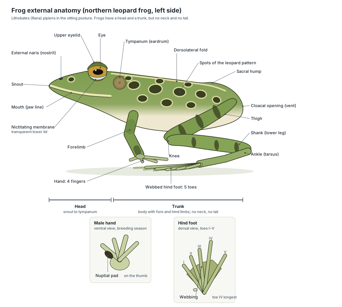
Three features identify a leopard frog: rows of rounded dark spots with pale borders, a pale dorsolateral fold running from behind each eye to the hip, and a pale stripe along the upper lip. The sharp hump in the middle of the back of a sitting frog is not a vertebra sticking up; it is the joint between the hip bones (ilia) and the sacral vertebra.
| External part | Where | What it does |
|---|---|---|
| External nares | Top of the snout | Let air in for breathing and smell; close during each lung-filling breath |
| Eye and eyelids | Top of the head | Vision; the thin lower lid and nictitating membrane protect the eye |
| Tympanum | Behind the eye | Eardrum; passes sound to the middle and inner ear |
| Forelimb and hand | Behind the head | Props up the body and absorbs the landing; 4 fingers |
| Hind limb and foot | Back of the body | Jumping and swimming; 5 webbed toes, toe IV longest |
| Cloacal opening | Back end of the body | Single exit for feces, urine and eggs or sperm |
| Skin | Whole body | Protection, water uptake and gas exchange |
Frog internal anatomy: labeled diagram
Inside a frog, the first thing you see is the large, three-lobed, reddish-brown liver, with the heart in front of it and the two spongy lungs on either side. Under the liver are the stomach on the frog's left, the coiled small intestine, the yellow fat bodies, and at the back the large intestine, urinary bladder and cloaca. The kidneys lie flat against the back wall, and the gonads sit on or beside them.

For a step-by-step guide to finding these organs in a real specimen, with a worksheet and answer key, see our frog dissection guide.
Frog anatomy labeled: every part and its function
This is the text version of the 3D model: every labeled structure of the leopard frog, grouped by body system, with what it does. Use it as a study list or to check a frog anatomy labeling worksheet.
External
- Skin
- Thin, moist skin with no scales, hair or claws. The leopard frog is green or brown with rows of dark, pale-edged spots. Protects the body, keeps it moist with mucus, and works as a second breathing surface: oxygen and carbon dioxide pass through it.
- Eye
- Large, bulging eyes on top of the head, with a golden iris and a horizontal pupil. Vision for spotting moving prey and predators. The eyes also help swallowing: the frog pulls them down into the mouth to push food toward the gullet.
- Nictitating membrane (Transparent lower eyelid, third eyelid)
- A thin, see-through fold that slides up over the eye from below. Protects and moistens the eye, on land and under water, while the frog can still see through it.
- Tympanum (Tympanic membrane, eardrum)
- A round, flat eardrum on the side of the head, just behind the eye. Picks up sound and passes vibrations through the middle-ear bone (columella) to the inner ear. The middle ear connects to the mouth by the Eustachian tube.
- External nares (Nostrils)
- Two small nostrils near the tip of the snout. Let air into the mouth for breathing and smelling. They open into the mouth through the internal nares, and close during each lung-filling breath.
- Dorsolateral folds (Dorsolateral ridges)
- A pale raised ridge of skin running down each side of the back. Glandular skin ridges; they are mainly useful to us for identifying the species.
- Forelimb (arm) (Brachium and antebrachium)
- The short front leg: upper arm and forearm. Props up the front of the body when sitting and absorbs the shock of landing after a jump.
- Hand (Manus, four fingers)
- A hand with four fingers and no webbing. Supports the body and helps push food into the mouth.
- Nuptial pad (Thumb pad) · males only
- A swollen, darkened pad on the inner side of the male's thumb. Gives the male a firm grip on the female's slippery body during amplexus, when he clasps her while she lays eggs.
- Vocal sacs · males only
- Paired balloons of thin skin that inflate when a male calls. Act as resonators that make the call louder and carry farther.
- Thigh (Femoral region)
- The upper hind leg, thick with jumping muscles. Holds the large muscles that straighten the hip and knee for jumping and swimming.
- Shank (Crus, lower leg)
- The lower leg, between knee and ankle. Holds the calf muscle (gastrocnemius) that extends the ankle for take-off.
- Webbed foot (Pes, five toes)
- A long foot with five toes joined by webbing. The webbing pushes against water like a paddle when the frog swims, and the long foot adds leverage when it jumps.
- Cloacal opening (Vent, anus)
- The single opening at the back of the body. Releases feces, urine, and eggs or sperm. All three empty into the cloaca first.
Mouth
- Buccal cavity (Mouth cavity, oral cavity, pharynx)
- The wide, flat mouth cavity. Its roof is the palate; its floor carries the tongue. Takes in food and also works as the frog's breathing pump: raising and lowering its floor moves air in and out of the lungs.
- Maxillary teeth
- A row of tiny, cone-shaped teeth around the edge of the upper jaw. Grip slippery prey so it cannot escape. Frogs do not chew; they swallow food whole.
- Vomerine teeth
- Two small patches of sharp teeth on the roof of the mouth. Hold struggling prey against the roof of the mouth while it is swallowed.
- Internal nares (Choanae, posterior nares)
- Two small openings in the roof of the mouth that connect to the nostrils. Carry air between the nostrils and the mouth cavity for breathing.
- Eustachian tube openings (Auditory tubes)
- Two large openings at the back corners of the roof of the mouth. Connect the middle ear with the mouth and equalise air pressure on both sides of the eardrum.
- Tongue
- A soft, sticky tongue attached at the front of the mouth, with a forked (bilobed) tip pointing back. Flips out over the lower jaw to catch insects and other small prey, then flips back to bring the prey into the mouth.
- Glottis
- A slit in the floor of the mouth that opens into the larynx and lungs. Opens to let air pass between the mouth and lungs, and closes to hold air in the lungs.
- Vocal sac openings (Vocal slits) · males only
- Small slits in the floor of the male's mouth that lead into the vocal sacs. Let air pass from the mouth into the vocal sacs when the frog calls.
Skeletal
- Cranium (braincase) (Sphenethmoid, prootics and exoccipitals)
- The narrow, box-like braincase in the middle of the skull. Protects the brain and inner ears. The sphenethmoid forms its front, the prootics hold the inner ears, and the exoccipitals form two condyles that join the first vertebra.
- Frontoparietals
- A pair of long, flat bones forming the roof of the braincase. Roof over and protect the brain.
- Nasals
- Two triangular bones over the nasal capsules. Cover and protect the olfactory (smell) organs.
- Upper jaw (Premaxillae, maxillae and quadratojugals)
- The curved bony rim of the upper jaw, from the snout tip to each jaw angle. Carries the maxillary teeth and forms the outer frame of the skull.
- Squamosal
- A T-shaped bone behind each eye. Braces the jaw joint against the braincase and supports the ring of the eardrum.
- Pterygoids
- Three-pronged bones in the roof of the mouth. Brace the upper jaw and jaw joint against the braincase.
- Parasphenoid
- A dagger-shaped bone forming the floor of the braincase. Floors and strengthens the braincase; it is visible through the thin lining of the palate.
- Vomers
- Small paired bones at the front of the palate. Carry the vomerine teeth.
- Lower jaw (Mandible: dentary, angulosplenial and mentomeckelian bones)
- A thin, toothless bony arc forming the lower lip. Opens and closes the mouth and frames the floor of the mouth for buccal pumping.
- Hyoid apparatus (Hyoid plate)
- A broad cartilage plate with bony and cartilage horns in the floor of the mouth. Supports the tongue and larynx and anchors the throat muscles that raise and lower the floor of the mouth during breathing.
- Presacral vertebrae (Vertebrae 1–8, including the atlas)
- Eight short vertebrae forming the backbone in front of the hips. There are no ribs. Protect the spinal cord and form a short, stiff column that transmits the push of the hind legs to the body.
- Sacral vertebra (Vertebra 9, sacrum)
- The ninth vertebra, with large transverse processes that join the hip bones. Connects the backbone to the pelvic girdle so jumping forces pass from the legs to the body.
- Urostyle (Coccyx)
- A long, rod-shaped bone behind the sacral vertebra, unique to frogs and toads. Stiffens the back of the body and helps transmit thrust from the hind limbs to the spine during jumping and swimming.
- Suprascapula
- A broad, partly cartilaginous plate at the top of the shoulder blade. Gives a wide area for back and shoulder muscles to attach.
- Pectoral girdle (Scapula, clavicle and coracoid)
- The shoulder girdle: a hoop of bones around the front of the body that carries the forelimbs. Anchors the forelimbs and absorbs landing forces. It also protects the heart.
- Sternum (Omosternum and xiphisternum)
- A small breastbone of bone and cartilage on the ventral midline. Anchors the chest (pectoralis) muscles.
- Humerus
- The upper-arm bone. Supports the upper arm and anchors the arm and chest muscles.
- Radio-ulna
- The single forearm bone, made of the radius and ulna fused together. Supports the forearm; a single bone is stronger for absorbing the shock of landing.
- Carpals (Wrist bones)
- A group of small wrist bones. Let the wrist bend.
- Metacarpals and phalanges (Hand bones)
- The long bones of the palm and the finger bones of the four fingers. Support the fingers.
- Ilium (Hip bone)
- A very long, rod-like hip bone on each side. Connects the hind limbs to the backbone and acts as a lever in jumping.
- Ischium and pubis (Pelvic disc, acetabulum)
- A small vertical disc of bone and cartilage at the back of the pelvis. Holds the two hip sockets that the heads of the femurs fit into.
- Femur (Thigh bone)
- The long, slightly S-shaped thigh bone. Supports the thigh and anchors the big jumping muscles.
- Tibiofibula (Os cruris)
- The single lower-leg bone, formed by fusion of the tibia and fibula. Supports the shank and anchors the calf and shin muscles.
- Astragalus and calcaneum (Tibiale and fibulare, elongated tarsals)
- Two long ankle bones lying side by side, joined at both ends. Give the hind leg an extra long segment, so the foot works as a long lever for jumping and swimming.
- Foot bones (Small tarsals, metatarsals, phalanges and prehallux)
- The bones of the five toes and the sole. Support the five toes and the webbing.
Muscular
- Mylohyoid (Submandibular, intermandibularis)
- A thin sheet of muscle under the floor of the mouth. Raises the floor of the mouth, pushing air into the lungs (buccal pumping) and food back toward the gullet.
- Pectoralis
- A fan-shaped chest muscle. Pulls the forelimb toward the body; used by males to hold females in amplexus.
- Deltoid
- A short muscle over the front of the shoulder. Moves the upper arm forward.
- Rectus abdominis (With the linea alba between its halves)
- A pair of long, flat belly muscles divided into segments. Supports the belly and the organs inside and helps bend the body.
- External oblique
- A thin muscle sheet on the side of the body. Compresses the body cavity; it helps squeeze air out of the lungs.
- Latissimus dorsi
- A triangular muscle behind the shoulder. Pulls the forelimb backward and upward.
- Longissimus dorsi
- A long back muscle running beside the backbone. Straightens and raises the back and head.
- Triceps femoris (Rectus anticus femoris, vastus internus and vastus externus)
- The large three-headed muscle on the front of the thigh. Straightens the knee: the main muscle that powers a jump.
- Sartorius
- A long, narrow strap crossing the underside of the thigh diagonally. Flexes the knee and pulls the thigh forward.
- Adductor magnus (With the adductor longus)
- A large muscle on the inner underside of the thigh, partly under the sartorius. Pulls the thigh toward the midline (adduction).
- Gracilis major and minor (Rectus internus major and minor)
- Two muscles along the back (flexor) edge of the underside of the thigh. Bend the knee and pull the thigh back and in.
- Semimembranosus
- A thick muscle on the back edge of the dorsal thigh. Bends the knee and extends the hip.
- Iliofibularis (Biceps femoris)
- A slender muscle in the middle of the dorsal thigh. Bends the knee.
- Gastrocnemius (Calf muscle)
- The large, spindle-shaped calf muscle. Extends the ankle (points the foot) for the final push of a jump; also bends the knee.
- Achilles tendon (Tendo Achillis, calcaneal tendon)
- The strong tendon at the end of the gastrocnemius. Transmits the pull of the calf muscle to the foot.
- Tibialis anticus longus
- A muscle on the front of the shank. Flexes the ankle and straightens the knee.
- Peroneus
- A muscle on the outer side of the shank. Extends the ankle and helps rotate the foot.
Nervous
- Olfactory lobes (Olfactory bulbs)
- The front-most part of the brain, partly fused at the midline. Process smell signals from the nose.
- Cerebral hemispheres (Cerebrum)
- Two long, smooth lobes behind the olfactory lobes. Process smell and control behaviour such as feeding and mating; much simpler than the folded human cerebrum.
- Diencephalon (Thalamencephalon, with pineal and pituitary)
- A small region between the cerebrum and the optic lobes. Relays signals and controls hormones. The pituitary controls growth and breeding; the pineal responds to light.
- Optic lobes (Optic tectum, midbrain)
- Two large, rounded lobes in the middle of the brain. Process vision. They control the fast strike at moving prey.
- Cerebellum
- A narrow ridge across the brain behind the optic lobes. Coordinates movement and balance.
- Medulla oblongata
- The tapering back of the brain that joins the spinal cord. Controls breathing, heart rate and other automatic functions. Most cranial nerves leave from here.
- Optic nerves
- The nerves from the eyes to the brain. Carry visual signals from the retina to the optic lobes.
- Spinal cord
- The nerve cord inside the vertebral column. Carries signals between the brain and body and controls reflexes such as the jump.
- Spinal nerves
- Ten pairs of white nerves leaving the spinal cord. Carry signals to and from the muscles and skin of the body and limbs.
- Brachial nerve (Brachial plexus)
- The large nerve to the forelimb. Controls the muscles of the forelimb.
- Sciatic plexus (Lumbosacral plexus)
- A network formed by spinal nerves 7, 8 and 9 on each side of the urostyle. Combines nerve fibres that then run into the hind limb as the sciatic nerve.
- Sciatic nerve
- The largest nerve in the body, running down the back of the thigh. Controls the muscles of the hind limb. Near the knee it splits into the tibial and peroneal nerves.
Digestive
- Esophagus (Gullet)
- A short, wide, stretchy tube from the back of the mouth to the stomach. Carries whole prey from the mouth to the stomach.
- Stomach
- A J-shaped, muscular sac on the frog's left side. Stores swallowed prey and starts digestion with acid and the enzyme pepsin.
- Pyloric sphincter (Pylorus)
- A ring of muscle where the stomach narrows into the small intestine. Controls the passage of food from the stomach into the duodenum.
- Duodenum
- The first part of the small intestine. Receives bile and pancreatic juice and continues digestion.
- Ileum (Small intestine)
- The long, coiled part of the small intestine. Finishes digestion and absorbs nutrients into the blood.
- Mesentery
- A thin, see-through membrane that holds the intestines in place. Suspends the gut and carries its blood vessels and nerves.
- Large intestine (Colon, rectum)
- A short, wide tube at the end of the gut. Absorbs water and stores feces.
- Cloaca
- A small chamber at the end of the gut shared by the digestive, urinary and reproductive systems. Collects feces, urine, and eggs or sperm before they leave the body.
- Liver (Right, left anterior and left posterior lobes)
- The largest organ in the body cavity: a reddish-brown organ with three lobes. Makes bile, stores glycogen and fat, and processes nutrients absorbed by the gut.
- Gallbladder
- A small, round, greenish sac. Stores bile from the liver and releases it into the duodenum through the bile duct.
- Pancreas
- A thin, whitish or pale-yellow, irregular gland. Makes digestive enzymes for the duodenum and the hormone insulin.
- Spleen
- A small, round, dark-red body. Part of the lymphatic and circulatory systems, not digestion: it filters blood, stores blood cells and makes white blood cells. It is listed here because it lies among the intestines.
Respiratory
- Larynx (Laryngotracheal chamber, with vocal cords)
- A short chamber that joins the glottis to the two lungs. Carries air to the lungs and holds the vocal cords that make the frog's call.
- Lungs
- Two thin-walled, elastic sacs with a honeycomb of folds inside. Exchange oxygen and carbon dioxide with the blood. They are filled by buccal pumping, not by ribs or a diaphragm.
Circulatory
- Sinus venosus
- A thin-walled sac on the dorsal side of the heart. Collects oxygen-poor blood from the three venae cavae and passes it to the right atrium. The heartbeat starts here.
- Right atrium (Right auricle)
- The larger of the two thin-walled upper chambers. Receives oxygen-poor blood from the body (through the sinus venosus) and pushes it into the ventricle.
- Left atrium (Left auricle)
- The smaller upper chamber on the frog's left. Receives oxygen-rich blood from the lungs through the pulmonary veins and passes it into the ventricle.
- Ventricle
- The single, thick-walled, cone-shaped lower chamber of the three-chambered heart. Pumps blood out through the conus arteriosus to the lungs, skin and body.
- Conus arteriosus (Truncus arteriosus, with spiral valve)
- A muscular tube leaving the front of the ventricle, which divides into left and right trunks. Carries blood from the ventricle to the arterial arches. A spiral valve inside helps steer oxygen-poor blood toward the lungs and skin and oxygen-rich blood toward the body.
- Carotid arches
- The first pair of arterial arches, running forward. Carry blood to the head and brain.
- Systemic arches (Aortic arches)
- The middle pair of arches, which curve around the esophagus. Join to form the dorsal aorta and supply the body and forelimbs.
- Pulmocutaneous arches
- The last pair of arches, each splitting into a pulmonary and a cutaneous artery. Carry oxygen-poor blood to the lungs and to the skin, the frog's two breathing surfaces.
- Dorsal aorta
- The main artery of the body, under the backbone. Carries blood to the organs and hind limbs.
- Anterior venae cavae (Precaval veins)
- Two veins that return blood from the head and forelimbs. Bring oxygen-poor blood, and oxygen-rich blood from the skin, back to the heart.
- Posterior vena cava (Postcaval vein)
- A large vein returning blood from the kidneys, gonads and liver. Returns blood from the back of the body to the heart.
- Pulmonary veins
- Small veins from the lungs. Return oxygen-rich blood from the lungs to the heart.
- Abdominal vein (Ventral abdominal vein)
- A large vein running along the inside of the belly wall. Carries blood from the hind limbs and bladder to the liver.
Urogenital
- Kidneys
- Two long, flat, dark-red organs against the roof of the body cavity. Filter wastes from the blood to make urine and control salt and water balance.
- Adrenal glands
- Thin yellowish-orange stripes on the ventral surface of each kidney. Make hormones such as adrenaline and corticosterone. They are endocrine glands, shown here because they lie on the kidneys.
- Ureters (Wolffian ducts) (Mesonephric ducts)
- Thin ducts that drain urine from the kidneys to the cloaca. Carry urine. In males they also carry sperm, so they are urogenital ducts.
- Urinary bladder
- A thin-walled, two-lobed sac at the back of the body cavity. Stores urine and lets the frog reabsorb water from it when the frog is on dry land.
- Fat bodies (Corpora adiposa)
- Yellow, finger-like bundles of fat. Store energy for the winter and for making eggs or sperm in spring.
- Testes · males only
- Two small, yellowish, bean-shaped organs. Make sperm. Tiny ducts (vasa efferentia) carry the sperm through the kidney into the Wolffian duct.
- Ovaries · females only
- Two lobed sacs full of black-and-white eggs. Make eggs. Mature eggs burst into the body cavity and are swept to the oviducts.
- Oviducts · females only
- Two long, white, coiled tubes. Carry eggs and coat them with layers of jelly.
- Ovisacs (Uteri) · females only
- The thin-walled, expanded ends of the oviducts. Store eggs just before they are laid.
Frog mouth anatomy and the tongue
A frog's mouth contains teeth on the upper jaw only, two patches of vomerine teeth on the roof, two internal nares that connect to the nostrils, two Eustachian tube openings that lead to the ears, a tongue attached at the front, the slit-like glottis that leads to the lungs, and the wide opening of the gullet. Males also have two vocal sac openings.

- Maxillary teeth: Grip slippery prey so it cannot escape. Frogs do not chew; they swallow food whole.
- Vomerine teeth: Hold struggling prey against the roof of the mouth while it is swallowed.
- Internal nares: Carry air between the nostrils and the mouth cavity for breathing.
- Eustachian tube openings: Connect the middle ear with the mouth and equalise air pressure on both sides of the eardrum.
- Tongue: Flips out over the lower jaw to catch insects and other small prey, then flips back to bring the prey into the mouth.
- Glottis: Opens to let air pass between the mouth and lungs, and closes to hold air in the lungs.
- Vocal sac openings: Let air pass from the mouth into the vocal sacs when the frog calls.
The tongue is the most famous part. Because it is attached at the front and folded back, it can flip out over the lower jaw like a catapult, hit an insect, and fold back in. The research section below explains why it is so sticky and how the frog uses its eyes to swallow.
Frog digestive system
Food passes from the mouth into the short esophagus, then the J-shaped stomach, through the pyloric sphincter into the duodenum and the coiled ileum (small intestine), then into the wide large intestine and out through the cloaca. The liver, gallbladder and pancreas add digestive juices to the duodenum.
- Esophagus: Carries whole prey from the mouth to the stomach.
- Stomach: Stores swallowed prey and starts digestion with acid and the enzyme pepsin.
- Pyloric sphincter: Controls the passage of food from the stomach into the duodenum.
- Duodenum: Receives bile and pancreatic juice and continues digestion.
- Ileum: Finishes digestion and absorbs nutrients into the blood.
- Mesentery: Suspends the gut and carries its blood vessels and nerves.
- Large intestine: Absorbs water and stores feces.
- Cloaca: Collects feces, urine, and eggs or sperm before they leave the body.
- Liver: Makes bile, stores glycogen and fat, and processes nutrients absorbed by the gut.
- Gallbladder: Stores bile from the liver and releases it into the duodenum through the bile duct.
- Pancreas: Makes digestive enzymes for the duodenum and the hormone insulin.
- Spleen: Part of the lymphatic and circulatory systems, not digestion: it filters blood, stores blood cells and makes white blood cells. It is listed here because it lies among the intestines.
Adult frogs are carnivores that swallow prey whole, so their gut is short compared with that of a plant-eating tadpole. The spleen is listed here because it sits in the mesentery among the intestines, but it belongs to the circulatory and immune systems.
Frog heart anatomy
The frog heart has three chambers: a right atrium, a left atrium and one ventricle. Blood from the body enters the right atrium through the thin sinus venosus; blood from the lungs enters the left atrium through the pulmonary veins. The ventricle pumps both out through the conus arteriosus into three pairs of arches: carotid (head), systemic (body) and pulmocutaneous (lungs and skin).

This is a double circulation, like ours, because blood passes through the heart twice per lap, once on the way to the lungs and skin and once on the way to the body. Unlike ours, it is incompletely divided: with one ventricle, some oxygen-rich and oxygen-poor blood mixes.
Frog skeleton
The frog skeleton is light and built for jumping. A leopard frog has nine vertebrae (eight presacral and one sacral), a rod-shaped urostyle instead of a tail, no ribs, very long hip bones (ilia), a single forearm bone (radio-ulna), a single shin bone (tibiofibula) and two long ankle bones (astragalus and calcaneum) that add an extra segment to the leg.

- Cranium (braincase): Protects the brain and inner ears. The sphenethmoid forms its front, the prootics hold the inner ears, and the exoccipitals form two condyles that join the first vertebra.
- Frontoparietals: Roof over and protect the brain.
- Upper jaw: Carries the maxillary teeth and forms the outer frame of the skull.
- Presacral vertebrae: Protect the spinal cord and form a short, stiff column that transmits the push of the hind legs to the body.
- Sacral vertebra: Connects the backbone to the pelvic girdle so jumping forces pass from the legs to the body.
- Urostyle: Stiffens the back of the body and helps transmit thrust from the hind limbs to the spine during jumping and swimming.
- Pectoral girdle: Anchors the forelimbs and absorbs landing forces. It also protects the heart.
- Radio-ulna: Supports the forearm; a single bone is stronger for absorbing the shock of landing.
- Ilium: Connects the hind limbs to the backbone and acts as a lever in jumping.
- Femur: Supports the thigh and anchors the big jumping muscles.
- Tibiofibula: Supports the shank and anchors the calf and shin muscles.
- Astragalus and calcaneum: Give the hind leg an extra long segment, so the foot works as a long lever for jumping and swimming.
Frog muscles and leg anatomy
Most of a frog's muscle is in its hind legs. The thigh holds the large triceps femoris on the front, which straightens the knee, and the sartorius, adductor magnus, gracilis, semimembranosus and iliofibularis on the underside and back. The calf is the spindle-shaped gastrocnemius, which ends in the Achilles tendon and gives the final push of a jump.
- Triceps femoris: Straightens the knee: the main muscle that powers a jump.
- Sartorius: Flexes the knee and pulls the thigh forward.
- Adductor magnus: Pulls the thigh toward the midline (adduction).
- Gracilis major and minor: Bend the knee and pull the thigh back and in.
- Semimembranosus: Bends the knee and extends the hip.
- Iliofibularis: Bends the knee.
- Gastrocnemius: Extends the ankle (points the foot) for the final push of a jump; also bends the knee.
- Achilles tendon: Transmits the pull of the calf muscle to the foot.
- Tibialis anticus longus: Flexes the ankle and straightens the knee.
- Peroneus: Extends the ankle and helps rotate the foot.
The body wall has a rectus abdominis down the belly with the white linea alba in the middle, external obliques on the sides, a fan-shaped pectoralis on the chest and a thin mylohyoid under the throat, which raises the floor of the mouth for breathing. The sciatic nerve runs down the back of the thigh between the iliofibularis and semimembranosus.
Frog brain and nervous system
The frog brain has six main parts from front to back: the olfactory lobes (smell), two cerebral hemispheres, the diencephalon with the pineal and pituitary, two large optic lobes (vision), a narrow cerebellum (balance and coordination) and the medulla oblongata, which joins the spinal cord. Frogs have ten pairs of cranial nerves and ten pairs of spinal nerves.
- Olfactory lobes: Process smell signals from the nose.
- Cerebral hemispheres: Process smell and control behaviour such as feeding and mating; much simpler than the folded human cerebrum.
- Diencephalon: Relays signals and controls hormones. The pituitary controls growth and breeding; the pineal responds to light.
- Optic lobes: Process vision. They control the fast strike at moving prey.
- Cerebellum: Coordinates movement and balance.
- Medulla oblongata: Controls breathing, heart rate and other automatic functions. Most cranial nerves leave from here.
- Spinal cord: Carries signals between the brain and body and controls reflexes such as the jump.
- Sciatic plexus: Combines nerve fibres that then run into the hind limb as the sciatic nerve.
Frog eye and ear
Frog eyes sit high on the head so the frog can watch above the water while hiding below it. Each eye has a thick upper lid that hardly moves and a thin, movable lower lid whose transparent part, the nictitating membrane, sweeps up to clean and protect the eye. The pupil is horizontal in leopard frogs and bullfrogs. With no bony floor under the eye, the eyeball can be pulled down into the mouth.
The ear has three parts. The tympanum on the outside vibrates with sound; a rod-shaped bone, the columella, carries the vibration across the middle ear; and the inner ear in the prootic bone senses sound and balance. The middle ear is connected to the mouth by the Eustachian tube, just as in humans.
Frog reproductive system: male vs female frog anatomy
You can sex a mature leopard frog from the outside: males have a swollen, dark nuptial pad on each thumb and paired vocal sacs, and females are usually larger. Inside, males have two small yellowish testes on the kidneys; females have two ovaries full of eggs and two long, coiled oviducts. Fertilization is external: the male clasps the female (amplexus) and releases sperm over the eggs as she lays them in water.
| Male frog anatomy | Female frog anatomy | |
|---|---|---|
| Gonads | Two small, yellowish, bean-shaped testes | Two large, lobed ovaries full of black-and-white eggs |
| Ducts | Wolffian duct carries both urine and sperm | Separate coiled oviducts with an ostium near the lungs and ovisacs at the end |
| Thumb | Swollen, dark nuptial pad (breeding season) | No pad |
| Vocal sacs | Paired sacs; slits in the floor of the mouth | None |
| Size | Smaller | Larger |
| Bullfrog clue | Tympanum much larger than the eye; yellow throat | Tympanum about eye-sized or smaller |
A female leopard frog lays one clump of 300 to 6,500 eggs per year, usually in spring. Switch the 3D model to Male or Female to compare the two sets of organs; the yellow fat bodies, which fuel egg and sperm production, are present in both.
Frog vs human anatomy
Frogs and humans share the same basic vertebrate plan: a skull and backbone, a brain and spinal cord, a heart and blood vessels, lungs, a liver, stomach, intestines, pancreas, kidneys and gonads. That is why frog dissection is used to teach human biology. The differences show how frogs are adapted to life in water and on land.
| Feature | Frog | Human |
|---|---|---|
| Heart | 3 chambers: 2 atria, 1 ventricle; sinus venosus and conus | 4 chambers: 2 atria, 2 ventricles |
| Breathing | Lungs filled by buccal pumping; skin and mouth lining also exchange gases | Lungs filled by ribs and diaphragm |
| Ribs and diaphragm | None | 12 pairs of ribs; muscular diaphragm |
| Vertebrae | 9 plus the urostyle | 33 (some fused in sacrum and coccyx) |
| Forearm and shin | Fused radio-ulna and tibiofibula | Separate radius/ulna and tibia/fibula |
| Teeth | Tiny teeth on upper jaw and vomers; food swallowed whole | 32 adult teeth in both jaws; food chewed |
| Tongue | Attached at the front, flips out | Attached at the back |
| Openings | One cloaca for feces, urine and gametes | Separate anus, urethra and genital openings |
| Kidney type | Opisthonephros (mesonephric type) | Metanephros |
| Fertilization | External, in water; larva (tadpole) with metamorphosis | Internal; direct development |
| Cranial nerves | 10 pairs | 12 pairs |
| Body temperature | Ectothermic (varies with surroundings) | Endothermic (about 37 °C) |
Bullfrog anatomy and other frogs compared
All true frogs share the anatomy on this page. These are the main differences between the frogs students usually meet, and between frogs and toads.
| Animal | How its anatomy differs from the leopard frog |
|---|---|
| American bullfrog (L. catesbeianus) | Much larger (9–15 cm); no dorsolateral fold on the body; male tympanum much larger than the eye; single vocal sac under the chin; more fully webbed feet |
| Green frog (L. clamitans) | Dorsolateral folds stop partway down the back; male tympanum larger than the eye; paired vocal sacs not visible externally |
| True toads (Bufo, Anaxyrus) | Dry, warty skin; large parotoid glands behind the eyes; shorter hind legs for hopping; no teeth |
| Tree frogs (Hylidae) | Sticky toe discs for climbing; often smaller and more slender |
| African clawed frog (Xenopus) | Fully aquatic; no tongue; claws on three inner toes; lateral-line organs on the skin |
| Poison dart frogs (Dendrobatidae) | Bright warning colors; skin glands store alkaloid toxins taken from their prey |
Why a frog is built for jumping: the anuran body plan
Every adult frog and toad shares the same basic body plan, and almost all of it is about jumping. Compared with a salamander or a lizard, a frog has lost its tail, has a very short backbone, very long hind limbs, and a pelvis that is tilted backward and joined to a rod-shaped bone, the urostyle, which lies between the two long hip bones (ilia). Researchers call this combination the anuran bauplan. (Senevirathne et al. (2020): ontogeny of the anuran urostyle, PNAS.)
The urostyle is the most unusual piece. In other animals, fused tail bones form structures such as the human coccyx or the bird pygostyle. The frog urostyle is different because it also contains an ossified hypochord, a rod of tissue that runs under the notochord in the embryo and turns into bone at the end of metamorphosis. The urostyle helps pass the thrust of the hind legs into the backbone, and it has hardly changed since frogs first appear in the fossil record in the Early Jurassic, about 190 million years ago. (Senevirathne et al. (2020): ontogeny of the anuran urostyle, PNAS.)
You can see the same plan in a lab specimen. The leopard frog's backbone has just nine vertebrae in front of the urostyle, the hind leg is longer than the head and body together, and each leg segment is a lever: thigh, shank, a long ankle made of two rod-like bones, and the foot. Each extra segment adds reach, which is why frogs can leap many times their own length.
Frog tongue anatomy: how the tongue sticks and how frogs swallow
A frog's tongue is attached at the front of the mouth and folded back, so it can flip forward over the lower jaw. It is unusually soft, and the saliva on it behaves in a surprising way. High-speed films and material tests showed that the saliva gets thinner when the tongue slams into an insect, spreading into every gap, and then thickens to grip as the tongue pulls back. Together, the soft tongue and this reversible saliva gave about 50 times the work of adhesion of a sticky-hand toy. (Noel et al. (2017): frogs use a viscoelastic tongue and non-Newtonian saliva, Journal of the Royal Society Interface.)
Swallowing is helped by the eyes. Frogs have no bony floor under the eye sockets, so muscles can pull the eyeballs down into the roof of the mouth. X-ray films of northern leopard frogs eating crickets showed the eyes pressing on the prey and pushing it toward the gullet. When the eye-retracting muscles were denervated, the frogs could still swallow, but they needed an average of 4.0 swallows per cricket instead of 2.3. (Levine, Monroy & Brainerd (2004): eye retraction and swallowing in Rana pipiens, Journal of Experimental Biology.)
An old idea held that the same eye movements also pump air into the lungs. X-ray motion studies of bullfrogs did not support this: the eyes moved inward during prey transport, not in time with breathing. So when you see a frog blink while it eats, it is swallowing, not breathing. (Witzmann, Brainerd & Konow (2019): eye movements in frogs and salamanders, Integrative Organismal Biology.)
To find the tongue, teeth and openings in a real specimen, follow step 3 of the frog dissection.
How frogs breathe: buccal pumping and skin breathing
Frogs have lungs, but no ribs and no diaphragm, so they cannot suck air in the way mammals do. Instead they use a buccal pump, an ancient method also used by air-breathing fish: they lower the floor of the mouth to draw air in, then raise it to push air into the lungs. This buccal pump is retained in frogs and toads, while reptiles, birds and mammals switched to aspiration breathing with the ribs or diaphragm. (Li & Wang (2021): hindbrain circuits for feeding and breathing across vertebrates, eNeuro.)
Two valves control the pump: the nostrils, which open and close the mouth to the outside air, and the glottis, which opens and closes the airway to the lungs. Between breaths, a frog makes long series of small throat movements with the glottis closed, which only refresh the air in the mouth. A recording study of cane toads found that the nostrils may not shut completely during a breath but instead increase their resistance. (Coelho, Baker & Smatresk (2003): ventilatory behaviors of the toad Bufo marinus, Brazilian Journal of Biology.)
The skin is a second breathing organ. In green frogs (Rana clamitans), over 80% of the carbon dioxide released left through the skin at every temperature tested. Oxygen was different: at 5 °C the skin took up more oxygen than the lungs, but at 25 °C the lungs took up most of it. Cold, inactive frogs can therefore rely heavily on their skin, which is why frogs can overwinter under water. (Vinegar & Hutchison (1965): pulmonary and cutaneous gas exchange in the green frog, Copeia.)
Inside the frog heart: three chambers and a spiral valve
The frog heart has three main chambers: two thin-walled atria and one thick ventricle with a spongy inner wall. A thin-walled sinus venosus on the dorsal side collects blood from the three venae cavae, and the conus arteriosus (also called the truncus arteriosus) carries blood out of the ventricle. A classic dissection guide describes a spiral valve running along the inside of the conus, and three arches leaving each side of the truncus: the carotid, systemic and pulmocutaneous arches. (Marshall & Gamble, The Frog: An Introduction to Anatomy, Histology and Embryology, 11th ed. (Internet Archive).)
Because both atria empty into one ventricle, oxygen-rich blood from the lungs and oxygen-poor blood from the body could mix. In practice they mix only partly: the spongy ventricle wall, the timing of filling and the spiral valve tend to send more oxygen-poor blood into the pulmocutaneous arches and more oxygen-rich blood into the carotid and systemic arches. How strongly the spiral valve separates the two streams is still debated, so treat textbook diagrams with perfectly separated arrows as a simplification.
This system has an advantage for an animal that breathes through its skin. Blood returning from the skin is already partly oxygenated, and it enters the right atrium with the body blood. A single ventricle that can send blood either to the lungs or to the body lets a frog adjust when it is under water and not using its lungs at all.
Leopard frog vs bullfrog: telling lab frogs apart
Northern leopard frogs are medium-sized, about 5 to 11 cm from snout to vent, with a pale dorsolateral fold running down each side of the back and rounded, pale-edged dark spots. Females are larger than males. During the breeding season, males have thickened thumb pads, and their paired vocal sacs inflate over the shoulders when they call. (Animal Diversity Web: Lithobates pipiens (northern leopard frog).)
In Illinois specimens the spots are mostly larger than the eye, there is usually a dark spot on the snout and the tympanum is unspotted. The male's vocal pouches show at the angles of the jaws when he calls, producing a long, rattling snore. (Illinois Natural History Survey: northern leopard frog.)
American bullfrogs are much bigger, about 9 to 15 cm snout to vent. Their fold curves around the back of the eardrum but does not run down the body. Males have a single vocal pouch under the lower jaw, a yellow throat and a particularly large tympanum. (Illinois Natural History Survey: American bullfrog.)
In male bullfrogs the tympanum is much larger than the eye; in females it is about the same size as the eye or smaller. Bullfrogs are native to eastern North America but have been introduced to the western United States, Mexico, southern Europe, South America and Asia. (Animal Diversity Web: Lithobates catesbeianus (American bullfrog).)
Poison dart frog anatomy: the poison is in the skin, and it comes from food
Poison dart frogs have the same internal anatomy as other frogs. What is special is their skin glands. Many species do not make their own toxins: they take alkaloids from the ants, termites and mites they eat, pass them unchanged through the gut, and store them in glands in the skin. Frogs raised in zoos on crickets and fruit flies are not poisonous, because those insects contain no alkaloids. (Smithsonian's National Zoo: can once-poisonous frogs get their toxins back?.)
Leopard frogs and bullfrogs also have granular skin glands, but their secretions are only mildly distasteful. Their main defenses are camouflage, a quick jump into water and, in some cases, inflating the lungs to look bigger.
Frog anatomy worksheet: test yourself
Use the 3D model and diagrams to answer these. Answers are in the lab answer key on the dissection page.
- How many fingers and how many toes does a frog have?
- Name the openings in the roof and floor of the frog's mouth.
- Which organ is the largest in the body cavity, and how many lobes does it have?
- Name the three chambers of the frog heart.
- Why does a frog need to pump air with its mouth to fill its lungs?
- What are the urostyle and the radio-ulna?
- Name two features that tell a male leopard frog from a female.
- What three systems empty into the cloaca?
Full lab questions with an answer key are in the frog dissection guide.
Frequently asked questions
How many chambers does a frog heart have?
Three: two atria and one ventricle. A thin-walled sinus venosus collects blood behind the atria, and a conus arteriosus with a spiral valve carries blood out of the ventricle to three pairs of arterial arches: carotid, systemic and pulmocutaneous.
Do frogs have ribs?
No. Leopard frogs, bullfrogs and other true frogs have no ribs. Long transverse processes stick out from the vertebrae instead. Without ribs or a diaphragm, frogs fill their lungs by pumping air in with the floor of the mouth.
How many vertebrae does a frog have?
A leopard frog has nine: eight presacral vertebrae and one sacral vertebra. Behind them is the urostyle, a rod-like bone formed from fused tail vertebrae and an ossified rod called the hypochord.
How do frogs breathe?
Three ways: through the lungs, through the skin, and through the lining of the mouth. The lungs are filled by buccal pumping: the frog lowers the floor of its mouth to draw in air through the nostrils, closes the nostrils and raises the floor to push the air into the lungs. Much of the carbon dioxide leaves through the skin.
Do frogs have teeth?
Most do. A leopard frog has a row of tiny maxillary teeth around the upper jaw and two patches of vomerine teeth on the roof of the mouth. The lower jaw has no teeth. The teeth only grip prey; frogs swallow food whole.
How can you tell a male frog from a female?
Male leopard frogs have a swollen, dark nuptial pad on each thumb and paired vocal sacs. Inside, males have small yellow testes and females have ovaries full of eggs and coiled oviducts. In bullfrogs and green frogs, the male's eardrum is much bigger than its eye.
Why is the frog tongue attached at the front?
So it can flip out like a catapult. The tongue lies folded back in the mouth with its tip toward the throat. When the frog strikes, the tongue swings forward over the lower jaw, sticks to the prey and swings back. Very soft tissue and saliva that thins on impact make it extremely sticky.
Why are frog organs similar to human organs?
Frogs and humans are both vertebrates and share a common ancestor, so they have the same basic organ systems: a brain and spinal cord, heart, lungs, liver, stomach, intestines, pancreas, kidneys and gonads. The main differences are the three-chambered heart, no ribs or diaphragm, skin breathing and a cloaca.
References
- Marshall, A. M. & Gamble, F. W. (1912/1916). The Frog: An Introduction to Anatomy, Histology and Embryology, 11th ed. London: Macmillan.
- Animal Diversity Web. Lithobates pipiens, northern leopard frog. University of Michigan Museum of Zoology.
- Animal Diversity Web. Lithobates catesbeianus, American bullfrog. University of Michigan Museum of Zoology.
- Illinois Natural History Survey. Northern leopard frog (Rana pipiens).
- Illinois Natural History Survey. American bullfrog.
- Senevirathne, G. et al. (2020). Ontogeny of the anuran urostyle and the developmental context of evolutionary novelty. Proceedings of the National Academy of Sciences.
- Noel, A. C., Guo, H.-Y., Mandica, M. & Hu, D. L. (2017). Frogs use a viscoelastic tongue and non-Newtonian saliva to catch prey. Journal of the Royal Society Interface 14, 20160764.
- Levine, R. P., Monroy, J. A. & Brainerd, E. L. (2004). Contribution of eye retraction to swallowing performance in the northern leopard frog, Rana pipiens. Journal of Experimental Biology.
- Witzmann, F., Brainerd, E. L. & Konow, N. (2019). Eye movements in frogs and salamanders: testing the palatal buccal pump hypothesis. Integrative Organismal Biology.
- Li, S. & Wang, F. (2021). Vertebrate evolution conserves hindbrain circuits despite diverse feeding and breathing modes. eNeuro.
- Coelho, F. C., Baker, T. L. & Smatresk, N. J. (2003). Ventilatory behaviors of the toad Bufo marinus revealed by coherence analysis. Brazilian Journal of Biology.
- Vinegar, A. & Hutchison, V. H. (1965). Pulmonary and cutaneous gas exchange in the green frog, Rana clamitans. Copeia 1965.
- Smithsonian's National Zoo and Conservation Biology Institute. After an insect detox, can once-poisonous frogs get their spice back?
More interactive anatomy models
- Frog dissectionGuided frog dissection with labeled organs and a lab answer key.
- Human anatomy1,000+ bones, muscles, organs, vessels and nerves from real anatomical data.
- Octopus anatomyThree hearts, a ring-shaped brain, eight arms and a beak, in 3D.
- Squid anatomyPen, eight arms, two tentacles, giant axons and three hearts of the longfin squid.
- Squid dissectionStep-by-step guided squid dissection with every structure labeled.
- Fish anatomyExternal and internal anatomy of a bony fish: fins, gills, swim bladder, heart and brain.