

The bird skeleton at a glance
A bird skeleton is a light but rigid frame built for flight: many bones are fused, many are hollow and filled with air, the breastbone carries a deep keel for the flight muscles, the collarbones are fused into a wishbone, and the hand bones and tail vertebrae are reduced and fused. The legs are built for standing on the toes, with a fused shin bone (tibiotarsus) and a fused foot bone (tarsometatarsus).
This page uses the rock pigeon, Columba livia, as its reference bird. It is the bird most often studied in zoology labs, it is a strong flier, and its skeleton is typical of flying birds. Where other birds differ, such as flightless ratites, hummingbirds or birds of prey, the text says so.
Directions: cranial (anterior) is towards the bill, caudal towards the tail, dorsal the back and ventral the belly. Proximal means nearer the body, distal further out along a limb.
Bird skeleton labeled diagram
The labeled diagram shows every major bone of the pigeon from the left side. The wing is drawn raised so that the humerus, forearm and hand bones do not overlap the body.
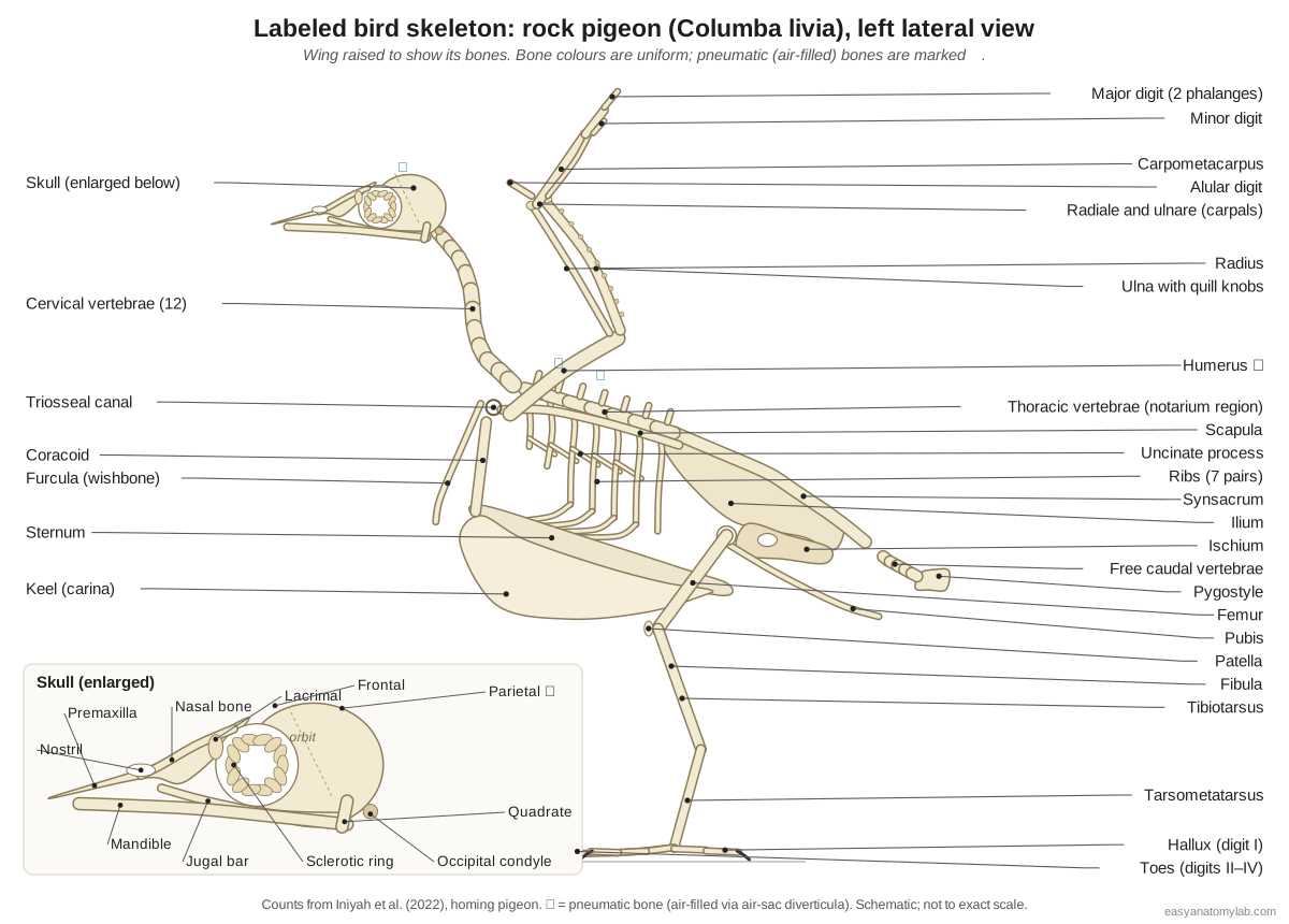
| Region | Bones | Pigeon detail |
|---|---|---|
| Skull | Premaxilla, nasal, frontal, parietal, occipital, lacrimal, jugal bar, quadrate, palatine and pterygoid, mandible, sclerotic ring, hyoid | Sutures fused in adults; single occipital condyle |
| Neck | Atlas, axis, cervical vertebrae | 12 cervical vertebrae |
| Trunk | Thoracic vertebrae (notarium region), ribs, uncinate processes, sternum and keel | 7 thoracic vertebrae, 7 pairs of ribs, uncinate processes on ribs 1–5 |
| Pelvis and tail | Synsacrum, ilium, ischium, pubis, free caudal vertebrae, pygostyle | Open pelvis; last caudal vertebrae fused into the pygostyle |
| Wing | Furcula, coracoid, scapula, humerus, radius, ulna, radiale, ulnare, carpometacarpus, alular, major and minor digits | Pneumatic humerus; quill knobs on the ulna |
| Leg | Femur, patella, tibiotarsus, fibula, tarsometatarsus, hallux and toe phalanges | Four toes, anisodactyl (three forward, one back) |
Bird skeleton anatomy: every bone and its function
This is the text version of the 3D skeleton: every labeled bone and bone group of the pigeon, with what it does.
Skeleton
- Premaxilla (Os premaxillare, upper bill bone)
- The main bone of the upper bill. Supports the horny upper bill.
- Nasal bone
- A forked bone between the premaxilla and the frontal that frames the nostril. Forms the back edge of the nasal opening and the roof of the nasal cavity.
- Frontal bone
- The broad roof of the skull between and above the orbits. Roofs the orbits and the front of the braincase.
- Parietal bone
- The rounded top-back of the braincase. Covers the cerebral hemispheres and cerebellum.
- Occipital bone and condyle (Occipital condyle, foramen magnum)
- The back of the skull, around the foramen magnum, with a single rounded occipital condyle below it. The single condyle forms a ball joint with the atlas, allowing very wide head rotation.
- Lacrimal bone (Prefrontal)
- A small bone forming the front rim of the orbit. Completes the bony rim in front of the eye.
- Jugal bar (Zygomatic arch (maxilla, jugal and quadratojugal fused))
- A thin, rod-like bony bar running from the upper bill back to the quadrate below the eye. Pushes the upper bill when the quadrate swings forward, helping it flex as the bill opens.
- Quadrate
- A small, movable bone at the back of the orbit that links skull, palate and lower jaw. The lower jaw hinges on it. Because it can swing, it transmits movement to the jugal bar and palate so the upper bill rises as the mouth opens (cranial kinesis).
- Palatine and pterygoid bones (Bony palate)
- Paired rods in the roof of the mouth linking the quadrate to the upper bill. Slide forward and back with the quadrate to raise and lower the upper bill.
- Mandible (lower jaw) (Dentary, angular and other fused jaw bones)
- The V-shaped lower jaw, made of several fused bones, covered by the horny lower bill. Opens and closes the bill.
- Sclerotic ring (Scleral ossicles)
- A ring of small overlapping bony plates in the wall of each eye. Stiffens the large eyeball and gives attachment to the muscles that change focus.
- Hyoid apparatus
- The slender bones inside the tongue and their two long horns. Support and move the tongue.
- Atlas and axis (Cervical vertebrae 1 and 2)
- The first two neck vertebrae: the ring-shaped atlas and the axis with its odontoid process. The atlas cups the single occipital condyle; together they allow the head to turn and nod.
- Cervical vertebrae (C3–C12) (Neck vertebrae)
- The long, flexible chain of neck vertebrae with saddle-shaped (heterocoelous) joints. Give the S-shaped neck its reach and flexibility for feeding and preening, and stabilise the head while walking.
- Thoracic vertebrae (notarium region) (Notarium in some birds)
- The rib-bearing vertebrae of the back; in many birds several fuse into a rigid notarium. Carry the ribs and form a stiff back that resists the forces of the flight muscles.
- Synsacrum (Lumbosacral mass)
- A long rod of fused vertebrae (the last thoracic, the lumbar, the sacral and the first caudal vertebrae), welded to the pelvis. Makes the back and pelvis one rigid frame that carries the body on the legs.
- Free caudal vertebrae (Free tail vertebrae, coccygeal vertebrae)
- A short series of movable tail vertebrae between the synsacrum and the pygostyle. Let the tail move up, down and sideways.
- Pygostyle (Ploughshare bone)
- A small blade of fused tail vertebrae at the end of the spine. Anchors the tail feathers and their muscles, so the tail fan can be spread and steered.
- Ribs (vertebral and sternal segments)
- Seven pairs of ribs, most with a dorsal (vertebral) and a ventral (sternal) bony segment. Form a rigid but hinged rib cage; the joint between segments lets the sternum swing down and up to pump the air sacs.
- Uncinate processes
- Backward-pointing hooks on the middle of the ribs that overlap the next rib. Brace the rib cage and give leverage to the breathing muscles.
- Sternum (breastbone)
- A large, curved bony shield under the thorax and abdomen. Anchors the flight muscles, protects the heart and liver, and moves during breathing.
- Keel (carina) (Carina sterni)
- The tall blade along the midline of the sternum. Gives a large area for the attachment of the pectoralis and supracoracoideus.
- Furcula (wishbone) (Fused clavicles)
- The V-shaped bone formed by the two fused clavicles. Acts as a spring brace between the shoulders during the wingbeat and helps form the triosseal canal.
- Coracoid
- A strong strut from each shoulder to the front edge of the sternum. Braces the shoulder against the pull of the huge pectoralis on the downstroke.
- Scapula (shoulder blade)
- A long, narrow, sabre-shaped blade lying over the ribs. Forms part of the shoulder joint and anchors wing muscles.
- Triosseal canal (Foramen triosseum)
- A bony tunnel at the shoulder where the coracoid, scapula and furcula meet. Acts as a pulley: the supracoracoideus tendon passes through it, so a muscle on the breast can lift the wing.
- Humerus
- The upper arm bone: short, stout and hollow (pneumatic). Transmits the force of the flight muscles to the wing. The pectoralis pulls it down; the supracoracoideus tendon lifts and twists it.
- Radius
- The thinner, straight bone of the forearm, on the leading edge. Slides along the ulna as the elbow flexes, automatically folding or spreading the hand.
- Ulna
- The thicker, slightly bowed forearm bone that carries the secondary feathers. Supports the secondaries, which attach along its trailing edge.
- Radiale and ulnare (free carpals) (Wrist bones)
- The two small free wrist bones left in the bird wing. Guide wrist folding; the other carpals are fused into the carpometacarpus.
- Carpometacarpus
- The fused bone of the hand: three metacarpals fused with some carpals. A rigid base for the primary feathers.
- Alular digit ('Thumb', digit carrying the alula)
- The short first digit at the front of the hand. Carries the alula feathers.
- Major digit
- The longest finger, of two phalanges, extending the hand to the wingtip. Supports the outer primaries.
- Minor digit
- A tiny single-phalanx finger beside the major digit. A small anchor for primary feathers.
- Ilium
- The large, plate-like upper bone of the pelvis, fused to the synsacrum. Carries the hip socket (acetabulum) and anchors the thigh muscles.
- Ischium
- The flat bone forming the back-lower part of the pelvis. Completes the side wall of the pelvis.
- Pubis
- A thin rod running backward along the lower edge of the ischium. Supports the abdominal wall.
- Femur (thigh bone)
- The short thigh bone, held almost horizontally and hidden inside the body outline. Carries the body weight to the knee; the forward-pointing femur keeps the feet under the centre of gravity.
- Patella (kneecap)
- A small sesamoid bone in front of the knee. Improves the leverage of the knee extensor muscles.
- Tibiotarsus (Drumstick bone)
- The long shin bone: the tibia fused with the upper row of ankle (tarsal) bones. Main bone of the lower leg; the 'drumstick' muscles surround it.
- Fibula
- A thin splint of bone along the outer side of the tibiotarsus. Anchors leg muscles; it is reduced and does not reach the ankle.
- Tarsometatarsus (Tarsus, shank)
- The bone of the bare lower leg: fused metatarsals and the lower row of tarsals. Adds a third long segment to the leg, so birds stand on their toes (digitigrade).
- Hallux (digit I) and metatarsal I (Hind toe)
- The backward-pointing first toe, attached by a small free metatarsal. Opposes the front toes, so the foot can grip a perch.
- Toe bones (digits II–IV) (Phalanges of the forward toes)
- The phalanges of the three forward-pointing toes. Spread the weight when standing and walking.
Bird skull and beak skeleton
A bird skull is a thin, rounded braincase with very large orbits, a toothless bill built on the premaxilla and mandible, and a single occipital condyle. In adult pigeons the sutures between the skull bones are ossified, so the cranium is effectively one bone; anatomists still name its regions (frontal, parietal, occipital) because they form separately in the embryo.
- Premaxilla: The main bone of the upper bill. Supports the horny upper bill.
- Nasal bone: A forked bone between the premaxilla and the frontal that frames the nostril. Forms the back edge of the nasal opening and the roof of the nasal cavity.
- Frontal bone: The broad roof of the skull between and above the orbits. Roofs the orbits and the front of the braincase.
- Lacrimal bone: A small bone forming the front rim of the orbit. Completes the bony rim in front of the eye.
- Quadrate: A small, movable bone at the back of the orbit that links skull, palate and lower jaw. The lower jaw hinges on it. Because it can swing, it transmits movement to the jugal bar and palate so the upper bill rises as the mouth opens (cranial kinesis).
- Jugal bar: A thin, rod-like bony bar running from the upper bill back to the quadrate below the eye. Pushes the upper bill when the quadrate swings forward, helping it flex as the bill opens.
- Mandible (lower jaw): The V-shaped lower jaw, made of several fused bones, covered by the horny lower bill. Opens and closes the bill.
- Sclerotic ring: A ring of small overlapping bony plates in the wall of each eye. Stiffens the large eyeball and gives attachment to the muscles that change focus.
Birds can raise the upper bill slightly as they open the mouth, a movement called cranial kinesis. When the lower jaw opens, the movable quadrate swings forward and pushes the jugal bars and the palatine and pterygoid bones, which flex the upper bill at a thin bending zone near the nostrils. The large eye is stiffened by a ring of small overlapping plates, the sclerotic ring; birds usually have 12–18 of these ossicles, most often 14.
The bird spine: neck, back, synsacrum and pygostyle
The bird vertebral column has a long, flexible neck, a short and stiff back, a long synsacrum welded to the pelvis, a few free tail vertebrae and a pygostyle. In the homing pigeon, Iniyah et al. (2022) counted 12 cervical vertebrae and 7 thoracic vertebrae.
| Region | Pigeon | Comparison |
|---|---|---|
| Cervical vertebrae | 12 | Domestic fowl 14; goose 17; birds in general about 8–25 |
| Thoracic vertebrae | 7 (rib-bearing) | A notarium of 2–6 fused vertebrae occurs in pigeons, galliforms, grebes and others; fusion varies |
| Synsacrum | Last thoracic, lumbar, sacral and first caudal vertebrae fused; about 10 synsacral bodies ossify by 2 days after hatching | Present in all living birds; Archaeopteryx had a much shorter sacrum of about five vertebrae |
| Free caudal vertebrae | A short movable series | Dinosaurs had long bony tails |
| Pygostyle | Last three caudal vertebrae fused (Iniyah et al.) | About four in birds generally |
The neck vertebrae have heterocoelous (saddle-shaped) joints: each end of the centrum is curved one way side to side and the opposite way top to bottom. Two saddles fit together so the neck can bend up, down and sideways without twisting out of line. This is why a pigeon can bob its head while walking and reach every part of its body with its bill while preening.
- Atlas and axis: The first two neck vertebrae: the ring-shaped atlas and the axis with its odontoid process. The atlas cups the single occipital condyle; together they allow the head to turn and nod.
- Cervical vertebrae (C3–C12): The long, flexible chain of neck vertebrae with saddle-shaped (heterocoelous) joints. Give the S-shaped neck its reach and flexibility for feeding and preening, and stabilise the head while walking.
- Thoracic vertebrae (notarium region): The rib-bearing vertebrae of the back; in many birds several fuse into a rigid notarium. Carry the ribs and form a stiff back that resists the forces of the flight muscles.
- Synsacrum: A long rod of fused vertebrae (the last thoracic, the lumbar, the sacral and the first caudal vertebrae), welded to the pelvis. Makes the back and pelvis one rigid frame that carries the body on the legs.
- Free caudal vertebrae: A short series of movable tail vertebrae between the synsacrum and the pygostyle. Let the tail move up, down and sideways.
- Pygostyle: A small blade of fused tail vertebrae at the end of the spine. Anchors the tail feathers and their muscles, so the tail fan can be spread and steered.
Bird ribs, sternum and keel
The bird rib cage is a rigid box: each rib has a dorsal (vertebral) and a ventral (sternal) bony segment, the middle ribs carry backward-pointing uncinate processes, and the ribs end on a broad sternum with a keel. In the pigeon the sternum is a flat four-sided plate, concave on its dorsal side, with facets for four sternal ribs and two long processes on each side of its back edge.
- Ribs (vertebral and sternal segments): Seven pairs of ribs, most with a dorsal (vertebral) and a ventral (sternal) bony segment. Form a rigid but hinged rib cage; the joint between segments lets the sternum swing down and up to pump the air sacs.
- Uncinate processes: Backward-pointing hooks on the middle of the ribs that overlap the next rib. Brace the rib cage and give leverage to the breathing muscles.
- Sternum (breastbone): A large, curved bony shield under the thorax and abdomen. Anchors the flight muscles, protects the heart and liver, and moves during breathing.
- Keel (carina): The tall blade along the midline of the sternum. Gives a large area for the attachment of the pectoralis and supracoracoideus.
Bird breathing depends on this design. Birds have no diaphragm. Instead, muscles swing the sternum down and forward and back up, hinging at the joints between the two rib segments; this changes the volume of the body cavity and pumps the air sacs. The keel, meanwhile, gives the huge pectoralis and supracoracoideus somewhere to attach. A deep keel is the clearest sign in a skeleton that a bird could fly.
The wishbone and shoulder: furcula, coracoid, scapula
The bird shoulder girdle has three bones on each side: the strut-like coracoid, the long blade-shaped scapula and the clavicle, which is fused with its partner into the V-shaped furcula (wishbone). Where they meet they leave a bony tunnel, the triosseal canal.
- Furcula (wishbone): The V-shaped bone formed by the two fused clavicles. Acts as a spring brace between the shoulders during the wingbeat and helps form the triosseal canal.
- Coracoid: A strong strut from each shoulder to the front edge of the sternum. Braces the shoulder against the pull of the huge pectoralis on the downstroke.
- Scapula (shoulder blade): A long, narrow, sabre-shaped blade lying over the ribs. Forms part of the shoulder joint and anchors wing muscles.
- Triosseal canal: A bony tunnel at the shoulder where the coracoid, scapula and furcula meet. Acts as a pulley: the supracoracoideus tendon passes through it, so a muscle on the breast can lift the wing.
The triosseal canal is the bird skeleton's pulley. The supracoracoideus lies on the breast, below the wing, yet it raises the wing: its tendon runs up through the canal and over the shoulder to the top of the humerus. In pigeons the muscle's main job is to twist the humerus at the start of the upstroke (Tobalske & Biewener, 2008).
Bird wing skeleton
A bird wing has the same basic bones as a human arm, but the hand is reduced to three digits and fused: humerus, radius and ulna, two free wrist bones (radiale and ulnare), the carpometacarpus, and the alular, major and minor digits. The primary flight feathers attach to the hand, the secondaries to the ulna.

- Humerus: The upper arm bone: short, stout and hollow (pneumatic). Transmits the force of the flight muscles to the wing. The pectoralis pulls it down; the supracoracoideus tendon lifts and twists it.
- Radius: The thinner, straight bone of the forearm, on the leading edge. Slides along the ulna as the elbow flexes, automatically folding or spreading the hand.
- Ulna: The thicker, slightly bowed forearm bone that carries the secondary feathers. Supports the secondaries, which attach along its trailing edge.
- Radiale and ulnare (free carpals): The two small free wrist bones left in the bird wing. Guide wrist folding; the other carpals are fused into the carpometacarpus.
- Carpometacarpus: The fused bone of the hand: three metacarpals fused with some carpals. A rigid base for the primary feathers.
- Alular digit: The short first digit at the front of the hand. Carries the alula feathers.
- Major digit: The longest finger, of two phalanges, extending the hand to the wingtip. Supports the outer primaries.
- Minor digit: A tiny single-phalanx finger beside the major digit. A small anchor for primary feathers.
Two details show up well on a real wing skeleton. The ulna carries a row of small bumps, the quill knobs, where the secondaries are anchored; the same bumps on a fossil ulna are evidence of wing feathers. And because the radius slides along the ulna as the elbow bends, a bird folds or spreads its hand automatically when it folds or spreads its elbow. Which ancestral fingers the three bird digits are is still debated: fossils and molecular data point to digits I–III, embryos to II–IV.
Bird pelvis skeleton
The bird pelvis is a long, light shield made of the ilium, ischium and pubis fused on each side and welded to the synsacrum. It is open below: the two pubic bones do not meet in the midline, which leaves room for a large egg to pass.
- Ilium: The large, plate-like upper bone of the pelvis, fused to the synsacrum. Carries the hip socket (acetabulum) and anchors the thigh muscles.
- Ischium: The flat bone forming the back-lower part of the pelvis. Completes the side wall of the pelvis.
- Pubis: A thin rod running backward along the lower edge of the ischium. Supports the abdominal wall.
Bird leg and foot skeleton
A bird leg has three long segments: the femur (thigh), the tibiotarsus (shin, the tibia fused with the upper ankle bones) and the tarsometatarsus (the fused foot bones), followed by the toes. The femur is short and held almost horizontally inside the body, so the visible "backward knee" is really the ankle.
- Femur (thigh bone): The short thigh bone, held almost horizontally and hidden inside the body outline. Carries the body weight to the knee; the forward-pointing femur keeps the feet under the centre of gravity.
- Patella (kneecap): A small sesamoid bone in front of the knee. Improves the leverage of the knee extensor muscles.
- Tibiotarsus: The long shin bone: the tibia fused with the upper row of ankle (tarsal) bones. Main bone of the lower leg; the 'drumstick' muscles surround it.
- Fibula: A thin splint of bone along the outer side of the tibiotarsus. Anchors leg muscles; it is reduced and does not reach the ankle.
- Tarsometatarsus: The bone of the bare lower leg: fused metatarsals and the lower row of tarsals. Adds a third long segment to the leg, so birds stand on their toes (digitigrade).
- Hallux (digit I) and metatarsal I: The backward-pointing first toe, attached by a small free metatarsal. Opposes the front toes, so the foot can grip a perch.
- Toe bones (digits II–IV): The phalanges of the three forward-pointing toes. Spread the weight when standing and walking.
Pigeons, like most perching birds, have an anisodactyl foot: three toes forward and the hallux backward. Other arrangements include zygodactyl feet (two toes forward, two back) in parrots and woodpeckers. In birds of prey the toes end in large talons; hawks and eagles have especially enlarged talons on digits I and II for holding struggling prey (Fowler et al., 2009).
Pneumatic bones: are bird bones hollow?
Many bird bones are hollow and air-filled. Diverticula (outpouchings) of the air sacs grow into the bones and replace the marrow: in the pigeon the clavicular air sac enters the humerus, the cervical sacs reach the vertebrae and the abdominal sacs reach the synsacrum and the region around the hip sockets.
Hollow does not mean fragile. The walls are thin but dense, and many pneumatic bones are braced inside by struts, like the trusses of a bridge. Bird skeletons are also strengthened by fusion. Female birds add another tissue during the laying season: medullary bone, a temporary calcium store inside the marrow cavity that supplies calcium for eggshells. Medullary bone has also been reported in Tyrannosaurus.
Axial and appendicular skeleton of a bird
Yes: the bird appendicular skeleton is the wings and legs plus the girdles that attach them. The axial skeleton is the skull, vertebral column, ribs and sternum.
| Part | Axial skeleton | Appendicular skeleton |
|---|---|---|
| Head and trunk | Skull, hyoid, vertebrae, ribs, sternum and keel | – |
| Girdles | – | Pectoral girdle (scapula, coracoid, furcula); pelvic girdle (ilium, ischium, pubis) |
| Limbs | – | Wing bones and leg bones |
Bird skeleton vs dinosaur skeleton
Birds are living theropod dinosaurs, and their skeleton shows it. Birds share with coelurosaurian dinosaurs such as dromaeosaurs a furcula, thin-walled hollow bones, a three-fingered grasping hand and a flexible wrist with a half-moon-shaped (semilunate) carpal. Without its feathers, Archaeopteryx looks much like a small dromaeosaur or troodontid (UCMP).
| Feature | Tyrannosaurus rex | Pigeon |
|---|---|---|
| Furcula (wishbone) | Present | Present |
| Air-filled (pneumatic) bones | Present in many bones | Present |
| Jaws | Teeth in bony jaws | Toothless keratin beak |
| Tail | Long tail of many vertebrae | Short; a few free vertebrae and a pygostyle |
| Forelimb | Short arms with clawed fingers | Wing: fused carpometacarpus with three reduced digits |
| Medullary bone in females | Reported | Present in laying females |
Feathered wings came before birds, too. Quill knobs, the bumps that anchor secondary feathers on a bird's ulna, have been found on the ulna of Velociraptor (Turner et al., 2007). What sets modern birds apart is the combination of a toothless beak, a fused and keeled flight skeleton, and a tail shortened to a pygostyle.
Bat wing vs bird wing
Bat and bird wings are made of the same forelimb bones, inherited from a common ancestor, but they became wings separately. As forelimbs they are homologous; as wings they are analogous, the result of convergent evolution.
| Bird wing | Bat wing | |
|---|---|---|
| Flight surface | Feathers along the arm and hand | Skin membrane (patagium) between the fingers, arm, body and leg |
| Fingers | Three, reduced and partly fused | Five; digits 2–5 very long, thumb free with a claw |
| Hand bones | Fused carpometacarpus | Separate long metacarpals |
| Lightening | Pneumatic (air-filled) bones | Thin, slender bones; no air sacs |
The flying bird skeleton: adaptations for flight
Every feature of a flying bird's skeleton either saves weight or stiffens the body against the forces of the wingbeat. Use this checklist when you compare a pigeon skeleton with a mammal's or a flightless bird's.
| Feature | Function in flight |
|---|---|
| Keeled sternum | Large attachment area for the flight muscles |
| Furcula | Spring-like brace between the shoulders |
| Coracoid strut | Holds the shoulder away from the sternum against the pull of the pectoralis |
| Uncinate processes | Brace the ribs; leverage for breathing muscles |
| Notarium and synsacrum | Rigid back and pelvis |
| Pneumatic bones | Lower weight for the same stiffness |
| Carpometacarpus | A rigid base for the primaries |
| Pygostyle | Anchor for the steerable tail fan |
| Toothless beak | Lighter head; grinding moved to the gizzard near the centre of mass |
Bird skeletons compared: dodo, kiwi, moa, hummingbird and more
The pigeon skeleton is a good template, but other birds show what happens when flight is lost, when birds become giants, or when they specialise in hovering, kicking or grasping prey.
| Bird | Distinctive skeleton |
|---|---|
| Dodo (Raphus cucullatus) | A giant flightless pigeon: reduced wing bones and shoulder blades, small sternum, robust legs and pelvis, heavy hooked bill. Extinct; last widely accepted sighting 1662. |
| Kiwi (Apteryx) | No keel, tiny hidden wings, no tail, marrow-filled bones and nostrils at the bill tip. The egg can reach about a fifth of the female's weight (sources give up to 20–25%). |
| Moa (Dinornithiformes) | The only completely wingless birds: no wing bones at all. Largest species about 3.6 m tall with the neck raised; extinct by about 1445. |
| Elephant bird (Aepyornis) | Heaviest known birds (estimates from about 275 kg to over 700 kg), vestigial wings, no keel; eggs up to about 40 cm long. |
| Terror bird (Andalgalornis) | A roughly 40 kg phorusrhacid with a rigid, deep skull and hooked beak, thought to have struck prey with repeated downward blows (Degrange et al., 2010). |
| Hummingbirds | Proportionally larger supracoracoideus than other birds; no alula; about 25% of weight support comes from the upstroke. The bee hummingbird weighs about 2 g. |
| Secretary bird (Sagittarius serpentarius) | About 1.3 m tall with legs nearly twice as long as other ground birds of its mass; kicks prey with a force about five times its body weight in 10–15 milliseconds. |
| Common raven (Corvus corax) | A typical perching-bird skeleton on a large scale: the heaviest passerine (about 0.7–2.25 kg), wingspan 116–153 cm, stout legs and a large bill. |
| Birds of prey | Hooked beak, strong tarsometatarsus and large talons; in hawks and eagles digits I and II carry the biggest talons, while falcons have modest talons and kill with the bill. |
Kiwi are often shown in museums as a "kiwi skeleton with egg": an X-ray or mount of a female with an egg that fills most of her abdomen. It is one of the largest eggs relative to body size of any bird. For the dodo, no complete skeleton of a single bird survives; the famous mounted skeletons are assembled from the bones of several individuals.
Counting the vertebrae of a pigeon skeleton
Bird vertebral counts are a common lab question, and the answer depends on the species and on how the regions are defined. For the homing pigeon, a veterinary osteology study counted 12 cervical vertebrae and 7 thoracic (rib-bearing) vertebrae, against 14 cervical vertebrae in the domestic fowl and 17 in the goose. The same study describes the lumbar and sacral vertebrae fused into a synsacrum wedged between the ilia, and the last caudal vertebrae fused into the pygostyle. (Iniyah, Kumaravel & Gnanadevi (2022): axial skeleton of the homing pigeon.)
Developmental work on pigeon hatchlings labels ossification centres in the bodies of at least ten synsacral vertebrae (S1 to S10) two days after hatching. That is a useful reminder that the synsacrum starts as a row of separate vertebrae that only later weld together. When you count on a cleaned skeleton, start at the atlas, stop the neck at the first vertebra whose rib reaches the sternum, and record how you made each decision, because textbooks draw the cervical–thoracic boundary differently. (Skawiński et al. (2022): ossification of the vertebral column in the domestic pigeon, Biology.)
Whether a pigeon has a notarium, a block of fused thoracic vertebrae, is a second point of disagreement. Pigeons (Columbidae) are listed among the bird families in which a notarium occurs, but the 2022 homing-pigeon study found the first thoracic vertebrae tightly bound by ligament rather than fused. Fusion may vary with age and individual, so describe what you see: a stiff block, or separate vertebrae that cannot bend. (Wikipedia: notarium (citing Storer 1982 on fused thoracic vertebrae in birds).)
How the bird skeleton resists the forces of flight
A flying bird needs a rigid trunk, because the flight muscles pull on the wing with great force at every beat. The pigeon skeleton gets that rigidity from several features working together: a broad, keeled sternum; seven pairs of ribs, each with a dorsal and a ventral bony segment; uncinate processes on the first five ribs that overlap the next rib; and the welded synsacrum and pelvis behind them. (Iniyah, Kumaravel & Gnanadevi (2022): axial skeleton of the homing pigeon.)
The shoulder is built as a tripod. The coracoid braces the shoulder joint against the front of the sternum, the furcula springs between the two shoulders, and the scapula lies along the ribs. Where the three bones meet they leave the triosseal canal, a tunnel through which the tendon of the supracoracoideus passes on its way to the top of the humerus. Tobalske and Biewener showed in pigeons that this arrangement mainly twists (supinates) the humerus at the start of the upstroke, rather than simply hauling the wing upward. (Tobalske & Biewener (2008): the pigeon supracoracoideus in flight, Journal of Experimental Biology.)
Lightness comes from pneumatisation. Diverticula of the air sacs invade bones and replace marrow: the clavicular air sac sends a branch into the humerus, and other sacs reach the vertebrae and the bones around the hip. A pneumatic bone is not weak; thin walls braced by internal struts make it stiff for its weight. (WikiVet: avian respiration, anatomy and physiology.)
Extinct bird skeletons: dodo, moa and elephant bird
The dodo (Raphus cucullatus) was a giant, flightless member of the pigeon family from Mauritius; its closest living relative is the Nicobar pigeon. Its skeleton shows how a pigeon body plan changes when flight is lost: the wing bones and shoulder blades are reduced, the sternum is small for the body size, the leg bones and pelvis are robust, and the skull is heavy with a hooked bill. No complete individual dodo skeleton exists; mounted skeletons are composites, and estimates of its weight vary widely. (Wikipedia: dodo.)
Moa were the only birds that lacked wings entirely: their skeletons show no trace of wing bones, whereas every other ratite keeps at least a vestigial wing. The tallest moa reached about 3.6 m with the neck stretched up, although the head was probably carried further forward than in older museum mounts. All moa were extinct by about 1445 CE, within roughly a century of human settlement of New Zealand. (Wikipedia: moa.)
Madagascar's elephant birds were the heaviest birds known, with mass estimates for Aepyornis maximus ranging from roughly 275 kg to well over 700 kg depending on the method. Their wings were vestigial and their sternum had no keel. Their eggs were the largest of any bird, up to about 40 cm long. Taxonomy is still debated: a 2018 study proposed a separate giant genus, Vorombe, which later genetic work questioned. (Wikipedia: elephant bird.)
Bird skeleton worksheet: test yourself
Use the 3D model or the labeled diagram to answer these. Answers are in the text above.
- Which bone forms the wishbone, and what is it made of?
- How many cervical vertebrae does a pigeon have?
- Name the three bones that meet at the triosseal canal.
- Which wing bone carries the quill knobs, and which feathers attach there?
- What two bones are fused to form the tibiotarsus?
- Which joint is the "backward knee" of a standing bird?
- Give three skeletal features birds share with theropod dinosaurs.
- Why does a kiwi or ostrich have no keel?
For the soft anatomy, see bird anatomy, and for a hands-on lab, the pigeon dissection guide.
Frequently asked questions
How many bones does a bird have?
There is no single number: it differs between species and with age, because many bones fuse as a bird grows (skull bones, the synsacrum, pygostyle, carpometacarpus, tibiotarsus and tarsometatarsus). Birds generally have fewer separate bones than their reptile ancestors because of this fusion. For the pigeon, standard counts are 12 cervical vertebrae, 7 thoracic vertebrae and 7 pairs of ribs.
Are bird bones hollow?
Many are. In flying birds such as pigeons, extensions of the air sacs enter bones like the humerus, vertebrae and pelvis and replace the marrow, so the bones are filled with air and braced inside by thin struts. Not every bone is pneumatic, and flightless kiwi have marrow-filled bones.
What is the keel on a bird?
The keel (carina) is the tall blade along the midline of the sternum. It anchors the two flight muscles, the pectoralis and supracoracoideus. Strong fliers have a large keel; flightless ratites such as the ostrich, emu and kiwi have a flat sternum with no keel.
What is the wishbone of a bird?
The wishbone, or furcula, is the two clavicles fused into a V. It braces the shoulders and flexes like a spring during the wingbeat. Theropod dinosaurs such as Tyrannosaurus and Velociraptor also had a furcula.
Is the bird appendicular skeleton the legs and wings?
Yes. The appendicular skeleton is the limbs and the girdles that attach them: the wings with the pectoral girdle (scapula, coracoid, furcula) and the legs with the pelvic girdle (ilium, ischium, pubis). The axial skeleton is the skull, vertebral column, ribs and sternum.
Why does a bird's knee bend backwards?
It doesn't. The joint you see bending backwards halfway down a standing bird's leg is its ankle (the intertarsal joint). The real knee is hidden inside the body feathers, close to the belly, and bends forward as in humans.
What bones are in a bird wing?
Humerus, radius and ulna, two free wrist bones (radiale and ulnare), the fused carpometacarpus, and three digits: a short alular digit, a long major digit and a tiny minor digit. The primaries attach to the hand and the secondaries to the ulna.
How is a bird skeleton similar to a dinosaur skeleton?
Birds are living theropod dinosaurs. They share with other coelurosaurs a furcula, hollow thin-walled bones, a three-fingered hand and a half-moon-shaped (semilunate) wrist bone. Velociraptor even had quill knobs for wing feathers on its ulna. Birds differ in having a toothless beak, a short tail ending in a pygostyle and a fused, keeled flight skeleton.
References
- Iniyah, K., Kumaravel, A. & Gnanadevi, R. (2022). Axial skeleton of homing pigeon (Columba livia). Biological Forum – An International Journal 14(3), 167–170.
- Skawiński, T. et al. (2022). Phylogenetic diversity of ossification patterns in the avian vertebral column: new data from the domestic pigeon and two grebes. Biology 11(2), 180.
- Cavitt, J. Weber State University. External and internal anatomy of the rock dove (ornithology lab guide).
- Tobalske, B. W. & Biewener, A. A. (2008). Contractile properties of the pigeon supracoracoideus during different modes of flight. Journal of Experimental Biology 211, 170–179.
- WikiVet. Avian respiration: anatomy and physiology (air sacs and pneumatised bones).
- University of California Museum of Paleontology. Introduction to the Aves: the dinosaur–bird link.
- Understanding Evolution, UC Berkeley. Homologies and analogies (bat and bird wings).
- Degrange, F. J. et al. (2010). Mechanical analysis of feeding behavior in the extinct 'terror bird' Andalgalornis steulleti. PLoS ONE 5(8), e11856.
- Fowler, D. W., Freedman, E. A. & Scannella, J. B. (2009). Predatory functional morphology in raptors. PLoS ONE 4(11), e7999.
- Wikipedia: Notarium (citing Storer, R. W., 1982, J. Yamashina Inst. Ornithol. 14, 86–95).
- Wikipedia: Bird wing; Quill knob (citing Turner, Makovicky & Norell, 2007, Science 317, 1721); Sclerotic ring; Medullary bone.
- Wikipedia: Dodo; Kiwi; Moa; Elephant bird; Secretarybird (citing Portugal et al., 2016, Current Biology); Hummingbird; Common raven.
More interactive anatomy models
- Bird anatomyBird digestive system, air sacs, heart and feather anatomy in 3D.
- Chicken anatomyCrop, gizzard, reproductive tract, skeleton and external parts of the chicken.
- Pigeon dissectionGuided pigeon dissection: flight muscles, crop, gizzard, air sacs and heart.
- Human anatomy1,000+ bones, muscles, organs, vessels and nerves from real anatomical data.
- Octopus anatomyThree hearts, a ring-shaped brain, eight arms and a beak, in 3D.
- Squid anatomyPen, eight arms, two tentacles, giant axons and three hearts of the longfin squid.