

Grasshopper dissection at a glance
A grasshopper dissection has two halves. First you study the outside: the three body regions, the head and its mouthparts, the legs and wings, the tympanum and spiracles, and the clues that tell a male from a female. Then you open the body along the back and work down through the layers: heart, air sacs, gonads, gut, and finally the nerve cord on the floor of the body.
Plan on one to two class periods. The specimen is small compared with a frog or fetal pig, so most mistakes come from cutting too deep. Keep a hand lens nearby; many structures, such as the ocelli, spiracles, tracheae and palp segments, are only a millimetre or two across.
Materials and safety
Materials
- Preserved adult grasshopper, ideally an eastern lubber (Romalea microptera); one male and one female per group if possible
- Dissecting pan with wax, or a cutting mat, and a supply of dissecting pins
- Fine-pointed scissors (iris scissors) and medium scissors
- Fine forceps, a blunt probe and a dissecting needle
- Hand lens or dissecting microscope (for ocelli, spiracles, mouthparts and tracheae)
- Shallow dish of water or 40% isopropyl alcohol to cover the opened specimen
- Microscope slide or index card and clear tape for mounting mouthparts
- Nitrile gloves, safety glasses, lab coat or apron, paper towels
Safety
- Wear gloves and eye protection. Rinse the preserved specimen in water first to reduce preservative fumes, and work in a ventilated room.
- Cut away from your fingers. Keep the lower scissor blade just under the exoskeleton so you do not cut the organs below.
- The hind tibiae have sharp spines: handle the jumping legs by the femur.
- Do not eat or drink in the lab. Dispose of tissue, pins and gloves as your teacher directs, and wash your hands.
External anatomy to observe before you cut
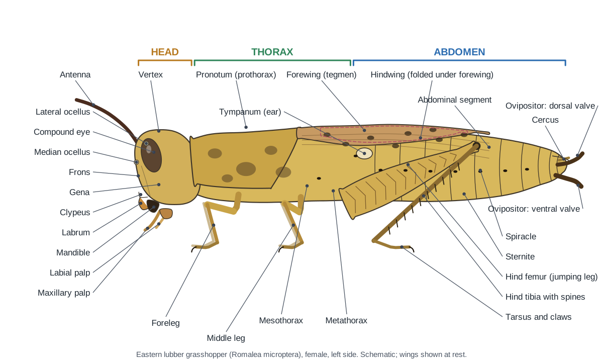
Before you make any cut, identify the external parts. Biology Corner's external lab, for example, asks students to count the abdominal segments, find the thorax where all appendages attach, count antennae and eyes, find walking and jumping legs and their claws, compare the forewing and hindwing, examine the mouthparts, raise the wings to find the tympanum, count spiracles and check for an ovipositor.
Grasshopper dissection procedure, step by step
These steps match the 3D guide above. They follow the order used by Fox's lubber laboratory text and school handouts: outside first, then open the back, then work from the top layers down.
1. Body regions and sense organs
- Place the grasshopper in the pan on its belly. Find the three body regions: head, thorax and abdomen.
- On the thorax, find the saddle-shaped pronotum (prothorax), then the mesothorax and metathorax behind it.
- On the head, find the two compound eyes and the one pair of antennae. With a hand lens, find the three ocelli: one in the middle of the face and one beside each antenna.
- Count the abdominal segments along the back and compare your count with your lab partner's.
Identify: Head capsule, Pronotum (prothorax), Mesothorax, Metathorax, Abdomen: tergites, Compound eyes, Ocelli, Antennae.
2. Regions of the head
- Look at the head from the front. The flat face between the eyes is the frons; the top of the head is the vertex.
- The cheek below each compound eye is the gena. The broad plate at the bottom of the face is the clypeus, and the flap hanging from it is the labrum.
- Note that the mouthparts point downwards (a hypognathous head).
Identify: Vertex, Frons, Gena, Clypeus, Labrum, Compound eyes, Ocelli.
3. Legs and wings
- Find the three pairs of legs, one pair on each thoracic segment. The front and middle pairs are walking legs; the hind pair are jumping legs.
- On a hind leg, name the segments from the body outwards: coxa, trochanter, femur, tibia, tarsus. Note the herringbone pattern on the femur and the spines on the tibia.
- With a hand lens, look at a foot: the tarsal segments with soft pads underneath, two claws, and the small arolium pad between them.
- Lift a forewing (tegmen) with forceps to see the thin, fan-folded hindwing beneath it. In the lubber both pairs are too short to fly.
Caution: The hind tibial spines are sharp. Hold the jumping leg by the femur.
Identify: Coxae, Trochanters, Femora (fore and middle legs), Hind femur, Tibiae (fore and middle legs), Hind tibia, Tarsi, Tarsal pads, Tarsal claws, Arolium, Forewings (tegmina), Hindwings.
4. Tympanum, spiracles and sexing
- Lift the wings and find the oval tympanum (eardrum) on the side of the first abdominal segment.
- Find the spiracles: small dark slits low on the side of each abdominal segment, one on the front edge of the tympanum, and one on the thorax between the meso- and metathorax. Count them on one side.
- Look at the tip of the abdomen. Find the triangular epiproct, the paraprocts and the short cerci.
- Sex your grasshopper. Two pairs of pointed, hard ovipositor valves mean female; a rounded, upturned subgenital plate means male. Use the Female/Male switch on the model to compare, and swap specimens with another group.
Identify: Tympanum, Spiracles, Epiproct, Paraprocts, Cerci, Ovipositor: dorsal valves, Ovipositor: ventral valves, Genital plate & egg guide, Subgenital plate.
5. Mouthparts dissection
- Hold the head with forceps. Remove the mouthparts from front to back on one side, and lay them out in order on a card or slide.
- First lift and pull off the labrum (upper lip). Behind it are the dark mandibles: work one free and note its cutting teeth and grinding surface.
- Next remove a maxilla with its long, five-segmented palp, then the labium (lower lip) with its two short, three-segmented palps.
- The soft, tongue-like hypopharynx is now visible in the middle of the mouth. Tape the parts in order and label them.
Identify: Labrum, Mandibles, Maxillae, Maxillary palps, Labium, Labial palps, Hypopharynx.
6. Open the body: heart and aorta
- Pin the grasshopper belly-down through the middle legs and the tip of the abdomen. Cut off the wings close to the body.
- Cut across the pronotum to remove its rear overhang.
- Slide the fine scissors under the back edge of an abdominal tergite about 2 mm to one side of the midline. Cut forward along the whole abdomen and thorax, keeping the blade just under the exoskeleton. Make a matching cut on the other side.
- Lift the narrow strip of dorsal exoskeleton slowly, freeing the tissue under it with a probe. The heart, a thin clear tube on the midline, should stay on the body. Cover the specimen with water.
Caution: Cut through the exoskeleton only. If the blade goes deeper you will cut the heart and gonads directly underneath.
Identify: Heart (dorsal vessel), Aorta, Alary muscles.
7. Tracheae, air sacs and fat body
- Look under the tympana and in the thorax for large, thin white air sacs.
- Find the silvery tracheae: tubes that run along each side and branch into every organ. Under a hand lens their walls show fine spiral rings.
- Note any loose, creamy fat body around the organs. You may remove some to see the organs better.
Identify: Tracheae, Air sacs, Spiracles, Fat body.
8. Reproductive organs
- The gonads lie on top of the gut in the abdomen, just under the heart.
- Female: find the two large yellow ovaries packed with long eggs, the lateral oviduct along each, the vagina below the gut and the small seminal receptacle near the tip.
- Male: find the testes, fused into one mass over the midline, the white vasa deferentia, the seminal vesicles, the tuft of accessory glands and the ejaculatory duct leading to the phallus.
- Swap specimens with a group that has the other sex, or use the Female/Male switch.
Caution: In a mature female the ovaries can fill most of the abdomen. Remove eggs only if your teacher asks you to.
Identify: Ovaries, Lateral oviducts, Vagina (common oviduct), Seminal receptacle, Testes, Vasa deferentia, Seminal vesicles, Accessory glands, Ejaculatory duct, Phallus.
9. Digestive and excretory systems
- Gently remove the gonads (or pull them to one side) to expose the gut, which runs straight from head to tip.
- In the thorax, find the large, thin-walled crop, often green with leaf pieces. Behind it is the narrow, muscular gizzard (proventriculus).
- Find the six finger-like gastric caeca around the front of the midgut, pointing forwards over the gizzard.
- Follow the midgut back to the many fine Malpighian tubules that join the gut where the hindgut begins. Then trace the ileum, the narrow colon and the ridged rectum to the anus.
- Look low in the thorax beside the crop for the white, grape-like salivary glands.
Identify: Oesophagus, Crop, Gizzard (proventriculus), Gastric caeca, Midgut (stomach), Malpighian tubules, Ileum, Colon, Rectum, Anus, Salivary glands.
10. Nervous system
- Cut the gut at the oesophagus and at the rectum, and lift it out. Remove the thin membrane (ventral diaphragm) on the floor of the abdomen.
- The ventral nerve cord is a white double cord on the midline. Follow it forward and count the ganglia: five in the abdomen and three large ones in the thorax.
- Pouring 40% isopropyl alcohol over the specimen turns the nerve tissue opaque white and easier to see.
- Optional: open the top of the head capsule and pick away muscle to find the brain above the oesophagus, the optic lobes beside it, and the connectives that loop down to the suboesophageal ganglion.
Caution: The thoracic part of the cord is hidden under the thoracic muscles; cut or pull them aside with care.
Identify: Ventral nerve cord, Abdominal ganglia, Thoracic ganglia, Suboesophageal ganglion, Circumoesophageal connectives, Brain, Optic lobes.
11. Jumping muscles
- Cut a window in the outer wall of a hind femur with fine scissors.
- The large, feather-like muscle filling the upper femur is the extensor tibiae, which kicks the leg straight in a jump. The smaller flexor tibiae lies below it and folds the leg.
- Look into the thorax for the large leg and wing muscles.
Identify: Extensor tibiae muscle, Flexor tibiae muscle, Thoracic (leg and wing) muscles, Hind femur.
Grasshopper dissection labeled diagram
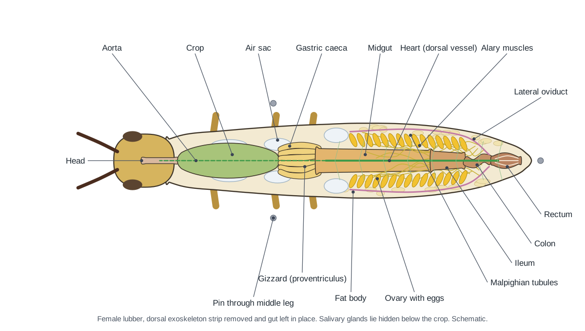
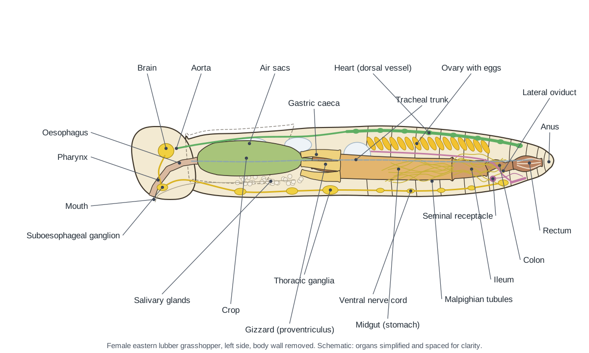
This is what an opened female should look like from above, with the dorsal strip of exoskeleton removed: the heart on the midline, yellow egg-filled ovaries on top of the gut, and the gut running straight from the crop to the rectum.
Grasshopper mouthparts dissection
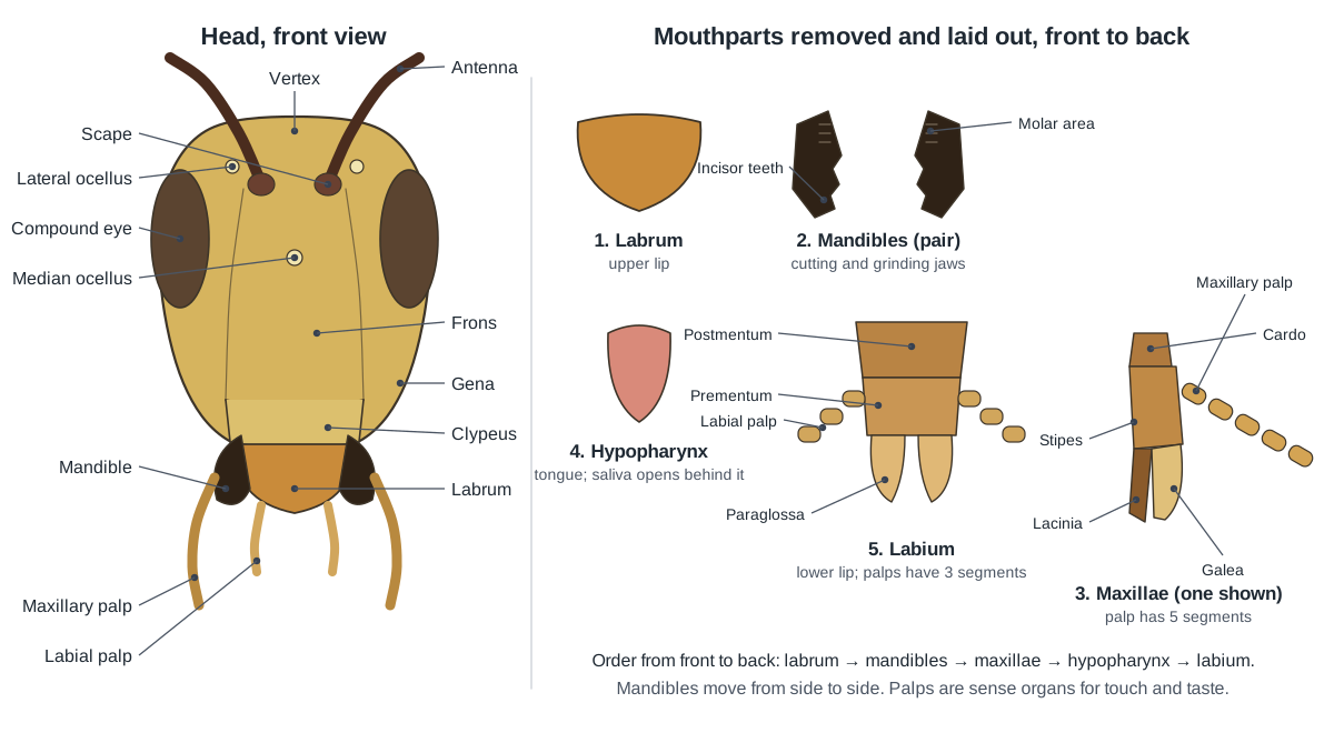
Remove the mouthparts from one side, from front to back, and tape them in order. The labrum comes off first, then a mandible, then a maxilla with its five-segmented palp, then the labium with its two three-segmented palps. The soft hypopharynx (tongue) is then visible in the middle of the mouth. Fox advises studying the mouthparts in place first and removing them later.
Crop, gizzard and gastric caeca: the digestive tract
The crop, gizzard and gastric caeca are the most-asked structures in a grasshopper dissection. The crop is the large, thin-walled sac in the thorax, usually green with leaf fragments. The gizzard (proventriculus) is the narrower, muscular section right behind it. The six gastric caeca are finger-like pouches that ring the front of the midgut and point forward over the gizzard. Further back, the hair-thin Malpighian tubules mark where the midgut ends and the hindgut begins.
| Structure | How to recognise it | Job |
|---|---|---|
| Crop | Biggest, thin-walled, often green; in the thorax | Stores food; some digestion begins |
| Gizzard | Short, narrow, muscular; behind the crop | Grinds food with tooth-like denticles |
| Gastric caeca | Six finger-like pouches pointing forward | Make enzymes, absorb nutrients |
| Midgut | Wide tube in the front abdomen | Main digestion and absorption |
| Malpighian tubules | Many fine yellowish threads at the midgut–hindgut junction | Excretion (like kidneys) |
| Ileum, colon | Wide then narrow hindgut | Carry waste, absorb water |
| Rectum | Ridged chamber near the tip, 6 rectal pads | Reabsorbs water; forms dry pellets |
Heart, testis and ovary: finding the organs on top
The heart is the first internal organ you meet. It is a transparent, slender tube on the dorsal midline of the abdomen, held by fan-shaped alary muscles in the dorsal diaphragm. If you cut too deep, it comes away with the exoskeleton strip; check the underside of the strip if you cannot find it.
Right below the heart are the gonads. A testis in a lubber is actually two testes fused over the midline; look for the white sperm ducts leaving its rear corners. The ovaries are bright yellow and full of long eggs; each oviduct runs along the outer edge of its ovary and the two join below the gut to form the vagina.
Male vs female grasshopper in the dissection
- Female: two pairs of hard, pointed ovipositor valves at the abdomen tip and an egg guide on the eighth sternite; inside, two large yellow ovaries packed with eggs, lateral oviducts, a vagina and a small seminal receptacle.
- Male: a rounded, upturned subgenital plate at the tip; inside, fused testes on top of the gut, vasa deferentia, seminal vesicles, a tuft of accessory glands, the ejaculatory duct and the phallus folded inside the subgenital plate.
Because the two sexes differ so much at the tip and inside, Fox recommends swapping specimens with another student to study both.
Comparison: crayfish vs grasshopper dissection
Crayfish and grasshoppers are both arthropods, so their dissections share a plan: an exoskeleton you cut through, a dorsal heart, a gut from mouth to anus, a brain above the gut and a ventral nerve cord. The differences come from living in water versus on land.
| Feature | Crayfish (crustacean) | Grasshopper (insect) |
|---|---|---|
| Body regions | 2: cephalothorax (under a carapace) and abdomen | 3: head, thorax, abdomen |
| Antennae | 2 pairs (antennules and antennae) | 1 pair |
| Legs | 5 pairs of walking legs, first pair large chelipeds | 3 pairs, on the thorax |
| Wings | None | 2 pairs (short in the lubber) |
| Breathing | Gills in chambers under the carapace | Spiracles, tracheae and air sacs |
| Excretion | Antennal (green) gland opening at the base of the antenna | Malpighian tubules |
| Abdomen appendages | Swimmerets, uropods and telson (tail fan) | Cerci, ovipositor or subgenital plate |
| Circulation | Dorsal heart, open system | Dorsal heart (dorsal vessel), open system |
Choosing and preparing a specimen
School suppliers sell preserved eastern lubber grasshoppers because the species is large and well known; UF/IFAS notes that it is widely used for dissection in biology classrooms. Adults are 50–75 mm long, with females about 75 mm and males about 55 mm, so the organs are big enough to see without a microscope. Order both sexes if you can, because male and female abdomens must be studied separately. (Fox, R. (2006). Romalea microptera, eastern lubber grasshopper. Invertebrate Anatomy OnLine, Lander University.)
Lubbers are common in the southeastern US and can damage crops and garden plants; they have one generation a year and spend the winter as eggs in the soil. Preserved specimens arrive in a fluid; rinse them in water before you start. A dissecting microscope or a good hand lens makes the ocelli, spiracles, tracheae and mouthpart segments much easier to see. (Capinera, J. L. & Scherer, C. W. Eastern lubber grasshopper, Romalea microptera. UF/IFAS EDIS publication IN132.)
How to open the grasshopper without destroying the heart
Fox's method opens the grasshopper from the back. Pin the middle legs flat, remove the wings with scissors, and cut across the pronotum to remove its rear overhang. Then slip fine scissors under the edge of a tergite about 2 mm to one side of the midline and cut forward, through the exoskeleton but no deeper, and repeat on the other side. Lifting the narrow strip between the cuts exposes the organs, and if you work carefully the heart stays on the body. (Fox, R. (2006). Romalea microptera, eastern lubber grasshopper. Invertebrate Anatomy OnLine, Lander University.)
Many school handouts instead cut slightly to one side of the mid-dorsal line along the whole length, then along the ventral side and up the front of the head, keeping the inner blade just under the body covering. They warn that a mature female's body cavity may be filled with slender eggs. Either method works; what matters is cutting shallowly, lifting the body wall with forceps as you go, and removing lateral muscles and tracheae only when they block your view. (Biology Junction. Grasshopper internal anatomy.)
What you will see inside, and in what order
Directly under the dorsal body wall lie the transparent heart, attached to a porous dorsal diaphragm, and below it the gonads. A female's two ovaries are large and bright yellow, made of many ovarioles that hold oval yellow mature eggs and smaller white immature ones; they sit above the gut in the posterior abdomen. A male's two testes lie together over the midline and look like a single organ. (Fox, R. (2006). Romalea microptera, eastern lubber grasshopper. Invertebrate Anatomy OnLine, Lander University.)
Large white air sacs show under the tympana and in the thorax. Under the gonads the gut runs straight from front to back: the large crop in the thorax, the narrow proventriculus, the ring of six gastric caeca, the midgut, the Malpighian tubules and the hindgut. The salivary glands are white clusters of spherical follicles, like bunches of grapes, low in the thorax under the ventral diaphragm. The nerve cord is the last thing you reach, on the floor of the body. (Fox, R. (2006). Romalea microptera, eastern lubber grasshopper. Invertebrate Anatomy OnLine, Lander University.)
Crayfish vs grasshopper: comparing two arthropods
Many courses dissect a crayfish and a grasshopper together because both are arthropods with an exoskeleton, jointed legs, a dorsal heart in an open blood system, a brain above the gut and a ventral nerve cord. The differences reflect water versus land. A crayfish's body is a cephalothorax covered by a carapace plus an abdomen (pleon); it has two pairs of antennae, five pairs of walking legs (the first pair the large chelipeds), and 17–18 pairs of gills in branchial chambers under the carapace. (Fox, R. Procambarus, crayfish. Invertebrate Anatomy OnLine, Lander University.)
The grasshopper has three body regions instead of two, one pair of antennae, three pairs of legs, wings, and breathes air through spiracles and tracheae. It excretes with Malpighian tubules, whereas the crayfish's excretory organ opens at the base of its second antenna (the antennal or green gland). The table on this page summarises the comparison for lab worksheets. (Fox, R. (2006). Romalea microptera, eastern lubber grasshopper. Invertebrate Anatomy OnLine, Lander University.)
For every structure in detail, with external, internal and mouthpart diagrams, see the grasshopper anatomy page.
Grasshopper dissection lab questions and answer key
Answer these during or after the dissection. Click a question to reveal the answer.
To which phylum and class does the grasshopper belong?
Phylum Arthropoda, class Insecta (order Orthoptera, suborder Caelifera, family Romaleidae for the lubber).
Name the three body regions and say what is attached to each.
Head: eyes, ocelli, antennae and mouthparts. Thorax: all three pairs of legs and both pairs of wings. Abdomen: tympana, most spiracles, cerci and the ovipositor or subgenital plate.
How many eyes does a grasshopper have, and what kinds?
Five: two compound eyes, which form images, and three simple eyes (ocelli), which sense light intensity.
On which thoracic segments are the wings attached?
The forewings (tegmina) on the mesothorax and the hindwings on the metathorax. The prothorax never has wings.
Which pair of legs is adapted for jumping, and what is unusual about it?
The hind (metathoracic) legs. The femur is greatly enlarged to hold the extensor tibiae muscle, and the tibia is long and spiny.
Where is the tympanum and what does it do?
On each side of the first abdominal segment. It is an eardrum: a thin membrane that vibrates with sound.
How many spiracles did you count, and what are they for?
Ten pairs: two on the thorax and eight on the abdomen (segments 1–8). They are the openings of the tracheal system, through which the grasshopper breathes.
Do the mandibles move up and down or side to side?
Side to side. They meet in the middle, cutting with their tips and grinding with their bases.
List the mouthparts in order from front to back.
Labrum, mandibles, maxillae (with maxillary palps), hypopharynx, and labium (with labial palps).
How did you tell whether your specimen was male or female?
A female has two pairs of short, hard, pointed ovipositor valves at the tip of the abdomen; a male has a rounded, upturned subgenital plate. Inside, a female has large yellow ovaries full of eggs; a male has testes.
Trace the path of food from mouth to anus.
Mouth → pharynx → oesophagus → crop → gizzard (proventriculus) → midgut, with gastric caeca at its front → ileum → colon → rectum → anus.
What is the function of the crop? Of the gizzard? Of the gastric caeca?
The crop stores food. The gizzard grinds it with tooth-like ridges. The gastric caeca secrete digestive enzymes and absorb nutrients for the midgut.
What do the Malpighian tubules do, and where are they attached?
They remove nitrogen wastes and salts from the hemolymph, like kidneys. They join the gut where the midgut meets the hindgut.
Where is the grasshopper's heart, and why is its blood not red?
The heart is a tube along the top (dorsal side) of the abdomen. The hemolymph is not red because it has no hemoglobin: insects deliver oxygen through the tracheae, not the blood.
Is the grasshopper nerve cord dorsal or ventral? How does this compare with a human?
Ventral, along the floor of the body, with a ganglion in each segment. A human spinal cord is dorsal, in the back.
What kind of metamorphosis do grasshoppers have?
Incomplete (simple) metamorphosis: egg, nymph and adult. Nymphs look like small, wingless adults and develop wing pads as they moult.
Virtual grasshopper dissection
The 3D viewer above is a free virtual grasshopper dissection. Each step sets the exoskeleton, hides the layers you would have removed, and lists the structures to find; click any one for its location and function. Students who opt out of a physical dissection, or who are absent on lab day, can complete the same steps and the same answer key. For the full anatomy, including the life cycle and every system in detail, see our grasshopper anatomy page, and for the three body parts shared by all insects, see insect body parts.
Frequently asked questions
What grasshopper is used for dissection?
Usually the eastern lubber grasshopper, Romalea microptera, from the southeastern United States. Adults are 5–7.5 cm long, so the organs are large and easy to find. Smaller Melanoplus species are sometimes used and have the same internal layout.
Where do you cut to dissect a grasshopper?
Along the back. Remove the wings, trim the rear of the pronotum, then cut forward through the exoskeleton about 2 mm to one side of the midline, and again on the other side, keeping the scissor blade just under the body wall. Lift the strip to expose the heart and organs.
Where is the heart in a grasshopper dissection?
Just under the dorsal exoskeleton on the midline of the abdomen. It is a thin, transparent tube, so it is easy to tear when you lift the body wall. Its front part, the aorta, continues forward through the thorax above the crop.
What is the crop in a grasshopper?
The crop is a large, thin-walled storage sac of the foregut in the thorax, often green with chewed leaves. Behind it is the muscular gizzard (proventriculus), then the six gastric caeca around the front of the midgut.
How do you find the testes in a grasshopper dissection?
In a male, the two testes lie together on top of the gut in the abdomen, just below the heart, and look like a single mass. Thin white sperm ducts (vasa deferentia) leave their rear corners and run down around the gut to the ejaculatory duct.
Is there a virtual grasshopper dissection?
Yes. The 3D model on this page follows the same 11 steps as the lab. Each step removes the right layers and highlights what to find, so students who opt out of a physical dissection can complete the same worksheet.
Why are the eggs yellow in my grasshopper?
Mature eggs in a lubber's ovaries are large, oval and bright yellow; immature eggs are white. A mature female can be so full of eggs that they fill most of the abdomen.
References
- Fox, R. (2006). Romalea microptera, eastern lubber grasshopper. Invertebrate Anatomy OnLine, Lander University
- Biology Junction. Grasshopper internal anatomy
- Biology Corner. Grasshopper external anatomy lab
- Capinera, J. L. & Scherer, C. W. Eastern lubber grasshopper, Romalea microptera. UF/IFAS EDIS publication IN132
- Fox, R. Procambarus, crayfish. Invertebrate Anatomy OnLine, Lander University
- NC State University, General Entomology: digestive system
- NC State University, General Entomology: circulatory system
- NC State University, General Entomology: respiratory system
- NC State University, General Entomology: nervous system
- NC State University, General Entomology: mouthparts
More interactive anatomy models
- Insect body partsHead, thorax and abdomen: the three body parts every insect shares.
- Grasshopper anatomyExternal and internal anatomy of the grasshopper, with mouthparts.
- Butterfly anatomyScaled wings, proboscis, compound eyes and internal organs of a butterfly.
- Ant anatomyAnt body parts: elbowed antennae, mandibles, petiole and stinger.
- Honeybee anatomyWorker bee anatomy: pollen baskets, honey stomach, sting and wax glands.
- Human anatomy1,000+ bones, muscles, organs, vessels and nerves from real anatomical data.