

Bird anatomy at a glance
A bird is a feathered, warm-blooded vertebrate with a toothless beak, front limbs turned into wings, a light skeleton, a crop and two-part stomach instead of teeth, small rigid lungs ventilated by air sacs, a four-chambered heart, and a single cloaca for droppings, urine and eggs. Most of its body plan is a trade-off between flying and carrying weight: heavy parts, such as the gizzard and the flight muscles, sit close to the centre of the body.
This page describes the rock pigeon (also called the rock dove or feral pigeon), Columba livia. Wild-type birds are 30–35 cm long with a wingspan of 62–68 cm and weigh about 240–380 g. They are blue-grey with an iridescent green and purple neck, two black bars on each wing and a white lower back. Pigeons are the standard bird of zoology labs; the chicken, covered on its own page, differs in several organs that are noted below.
For the bones, see the bird skeleton page; for a step-by-step lab, the pigeon dissection guide.
External anatomy of a bird: parts and topography
Bird topography divides the body into named regions so plumage can be described exactly. On the head: forehead, crown, nape, lores (between eye and bill), cheek, chin and throat. On the body: mantle and back, rump, breast, belly, flanks and vent. On the wing: lesser, median and greater coverts, the alula, primaries, secondaries and tertials. The tail has rectrices with upper- and undertail coverts, and the legs end in a bare, scaly tarsus and toes.
- Bill (beak): The toothless bill: a horny keratin sheath (rhamphotheca) over the bones of the upper and lower jaws. Picks up seeds and grit, drinks (pigeons can suck water up without tipping the head back), preens feathers and feeds the squabs.
- Cere (operculum): The soft, swollen, whitish patch of skin at the base of the upper bill, over the nostrils. Covers and partly shields the nostrils.
- Nostrils (external nares): Slit-like openings into the nasal cavity, at the base of the upper bill under the cere. Let air into the nasal cavity, which opens into the mouth through a slit in the palate (the choana).
- Eye: A very large eye on each side of the head, with an orange iris in the rock pigeon. Wide field of view for spotting predators; good colour vision.
- Ear opening (external acoustic meatus): A small opening behind and below the eye, hidden under feathers (ear coverts). Leads to the eardrum and middle ear; birds have no external ear flap (pinna).
- Uropygial (preen) gland: A small skin gland on the back at the base of the tail that secretes preen oil. The bird squeezes oil from it with the bill and spreads it through the plumage while preening.
- Feet and scales (podotheca): The bare, red, scaly skin of the tarsus and the four toes. Support the bird while perching and walking; the scales are keratin, like the bill.
- Vent (cloacal opening): The single external opening of the cloaca, under the base of the tail. Releases droppings (faeces plus white urates), and eggs or sperm.
Pigeons show a few features that students often miss. The soft white swelling at the base of the upper bill is the cere (operculum), small in wild rock pigeons and much larger in many domestic breeds. The ear opening is hidden under the feathers behind the eye; birds have no external ear flap. And the preen gland at the base of the tail is small in the rock pigeon, and Columbidae is one of the families in which some species lack it entirely.
Bird digestive system diagram
The bird digestive system runs, in order, from the bill and mouth through the oesophagus, crop, proventriculus (glandular stomach), gizzard (muscular stomach), duodenum, small intestine, caeca and rectum to the cloaca, which opens at the vent. The liver and pancreas empty into the duodenum. Birds have no teeth: food is swallowed whole, stored in the crop, softened by acid and enzymes in the proventriculus and ground up in the gizzard.
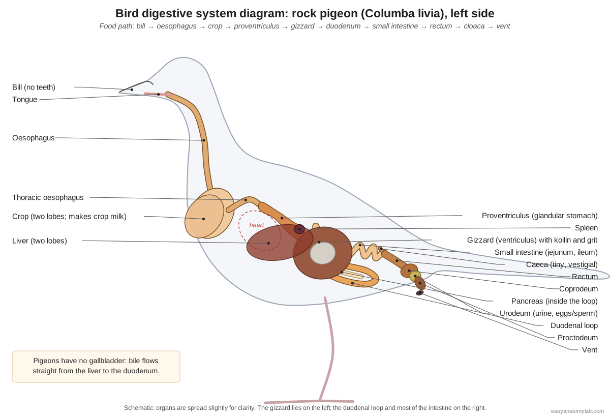
- Oesophagus (esophagus): The muscular food tube from the pharynx down the neck to the crop and on to the stomach. Carries swallowed food by peristalsis; its folded wall stretches to take whole seeds.
- Crop (ingluvies): A thin-walled, two-lobed pouch of the oesophagus at the base of the neck. Stores and softens food; during breeding its lining produces crop milk for the squabs.
- Proventriculus (glandular stomach): The first, spindle-shaped part of the stomach, with a thick glandular lining. Secretes hydrochloric acid and pepsinogen that start the digestion of protein.
- Gizzard (ventriculus): The round, very muscular second stomach that grinds food with swallowed grit. Crushes seeds against grit, acting as the bird's teeth.
- Duodenum (duodenal loop): A U-shaped loop of small intestine leaving the gizzard, with the pancreas held inside it. Receives bile and pancreatic juice and begins most chemical digestion.
- Pancreas: A thin, pale gland lying between the two limbs of the duodenal loop. Makes digestive enzymes (and hormones such as insulin and glucagon).
- Liver: A large, dark red-brown organ with two lobes. Makes bile, stores glycogen and processes absorbed nutrients.
- Small intestine (jejunum and ileum): The long, coiled middle part of the intestine after the duodenal loop. Completes digestion and absorbs nutrients.
- Caeca: A pair of tiny blind pouches where the small intestine joins the rectum. In pigeons they are vestigial and contain lymphoid tissue; in chickens and grouse they are long and ferment plant fibre.
- Rectum (colon): The short, straight large intestine leading to the cloaca. Absorbs water and salts, including water returned backward from the urine.
The cloaca is divided into three chambers. The coprodeum receives faeces from the rectum, the urodeum receives the ureters and the reproductive ducts, and the proctodeumopens to the outside through the vent. Bird droppings are therefore a mixture: dark faeces capped with white uric acid (urates) from the kidneys.
Bird digestive system vs human digestive system
Birds and mammals digest food with the same chemistry, but birds replace teeth with a gizzard, add a crop for storage, split the stomach in two, and end the tract in a cloaca.
| Feature | Pigeon | Human |
|---|---|---|
| Chewing | None: toothless beak; grinding in the gizzard with grit | Teeth and jaws |
| Food storage | Crop (bilobed in pigeons) | Stomach only |
| Stomach | Proventriculus (glandular) + gizzard (muscular) | Single glandular stomach |
| Gallbladder | Absent in pigeons | Present |
| Caeca | A pair, tiny and vestigial in pigeons | One caecum with the appendix |
| Large intestine | Short rectum | Long colon |
| Exit | Cloaca shared with urinary and reproductive tracts | Separate anus, urethra and (in females) vagina |
| Nitrogen waste | Uric acid, little water | Urea in liquid urine |
Seed-eaters vs birds of prey: grit, gizzards and pellets
A bird's digestive system matches its diet. Seed-eaters such as pigeons have a large crop, a very thick, muscular gizzard with a hard koilin lining, and swallow grit that the gizzard uses as millstones. Birds of prey and owls eat meat, have thinner-walled gizzards, and cast pellets: the gizzard compacts fur, feathers, bones and claws into a pellet that the bird coughs up, within about six to ten hours of a meal.
Caeca vary in the same way. Grouse and chickens have long paired caeca that ferment plant fibre; in pigeons the caeca are vestigial and contain lymphoid (immune) tissue. Pellets are cast by many other birds too, including herons, gulls, kingfishers, crows and most shorebirds.
Feather anatomy
A feather has a hollow base, the calamus, held in a follicle in the skin, and a shaft, the rachis, carrying two vanes. Each vane is a row of barbs. Each barb carries two rows of barbules: the barbules facing the feather tip carry tiny hooks (hooklets, hamuli or barbicels) that catch the grooved barbules of the next barb, zipping the vane into a flat, airtight surface.
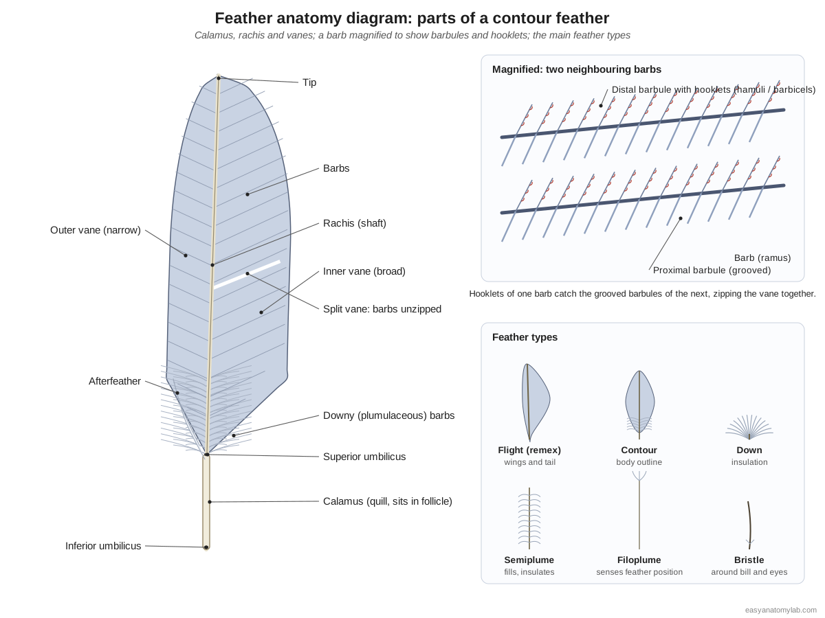
The barbs near the base of most contour feathers are fluffy, plumulaceous barbs without interlocking hooks; they trap air for warmth. Many contour feathers also have a small downy afterfeather attached near the calamus. Under a microscope, the nodes and pigment along downy barbules differ between bird groups, which is how Smithsonian scientists identify birds from feather fragments after aircraft bird strikes.
| Feather type | Structure | Main job |
|---|---|---|
| Flight feathers (remiges and rectrices) | Long, stiff, asymmetric vanes with interlocking barbules | Lift, thrust and steering |
| Contour feathers | Vaned tip, downy base | Body outline, waterproofing, colour |
| Down | Little or no rachis; loose barbs | Insulation |
| Semiplumes | Rachis with loose, unhooked barbs | Insulation and filling out the outline |
| Filoplumes | Hair-like shaft with a few barbs at the tip | Associated with contour feathers; thought to sense feather position |
| Bristles | Stiff shaft, few or no barbs | Around the bill, eyes and nostrils |
Wing and tail feathers: primaries, secondaries, coverts
The flight feathers of the wing (remiges) are the primaries on the hand, the secondaries on the forearm and the tertials near the body. Rows of shorter coverts cover their bases, and the alula sits on the thumb. The tail feathers (rectrices) fan out from the pygostyle.
- Primary flight feathers (primaries): The long outer flight feathers of the wing, attached to the bones of the hand. Produce thrust on the downstroke and shape the wingtip; they can spread like fingers or close.
- Secondary flight feathers (secondaries): The flight feathers of the forearm, attached to the ulna. Form the lift-producing inner wing surface.
- Tertials: The innermost long wing feathers, beside the body, over the upper arm. Close the gap between wing and body and cover the folded secondaries.
- Wing coverts: Rows of short feathers that cover the bases of the flight feathers and the leading edge of the wing. Smooth the airflow over the wing and protect the bones and patagium (wing skin fold) underneath.
- Alula (bastard wing): A small tuft of stiff feathers on the alular digit (the 'thumb') at the front of the wrist. Lifted like an aircraft's leading-edge slat, it keeps airflow attached at slow speeds and steep angles, as in landing.
- Tail feathers (rectrices): The long, paired flight feathers of the tail, rooted around the pygostyle. Steer, brake and add lift; the tail spreads wide on landing.
Most non-passerine birds, including pigeons, have 9–11 primaries; secondary counts range from about six in hummingbirds to around 40 in some albatrosses. Most birds have 12 rectrices (six pairs), but centuries of breeding have made the tail-feather count highly variable in domestic pigeons.
Blood feathers, pin feathers and molt
A blood feather (pin feather) is a new feather that is still growing. It develops inside a keratin sheath, with a blood supply running up its shaft. As it grows, the tip matures first and the blood withdraws towards the follicle; once the feather is full-grown its blood supply stops and the bird preens away the sheath. A broken blood feather can bleed heavily, so birds in molt are handled gently.
Molt is the regular replacement of feathers. In most passerines the wing molt starts between the innermost primary and the first secondary and spreads in both directions, a few feathers at a time, so the bird can keep flying. Some waterbirds, such as most ducks, loons and grebes, drop all their flight feathers at once and are flightless for three to four weeks.
Bird air sacs and respiration
Birds breathe with small, rigid lungs and a system of thin-walled air sacs. The sacs work as bellows; the lungs, which barely change volume, only exchange gas. Air flows through the lungs' parabronchi in the same direction during both inspiration and expiration, which is why bird lungs extract oxygen so efficiently. Birds have no diaphragm.
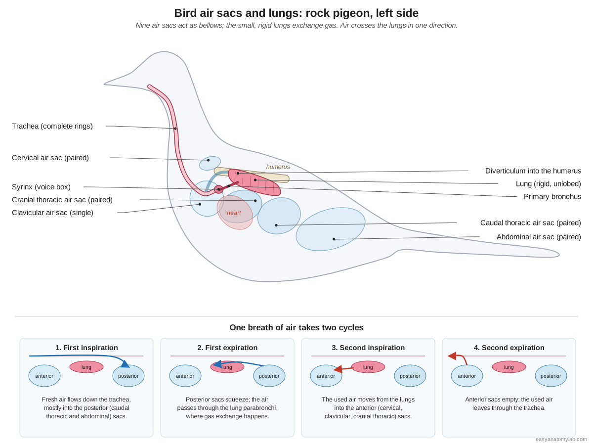
- Trachea (windpipe): A long tube of complete, overlapping cartilage rings running down the neck. Carries air between the larynx and the syrinx and bronchi.
- Syrinx: The bird's voice box, at the fork of the trachea into the two bronchi. Vibrating tympaniform membranes in its walls produce the pigeon's coo.
- Lungs: Small, bright pink, spongy lungs pressed into the ribs on the roof of the thorax. Gas exchange in the air capillaries around the parabronchi. The lungs barely change volume: the air sacs do the pumping.
- Cervical air sacs: A pair of small air sacs at the base of the neck, above the lungs. Part of the anterior air sac group; their extensions pneumatise the neck and thoracic vertebrae.
- Clavicular air sac: A single air sac at the thoracic inlet, around the syrinx and the base of the heart. Sends diverticula into the humerus and around the shoulder, and surrounds the syrinx.
- Cranial thoracic air sacs: A pair of air sacs on the sides of the thorax, ventral to the lungs. Anterior air sacs: they receive air that has already passed through the lungs and send it out on expiration.
- Caudal thoracic air sacs: A pair of air sacs behind the cranial thoracic sacs, on the sides of the body cavity. Posterior air sacs: they fill with fresh air on inspiration and push it through the lungs on expiration.
- Abdominal air sacs: The largest air sacs, filling spaces among the intestines in the rear abdomen. Posterior air sacs that store fresh air; their extensions reach into the synsacrum and around the hip sockets.
Air sacs also reach into the bones. In pigeons and chickens the clavicular sac sends a diverticulum into the humerus, the cervical sacs extend along the vertebrae, and the abdominal sacs reach into the synsacrum and around the hip sockets. Sound, finally, comes not from the larynx but from the syrinx, deep in the chest where the trachea divides, surrounded by the clavicular air sac.
Bird heart and circulation
Birds have a four-chambered heart: two atria and two ventricles, with oxygen-rich and oxygen-poor blood kept fully separate, as in mammals. The bird heart is large for the body size, and its aorta curves to the right instead of the left.
- Right atrium: The thin-walled chamber that receives oxygen-poor blood from the body. Receives blood from the two cranial venae cavae and the caudal vena cava and passes it to the right ventricle.
- Left atrium: The chamber that receives oxygen-rich blood from the lungs. Passes blood from the pulmonary veins to the left ventricle.
- Right ventricle: The crescent-shaped chamber wrapped around the right side of the left ventricle. Pumps oxygen-poor blood to the lungs through the pulmonary arteries.
- Left ventricle: The thick-walled, cone-shaped main pumping chamber forming the heart's apex. Pumps oxygen-rich blood into the aorta for the whole body.
- Right aortic arch and dorsal aorta: The main artery: it leaves the left ventricle, arches over to the right, and runs back under the spine. Carries oxygen-rich blood to the body.
- Brachiocephalic arteries: Two large arteries from the aortic arch, each splitting into a common carotid and a subclavian artery. Supply the head and neck (carotids) and the wing and huge breast muscles (subclavian, pectoral arteries).
- Venae cavae: The great veins returning oxygen-poor blood to the right atrium. Return blood from the body to the heart.
Blood returns to the right atrium through three great veins (two cranial venae cavae and one caudal vena cava), passes to the right ventricle and the lungs, returns to the left atrium and is pumped by the thick left ventricle into the right aortic arch. Two large brachiocephalic arteries leave the arch for the head and the wings; their pectoral branches feed the flight muscles.
Bird kidneys and reproductive system: male vs female
Bird kidneys are three-lobed and pressed into the pelvis; their ureters run straight to the cloaca, because birds have no urinary bladder. Males have two internal testes; females of most species, including pigeons, have only a left ovary and left oviduct.
| Male pigeon | Female pigeon | |
|---|---|---|
| Gonads | Two bean-shaped testes in front of the kidneys, the left often larger | Single left ovary with yolk follicles |
| Ducts | Zigzag ductus deferens to the urodeum, widening into a seminal vesicle | Coiled left oviduct: infundibulum, magnum, isthmus, shell gland, vagina |
| Copulatory organ | None (cloacal contact) | None |
| Breeding role | Shares incubation; makes crop milk | Lays a clutch of two eggs; shares incubation; makes crop milk |
Switch the 3D model to Female or Male to compare the organs.
Brain and senses of a bird
The pigeon brain has two smooth cerebral hemispheres, a folded cerebellum, large optic lobes on its lower sides and a medulla that leads into the spinal cord. The size of the optic lobes reflects how much the pigeon relies on vision.
- Cerebral hemispheres: The two smooth, unfolded hemispheres at the front of the brain. Learning, memory, navigation and complex behaviour.
- Optic lobes (optic tectum): Two large rounded swellings on the lower sides of the brain. Process vision, the pigeon's dominant sense.
- Cerebellum: A single, ridged (folded) lobe at the back of the brain. Coordinates balance and movement, crucial in flight.
- Medulla oblongata: The base of the brain, tapering into the spinal cord. Controls breathing, heart rate and other automatic functions.
Bird eyes have three eyelids, the third being the nictitating membrane that sweeps across from the front corner, and inside, a pleated, blood-rich comb called the pecten. Each eye is stiffened by a ring of bony plates, the sclerotic ring.
Bird muscles: the flight engine
The largest muscles of a pigeon are the two flight muscles on the breast. The pectoralis pulls the wing down; the supracoracoideus, underneath it, lifts the wing using a tendon that runs through a bony pulley at the shoulder.
- Pectoralis (breast muscle): The huge, dark red, superficial breast muscle that powers the downstroke. Pulls the wing down and forward: the power stroke of flight.
- Supracoracoideus: The deep breast muscle, under the pectoralis, that lifts the wing through a pulley at the shoulder. Raises the wing and twists (supinates) the humerus during the upstroke.
- Thigh and drumstick muscles: The muscles around the femur and tibiotarsus that move the leg and toes. Walk, perch and launch the bird into the air; long tendons run down to the toes.
Bird anatomy labeled: every part and its function
This is the text version of the 3D model: every labeled structure of the rock pigeon, grouped by body system.
Feathers & skin
- Contour feathers (body plumage) (Body feathers, plumage)
- The overlapping vaned feathers that cover the neck, back, breast and belly and give the bird its smooth outline. Streamline the body, shed water, insulate, and carry colour and pattern. Their fluffy (plumulaceous) bases trap air for warmth.
- Head (Crown, forehead, face)
- The small, rounded, feathered head that carries the bill, eyes and ear openings. Holds the brain and the main sense organs and positions the bill for feeding.
- Bill (beak) (Rhamphotheca over the upper and lower mandibles)
- The toothless bill: a horny keratin sheath (rhamphotheca) over the bones of the upper and lower jaws. Picks up seeds and grit, drinks (pigeons can suck water up without tipping the head back), preens feathers and feeds the squabs.
- Cere (operculum) (Wattle, nasal operculum)
- The soft, swollen, whitish patch of skin at the base of the upper bill, over the nostrils. Covers and partly shields the nostrils.
- Nostrils (external nares)
- Slit-like openings into the nasal cavity, at the base of the upper bill under the cere. Let air into the nasal cavity, which opens into the mouth through a slit in the palate (the choana).
- Primary flight feathers (primaries) (Primary remiges)
- The long outer flight feathers of the wing, attached to the bones of the hand. Produce thrust on the downstroke and shape the wingtip; they can spread like fingers or close.
- Secondary flight feathers (secondaries) (Secondary remiges)
- The flight feathers of the forearm, attached to the ulna. Form the lift-producing inner wing surface.
- Tertials (Tertiary feathers)
- The innermost long wing feathers, beside the body, over the upper arm. Close the gap between wing and body and cover the folded secondaries.
- Wing coverts (Lesser, median and greater coverts; primary coverts)
- Rows of short feathers that cover the bases of the flight feathers and the leading edge of the wing. Smooth the airflow over the wing and protect the bones and patagium (wing skin fold) underneath.
- Alula (bastard wing)
- A small tuft of stiff feathers on the alular digit (the 'thumb') at the front of the wrist. Lifted like an aircraft's leading-edge slat, it keeps airflow attached at slow speeds and steep angles, as in landing.
- Tail feathers (rectrices) (Rectrices, tail fan)
- The long, paired flight feathers of the tail, rooted around the pygostyle. Steer, brake and add lift; the tail spreads wide on landing.
- Upper- and undertail coverts
- Short feathers that cover the bases of the tail feathers above and below. Smooth the junction between body and tail.
- Down feathers (Plumules)
- Soft, fluffy feathers without interlocking hooklets, hidden under the contour feathers. Trap air for insulation.
- Uropygial (preen) gland (Oil gland)
- A small skin gland on the back at the base of the tail that secretes preen oil. The bird squeezes oil from it with the bill and spreads it through the plumage while preening.
- Feet and scales (podotheca) (Tarsus and toes, scutes)
- The bare, red, scaly skin of the tarsus and the four toes. Support the bird while perching and walking; the scales are keratin, like the bill.
- Claws (Ungues)
- Curved keratin sheaths over the last bone of each toe. Grip perches and the ground.
- Vent (cloacal opening)
- The single external opening of the cloaca, under the base of the tail. Releases droppings (faeces plus white urates), and eggs or sperm.
Skeleton
- Premaxilla (Os premaxillare, upper bill bone)
- The main bone of the upper bill. Supports the horny upper bill.
- Nasal bone
- A forked bone between the premaxilla and the frontal that frames the nostril. Forms the back edge of the nasal opening and the roof of the nasal cavity.
- Frontal bone
- The broad roof of the skull between and above the orbits. Roofs the orbits and the front of the braincase.
- Parietal bone
- The rounded top-back of the braincase. Covers the cerebral hemispheres and cerebellum.
- Occipital bone and condyle (Occipital condyle, foramen magnum)
- The back of the skull, around the foramen magnum, with a single rounded occipital condyle below it. The single condyle forms a ball joint with the atlas, allowing very wide head rotation.
- Lacrimal bone (Prefrontal)
- A small bone forming the front rim of the orbit. Completes the bony rim in front of the eye.
- Jugal bar (Zygomatic arch (maxilla, jugal and quadratojugal fused))
- A thin, rod-like bony bar running from the upper bill back to the quadrate below the eye. Pushes the upper bill when the quadrate swings forward, helping it flex as the bill opens.
- Quadrate
- A small, movable bone at the back of the orbit that links skull, palate and lower jaw. The lower jaw hinges on it. Because it can swing, it transmits movement to the jugal bar and palate so the upper bill rises as the mouth opens (cranial kinesis).
- Palatine and pterygoid bones (Bony palate)
- Paired rods in the roof of the mouth linking the quadrate to the upper bill. Slide forward and back with the quadrate to raise and lower the upper bill.
- Mandible (lower jaw) (Dentary, angular and other fused jaw bones)
- The V-shaped lower jaw, made of several fused bones, covered by the horny lower bill. Opens and closes the bill.
- Sclerotic ring (Scleral ossicles)
- A ring of small overlapping bony plates in the wall of each eye. Stiffens the large eyeball and gives attachment to the muscles that change focus.
- Hyoid apparatus
- The slender bones inside the tongue and their two long horns. Support and move the tongue.
- Atlas and axis (Cervical vertebrae 1 and 2)
- The first two neck vertebrae: the ring-shaped atlas and the axis with its odontoid process. The atlas cups the single occipital condyle; together they allow the head to turn and nod.
- Cervical vertebrae (C3–C12) (Neck vertebrae)
- The long, flexible chain of neck vertebrae with saddle-shaped (heterocoelous) joints. Give the S-shaped neck its reach and flexibility for feeding and preening, and stabilise the head while walking.
- Thoracic vertebrae (notarium region) (Notarium in some birds)
- The rib-bearing vertebrae of the back; in many birds several fuse into a rigid notarium. Carry the ribs and form a stiff back that resists the forces of the flight muscles.
- Synsacrum (Lumbosacral mass)
- A long rod of fused vertebrae (the last thoracic, the lumbar, the sacral and the first caudal vertebrae), welded to the pelvis. Makes the back and pelvis one rigid frame that carries the body on the legs.
- Free caudal vertebrae (Free tail vertebrae, coccygeal vertebrae)
- A short series of movable tail vertebrae between the synsacrum and the pygostyle. Let the tail move up, down and sideways.
- Pygostyle (Ploughshare bone)
- A small blade of fused tail vertebrae at the end of the spine. Anchors the tail feathers and their muscles, so the tail fan can be spread and steered.
- Ribs (vertebral and sternal segments)
- Seven pairs of ribs, most with a dorsal (vertebral) and a ventral (sternal) bony segment. Form a rigid but hinged rib cage; the joint between segments lets the sternum swing down and up to pump the air sacs.
- Uncinate processes
- Backward-pointing hooks on the middle of the ribs that overlap the next rib. Brace the rib cage and give leverage to the breathing muscles.
- Sternum (breastbone)
- A large, curved bony shield under the thorax and abdomen. Anchors the flight muscles, protects the heart and liver, and moves during breathing.
- Keel (carina) (Carina sterni)
- The tall blade along the midline of the sternum. Gives a large area for the attachment of the pectoralis and supracoracoideus.
- Furcula (wishbone) (Fused clavicles)
- The V-shaped bone formed by the two fused clavicles. Acts as a spring brace between the shoulders during the wingbeat and helps form the triosseal canal.
- Coracoid
- A strong strut from each shoulder to the front edge of the sternum. Braces the shoulder against the pull of the huge pectoralis on the downstroke.
- Scapula (shoulder blade)
- A long, narrow, sabre-shaped blade lying over the ribs. Forms part of the shoulder joint and anchors wing muscles.
- Triosseal canal (Foramen triosseum)
- A bony tunnel at the shoulder where the coracoid, scapula and furcula meet. Acts as a pulley: the supracoracoideus tendon passes through it, so a muscle on the breast can lift the wing.
- Humerus
- The upper arm bone: short, stout and hollow (pneumatic). Transmits the force of the flight muscles to the wing. The pectoralis pulls it down; the supracoracoideus tendon lifts and twists it.
- Radius
- The thinner, straight bone of the forearm, on the leading edge. Slides along the ulna as the elbow flexes, automatically folding or spreading the hand.
- Ulna
- The thicker, slightly bowed forearm bone that carries the secondary feathers. Supports the secondaries, which attach along its trailing edge.
- Radiale and ulnare (free carpals) (Wrist bones)
- The two small free wrist bones left in the bird wing. Guide wrist folding; the other carpals are fused into the carpometacarpus.
- Carpometacarpus
- The fused bone of the hand: three metacarpals fused with some carpals. A rigid base for the primary feathers.
- Alular digit ('Thumb', digit carrying the alula)
- The short first digit at the front of the hand. Carries the alula feathers.
- Major digit
- The longest finger, of two phalanges, extending the hand to the wingtip. Supports the outer primaries.
- Minor digit
- A tiny single-phalanx finger beside the major digit. A small anchor for primary feathers.
- Ilium
- The large, plate-like upper bone of the pelvis, fused to the synsacrum. Carries the hip socket (acetabulum) and anchors the thigh muscles.
- Ischium
- The flat bone forming the back-lower part of the pelvis. Completes the side wall of the pelvis.
- Pubis
- A thin rod running backward along the lower edge of the ischium. Supports the abdominal wall.
- Femur (thigh bone)
- The short thigh bone, held almost horizontally and hidden inside the body outline. Carries the body weight to the knee; the forward-pointing femur keeps the feet under the centre of gravity.
- Patella (kneecap)
- A small sesamoid bone in front of the knee. Improves the leverage of the knee extensor muscles.
- Tibiotarsus (Drumstick bone)
- The long shin bone: the tibia fused with the upper row of ankle (tarsal) bones. Main bone of the lower leg; the 'drumstick' muscles surround it.
- Fibula
- A thin splint of bone along the outer side of the tibiotarsus. Anchors leg muscles; it is reduced and does not reach the ankle.
- Tarsometatarsus (Tarsus, shank)
- The bone of the bare lower leg: fused metatarsals and the lower row of tarsals. Adds a third long segment to the leg, so birds stand on their toes (digitigrade).
- Hallux (digit I) and metatarsal I (Hind toe)
- The backward-pointing first toe, attached by a small free metatarsal. Opposes the front toes, so the foot can grip a perch.
- Toe bones (digits II–IV) (Phalanges of the forward toes)
- The phalanges of the three forward-pointing toes. Spread the weight when standing and walking.
Muscles
- Pectoralis (breast muscle) (Pectoralis major)
- The huge, dark red, superficial breast muscle that powers the downstroke. Pulls the wing down and forward: the power stroke of flight.
- Supracoracoideus (Pectoralis minor)
- The deep breast muscle, under the pectoralis, that lifts the wing through a pulley at the shoulder. Raises the wing and twists (supinates) the humerus during the upstroke.
- Thigh and drumstick muscles
- The muscles around the femur and tibiotarsus that move the leg and toes. Walk, perch and launch the bird into the air; long tendons run down to the toes.
Digestive
- Tongue
- A narrow, pointed tongue on the floor of the mouth, supported by the hyoid. Moves seeds back to the pharynx for swallowing.
- Oesophagus (esophagus)
- The muscular food tube from the pharynx down the neck to the crop and on to the stomach. Carries swallowed food by peristalsis; its folded wall stretches to take whole seeds.
- Crop (ingluvies)
- A thin-walled, two-lobed pouch of the oesophagus at the base of the neck. Stores and softens food; during breeding its lining produces crop milk for the squabs.
- Proventriculus (glandular stomach)
- The first, spindle-shaped part of the stomach, with a thick glandular lining. Secretes hydrochloric acid and pepsinogen that start the digestion of protein.
- Gizzard (ventriculus) (Muscular stomach)
- The round, very muscular second stomach that grinds food with swallowed grit. Crushes seeds against grit, acting as the bird's teeth.
- Duodenum (duodenal loop)
- A U-shaped loop of small intestine leaving the gizzard, with the pancreas held inside it. Receives bile and pancreatic juice and begins most chemical digestion.
- Pancreas
- A thin, pale gland lying between the two limbs of the duodenal loop. Makes digestive enzymes (and hormones such as insulin and glucagon).
- Liver
- A large, dark red-brown organ with two lobes. Makes bile, stores glycogen and processes absorbed nutrients.
- Small intestine (jejunum and ileum)
- The long, coiled middle part of the intestine after the duodenal loop. Completes digestion and absorbs nutrients.
- Caeca (Ceca)
- A pair of tiny blind pouches where the small intestine joins the rectum. In pigeons they are vestigial and contain lymphoid tissue; in chickens and grouse they are long and ferment plant fibre.
- Rectum (colon) (Large intestine)
- The short, straight large intestine leading to the cloaca. Absorbs water and salts, including water returned backward from the urine.
- Coprodeum
- The first and largest chamber of the cloaca, continuous with the rectum. Receives and briefly holds faeces.
- Proctodeum
- The last chamber of the cloaca, opening to the outside through the vent. Passes droppings, eggs and sperm to the vent.
Lungs & air sacs
- Larynx and glottis
- The slit-like opening into the trachea on a mound at the back of the tongue. Closes the airway during swallowing; it does not make sound.
- Trachea (windpipe)
- A long tube of complete, overlapping cartilage rings running down the neck. Carries air between the larynx and the syrinx and bronchi.
- Syrinx
- The bird's voice box, at the fork of the trachea into the two bronchi. Vibrating tympaniform membranes in its walls produce the pigeon's coo.
- Primary bronchi
- The two tubes from the syrinx into the lungs. Carry air into each lung and on to the air sacs.
- Lungs
- Small, bright pink, spongy lungs pressed into the ribs on the roof of the thorax. Gas exchange in the air capillaries around the parabronchi. The lungs barely change volume: the air sacs do the pumping.
- Cervical air sacs
- A pair of small air sacs at the base of the neck, above the lungs. Part of the anterior air sac group; their extensions pneumatise the neck and thoracic vertebrae.
- Clavicular air sac (Interclavicular air sac)
- A single air sac at the thoracic inlet, around the syrinx and the base of the heart. Sends diverticula into the humerus and around the shoulder, and surrounds the syrinx.
- Cranial thoracic air sacs (Anterior thoracic air sacs)
- A pair of air sacs on the sides of the thorax, ventral to the lungs. Anterior air sacs: they receive air that has already passed through the lungs and send it out on expiration.
- Caudal thoracic air sacs (Posterior thoracic air sacs)
- A pair of air sacs behind the cranial thoracic sacs, on the sides of the body cavity. Posterior air sacs: they fill with fresh air on inspiration and push it through the lungs on expiration.
- Abdominal air sacs
- The largest air sacs, filling spaces among the intestines in the rear abdomen. Posterior air sacs that store fresh air; their extensions reach into the synsacrum and around the hip sockets.
Heart & vessels
- Right atrium
- The thin-walled chamber that receives oxygen-poor blood from the body. Receives blood from the two cranial venae cavae and the caudal vena cava and passes it to the right ventricle.
- Left atrium
- The chamber that receives oxygen-rich blood from the lungs. Passes blood from the pulmonary veins to the left ventricle.
- Right ventricle
- The crescent-shaped chamber wrapped around the right side of the left ventricle. Pumps oxygen-poor blood to the lungs through the pulmonary arteries.
- Left ventricle
- The thick-walled, cone-shaped main pumping chamber forming the heart's apex. Pumps oxygen-rich blood into the aorta for the whole body.
- Right aortic arch and dorsal aorta
- The main artery: it leaves the left ventricle, arches over to the right, and runs back under the spine. Carries oxygen-rich blood to the body.
- Brachiocephalic arteries (Innominate arteries, with carotid and subclavian branches)
- Two large arteries from the aortic arch, each splitting into a common carotid and a subclavian artery. Supply the head and neck (carotids) and the wing and huge breast muscles (subclavian, pectoral arteries).
- Pulmonary arteries
- Vessels from the right ventricle to the lungs. Carry oxygen-poor blood to the lungs.
- Venae cavae (Two precavae and one postcava)
- The great veins returning oxygen-poor blood to the right atrium. Return blood from the body to the heart.
- Jugular veins
- The large veins running down each side of the neck. Drain blood from the head and neck into the cranial venae cavae.
- Spleen
- A small, rounded, red-brown lymphoid organ. Filters old red blood cells and blood-borne microbes from the blood and makes immune cells. Birds have no lymph nodes, so the spleen and bursa are key immune organs.
Urinary
- Urodeum
- The middle chamber of the cloaca, which receives the ureters and the reproductive ducts. Collects urine (urates) and sperm or eggs.
- Kidneys
- Paired, dark red-brown, three-lobed kidneys pressed into the hollows of the synsacrum. Filter the blood and excrete nitrogen as uric acid (white urates) instead of urea, saving water.
- Ureters
- Two thin tubes from the kidneys to the urodeum of the cloaca. Carry urine and urates to the cloaca.
Reproductive
- Testes · males only
- Paired, bean-shaped, cream-coloured testes inside the body cavity. Produce sperm; they swell greatly in the breeding season.
- Ductus deferens and seminal vesicle (Vas deferens) · males only
- A white, zigzag sperm duct from each testis to the cloaca, widening at its end. Carries and stores sperm; the expanded end (seminal vesicle) holds sperm before mating.
- Ovary (left) · females only
- A single left ovary, like a small bunch of grapes of yolk follicles. Produces ova (yolks) and sex hormones.
- Oviduct (left) · females only
- A long, coiled tube from the ovary to the cloaca where the egg is completed. Fertilisation, then addition of albumen, shell membranes and shell as the egg travels down it.
Glands & immune organs
- Thyroid glands
- A pair of small, oval, dark red glands at the base of the neck. Make thyroid hormones (thyroxine and triiodothyronine) that set the metabolic rate and control moult and growth of feathers.
- Adrenal glands
- Small, paired, yellow-orange glands at the front of the kidneys. Make corticosterone (the main bird stress and metabolic hormone), aldosterone, and adrenaline and noradrenaline.
- Bursa of Fabricius (Cloacal bursa)
- A small, round lymphoid sac on the dorsal wall of the cloaca, present only in young birds. Site where B lymphocytes (antibody-making white blood cells) mature.
Nervous
- Cerebral hemispheres (Cerebrum, telencephalon)
- The two smooth, unfolded hemispheres at the front of the brain. Learning, memory, navigation and complex behaviour.
- Optic lobes (optic tectum)
- Two large rounded swellings on the lower sides of the brain. Process vision, the pigeon's dominant sense.
- Cerebellum
- A single, ridged (folded) lobe at the back of the brain. Coordinates balance and movement, crucial in flight.
- Medulla oblongata (Brainstem)
- The base of the brain, tapering into the spinal cord. Controls breathing, heart rate and other automatic functions.
- Spinal cord
- The nerve cord running through the vertebral canal from the brain to the tail. Carries signals between brain and body; enlarged where the wing and leg nerves leave.
Sense organs
- Eye
- A very large eye on each side of the head, with an orange iris in the rock pigeon. Wide field of view for spotting predators; good colour vision.
- Ear opening (external acoustic meatus)
- A small opening behind and below the eye, hidden under feathers (ear coverts). Leads to the eardrum and middle ear; birds have no external ear flap (pinna).
Crop milk: how pigeons feed their squabs
Pigeons and doves are unusual among birds in feeding their young a secretion from the crop. In both males and females, the hormone prolactin makes the lining of the crop thicken during brooding; its surface cells fill with fat and are shed into the crop as a thick, protein- and fat-rich fluid that also carries immunoglobulins. During production the crop lining becomes visibly congested and thickened. (Crespo et al. (2018): Galliformes and Columbiformes, Pathology of Wildlife and Zoo Animals.)
The anatomy supports this function. In domestic pigeons the crop is a large, thin-walled sac with two lateral diverticula at the entrance to the thorax, just in front of the breast muscles. Squabs start on crop milk alone; over roughly 4–7 days the parents switch to feeding them mostly regurgitated seed. Crop milk is rich in fat and protein and contains essentially no carbohydrate. Flamingos and male emperor penguins also produce a crop or oesophageal milk, an example of the same solution evolving more than once. (Wikipedia: crop milk.)
Why the pigeon has no gallbladder
Pigeons lack a gallbladder, although many birds, including chickens and gulls, have one. A comparative study of pigeon and gull livers found the gallbladder absent in every pigeon examined; bile leaves the two-lobed liver through two bile ducts that open into the duodenum. The Weber State dissection guide makes the same observation for the rock dove. (Khoder & Hameed (2025): liver and biliary system of the pigeon and gull, Iraqi Journal of Veterinary Sciences.)
For a student, the practical point is not to search for a missing organ: the liver's two hepatic ducts are the whole biliary route. Many textbook diagrams are drawn from the chicken and show a gallbladder, so check which species a diagram shows before you label a pigeon. (Cavitt, Weber State University: external and internal anatomy of the rock dove (PDF).)
Air sacs in pigeons: what the dissection studies found
A morphological study of domestic pigeons described paired cervical air sacs, a single clavicular sac formed by the fusion of two bilateral diverticula, cranial thoracic sacs whose diverticula extend into the second to seventh ribs, smaller cranial than caudal thoracic sacs, caudal thoracic sacs with no diverticula into bone, and abdominal sacs with cranial and caudal parts. That gives nine air sacs, the usual figure for birds. (Hussein, Ibrahim & Ali (2017): morphology of the air sacs in domestic pigeons.)
Some veterinary texts based on the chicken describe eight air sacs, because sacs joined across the midline are counted as one; poultry sources such as Poultry Hub count nine. The difference is a matter of counting, not of function: in every bird the sacs work as bellows, the lungs barely change volume, and there is no diaphragm. Air sac walls are only a few cells thick, which is why they appear as glistening, cobweb-like films when the body cavity is opened. (WikiVet: avian respiration, anatomy and physiology.)
Flight muscles: pectoralis and supracoracoideus
In flying birds the pectoral muscles make up roughly 15–25% of body mass, and together with the supracoracoideus 25–40%. The pectoralis pulls the wing down and forward in the power stroke; the smaller supracoracoideus, lying under it against the keel, lifts and twists the wing through its tendon pulley at the shoulder. (Wikipedia: supracoracoideus and flight muscle mass.)
Measurements in pigeons show the size difference: Tobalske and Biewener recorded single-side muscle masses averaging about 53 g for the pectoralis and 10 g for the supracoracoideus. Hummingbirds, which hover and generate about a quarter of their weight support on the upstroke, have a proportionally larger supracoracoideus than other birds. (Tobalske & Biewener (2008): the pigeon supracoracoideus in flight, Journal of Experimental Biology.)
Frequently asked questions
What are the parts of a bird's digestive system in order?
Bill and mouth, oesophagus, crop, proventriculus (glandular stomach), gizzard (muscular stomach), duodenum with the pancreas, small intestine (jejunum and ileum), caeca, rectum and cloaca, which opens at the vent. The liver empties bile into the duodenum.
Why do birds need a gizzard?
Birds have no teeth. The gizzard is a thick muscular stomach lined with a hard koilin layer; with swallowed grit it grinds seeds and other hard food that the bill swallowed whole.
Do pigeons have a gallbladder?
No. Pigeons lack a gallbladder. Bile flows from the two-lobed liver directly into the duodenum through two bile ducts. Many other birds, including chickens and gulls, do have one.
How many air sacs does a bird have?
Typically nine: paired cervical, an unpaired clavicular, paired cranial thoracic, paired caudal thoracic and paired abdominal sacs. Numbers range from about seven to twelve between species; for the chicken, poultry sources count nine, while veterinary texts often count eight because they treat sacs joined across the midline as one.
Why do birds breathe in one direction?
The air sacs act as bellows. Fresh air goes mostly to the posterior air sacs on inspiration and is pushed through the lungs' parabronchi on expiration, while used air from the lungs moves to the anterior sacs and out. Air flows through the gas-exchange tubes in the same direction during both phases.
What are the parts of a feather?
A feather has a hollow base, the calamus, set in a follicle in the skin; a central shaft, the rachis; and two vanes made of barbs. Each barb carries barbules, and hooklets (barbicels) on the barbules zip neighbouring barbs together into a flat vane. Downy barbs at the base lack hooklets.
What is a blood feather?
A growing feather (pin feather) still wrapped in its keratin sheath and supplied by blood vessels inside the shaft. The blood supply withdraws as the feather matures and the bird preens off the sheath. A broken blood feather can bleed heavily.
Do birds have a four-chambered heart?
Yes. Birds have two atria and two ventricles with complete separation of oxygen-rich and oxygen-poor blood, like mammals. Unlike mammals, the aorta arches to the right.
Why don't birds have a bladder?
Birds excrete nitrogen as uric acid, a white paste that needs little water, and send urine straight from the kidneys to the cloaca. Not storing liquid urine keeps the body light for flight.
Do female birds have two ovaries?
Usually only one. In most birds, including pigeons and chickens, only the left ovary and oviduct develop. Some birds of prey often have two functional ovaries.
References
- Cavitt, J. Weber State University. External and internal anatomy of the rock dove (ornithology lab guide).
- Crespo, R., França, M. S., Fenton, H. & Shivaprasad, H. L. (2018). Galliformes and Columbiformes. In Pathology of Wildlife and Zoo Animals, 747–773. Elsevier.
- Mahdy, M. A. A. & Mohammed, E. S. I. (2024). Anatomical, histological and SEM features of the esophagus and crop in young and adult domestic pigeons. BMC Veterinary Research 20, 428.
- Khoder, D. A. & Hameed, B. K. (2025). Macro- and microscopic anatomy of the liver and biliary system in pigeon and gull. Iraqi Journal of Veterinary Sciences 39(4), 701–709.
- Hussein, A. A., Ibrahim, R. S. & Ali, H. K. (2017). Morphological study of the air sacs in domestic pigeons. Diyala Agricultural Sciences Journal 9, 47–51.
- WikiVet. Avian respiration: anatomy and physiology.
- Tobalske, B. W. & Biewener, A. A. (2008). Contractile properties of the pigeon supracoracoideus during different modes of flight. Journal of Experimental Biology 211, 170–179.
- Dove, C. J. Forensic ornithology lab exercise (feather structure). Division of Birds, Smithsonian Institution.
- Jacob, J. Anatomy of a feather. Small and Backyard Poultry, University of Kentucky extension.
- VCA Animal Hospitals. Blood feathers in birds.
- Wikipedia: Flight feather; Crop milk; Rock dove; Preen gland; Pellet (ornithology); Bird anatomy.
More interactive anatomy models
- Bird skeletonHollow bones, keeled sternum, wishbone and fused wing bones of a bird.
- Chicken anatomyCrop, gizzard, reproductive tract, skeleton and external parts of the chicken.
- Pigeon dissectionGuided pigeon dissection: flight muscles, crop, gizzard, air sacs and heart.
- Human anatomy1,000+ bones, muscles, organs, vessels and nerves from real anatomical data.
- Octopus anatomyThree hearts, a ring-shaped brain, eight arms and a beak, in 3D.
- Squid anatomyPen, eight arms, two tentacles, giant axons and three hearts of the longfin squid.