

Bat skeleton at a glance
A bat skeleton is a mammal skeleton rebuilt for flight. It has the usual mammal skull, backbone, ribs, shoulder and hip girdles and four limbs, but the arm and hand have become a wing: the forearm and four fingers are enormously long, a skin membrane stretches between them, and the hind legs are turned outward so that the knees bend sideways and backward. A spur on each heel, the calcar, supports the tail membrane.
This page uses the little brown bat, Myotis lucifugus, as the reference species. It is a typical insect-eating, echolocating microbat of the family Vespertilionidae (evening bats), weighing about 5–14 g, with a forearm of about 33–41 mm and a wingspan of about 22–27 cm. Its close relative the big brown bat (Eptesicus fuscus), a common bat in buildings in North America, is very similar, and the differences are noted where they matter. Fruit bats, vampire bats and the hammer-headed bat are compared further down.
Directions: dorsal is the back, ventral the belly, anterior towards the head and posterior towards the tail. Wing digits are numbered like fingers: digit I is the thumb and digit V the little finger.
Bat skeleton labeled: diagram
A labeled bat skeleton shows the skull, 7 cervical, 11 thoracic and 5 lumbar vertebrae, a fused sacrum and the tail vertebrae; the ribs, sternum, clavicles and scapulae; and in each wing the humerus, radius, reduced ulna, carpals, clawed thumb and the metacarpals and phalanges of digits II to V. The hindlimb has a small pelvis, femur, tibia, a thin fibula, ankle bones with the calcar, and five clawed toes.
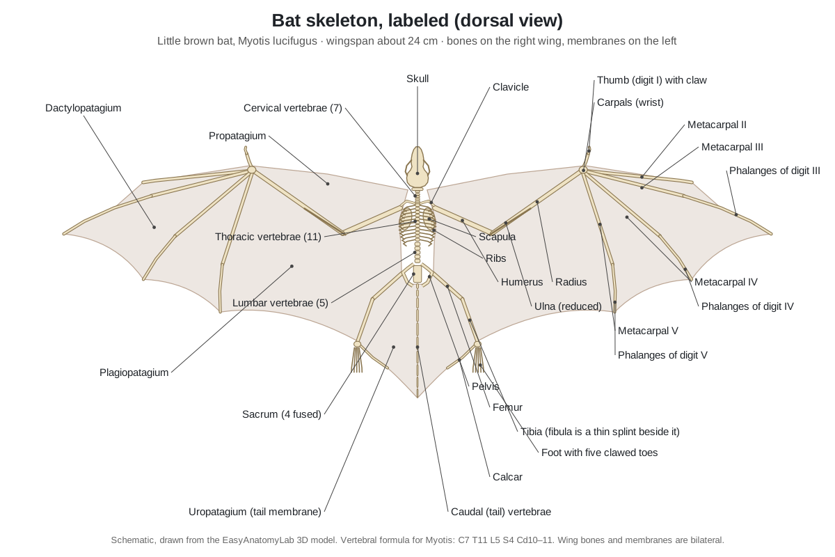
When you draw a bat skeleton, three proportions make it look right. The radius (forearm) is much longer than the humerus; the third finger is longer than the whole arm; and the legs are short and thin, pointing out to the sides and back.
Bat skeleton anatomy: every part and its function
This is the text version of the 3D model: every labeled structure of the little brown bat, grouped by body region, with what it does.
Skin & wing membranes
- Body (fur and skin)
- The furred skin of the head and body, shown faded so the skeleton inside can be seen. Insulates the small body; the wing membranes are extensions of this same skin from the back and belly.
- Propatagium (leading-edge membrane)
- The membrane in front of the arm, from the neck and shoulder to the wrist. Forms the front edge of the wing; muscles inside it can tense it to change the wing's camber.
- Dactylopatagium (hand-wing membrane)
- The membrane stretched between the fingers, digits II to V. Produces most of the thrust; the spread fingers tension it like the ribs of an umbrella.
- Plagiopatagium (arm-wing membrane)
- The large membrane between the fifth finger, the side of the body and the hind leg. Gives most of the lift; small plagiopatagial muscles in it adjust its stiffness.
- Uropatagium (tail membrane, interfemoral membrane)
- The membrane between the hind legs that encloses the tail. Adds lift and stability and helps steer; many insect-eating bats use it as a scoop to catch prey.
Skull & mandible
- Rostrum (snout: maxillae and nasals) (maxilla, nasal bones)
- The short front part of the skull, made mainly of the paired maxillae and nasal bones. Holds the upper canines and cheek teeth and surrounds the nasal cavity.
- Premaxillae (incisive bones)
- Two small bones at the very front of the upper jaw that hold the upper incisors. Carry the upper incisors.
- Frontal bones
- The bones of the forehead, between and above the eye sockets. Roof the front of the braincase and form the upper rim of the orbits.
- Braincase (parietals) (parietal bones, cranium)
- The rounded back part of the skull that encloses the brain. Protects the brain, including the large auditory centres used for echolocation.
- Occipital bone
- The back and base of the skull around the opening for the spinal cord. Its two occipital condyles form the joint between skull and neck.
- Zygomatic arch (cheekbone)
- A thin bony bar on each side of the skull below the eye socket. Anchors the masseter (chewing) muscle and frames the space for the temporalis muscle.
- Auditory bulla (tympanic bone) (tympanic bulla, ear capsule)
- The rounded bony capsule under each side of the skull that holds the middle ear. Houses the eardrum ring and middle-ear bones; next to it lies the large cochlea that hears echoes.
- Stylohyal bone (hyoid) (hyoid apparatus)
- A thin rod of the hyoid apparatus that runs from the throat up to the ear region. Suspends the tongue and larynx; in echolocating bats it links the larynx, which makes the calls, to the ear capsule.
- Mandible (dentary) (lower jaw)
- The lower jaw: two dentary bones joined at the chin. Carries the lower teeth; its tall coronoid process anchors the temporalis muscle for a strong bite.
Teeth
- Incisors
- Small front teeth: 2 upper and 3 lower on each side in Myotis. Grip and groom; they do little cutting in insect-eating bats.
- Canines
- One long, pointed canine in each jaw quarter, 4 in total. Pierce and hold struggling insects, such as hard-shelled beetles and moths.
- Premolars
- Cheek teeth between canines and molars: 3 upper and 3 lower on each side in Myotis. Help cut prey before the molars crush it.
- Molars
- Three upper and three lower molars on each side, with W-shaped crests. Shear and crush insect exoskeletons; the W-shaped (dilambdodont) crests work like scissors.
Vertebrae, ribs & sternum
- Atlas and axis (C1 and C2)
- The first two neck vertebrae, which carry and turn the head. The atlas lets the head nod; the axis, with its peg (dens), lets it rotate.
- Cervical vertebrae (C3–C7)
- The rest of the neck vertebrae; bats, like almost all mammals, have seven in total. Support the large head and let the bat bend its neck backward while hanging.
- Thoracic vertebrae (T1–T11)
- Eleven chest vertebrae in Myotis, each carrying a pair of ribs. Form a stiff back for the flight muscles to pull against.
- Lumbar vertebrae (L1–L5)
- Five lower-back vertebrae without ribs. Link chest and pelvis; they are short and stiffly jointed, so the back flexes little in flight.
- Sacrum
- Four fused sacral vertebrae in Myotis, joined to the pelvis. Locks the pelvis to the spine so the hind legs can carry the hanging body weight.
- Caudal (tail) vertebrae
- About 10–11 long, slim tail vertebrae in Myotis, inside the tail membrane. Support the tail membrane like the central rod of a kite.
- Ribs
- Eleven pairs of thin, flattened ribs, one per thoracic vertebra. Protect heart and lungs and form a rigid box that resists the pull of the flight muscles.
- Manubrium with keel (presternum, sternal keel)
- The front piece of the breastbone, with a small blade-like keel along its underside. Anchors the pectoralis, the main downstroke muscle, and the clavicles.
- Sternum (body and xiphoid) (mesosternum, sternebrae, xiphisternum)
- The rest of the breastbone: a rod of small sternebrae ending in the xiphoid process. Connects the rib cartilages and gives more area for the pectoralis.
Shoulder girdle
- Clavicle (collarbone)
- A long, strong, curved collarbone on each side. Braces the shoulder away from the chest so the wing can beat without the shoulder collapsing inward.
- Scapula (shoulder blade)
- The large, flat shoulder blade lying on the back of the rib cage. Gives attachment to the muscles that raise the wing in the upstroke and holds the shoulder joint.
Wing bones (forelimb)
- Humerus (upper arm bone)
- The upper arm bone, short and stout compared with the forearm. Carries the attachments of the pectoralis and other flight muscles, which drive the wingbeat.
- Radius (forearm bone)
- The main forearm bone, much longer than the humerus. Is the main strut of the arm wing; bat biologists measure it as 'forearm length' to identify species.
- Ulna (reduced)
- A thin splint of bone fused to the radius near the elbow. Forms part of the elbow joint; most of its length is lost, so the forearm cannot twist.
- Carpals (wrist bones, scapholunate)
- A compact group of small wrist bones; scaphoid and lunate are fused into one scapholunate. Let the hand fold against the forearm and spread again, controlling the shape of the handwing.
- Thumb (digit I) with claw (pollex)
- A short, free thumb with two phalanges and a hooked claw. Used for climbing, crawling and holding onto the roost; in some bats to hold food.
- Metacarpal II (index finger)
- The long palm bone of the index finger, with one tiny phalanx at its tip. Stiffens the leading edge of the handwing.
- Metacarpal III
- The palm bone of the middle finger, almost as long as the forearm. With digit II, forms the stiff leading edge and spar of the handwing.
- Metacarpal IV
- The palm bone of the ring finger, angled backward across the wing. Spreads the dactylopatagium and controls its camber.
- Metacarpal V
- The palm bone of the little finger, pointing back toward the trailing edge. Supports the trailing edge and divides the handwing from the plagiopatagium.
- Phalanges of digit III
- Three finger bones that form the wingtip; the last is mostly cartilage. Extend the longest finger to the wingtip; their low mineral content lets the tip bend.
- Phalanges of digit IV
- Two phalanges that continue the ring finger to the trailing edge. Hold the trailing edge of the handwing taut.
- Phalanges of digit V
- Two phalanges at the end of the little finger. Support the trailing edge between digit IV and the plagiopatagium.
Pelvis & hindlimb
- Pelvis (os coxae) (hip bones: ilium, ischium, pubis)
- The two hip bones, each made of fused ilium, ischium and pubis, joined to the sacrum. Carries the hip joints; the whole body hangs from it when the bat roosts.
- Femur (thigh bone)
- The slender thigh bone, rotated so the knee points up and out. Holds the leg out sideways to spread the plagiopatagium and uropatagium.
- Tibia (shin bone)
- The main lower-leg bone, thin and slightly longer than the femur. Carries the foot and supports the membranes along the leg.
- Fibula (reduced)
- A hair-thin, often incomplete bone beside the tibia. Little load-bearing role; reduced to save weight.
- Tarsals and calcaneus (ankle bones)
- The ankle bones, including the heel bone (calcaneus) that carries the calcar. Form the ankle joint and anchor the calcar.
- Calcar (heel spur)
- A thin spur of cartilage or bone that projects from the ankle into the edge of the tail membrane. Spreads and stiffens the tail membrane; it is a skeletal element found only in bats.
- Metatarsals
- Five slender bones of the sole, all about the same length. Make a narrow foot that can hook onto a rough surface.
- Toe bones (phalanges) and claws
- The toe bones of all five toes, each ending in a sharp, curved claw. Hook onto cracks and bark so the bat can hang upside down.
Muscles, senses & tendons
- Pectoralis muscle
- The large breast muscle that powers the downstroke. Pulls the wing down and forward in the downstroke, the power stroke of flight.
- Ear (pinna)
- The external ear, a thin, mobile funnel of skin and cartilage. Collects returning echoes and helps the bat tell where they come from.
- Tragus
- A narrow, pointed flap standing up inside the ear opening. Creates extra echoes inside the ear that help the bat judge the height of a target.
- Eye
- A small but working eye; no bat is blind. Detects light levels and landmarks at dusk; echolocation does the close-range work.
- Tendon locking mechanism (passive digital lock)
- A ratchet between each toe's flexor tendon and its sheath that holds the toes curled. Locks the claws in a grip, so a hanging bat uses almost no muscle energy to stay attached.
The bat backbone, ribs and breastbone
Bats have the standard seven neck vertebrae of mammals, but the rest of the column varies between species. Across bats the range is 11–13 thoracic, 4–7 lumbar, 1–5 sacral and 0–17 caudal vertebrae (Farina & Lankton, 2018, citing Walton & Walton). The vertebral formula of Myotis is given as C7 T11 L5 S4 Cd10–11.
| Region | Little brown bat (Myotis) | Common vampire bat (Desmodus) | Flying foxes (Pteropus) |
|---|---|---|---|
| Cervical | 7 | 7 | 7 |
| Thoracic | 11 | 12 | 12–14 |
| Lumbar | 5 | 6 | 5–6 |
| Sacral | 4 | 5, fused | fused |
| Caudal (tail) | 10–11 | 0 (no tail) | short or absent |
The chest is broad and strong. Eleven pairs of thin ribs meet a breastbone whose front piece, the manubrium, carries a small ventral keel. The pectoralis muscle that pulls the wing down arises mostly here. Two long clavicles run from the manubrium out to the shoulder and brace it away from the chest, and the scapulae lie flat on the back of the rib cage.
- Manubrium with keel: Anchors the pectoralis, the main downstroke muscle, and the clavicles.
- Clavicle: Braces the shoulder away from the chest so the wing can beat without the shoulder collapsing inward.
- Scapula: Gives attachment to the muscles that raise the wing in the upstroke and holds the shoulder joint.
- Sacrum: Locks the pelvis to the spine so the hind legs can carry the hanging body weight.
- Caudal (tail) vertebrae: Support the tail membrane like the central rod of a kite.
Bat wing skeleton: the wing is a hand
The bat wing skeleton is the bones of the arm and hand. From the body outward it has the humerus, the long radius with a reduced ulna fused to it, a compact group of carpal bones at the wrist, a short clawed thumb, and four very long fingers (digits II–V), each made of a metacarpal and one to three phalanges. The order Chiroptera is named for this: it means "hand-wing".
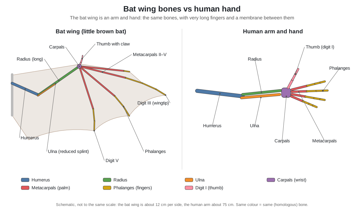
- Humerus: Carries the attachments of the pectoralis and other flight muscles, which drive the wingbeat.
- Radius: Is the main strut of the arm wing; bat biologists measure it as 'forearm length' to identify species.
- Ulna (reduced): Forms part of the elbow joint; most of its length is lost, so the forearm cannot twist.
- Carpals: Let the hand fold against the forearm and spread again, controlling the shape of the handwing.
- Thumb (digit I) with claw: Used for climbing, crawling and holding onto the roost; in some bats to hold food.
- Metacarpal II (index finger): Stiffens the leading edge of the handwing.
- Metacarpal III: With digit II, forms the stiff leading edge and spar of the handwing.
- Phalanges of digit III: Extend the longest finger to the wingtip; their low mineral content lets the tip bend.
- Metacarpal V: Supports the trailing edge and divides the handwing from the plagiopatagium.
The skin of the wing is divided into named membranes, each supported by different bones:
| Membrane | Where it is | Supported by |
|---|---|---|
| Propatagium | In front of the arm, from neck to wrist | Humerus, radius, thumb base |
| Dactylopatagium | Between the fingers (handwing) | Digits II–V |
| Plagiopatagium | From digit V to the body side and leg | Digit V, forearm, body, hindlimb |
| Uropatagium | Between the legs, around the tail | Legs, tail vertebrae, calcars |
Because the fingers can spread, fold and bend, a bat can change the shape of its wing far more than a bird can. The membranes also contain tiny muscles and elastic fibres that tune their tension during each wingbeat.
Bat vs bird skeleton and wing: homology vs analogy
Bat and bird wing bones are homologous: both are the humerus, radius, ulna, wrist and hand bones inherited from a common four-legged ancestor, the same bones as in your arm. The wings themselves are analogous: bats and birds evolved flight separately and built different wings from those bones. Pterosaurs did it a third way.
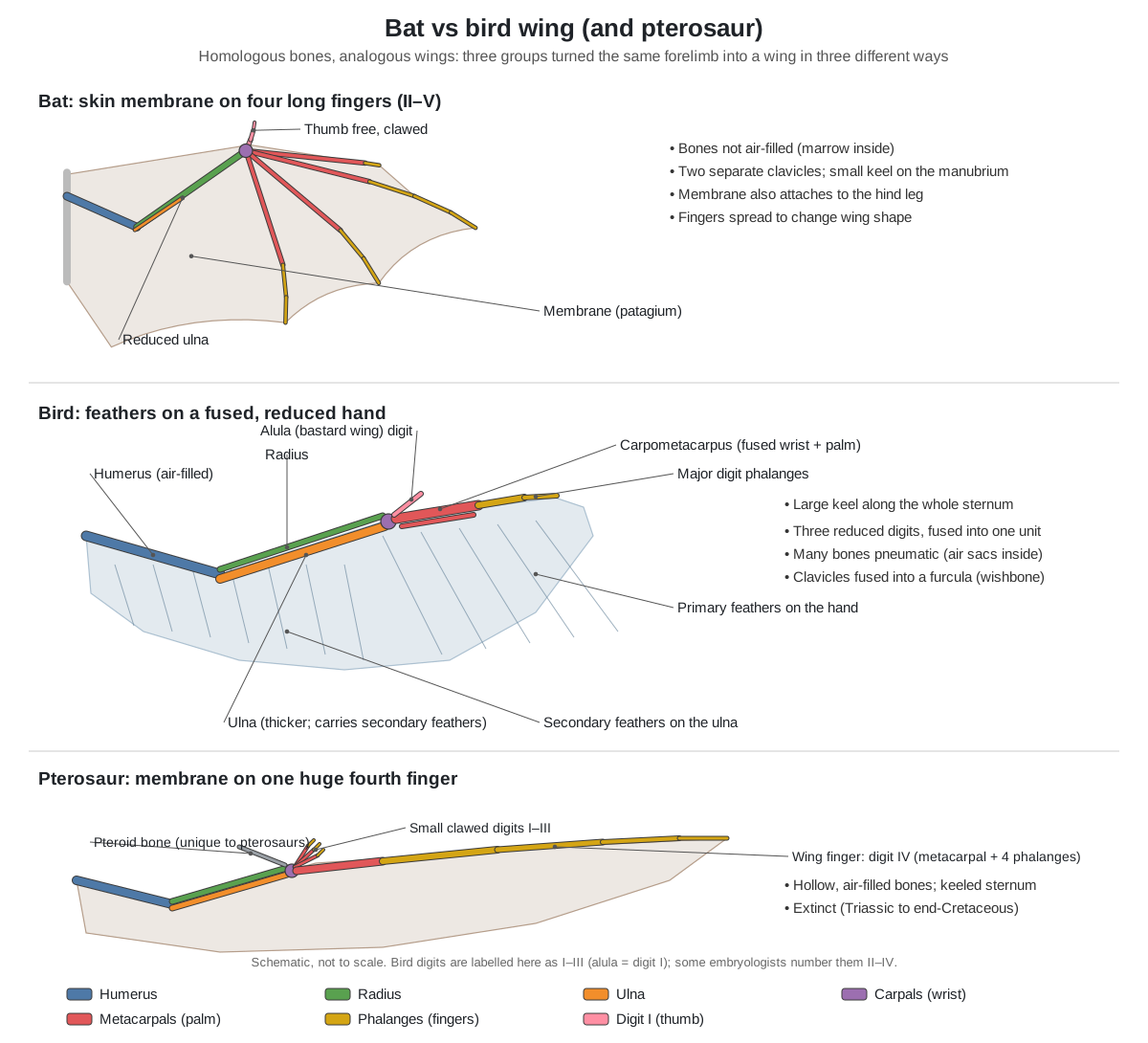
| Feature | Bat | Bird | Pterosaur | Human arm |
|---|---|---|---|---|
| Flight surface | Skin membrane | Feathers | Skin membrane | None |
| Main wing support | Digits II–V | Ulna and fused hand | Digit IV only | — |
| Hand bones | Separate, very long | Fused into a carpometacarpus | Digit IV huge, I–III small | Separate, short |
| Thumb | Free, clawed | Alula (bastard wing) | Clawed; plus a pteroid bone | Opposable |
| Forearm | Radius large, ulna reduced | Ulna larger than radius | Radius and ulna both present | Radius and ulna equal partners |
| Clavicles | Two, separate, robust | Fused into furcula | — | Two |
| Sternum keel | Small, on the manubrium | Large, whole sternum | Present | None |
| Bone interior | Marrow (not pneumatic) | Many bones air-filled | Hollow | Marrow |
| Hind legs in flight | Part of the wing (membrane) | Free | Attached to membrane | — |
Birds also fuse many bones (the synsacrum, pygostyle and carpometacarpus) and have a toothless beak, while bats keep separate finger bones and a full mammal dentition. A shared keel and similar tendon locks in the toes are examples of convergent evolution: similar solutions to the same physical problem.
Bat skull and teeth
A bat skull has a rounded braincase, a short snout (rostrum), large ear capsules (auditory bullae), slender zygomatic arches and a full set of sharp teeth. In adults the skull bones fuse early, so the sutures between them are hard to see. The little brown bat skull is about 14 mm long, has no sagittal crest and has a forehead that slopes up from a short rostrum.
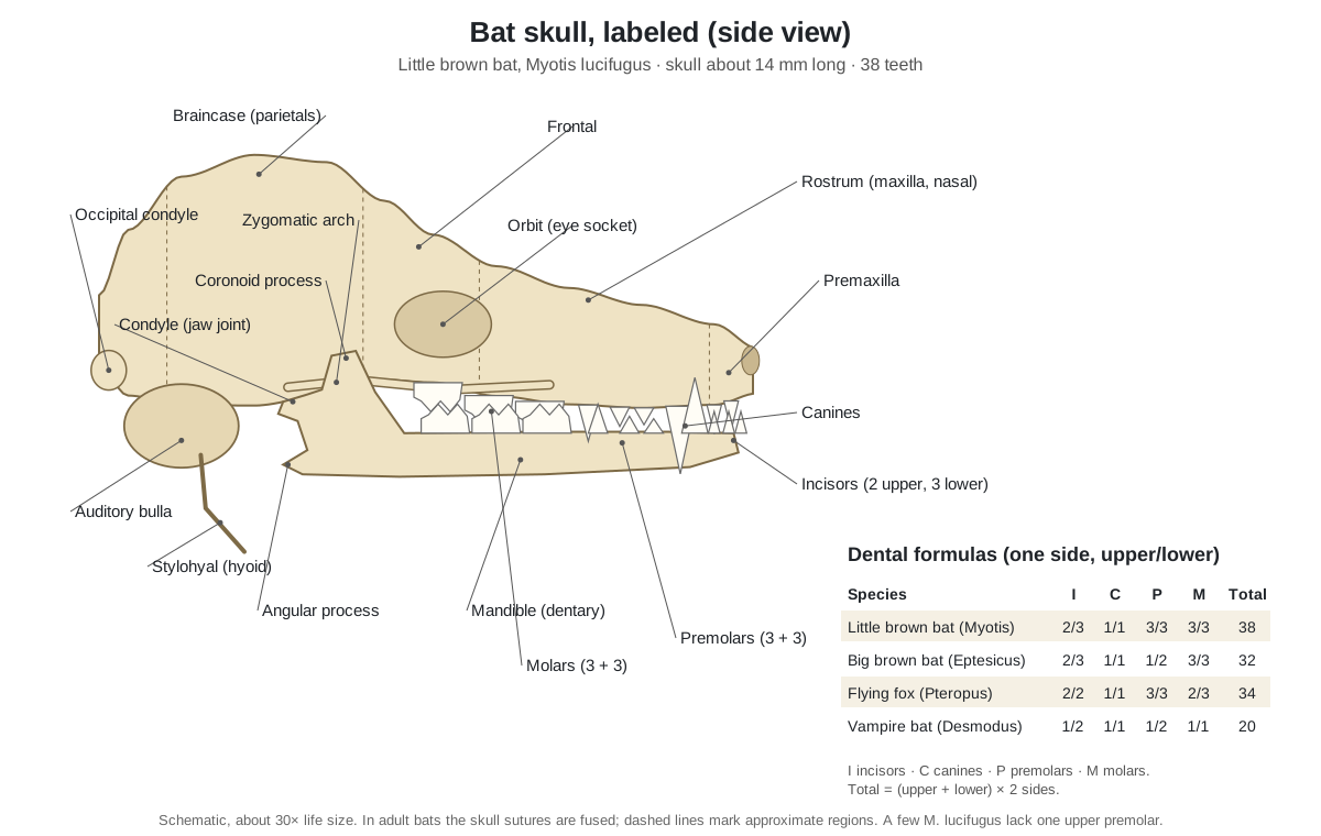
Dental formula. The basic formula for Myotis is I 2/3, C 1/1, P 3/3, M 3/3 = 38 teeth, the most of any bat; a small percentage of little brown bats lack one upper premolar. The big brown bat has I 2/3, C 1/1, P 1/2, M 3/3 = 32. Across all bats adults have from 20 to 38 teeth. The molars of insect-eating bats have W-shaped (dilambdodont) shearing crests that slice insect cuticle.
- Auditory bulla (tympanic bone): Houses the eardrum ring and middle-ear bones; next to it lies the large cochlea that hears echoes.
- Stylohyal bone (hyoid): Suspends the tongue and larynx; in echolocating bats it links the larynx, which makes the calls, to the ear capsule.
- Mandible (dentary): Carries the lower teeth; its tall coronoid process anchors the temporalis muscle for a strong bite.
- Premaxillae: Carry the upper incisors.
- Canines: Pierce and hold struggling insects, such as hard-shelled beetles and moths.
Bat legs: why bat knees bend backwards
Bat knees seem to bend backwards because the whole hind limb is rotated at the hip. Compared with other mammals, bat hind limbs are turned 90–180°, so the knee bends sideways or toward the back (Farina & Lankton, 2018). In the spread-wing pose of the 3D model, the knees point up and out and the soles face down and inward.
There are two reasons. First, the legs are part of the wing: they hold the back edge of the plagiopatagium and the uropatagium, and the rotation lets them spread these membranes sideways and adjust their tension in flight. Second, it suits hanging: with the legs turned this way, a bat hanging from a ceiling has its toes curled toward the surface while its belly faces the wall. The price is clumsy walking; most bats shuffle on the ground, with the clear exception of vampire bats.
- Pelvis (os coxae): Carries the hip joints; the whole body hangs from it when the bat roosts.
- Femur: Holds the leg out sideways to spread the plagiopatagium and uropatagium.
- Tibia: Carries the foot and supports the membranes along the leg.
- Fibula (reduced): Little load-bearing role; reduced to save weight.
- Calcar: Spreads and stiffens the tail membrane; it is a skeletal element found only in bats.
How bats hang upside down
A bat hangs upside down by hooking the claws of its five toes into a rough surface and letting its body weight pull the toes shut. In most bats a tendon locking mechanism then holds the grip: tubercles on each flexor tendon catch on ridges inside the tendon sheath, like a cable tie. The bat needs muscle effort to let go, not to hang on.
Hanging is also how bats take off. Many bats take off by simply releasing the grip, dropping and opening their wings, which is much easier than launching from flat ground. New World leaf-nosed bats (Phyllostomidae, which include the vampire bats) lack the ratchet-type lock but still hang well; in them, changes to the tendon sheath seem to work as a friction lock instead (Simmons & Quinn, 1994).
Are bat bones hollow? Thin, light, but not pneumatic
No. Bat bones are thin-walled, but they are not hollow and air-filled like bird bones. Air sacs from a bird's lungs extend into many of its bones (pneumatisation). Bats are mammals and have no air sacs; their long bones contain marrow. In a comparative study, bat skeletons made up a larger share of body mass than rodent skeletons, and skull and limb bones were on average densest in birds, followed closely by bats (Dumont, 2010). Light skeletons in fliers come from shape and distribution of bone, not from simply being hollow.
Bats save weight where it matters most: in the hand. The finger bones are flattened and become steadily less mineralised toward the wingtip, so the tip is light and flexible. The humerus near the body is the stiffest, roundest bone in the wing.
Echolocation anatomy in the skeleton
Echolocation leaves marks on the skull. Insect-eating bats like Myotis make calls in the larynx and hear the echoes with large ears, a tall tragus and a proportionally large cochlea housed beside the auditory bulla. In all laryngeally echolocating bats studied, the stylohyal bone of the hyoid connects to the tympanic bone of the ear (Veselka et al., 2010). Many other bats (horseshoe bats, leaf-nosed bats) add a fleshy noseleaf around the nostrils to focus calls emitted through the nose; it has no bone and leaves no trace on the skeleton except a modified snout.
- Ear (pinna): Collects returning echoes and helps the bat tell where they come from.
- Tragus: Creates extra echoes inside the ear that help the bat judge the height of a target.
- Eye: Detects light levels and landmarks at dusk; echolocation does the close-range work.
Do bats have skeletons?
Yes. Every bat has a complete internal skeleton of bone, because bats are mammals. Their bones are small and delicate, which is one reason fossil bats are rare, but the plan is the same as a mouse's or a human's. There is no fixed "number of bones" for a bat: the count varies with the number of tail vertebrae, fused bones and finger and toe bones in each species.
Fruit bat and flying fox skeleton
A fruit bat skeleton differs from a little brown bat's in the head, the hand and the tail. Old World fruit bats and flying foxes (family Pteropodidae) are much larger: the large flying fox (Pteropus vampyrus) has a wingspan of about 1.5 m, weighs 0.6–1.1 kg and has a forearm of about 18–22 cm.
- Two wing claws. Most pteropodids have a claw on the second finger as well as on the thumb; in pteropodids the thumb is also opposable.
- Big eyes, dog-like face. Large orbits for large, forward-facing eyes that see well in dim light, and a long muzzle; hence "flying fox".
- Teeth for fruit. Pteropus has 34 teeth (I 2/2, C 1/1, P 3/3, M 2/3), with simplified, flattened cheek teeth.
- No laryngeal echolocation. Fruit bats lack the stylohyal–tympanic link. Most do not echolocate at all; Rousettus fruit bats echolocate with tongue clicks.
- Short or no tail, small or no calcar. The tail membrane is often a narrow strip, and the calcar, when present, does not articulate with the calcaneus.
Vampire bat skeleton
The common vampire bat (Desmodus rotundus) has a skeleton built for walking as well as flying. Its thumbs are long and strong, with pads that act as extra feet, and its hind legs are sturdy, so it can walk, run and jump on the ground as it approaches sleeping animals. It has no tail.
Its skull has a very short rostrum and only 20 teeth (I 1/2, C 1/1, P 1/2, M 1/1). The two upper incisors and the canines are large and razor-sharp, used to make a small cut from which the bat laps blood; the cheek teeth are tiny and blade-like, with a single minute molar in each jaw quarter, because it never chews. Vampire bats also lack the toe tendon lock found in most other bats.
Hammerhead bat
The hammer-headed bat (Hypsignathus monstrosus) of central Africa is the largest bat on the continent and a fruit bat. Its skeleton follows the pteropodid plan, but males have an enlarged rostrum and a greatly enlarged larynx, with huge pendulous lips, used to make loud honking calls to attract females. Males weigh about 228–450 g and have wingspans of about 69–97 cm; females are smaller and have a fox-like face.
Bat skeletons compared
| Bat | Teeth | Distinctive skeletal features |
|---|---|---|
| Little brown bat (Myotis lucifugus) | 38 | No sagittal crest; calcar without a keel; 10–11 tail vertebrae; tendon lock present |
| Big brown bat (Eptesicus fuscus) | 32 | Larger (forearm 41–50 mm); keeled calcar; only 1 upper and 2 lower premolars per side |
| Flying foxes (Pteropus) | 34 | Claw on digit II; large orbits; long muzzle; short or no tail |
| Egyptian fruit bat (Rousettus) | — | Pteropodid skeleton; echolocates with tongue clicks, no stylohyal–tympanic link |
| Common vampire bat (Desmodus rotundus) | 20 | Strong thumbs and legs for running; blade-like upper incisors; tiny cheek teeth; no tail |
| Hammer-headed bat (Hypsignathus monstrosus) | — | Males with enlarged rostrum and larynx; extreme size difference between sexes |
| Slit-faced bats (Nycteridae) | — | No fibula at all |
Why bat wing bones bend: the mineral gradient
Bat wing bones are not equally stiff from shoulder to fingertip. Cleland and colleagues compared the humerus with the finger bones of two flying foxes (Pteropus poliocephalus and P. hypomelanus). Mineral crystals became thinner, less aligned and less densely packed from the humerus to the phalanges, and the stiffness of the bone material (Young's modulus) fell along the wing. The tips are low-density bone, not pure cartilage as older accounts suggested. (Cleland et al. (2021), Mechano-chemical regulation of bat wing bones for flight, J. Mech. Behav. Biomed. Mater..)
This matters for flight energy. Riskin and co-workers modelled six bat species and found that accelerating the metacarpals and phalanges accounted for around 44% of the inertial cost of flapping, although these bones make up only about 12% of wing weight. Lighter, less mineralised distal bones therefore save the bat a large share of the work of moving its wings. (Riskin et al. (2012), Upstroke wing flexion and the inertial cost of bat flight, Proc. R. Soc. B (PMC).)
The tendon locking mechanism: how the grip works
Most mammals keep friction between a tendon and its sheath as low as possible. Many bats do the opposite. Quinn and Baumel studied the third toe and the thumb in species from 15 bat families. Most had a patch of tubercles on the underside of the flexor tendon, opposite the first phalanx, and a series of transverse folds on the inside of the tendon sheath. When the two engage, the tendon is locked in place, like a ratchet, so the bat hangs with little muscular effort. The mechanism was missing in some microbats, most notably the phyllostomids (New World leaf-nosed bats), and many birds have a similar lock, an example of convergent evolution. (Quinn & Baumel (1993), Chiropteran tendon locking mechanism, J. Morphol..)
Schutt examined 10 species from 5 families by dissection and scanning electron microscopy and proposed that hibernation and body weight were the main selection pressures for this passive digital lock. It is absent in vampire bats, whose feeding requires very different ways of moving. A hibernating little brown bat that hangs for months on a cave wall is exactly the kind of bat that benefits. (Schutt (1993), Digital morphology in the Chiroptera: the passive digital lock, Acta Anat..)
Echolocation written in bone: the stylohyal link
You can tell from a skeleton whether a bat called with its larynx. Veselka and colleagues micro-CT scanned 35 fluid-preserved bats of 26 species. In every laryngeally echolocating bat, the upper end of the stylohyal bone (part of the hyoid apparatus) articulated directly with the tympanic bone of the ear, and was often fused to it. Non-echolocating fruit bats and the tongue-clicking fruit bats lacked this connection. The authors suggest it gives an independent anatomical test for echolocation in fossils. (Veselka et al. (2010), A bony connection signals laryngeal echolocation in bats, Nature.)
The oldest well-preserved bats show that flight evolved first. Onychonycteris finneyi, from the Early Eocene Green River Formation of Wyoming, had wings capable of powered flight but ear features suggesting it could not echolocate like other Eocene bats. It also kept claws on all five fingers and had a long calcar, which suggests a broad tail membrane evolved early in bats. (Simmons et al. (2008), Primitive Early Eocene bat from Wyoming, Nature.)
The long wing fingers are also ancient. Sears and colleagues found that the metacarpals of the roughly 50-million-year-old Icaronycteris index were already elongated and closely resemble those of modern bats, and that relative digit length has stayed constant for 50 million years. In embryos, the zone of enlarging cartilage cells in the bat metacarpal growth plate grows to more than 30% of the plate, against about 12% at most in mice. (Sears et al. (2006), Development of bat flight: evolution of bat wing digits, PNAS (PMC).)
The calcar: a bone found only in bats
The calcar is a neomorph: a new skeletal element with no equivalent in other mammals. Stanchak, Arbour and Santana showed that it diversified early in bat evolution and varies widely in length relative to the tibia and forearm, which affects flight behaviour. Its tissue also differs among species (cartilage, bone or a mix), and in some species a true synovial joint links it to the calcaneus, so it can move rather than act as a fixed strut. (Stanchak, Arbour & Santana (2019), Anatomical diversification of a skeletal novelty in bat feet, Evolution.)
Within one genus the calcar can be an identification clue. The big brown bat has a calcar with a keel-shaped extension, while the little brown bat lacks a calcar keel. (Animal Diversity Web: Eptesicus fuscus, big brown bat.)
The flight muscles on the skeleton
In bats, the pectoralis major arises from the sternum, especially the manubrium, and inserts on the crest of the greater tubercle of the humerus; it is the main downstroke muscle. The serratus ventralis thoracis runs from the sternum and upper ribs to the scapula and stabilises the wing in the downstroke, and the trapezius group from the skull and spine to the scapula and clavicle steadies the shoulder girdle. The membranes have their own muscles too: the occipito-pollicalis along the propatagium and small plagiopatagial and uropatagial muscles that adjust membrane tension. (Zhang et al. (2025), What factors shape the flyability in bats? Biology (PMC).)
Vampire and fruit bat skeletons: what the research shows
Bats are so specialised for flight that most can barely move on land. Common vampire bats are an exception: Riskin and Hermanson showed that they can run with a bounding gait in which the forelimbs, not the hind limbs, produce most of the force, because the wing muscles are far more powerful than the legs. Running seems to have evolved independently within bats. (Riskin & Hermanson (2005), Independent evolution of running in vampire bats, Nature.)
Egyptian fruit bats (Rousettus aegyptiacus) echolocate with tongue clicks rather than the larynx. Their clicks are extremely short, about 50–100 microseconds, and in landing and object-detection tasks they performed about as well as big brown bats, the model laryngeal echolocator. (Yovel, Geva-Sagiv & Ulanovsky (2011), Click-based echolocation in bats: not so primitive after all, J. Comp. Physiol. A.)
These fruit bats can even steer their sonar beam without moving the head. Lee and colleagues found that the beam sweeps with frequency and modelled the tongue as an array of sound sources along the side of the mouth, working like a phased-array sonar. (Lee et al. (2017), Tongue-driven sonar beam steering by a lingual-echolocating fruit bat, PLoS Biology.)
Little brown bats, white-nose syndrome and protected species
The reference species on this page is in trouble. White-nose syndrome, caused by the fungus Pseudogymnoascus destructans, infects the skin of the muzzle, ears and wings of hibernating bats. It has killed millions of insect-eating bats in North America since the winter of 2007–2008 and contributed to population declines of over 90% in little brown, northern long-eared and tri-colored bats. (USGS National Wildlife Health Center: white-nose syndrome.)
Several bats are protected by law because of such declines. The U.S. Fish and Wildlife Service reclassified the northern long-eared bat as endangered under the Endangered Species Act in a final rule published on 29 November 2022, naming white-nose syndrome as by far the main threat. Collecting or keeping protected animals or their remains can be illegal, which is one more reason to study bat skeletons from museum collections, scans or replicas. (U.S. Fish & Wildlife Service: northern long-eared bat.)
Real vs replica bat skeletons: what students should know
Students meet both real (museum) bat skeletons and resin or plastic replicas. For study, a good replica, a museum specimen or a published CT scan is as useful as a specimen you prepare yourself, and avoids three problems:
- Disease. Bats can carry rabies. The CDC advises never handling bats with bare hands and reporting any contact, including bites too small to notice, to the health department. Do not pick up a dead bat to make your own skeleton.
- Law. Many bats are protected, and several, such as the northern long-eared bat, are listed as endangered. Collecting or trading their remains can be illegal.
- Accuracy. Real bat skeletons are tiny and fragile; the thin fibula, cartilaginous wingtips and calcar are often lost or glued in the wrong place. Check a replica against a labeled diagram: the thumb should be free and clawed, the knees should face backward, and the calcar should point from the heel toward the tail.
Bat skeleton worksheet: test yourself
- Which bones form most of the bat wing? (The metacarpals and phalanges of digits II–V.)
- Which forearm bone is reduced in bats? (The ulna.)
- Which digit has a claw in every bat? (Digit I, the thumb.)
- What bone supports the tail membrane from the ankle? (The calcar.)
- How many teeth does a little brown bat have? (38.)
- Are bat wings and bird wings homologous or analogous? (Homologous as forelimbs, analogous as wings.)
- Why do bat knees point backwards? (The hind limbs are rotated 90–180° to spread the membranes and to hang.)
- Name one skeletal difference between a fruit bat and a little brown bat. (For example a claw on digit II, or no stylohyal–tympanic link.)
Frequently asked questions
Do bats have skeletons?
Yes. Bats are mammals, so they have a full internal bone skeleton: a skull with teeth, a backbone, ribs, shoulder and hip girdles and four limbs. Their wings are arms and hands with very long fingers, and their bones are thin but not hollow like bird bones.
How many bones does a bat have?
There is no single number. Bats share the basic mammal set of bones, but the count differs between species, mostly in the tail (0 to about 17 vertebrae), the sacrum (1–5 vertebrae), the number of finger and toe bones and how many bones fuse. A little brown bat, for example, has 7 neck, 11 chest, 5 lumbar, 4 fused sacral and about 10–11 tail vertebrae.
Is a bat wing a hand?
Mostly, yes. The wing is the whole forelimb: humerus, a long radius with a reduced ulna, wrist bones, a small clawed thumb and four very long fingers (digits II–V). Skin membranes stretch between the fingers, the arm, the body and the legs.
Why do bat knees bend backwards?
Bat legs are rotated at the hip by 90–180° compared with other mammals, so the knee points sideways or up instead of forward. This lets the legs spread and control the wing and tail membranes in flight, and lets the feet hang from a ceiling with the toes pointing the right way. The cost is that most bats walk clumsily.
How do bats hang upside down without falling?
Their weight does the work. When a bat hangs, its weight pulls on the toe flexor tendons, and in most bats a tendon locking mechanism (tubercles on the tendon catching ridges in its sheath) holds the claws curled. The bat needs muscle effort to let go, not to hold on.
Are bat bones hollow like bird bones?
No. Bird bones are pneumatic: air sacs from the lungs extend into them. Bat bones are not pneumatised and contain marrow like other mammal bones. They save weight by being thin-walled and, in the wing, by becoming less mineralised toward the fingertips.
What is the calcar on a bat?
The calcar is a thin spur of cartilage or bone that sticks out from the heel (calcaneus) along the edge of the tail membrane. It spreads and stiffens the uropatagium. It is unique to bats; some species have a small keel on it, and some bats have none.
What is the difference between a bat wing and a bird wing?
Both are modified forelimbs, so the bones are homologous, but the wings evolved independently. A bat wing is a skin membrane stretched over four long fingers; a bird wing is a fused, three-fingered hand that carries feathers. Birds have hollow, air-filled bones and a fused wishbone; bats have marrow-filled bones and two separate clavicles.
How is a fruit bat skeleton different?
Flying foxes and other Old World fruit bats (Pteropodidae) usually have a claw on the second finger as well as the thumb, large eye sockets, a dog-like snout, flat cheek teeth (34 teeth in Pteropus), a short or absent tail and little or no calcar. They also lack the stylohyal–tympanic link of echolocating bats.
Is it safe to pick up a dead bat or bat skeleton?
Do not touch bats, alive or dead, with bare hands: bats can carry rabies, and the CDC advises reporting any contact to the health department. Many bat species are also legally protected. For study, use museum specimens, published CT scans or licensed teaching replicas.
References
- Farina, L. L. & Lankton, J. S. (2018). Chiroptera. In Terio, K. A. et al. (eds), Pathology of Wildlife and Zoo Animals. Academic Press. (Vertebral formulas, dental formulas, limb and calcar anatomy.)
- Dumont, E. R. (2010). Bone density and the lightweight skeletons of birds. Proceedings of the Royal Society B 277, 2193–2198.
- Cleland, T. P. et al. (2021). Mechano-chemical regulation of bat wing bones for flight. Journal of the Mechanical Behavior of Biomedical Materials 124, 104809.
- Riskin, D. K., Bergou, A., Breuer, K. S. & Swartz, S. M. (2012). Upstroke wing flexion and the inertial cost of bat flight. Proceedings of the Royal Society B 279, 2945–2950.
- Quinn, T. H. & Baumel, J. J. (1993). Chiropteran tendon locking mechanism. Journal of Morphology 216, 197–208.
- Simmons, N. B. & Quinn, T. H. (1994). Evolution of the digital tendon locking mechanism in bats and dermopterans: a phylogenetic perspective. Journal of Mammalian Evolution 2, 231–254.
- Wilson, D. E. & Mittermeier, R. A. (eds) (2019). Handbook of the Mammals of the World, vol. 9, Bats: Desmodus rotundus account (via Plazi; dental formula).
- Schutt, W. A. (1993). Digital morphology in the Chiroptera: the passive digital lock. Acta Anatomica 148, 219–227.
- Veselka, N. et al. (2010). A bony connection signals laryngeal echolocation in bats. Nature 463, 939–942.
- Stanchak, K. E., Arbour, J. H. & Santana, S. E. (2019). Anatomical diversification of a skeletal novelty in bat feet. Evolution 73, 1591–1603.
- Simmons, N. B., Seymour, K. L., Habersetzer, J. & Gunnell, G. F. (2008). Primitive Early Eocene bat from Wyoming and the evolution of flight and echolocation. Nature 451, 818–821.
- Sears, K. E. et al. (2006). Development of bat flight: morphologic and molecular evolution of bat wing digits. PNAS 103, 6581–6586.
- Riskin, D. K. & Hermanson, J. W. (2005). Independent evolution of running in vampire bats. Nature 434, 292.
- Yovel, Y., Geva-Sagiv, M. & Ulanovsky, N. (2011). Click-based echolocation in bats: not so primitive after all. Journal of Comparative Physiology A 197, 515–530.
- Lee, W.-J. et al. (2017). Tongue-driven sonar beam steering by a lingual-echolocating fruit bat. PLoS Biology 15, e2003148.
- Zhang, M. et al. (2025). What factors shape the flyability in bats? The perspective from bat's wing development. Biology 14, 1524.
- Animal Diversity Web (University of Michigan): Myotis lucifugus, Eptesicus fuscus, Desmodus rotundus, Pteropus vampyrus and Hypsignathus monstrosus species accounts.
- Texas Tech University, The Mammals of Texas (online edition): Eptesicus fuscus, big brown bat.
- University of Texas at El Paso, Laboratory for Environmental Biology: Myotis (dental formula and premolar variation).
- University of California Museum of Paleontology: bat, bird and pterosaur flight exhibits.
- Centers for Disease Control and Prevention: rabies and bats.
- U.S. Geological Survey, National Wildlife Health Center: white-nose syndrome.
More interactive anatomy models
- Human anatomy1,000+ bones, muscles, organs, vessels and nerves from real anatomical data.
- Octopus anatomyThree hearts, a ring-shaped brain, eight arms and a beak, in 3D.
- Squid anatomyPen, eight arms, two tentacles, giant axons and three hearts of the longfin squid.
- Squid dissectionStep-by-step guided squid dissection with every structure labeled.
- Fish anatomyExternal and internal anatomy of a bony fish: fins, gills, swim bladder, heart and brain.
- Perch dissectionGuided yellow perch (fish) dissection, from scales to the swim bladder.