

Cow eye dissection at a glance
A cow eye dissection shows the three layers of the eye wall and the three clear media that focus light. On the outside you find fat, muscle stumps, the white sclera, the cornea and the optic nerve. After cutting the eye in half around its middle you find the vitreous humor, the lens hanging from its zonule fibres, the ciliary body, the iris, the retina, the blind spot and the shiny tapetum lucidum.
- Outer (fibrous) layer: sclera and cornea.
- Middle (vascular) layer, the uvea: choroid with the tapetum, ciliary body and iris.
- Inner (neural) layer: retina, which leaves the eye as the optic nerve.
- Clear media: cornea, aqueous humor, lens and vitreous humor.
A pair of students usually shares one eye. The same procedure works for a sheep eye, which is smaller but has the same parts.
Materials and safety
Materials
- One cow eye per pair of students: preserved (from a biological supply company) or fresh (from a butcher or slaughterhouse), ideally with fat and muscle still attached
- Dissecting tray, cutting board or wax paper on a tray
- Sharp scalpel or single-edged razor blade, and small pointed scissors
- Forceps and a blunt probe (or a wooden stick)
- Nitrile gloves, safety glasses or goggles and a lab apron
- A sheet of newspaper or printed text (to test the lens)
- Paper towels, a plastic bag for waste, soap and water
- Optional: hand lens, small torch (flashlight), ruler, camera
Safety
- The sclera and cornea are tough, so scalpels slip. Hold the eye with your fingers well away from the line of the cut and cut away from yourself.
- Puncture the cornea with care: aqueous humor can squirt out. Wear eye protection, especially with preserved eyes.
- Preserved eyes are stored in chemical preservative. Rinse the eye in water, work in a ventilated room and wear gloves.
- Treat fresh eyes like raw meat: they can carry bacteria. Wear gloves if you have cuts and wash your hands thoroughly afterwards (Exploratorium).
- Never eat or drink in the lab. Dispose of tissue in a sealed bag and blades in a sharps container, as your teacher directs.
Cow eyes for dissection: fresh or preserved?
Either works, but they look different, so know which one you have. Fresh eyes come from a butcher or slaughterhouse as a by-product of beef production. Preserved eyes come from biological supply companies, packed in a preservative solution, and keep for months.
- Fresh eye: clear cornea, liquid aqueous humor that spurts out, a clear, flexible lens you can read print through, and a jelly-like vitreous. Fresh eyes are easiest to cut on the day you get them.
- Preserved eye: cloudy or blue-grey cornea, little aqueous humor, a hard, yellow, opaque lens and a firmer vitreous. The structures are tougher, so they hold their shape when you cut them.
Ask for eyes with the fat and muscle still attached: you need them for the external examination. A lab usually orders one eye per two students, plus a few spares.
External anatomy of the cow eye
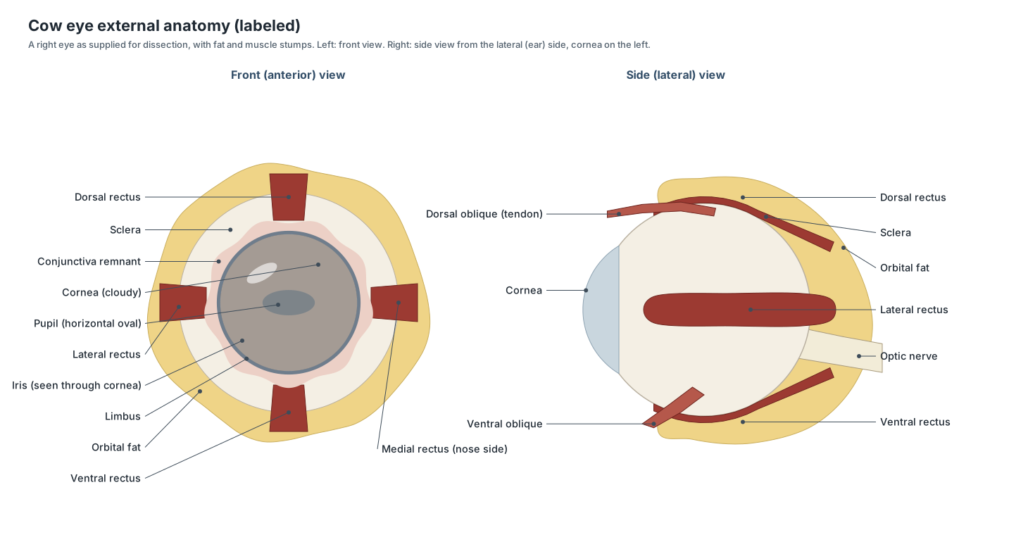
Before you cut, identify the cornea, sclera, fat, muscle stumps and optic nerve. The cornea is the domed window at the front; in a preserved eye it is cloudy, so you may only just see the dark iris and the horizontal oval pupil behind it. The white sclera covers the rest of the eyeball. Around it are yellow fat and dark red muscle stumps, and a ragged pink fringe of conjunctiva lies near the edge of the cornea.
Turn the eye over to find the optic nerve: a firm, white, rope-like cord coming out of the back slightly below centre. It feels firmer than the fat around it. Around it, deeper than the other muscles, lies the cone-shaped retractor bulbi.
Cow eye dissection procedure, step by step
This method follows the Exploratorium's classic cow's eye dissection, with the cut placed slightly behind the middle of the eye as in Carolina's procedure. Each numbered step matches a step in the 3D viewer above.
1. Examine the outside of the eye
- Rinse the eye and place it on the tray. Find the front: the domed, bluish or cloudy cornea.
- Identify the white sclera, the fat around the back and the ragged pink conjunctiva near the cornea.
- Look through the cornea. If it is clear enough you can see the dark iris and the oval pupil.
- Turn the eye over and find the optic nerve leaving the back. It is firmer and more rope-like than the fat.
Identify: Cornea, Sclera, Limbus, Conjunctiva, Orbital fat, Optic nerve.
2. Trim the fat; identify muscles and optic nerve
- With scissors, trim away the fat close to the sclera. Do not cut the optic nerve.
- Find the stumps of the extraocular muscles. Cattle have seven: four recti, two obliques and the retractor bulbi around the optic nerve.
- Use the muscles to orient the eye: the dorsal oblique tendon arrives at the top from the nasal side, and the ventral oblique crosses underneath.
- Look for the posterior ciliary arteries and the vorticose vein stumps on the sclera.
Identify: Dorsal rectus, Ventral rectus, Medial rectus, Lateral rectus, Dorsal oblique, Ventral oblique, Retractor bulbi, Optic nerve, Optic nerve sheath, Posterior ciliary arteries, Vorticose veins.
3. Puncture the cornea: aqueous humor
- Hold the eye by its sides. With the scalpel tip, make a small cut in the cornea. It is tougher than it looks.
- Clear fluid leaks or squirts out: this is the aqueous humor from the anterior chamber.
- Note that the cornea wrinkles once the fluid has gone: aqueous pressure kept it domed.
Caution: Aim the eye away from faces when you puncture the cornea.
Identify: Cornea, Aqueous humor, Posterior chamber, Iridocorneal angle, Iris.
4. Cut the eye in half around the middle
- Make a small incision through the sclera slightly behind the middle (equator) of the eye.
- Insert the scissors and cut all the way around the eye, keeping the same distance from the cornea.
- Gently separate the two halves. The front (anterior) half carries the cornea, iris and usually the lens; the back (posterior) half holds the vitreous humor.
- Lay both halves cut side up, as in the 3D view.
Caution: The scalpel can skid on the round, slippery sclera. Pinch up a fold of sclera before you start the cut.
Identify: Sclera, Choroid, Retina, Vitreous humor, Lens, Ciliary body.
5. Vitreous humor
- Look into the posterior half: it is filled with clear jelly, the vitreous humor.
- Let the vitreous slide out onto the tray. Tease it gently away from the lining with a probe.
- Feel its texture: it is a gel, not a liquid.
Identify: Vitreous humor, Retina.
6. The lens and zonule fibres
- Find the lens in the anterior half (sometimes it stays in the vitreous). It is about the size and shape of a squashed marble.
- Before lifting it out, look for the zonule fibres that hang it from the ciliary processes.
- Lift the lens out. Press it: a fresh lens is soft outside and hard in the middle; a preserved lens is hard, yellow and cloudy.
- Put the lens on a newspaper and look through it. A fresh lens magnifies the print.
Identify: Lens, Lens nucleus, Zonule fibres, Ciliary processes, Posterior chamber.
7. Iris, pupil and ciliary body
- With the lens removed, look into the anterior half from behind. Find the dark ring of the ciliary body with its fine radial ciliary processes.
- Find the iris and the oval opening in it, the pupil. The iris may come out in one piece if you pull gently.
- Note that the back of the iris is black: it is covered by dense pigment.
Identify: Iris, Pupil, Ciliary body, Ciliary muscle, Ciliary processes, Ora serrata.
8. The retina
- In the posterior half, find the thin, cream-coloured film with blood vessels in it: the retina.
- Without the vitreous pressing on it, it may have crumpled into a wad.
- Push the retina around with a probe. It stays attached at one spot: the optic disc, or blind spot.
Identify: Retina, Retinal blood vessels, Optic disc, Ora serrata.
9. Tapetum lucidum and choroid
- Peel the retina back to the optic disc. Underneath, the upper back of the eye shines blue-green: the tapetum lucidum.
- Tilt the half under the light and watch the colour change.
- Below it the fundus is dark: the pigmented, nontapetal part of the choroid.
Identify: Tapetum lucidum, Nontapetal fundus, Choroid, Optic disc.
10. The cornea and its layers
- Cut around the limbus to free the cornea from the anterior half. Lay it on the board.
- Cut it with the scalpel and listen for the crunch: you are slicing through many layers of tissue.
- Try to peel off the thin outer epithelium. Note how thick the cornea is compared with the sclera.
Identify: Cornea, Corneal epithelium, Corneal endothelium, Limbus.
11. The optic nerve
- Turn the posterior half over and find the optic nerve leaving the back.
- Pinch the cut end with forceps or your fingers. White goop may come out: myelin, the fatty insulation of the nerve fibres.
- Match the nerve's position outside with the optic disc inside.
Identify: Optic nerve, Optic nerve sheath, Optic disc, Posterior ciliary arteries.
Clean up by wrapping all tissue in a plastic bag for disposal, putting blades in the sharps container and washing your hands, tray and tools with soap and water.
Dissection of a cow eye: labeled diagrams
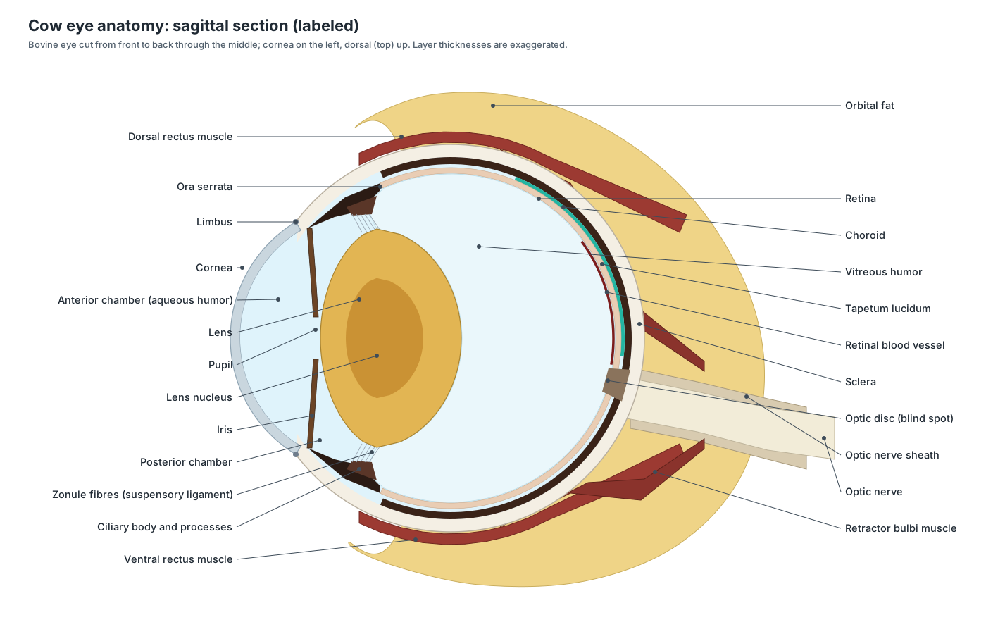
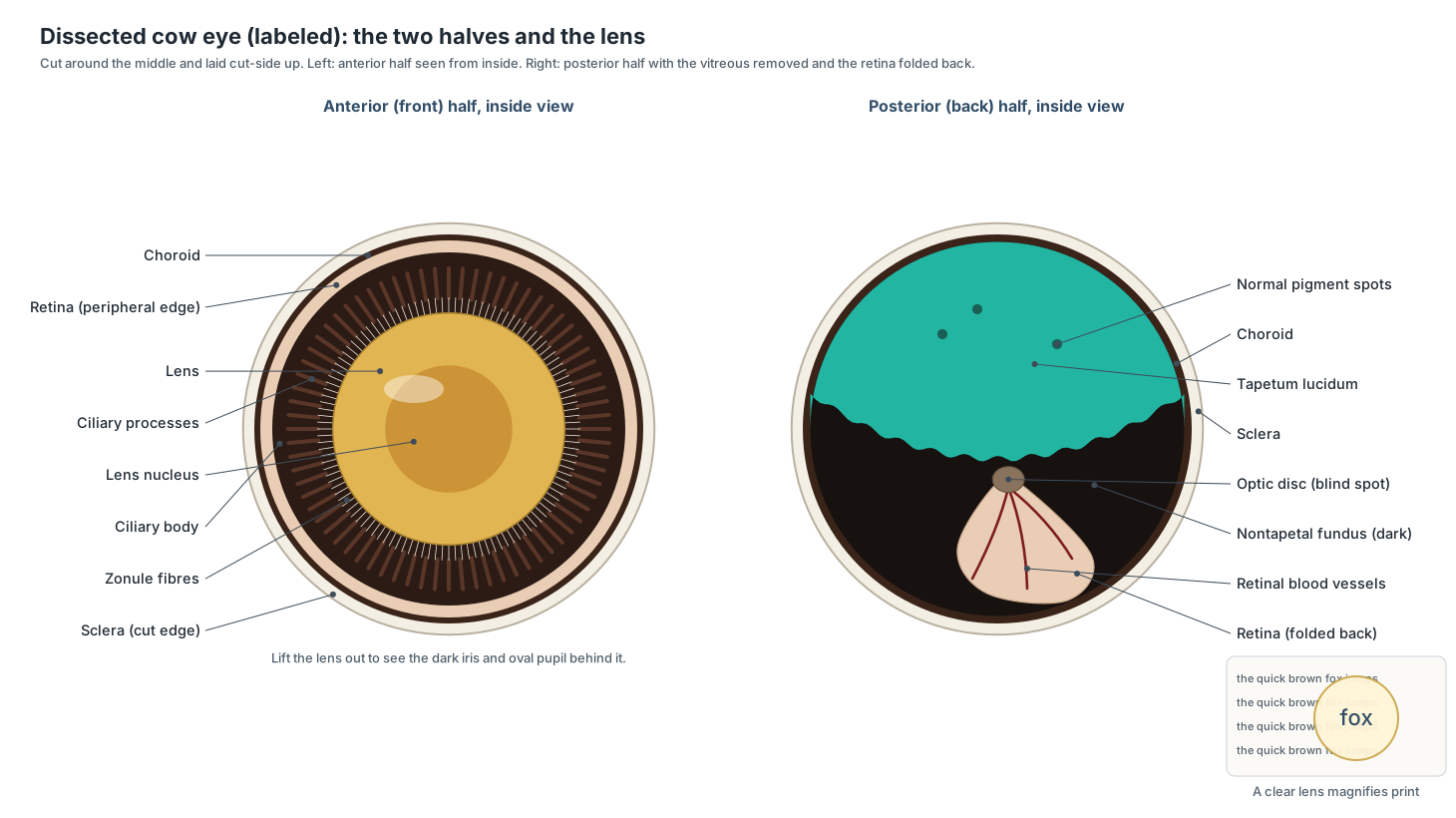
Use the section diagram to see where each part sits, and the halves diagram to match what is on your tray. The section shows the eye as if cut from front to back through the middle; the halves show the eye as you actually cut it, around the middle.
Parts of the cow eye and their functions
Every structure in the 3D model, grouped by layer. Click any of them in the viewer to zoom to it.
Fat & conjunctiva
- Orbital fat (Periorbital fat, adipose tissue)
- The soft, yellow-white fat packed around the back of the eyeball and its muscles. Cushions the eye inside the bony orbit and lets it rotate smoothly. In a dissection it is trimmed away to expose the muscles and optic nerve.
- Conjunctiva (Conjunctival remnant)
- A thin, pinkish membrane left as a ragged fringe around the front of the sclera. In life it lined the inside of the eyelids and folded back over the front of the sclera, keeping the eye surface moist and sealing off the space behind the eye.
Extraocular muscles
- Dorsal rectus (Superior rectus (human term))
- A flat strap of muscle on the top of the eyeball. Rotates the eye upward (elevation).
- Ventral rectus (Inferior rectus (human term))
- A strap of muscle under the eyeball, opposite the dorsal rectus. Rotates the eye downward (depression).
- Medial rectus
- A strap of muscle on the nasal (nose) side of the eyeball. Turns the eye toward the nose (adduction).
- Lateral rectus
- A strap of muscle on the temporal (ear) side of the eyeball. Turns the eye away from the nose (abduction).
- Dorsal oblique (Superior oblique (human term))
- A thin muscle whose tendon reaches the top of the eye from the nasal side. Rotates (twists) the top of the eye toward the nose and helps turn it down and out.
- Ventral oblique (Inferior oblique (human term))
- A short muscle that crosses under the eyeball from the nasal side. Rotates the bottom of the eye toward the nose and helps turn it up and out.
- Retractor bulbi (Retractor of the eyeball)
- A cone of muscle wrapped around the optic nerve behind the eye. Humans do not have it. Pulls the eyeball back into the orbit, which also makes the third eyelid slide across the cornea: a protective reflex.
Fibrous tunic (sclera, cornea)
- Sclera (White of the eye)
- The tough, white outer coat that covers most of the eyeball. Gives the eye its shape, resists the pressure of the fluids inside, and anchors the extraocular muscles.
- Cornea (Corneal stroma)
- The clear, domed window at the front of the eye. In a preserved eye it is cloudy or bluish. Lets light in and does most of the eye's focusing (bending light), because it is curved and has a much higher refractive index than air.
- Corneal epithelium
- The thin outer skin of the cornea, a few cells thick. A smooth, renewable barrier against dust, germs and water; damaged cells are replaced quickly from stem cells at the limbus.
- Corneal endothelium (With Descemet's membrane)
- A single layer of cells on the inner surface of the cornea, facing the aqueous humor. Pumps fluid out of the corneal stroma into the aqueous humor. This 'pump-leak' balance keeps the stroma thin and clear (Bonanno 2012).
- Limbus (Corneoscleral junction)
- The ring where the clear cornea meets the white sclera. Holds the stem cells that renew the corneal epithelium, and overlies the drainage angle where aqueous humor leaves the eye.
Vascular tunic (uvea, tapetum)
- Choroid
- A thin, dark, blood-rich layer lining the inside of the sclera. Supplies oxygen and nutrients to the outer retina (the light-sensitive photoreceptors). Its dark pigment absorbs stray light outside the tapetum.
- Tapetum lucidum (Tapetum fibrosum; the 'shiny blue-green layer')
- The iridescent blue-green layer at the back of the eye that makes cow eyes shine at night. Reflects light that has passed through the retina back through the photoreceptors, giving them a second chance to catch it. This improves vision in dim light.
- Nontapetal fundus (Pigmented (black) choroid and pigment epithelium)
- The dark, non-reflective lower part of the back of the eye, below the tapetum. Its pigment absorbs light instead of reflecting it, as the choroid does in eyes without a tapetum.
- Ciliary body
- A dark ring of tissue between the choroid and the iris. Its ciliary muscle changes the tension on the zonule to adjust focus, and its epithelium makes aqueous humor.
- Ciliary muscle
- The band of smooth muscle in the outer part of the ciliary body. When it contracts, the ring of the ciliary body narrows, the zonule slackens and the lens can round up to focus on near objects (accommodation).
- Ciliary processes
- Many fine, radial folds on the inner face of the ciliary body. Secrete aqueous humor into the posterior chamber, and anchor the zonule fibres that hold the lens.
- Iris
- The coloured, muscular ring in front of the lens. A cow's iris is dark brown. Its sphincter and dilator muscles change the size of the pupil to control how much light enters the eye.
- Pupil
- The opening in the centre of the iris. In cattle it is a horizontal oval, not round. Lets light reach the lens and retina; it narrows in bright light and widens in dim light.
- Iridocorneal angle (Drainage angle, filtration angle)
- The narrow angle where the edge of the iris meets the back of the cornea. Contains the trabecular meshwork, the sieve through which aqueous humor drains out of the eye.
Retina
- Retina
- The thin, cream-coloured layer of nerve tissue lining the back of the eye. It falls away easily. Its rods and cones turn light into nerve signals; other retinal neurons process them and send them to the brain through the optic nerve.
- Ora serrata
- The wavy front edge of the light-sensitive retina. Marks the boundary between the seeing retina and the non-seeing epithelium of the ciliary body; the retina is anchored here.
- Retinal blood vessels (Retinal arterioles and venules)
- Dark branching vessels that spread over the retina from the optic disc. Supply blood to the inner layers of the retina.
Optic nerve, disc & vessels
- Optic disc (Blind spot, optic nerve head)
- The spot where the retina is attached and its nerve fibres leave the eye: the blind spot. All the retina's nerve fibres gather here to form the optic nerve. There are no rods or cones on it, so an image falling here cannot be seen.
- Optic nerve (Cranial nerve II)
- The thick, white, rope-like cord leaving the back of the eye. Carries visual signals from the retina to the brain.
- Optic nerve sheath (Dural sheath (meninges))
- The tough outer wrapping of the optic nerve, continuous with the sclera. Protects the nerve; it is an extension of the brain's meninges, because the optic nerve is really part of the brain.
- Vorticose veins (Vortex veins)
- A few veins that leave the eyeball through the sclera near its equator. Drain blood from the choroid, ciliary body and iris.
- Posterior ciliary arteries
- Small arteries that enter the sclera around the optic nerve. Supply the choroid, ciliary body and iris (and so the tapetum and outer retina).
Lens & zonule
- Lens (Crystalline lens (with capsule and cortex))
- The clear, bean-shaped lens behind the iris. In a preserved eye it is yellow-brown, hard and cloudy. Fine-tunes the focus so that a sharp image lands on the retina; the cornea does most of the bending, the lens finishes the job.
- Lens nucleus
- The hard central core of the lens. The oldest, most compact lens fibres; it has the highest refractive index, which strengthens the lens.
- Zonule fibres (Suspensory ligaments, zonule of Zinn)
- Hundreds of fine fibres that hang the lens from the ciliary body. Hold the lens centred behind the pupil and pass the pull of the ciliary muscle to the lens to change its shape for focusing.
Humors & chambers
- Aqueous humor (Fluid of the anterior chamber)
- The clear, watery fluid that squirts out when you puncture the cornea. Keeps the cornea domed by its pressure and feeds the cornea and lens, which have no blood vessels.
- Posterior chamber
- The narrow, ring-shaped space between the back of the iris and the lens. Receives the aqueous humor made by the ciliary processes; it flows through the pupil into the anterior chamber.
- Vitreous humor (Vitreous body)
- The clear jelly that fills the large space behind the lens. Holds the eyeball's shape, presses the retina flat against the back of the eye, and lets light pass through to the retina.
Why cow eyes are used for dissection
Cow eyes are used because they are big, cheap and built like ours. A cow's eyeball is about 3.3–3.5 cm long from front to back: ultrasound of live cows gave a mean axial length of 3.46 cm in Holstein Friesians and 3.27 cm in Jerseys, and the authors note that these values match earlier measurements of dead (cadaver) eyes. A human eye is about 2.5 cm long (24.6 mm on average in a study of 135 removed human eyes). The larger size makes the lens, iris and retina easy to see and handle without a microscope. (Potter, Hallowell & Bowen (2008): ultrasonographic anatomy of the bovine eye, Vet. Radiol. Ultrasound 49.)
The eyes are a by-product of the meat industry: no cow is killed to supply them. The Exploratorium suggests buying them from a butcher or straight from a slaughterhouse, ideally on the day of the dissection with the muscle and fat still attached, because fresh eyes are easier to cut. Biological supply companies also sell cow eyes preserved in a chemical solution, which keeps for months and is what most school labs use. (Exploratorium: Dissecting a Cow's Eye, step-by-step instructions (PDF).)
Cow eyes for dissection come in two forms, and they look different. A fresh eye has a clear cornea, a soft, clear lens and liquid aqueous humor. A preserved eye usually has a cloudy or bluish cornea, very little aqueous humor and a hard, yellowish lens. Both show every structure you need, but if your teacher asks you to read print through the lens, a fresh eye works far better. (Biology Corner: cow eye dissection.)
Sheep eyes are the other common choice. A sheep eye is smaller, but it has the same parts and is dissected the same way, step for step. Like the cow, the sheep has a fibrous (collagen) tapetum, so the shiny blue-green layer looks much the same. The sheep also has seven extraocular muscles: four recti, two obliques and a retractor bulbi, the same set as in cattle. (Kirbaş et al. (2025): eye muscles of Tuj sheep, Arch. Razi Inst. (PMC).)
Why the cornea of a preserved cow eye is cloudy
The cornea is clear in life only because its collagen is extremely well ordered. The stroma, which makes up most of the cornea, is built from thin collagen fibrils laid down at regular spacing in stacked layers (lamellae). That order makes the light waves scattered by neighbouring fibrils cancel out in every direction except straight through. Small changes in the spacing can be tolerated, but larger ones make the cornea scatter light and look milky. (Meek et al. (2025): structural control of corneal transparency, refractive power and dynamics, Eye (PMC).)
The spacing is kept steady by the corneal endothelium, the single cell layer on the inner surface. It works as a 'pump-leak' system: the stroma keeps drawing water in (the leak), and the endothelium keeps pumping it out again (the pump). When the endothelial cells stop working, the stroma soaks up water, swells and turns cloudy. (Bonanno (2012): molecular mechanisms underlying the corneal endothelial pump, Exp. Eye Res. (PMC).)
After the cow dies, the endothelial pump stops, so the cornea swells and clouds within hours. Preservative then fixes the tissue in that state: formaldehyde links proteins together with chemical cross-links, so the change can't be undone. A cloudy cornea is therefore normal in a lab specimen and doesn't mean the eye was diseased. A cow's cornea is also thick, about 0.72 mm at the centre in Holstein cattle, compared with roughly half a millimetre in people, which is one reason it crunches when you cut it. (Kibar Kurt & Gülendağ (2026): central corneal thickness and intraocular pressure in Holstein cattle, Vet. Ophthalmol..)
Why the lens is hard and yellow
The lens is mostly protein. Its fibre cells are packed with crystallins, which make up over 90% of the lens's protein and are arranged so evenly that light passes straight through. Anything that makes those proteins clump or cross-link makes them scatter light, which is what a cataract is. (Ma et al. (2026): human crystallin variation and cataract, Invest. Ophthalmol. Vis. Sci. (PMC).)
Preservation does this on purpose to the whole eye. Formaldehyde forms bridges between protein molecules, which is why fixed tissue becomes firm. In the protein-dense lens the effect is dramatic: a preserved cow lens is hard, opaque and yellow to brown, much like a boiled egg white compared with a raw one. (Thavarajah et al. (2012): chemical and physical basics of routine formaldehyde fixation (PMC).)
Even a fresh cow lens is firm in the middle. The Exploratorium guide describes it as soft on the outside and hard in the centre, about the size and shape of a squashed marble. Laboratory tests agree: in bovine lenses stiffness is highest in the nucleus, and the overall stiffness profile resembles that of an older, presbyopic human lens. A lens grows by adding new fibre cells to its outside throughout life and never sheds the old ones, so the nucleus holds the oldest, most compressed fibres. (Qiu et al. (2025): lens water content and the stiffness of ex vivo bovine lenses, Front. Ophthalmol. (PMC).)
To see what a lens does, put a clear one on a sheet of newspaper. The letters look larger, because the lens is convex and bends light rays together, as a magnifying glass does. Hold it up and look through it at the room: the image appears upside down. In the eye, the cornea does most of the bending of light and the lens finishes the job, focusing an upside-down image onto the retina. (Exploratorium: Dissecting a Cow's Eye, step-by-step instructions (PDF).)
Why the retina falls off (and where it stays attached)
The retina comes away easily because, apart from two places, nothing strong holds it down. The neural retina sits on a layer of pigment epithelium cells. Near its front edge, the ora serrata, the two are joined by cell junctions, but further back the retina is held only by fine processes of the pigment epithelium that interlock with the tips of the photoreceptors. In the living eye, the vitreous gel presses the retina flat, so this weak attachment is enough. (EyeWiki (American Academy of Ophthalmology): Ora serrata.)
When you cut the eye open and the vitreous slides out, the retina loses that support and crumples into a pale, wrinkled wad. Push it around with a probe and you will find it is still fixed at one spot: the optic disc, where all the nerve fibres of the retina leave the eye to form the optic nerve. That spot has no light-sensitive cells, so it is the blind spot. (Exploratorium: Dissecting a Cow's Eye, step-by-step instructions (PDF).)
The same weak attachment is why people can suffer a retinal detachment, where fluid gets between the neural retina and the pigment layer. The human retina is about 0.5 mm thick, and its optic disc is a pale oval about 2 × 1.5 mm across. Unlike the cow, the human retina has a fovea: a pit packed with cones where vision is sharpest. (Kolb, Webvision: simple anatomy of the retina.)
The blue-green shiny layer: why cows have a tapetum lucidum and humans don't
The shiny blue-green layer at the back of a cow eye is the tapetum lucidum ('bright carpet'). It sits under the retina and reflects light that has already passed through the photoreceptors back through them, so each photon gets a second chance to be detected. This raises sensitivity in dim light. It is also what makes animals' eyes shine in headlights; the Exploratorium notes that a cow's eyes shine blue-green in a torch beam at night. (Exploratorium: Dissecting a Cow's Eye, step-by-step instructions (PDF).)
Tapeta come in several kinds. Cattle, sheep, goats and horses have a choroidal tapetum fibrosum, made of layers of very regular collagen fibres. Carnivores such as cats and dogs have a tapetum cellulosum made of reflective cells, and sharks use guanine crystals. Primates, birds, squirrels, the red kangaroo and the pig have no tapetum at all; the authors note that these are usually day-active animals. (Ollivier et al. (2004): comparative morphology of the tapetum lucidum, Vet. Ophthalmol. 7.)
Cattle and other hoofed animals are active by day, at dusk, at dawn and at night, so a tapetum pays off. Humans are a day-active species that relies on sharp, colour-rich central vision from a fovea, and we have no tapetum. (Carroll et al. (2001): photopigment basis for dichromatic color vision in the horse, J. Vision 1.)
In cattle the tapetum covers most of the upper back of the eye, above the optic disc, and the lower fundus is dark. Veterinarians describe normal dark pigment spots in the bovine tapetum about one to two disc widths above the optic disc, very large retinal vessels and a naturally dark optic disc. Do not mistake these spots for damage in your specimen. (Stehman, Rebhun & Riis (1987): progressive retinal atrophy in related cattle, The Bovine Practitioner.)
Horizontal pupils and two-colour vision
A cow's pupil is a horizontal oval rather than a circle. A comparative study of land animals found that horizontally elongated pupils go with grazing prey species whose eyes are on the sides of the head. The wide slot gives a sharp, panoramic image along the ground, which helps them spot predators and move over rough ground, and it reduces the amount of overhead sunlight entering the eye. When grazing animals lower their heads, their eyes rotate in the orbit to keep the pupil level with the ground. (Banks et al. (2015): why do animal eyes have pupils of different shapes? Science Advances 1 (PMC).)
Cattle see in two colour channels, not three. Electrical recordings from the retina found two cone types in cattle: one most sensitive to blue (about 444–455 nm) and one to yellow-green (about 552–555 nm). Most humans have three cone types. So cattle have the photopigment basis for dichromatic colour vision, roughly like a person with red-green colour blindness. (Jacobs, Deegan & Neitz (1998): photopigment basis for dichromatic color vision in cows, goats and sheep, Vis. Neurosci. 15.)
The extraocular muscles: seven in a cow, six in a human
The muscle stumps on a cow eye belong to seven extraocular muscles. There are four recti (dorsal, ventral, medial and lateral), two obliques (dorsal and ventral) and the retractor bulbi. A study of sheep eye muscles lists exactly this set, and cattle share it. Humans have the four recti and two obliques but no retractor bulbi. (Kirbaş et al. (2025): eye muscles of Tuj sheep, Arch. Razi Inst. (PMC).)
The retractor bulbi forms a cone around the optic nerve, deep to the recti. In cattle it has three slips (medial, dorsolateral and ventrolateral) that insert on the sclera at the equator, plus a fourth deep slip that inserts above the optic nerve near the back of the eye. It is supplied by the abducens nerve and pulls the eyeball back into the orbit. When the eye is pulled back, the third eyelid slides across it, a quick way to protect the cornea. (IMAIOS vet-Anatomy: retractor bulbi muscle.)
Aqueous humor, the drainage angle and glaucoma
Aqueous humor is made by the ciliary body, mainly by active secretion from the epithelium of the ciliary processes. It flows from the posterior chamber through the pupil into the anterior chamber, and leaves through the trabecular meshwork in the angle between the iris and the cornea. The balance between production and drainage sets the pressure inside the eye. If drainage is blocked, pressure rises and can damage the optic nerve: this is glaucoma. (Goel et al. (2010): aqueous humor dynamics, a review, Open Ophthalmol. J. (PMC).)
Cow eye vs human eye
A cow eye has the same basic parts as a human eye, but it is larger and adapted to a grazing prey animal. The table lists the differences you can see or measure in the lab.
| Feature | Cow eye | Human eye |
|---|---|---|
| Eyeball length (axial) | About 3.3–3.5 cm (3.46 cm Holstein, 3.27 cm Jersey, ultrasound) | About 2.4–2.5 cm (24.6 mm mean in removed eyes) |
| Central corneal thickness | About 0.72 mm (Holstein) | About 0.5–0.55 mm |
| Pupil | Horizontal oval | Round |
| Iris colour | Dark brown | Brown, blue, green, grey or hazel |
| Tapetum lucidum | Present: tapetum fibrosum (collagen), blue-green, in the upper fundus | Absent |
| Fovea (pit of sharpest vision) | Absent | Present, in the centre of the macula |
| Cone types (colour vision) | Two (blue and yellow-green): dichromat | Usually three: trichromat |
| Optic disc colour | Normally dark | Pale pink-white |
| Extraocular muscles | Seven: 4 recti, 2 obliques, retractor bulbi | Six: 4 recti, 2 obliques (no retractor bulbi) |
| Third eyelid | Present; swept across the eye when the retractor bulbi pulls the eye back | Only a small fold (plica semilunaris) |
| Lens in a lab specimen | Large; hard, yellow and opaque when preserved | Smaller; transparent in a healthy young eye |
Sources: Potter et al. 2008 and Jonas et al. 2017 (eye length), Kibar Kurt & Gülendağ (cow cornea), Ollivier et al. 2004 (tapetum), Banks et al. 2015 (pupil), Jacobs et al. 1998 (cones), Stehman et al. 1987 (optic disc), IMAIOS (retractor bulbi). See the reference list below.
Sheep eye dissection: what changes?
Almost nothing: a sheep eye is dissected exactly like a cow eye. It is smaller, so cut more carefully, but it has the same seven extraocular muscles, a horizontally elongated pupil, a collagen (fibrous) tapetum lucidum and the same layers inside. Every step, diagram and lab question on this page applies to a sheep eye as well.
Cow eye dissection lab questions and answer key
Use these as pre-lab or post-lab questions. Click a question to reveal the answer.
Name the three layers (tunics) of the eye wall, from outside in, and one structure in each.
Fibrous tunic (sclera and cornea), vascular tunic or uvea (choroid with tapetum, ciliary body, iris) and neural tunic (retina).
Why is the cornea of your cow eye cloudy, when it was clear in the living cow?
Corneal clarity depends on the endothelium pumping water out of the stroma so its collagen fibrils stay evenly spaced. After death the pump stops, the stroma swells and scatters light, and preservative fixes the cornea in that cloudy state.
What fluid came out when you punctured the cornea, and what is its job?
Aqueous humor. It keeps the cornea domed by its pressure and nourishes the cornea and lens, which have no blood vessels.
Describe the lens of the dissected cow eye. What happened when you put it on newsprint?
A biconvex, bean- or squashed-marble-shaped body, soft outside and hard in the middle (fresh) or hard, yellow and cloudy (preserved). A clear lens magnifies the print because it is a converging lens.
What holds the lens in place, and what changes its shape?
The zonule fibres (suspensory ligaments) hang it from the ciliary processes. The ciliary muscle changes the tension on the zonule, which lets the lens change shape to focus.
Why did the retina fall away from the back of the eye?
The neural retina is held to the layer behind it only loosely, and in life the vitreous humor pressed it flat. Once the vitreous is removed it detaches, except at the optic disc, where its nerve fibres leave to form the optic nerve.
Where is the blind spot, and why can't it detect light?
At the optic disc, where the retina is still attached. It has no rods or cones, because the whole spot is taken up by nerve fibres leaving the eye.
What is the shiny blue-green layer, and why do cows have it but humans do not?
The tapetum lucidum (a tapetum fibrosum in cattle). It reflects light back through the retina to boost vision in dim light. Cattle are active by day and at night; humans and other primates, which are mainly active by day, have no tapetum.
What shape is the cow's pupil, and why might that help a grazing animal?
A horizontal oval. Horizontal pupils give a sharp panoramic view along the ground for spotting predators, and cut down the overhead sunlight entering the eye.
Which extraocular muscle does a cow have that you do not, and what does it do?
The retractor bulbi. It pulls the eyeball back into the orbit, which also sweeps the third eyelid across the eye.
Trace the path of light through the eye to the brain.
Cornea → aqueous humor (anterior chamber) → pupil → lens → vitreous humor → retina (rods and cones) → optic nerve → brain.
Which structure does most of the focusing: the cornea or the lens?
The cornea, because light bends most where it passes from air into the curved cornea. The lens fine-tunes the focus.
Cow eye dissection worksheet
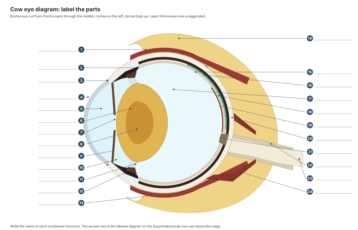
Part A: label the diagram. Print the blank diagram and write the name of each numbered structure. Check your answers against the labeled section diagram above: the numbers run from top to bottom down the left side (1–13), then down the right side (14–24).
Part B: match the structure to its function. Cover the left column and name the structure from its description.
| Structure (answer) | Description |
|---|---|
| Sclera | Tough white outer coat; keeps the eye's shape and anchors the muscles |
| Cornea | Clear front window; does most of the focusing |
| Aqueous humor | Watery fluid in front of the lens; keeps the cornea domed |
| Iris | Muscular ring that controls the size of the pupil |
| Pupil | Opening that lets light in; oval in a cow |
| Lens | Fine-focuses light on the retina; magnifies print |
| Ciliary body | Ring of muscle and folds that makes aqueous humor and holds the zonule |
| Vitreous humor | Clear jelly behind the lens; holds the retina flat |
| Retina | Thin light-sensitive layer that detects the image |
| Tapetum lucidum | Shiny reflective layer that helps night vision |
| Choroid | Dark, blood-rich layer that feeds the retina |
| Optic disc | Blind spot where the retina is attached |
| Optic nerve | Carries signals from the retina to the brain |
Part C: observations. Record the colour of the cornea, the shape of the pupil, the colour, hardness and size of the lens, what happened when you put the lens on print, the colour of the tapetum, and where the retina stayed attached. Then explain each observation using the sections above.
Virtual cow eye dissection
The 3D viewer at the top of this page is a complete virtual cow eye dissection. Step 1 starts with the whole eye in its fat; step 2 trims the fat; step 4 cuts the eye into halves; later steps remove the vitreous, lens, retina and cornea, just as you would on the tray. Click any part for its name, location and function, or open the "All structures" tab to find it from a list. Students who opt out of a physical dissection can complete the same steps, labeled diagrams and lab questions. For whole-animal anatomy, see our cow digestive system page.
Frequently asked questions
Why is the cornea of a cow eye cloudy?
In the living cow the cornea is clear because its collagen fibrils are evenly spaced, and the endothelium on its inner surface constantly pumps water out to keep them that way. After death the pump stops, the cornea absorbs water, swells and scatters light. Preservative then fixes it in that cloudy, sometimes bluish state. It is normal and does not mean the eye was diseased.
Why is the lens of a preserved cow eye hard and yellow?
The lens is packed with proteins called crystallins. Preservatives such as formaldehyde cross-link proteins, so the lens sets hard and turns yellow to brown and opaque. A fresh cow lens is clear, softer on the outside and hard in the middle, because the oldest lens fibres are compressed into a firm nucleus.
What is the blue shiny layer in a cow's eye?
It is the tapetum lucidum, a reflective layer in the choroid under the retina. In cattle it is a tapetum fibrosum made of very regular collagen fibres. It reflects light back through the retina to improve vision in dim light and makes cows' eyes shine blue-green in a torch beam at night. Humans do not have one.
Why did the retina fall off the back of the eye?
The neural retina is only loosely held against the layer behind it; in life the vitreous jelly presses it flat. When you cut the eye open and the vitreous comes out, the retina crumples away. It stays attached at the optic disc (the blind spot), where its nerve fibres leave the eye as the optic nerve.
Where do cow eyes for dissection come from?
They are a by-product of beef production: eyes are collected at slaughterhouses from cattle killed for meat. You can buy them fresh from a butcher or abattoir, or preserved from biological supply companies. No animal is killed for its eyes.
Is a cow eye like a human eye?
Yes, in its basic plan: both have a sclera and cornea, a choroid, ciliary body and iris, a lens held by zonule fibres, aqueous and vitreous humors, and a retina connected to the optic nerve. The main differences are that the cow eye is bigger, has a horizontal oval pupil, a reflective tapetum lucidum, a retractor bulbi muscle and two cone types instead of three, and lacks a fovea.
How do you cut a cow eye in half?
Trim off the fat, then make a small cut through the sclera slightly behind the middle of the eye with a scalpel. Insert scissors into the slit and cut all the way around, keeping the same distance from the cornea. Separate the front and back halves and lay them cut side up.
Can I do a cow eye dissection online?
Yes. The 3D model on this page is a free virtual cow eye dissection. Its 11 steps follow the real procedure: the fat is trimmed, the eye is cut into halves and each layer is removed in turn, with the structures to find listed at every step.
References
- Exploratorium: Dissecting a Cow's Eye, step-by-step instructions (PDF)
- Carolina Biological Supply: Perfect Solution cow eye dissection quick tips (PDF)
- Biology Corner: cow eye dissection
- Potter, Hallowell & Bowen (2008): ultrasonographic anatomy of the bovine eye, Vet. Radiol. Ultrasound 49
- Ollivier et al. (2004): comparative morphology of the tapetum lucidum, Vet. Ophthalmol. 7
- Banks et al. (2015): why do animal eyes have pupils of different shapes? Science Advances 1 (PMC)
- Jacobs, Deegan & Neitz (1998): photopigment basis for dichromatic color vision in cows, goats and sheep, Vis. Neurosci. 15
- Carroll et al. (2001): photopigment basis for dichromatic color vision in the horse, J. Vision 1
- Kibar Kurt & Gülendağ (2026): central corneal thickness and intraocular pressure in Holstein cattle, Vet. Ophthalmol.
- Meek et al. (2025): structural control of corneal transparency, refractive power and dynamics, Eye (PMC)
- Bonanno (2012): molecular mechanisms underlying the corneal endothelial pump, Exp. Eye Res. (PMC)
- Thavarajah et al. (2012): chemical and physical basics of routine formaldehyde fixation (PMC)
- Qiu et al. (2025): lens water content and the stiffness of ex vivo bovine lenses, Front. Ophthalmol. (PMC)
- Goel et al. (2010): aqueous humor dynamics, a review, Open Ophthalmol. J. (PMC)
- Ma et al. (2026): human crystallin variation and cataract, Invest. Ophthalmol. Vis. Sci. (PMC)
- EyeWiki (American Academy of Ophthalmology): Ora serrata
- Kolb, Webvision: simple anatomy of the retina
- IMAIOS vet-Anatomy: retractor bulbi muscle
- Kirbaş et al. (2025): eye muscles of Tuj sheep, Arch. Razi Inst. (PMC)
- Jonas et al. (2017): axial length and horizontal and vertical globe diameters of human eyes, Graefe's Arch.
- Stehman, Rebhun & Riis (1987): progressive retinal atrophy in related cattle, The Bovine Practitioner
More interactive anatomy models
- Sheep brain dissectionGuided sheep brain dissection with cerebrum, cerebellum and cranial nerves labeled.
- Sheep heart dissectionGuided sheep (and pig) heart dissection: chambers, valves and great vessels.
- Human anatomy1,000+ bones, muscles, organs, vessels and nerves from real anatomical data.
- Octopus anatomyThree hearts, a ring-shaped brain, eight arms and a beak, in 3D.
- Squid anatomyPen, eight arms, two tentacles, giant axons and three hearts of the longfin squid.
- Squid dissectionStep-by-step guided squid dissection with every structure labeled.