

Grasshopper anatomy at a glance
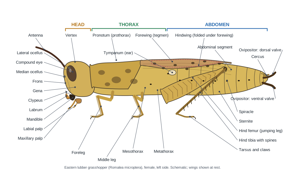
A grasshopper is an insect, so its body has three parts: the head, with the eyes, antennae and chewing mouthparts; the thorax, which carries all six legs and four wings; and the abdomen, with 11 segments, the ears (tympana), most of the breathing holes (spiracles) and the egg-laying or mating organs. Inside, a straight gut runs from mouth to anus, a tube-shaped heart runs along the back, and a double nerve cord runs along the belly.
Grasshoppers belong to the order Orthoptera, suborder Caelifera, together with locusts. Crickets and katydids are in the other suborder, Ensifera. The reference species here is the eastern lubber grasshopper, a big, flightless grasshopper of the southeastern United States, 5–7 cm long as an adult female. Where the common field grasshoppers (Melanoplus) differ, the text says so.
Directions used on this page: anterior means towards the head, posterior towards the abdomen tip, dorsal the back and ventral the belly. Left and right are the grasshopper's own.
Grasshopper diagram: external anatomy labeled
The labeled grasshopper diagram below shows every external part a lab worksheet asks for: one pair of antennae, two compound eyes, three ocelli, the mouthparts, the pronotum, three pairs of legs, two pairs of wings, the tympanum, the spiracles, the segmented abdomen, the cerci and, in a female, the ovipositor.
| Body part | Segments | What attaches to it |
|---|---|---|
| Head | Fused into one capsule | Antennae, compound eyes, 3 ocelli, mouthparts |
| Thorax | Prothorax, mesothorax, metathorax | 3 pairs of legs (one per segment), 2 pairs of wings (meso- and metathorax) |
| Abdomen | 11 segments | Tympana (segment 1), 8 pairs of spiracles, cerci, ovipositor or subgenital plate |
The exoskeleton is made of hard plates (sclerites) joined by soft membranes, so the body can bend and the abdomen can stretch. Grasshoppers grow by moulting this exoskeleton.
Grasshopper head diagram and mouthparts
The grasshopper head is a hard capsule divided into named regions: the vertex on top, the frons (face) in front, a gena (cheek) on each side and the clypeus at the bottom of the face. It carries two compound eyes, three ocelli and two antennae, and the mouthparts point straight down.
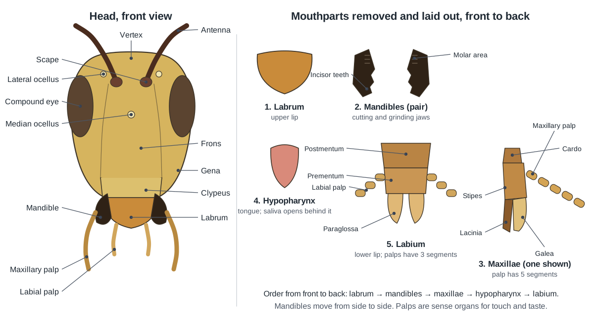
Grasshoppers have chewing (mandibulate) mouthparts, built for biting and grinding leaves. From front to back they are:
- Labrum: A broad, movable flap that forms the front wall of the mouth. Holds food in place between the jaws while they chew.
- Mandibles: A pair of hard, dark jaws with cutting edges and grinding surfaces. Cut and grind leaves. They move from side to side, not up and down.
- Maxillae: A pair of jointed mouthparts behind the mandibles that handle food. Hold and push food towards the mandibles. Their lacinia and galea manipulate the food.
- Maxillary palps: Long, five-segmented sensory feelers on the maxillae. Taste and touch food before it is eaten.
- Hypopharynx: A soft, tongue-like lobe in the middle of the mouthparts. Mixes food with saliva; the salivary duct opens just behind it.
- Labium: The lower lip, formed from a pair of appendages fused together on the midline. Closes the back of the mouth and pushes food forward.
- Labial palps: A pair of short, three-segmented sensory feelers on the labium. Taste and touch food.
Two clues help on a mouthparts quiz: the mandibles are the only dark, hard jaws, and the palps are the only jointed feelers. Five segments means a maxillary palp; three means a labial palp.
- Compound eyes: Two large oval eyes, each made of thousands of tiny units called ommatidia. Form an image and detect movement over a very wide field of view.
- Ocelli: Three tiny simple eyes: one median ocellus on the face and two lateral ocelli. Sense light intensity and changes in brightness rather than forming detailed images.
- Antennae: One pair of jointed, thread-like feelers on the front of the head. Smell and touch: they carry many sense organs that detect odours, food plants and contact.
Grasshopper thorax diagram: pronotum, wings and legs
The thorax is the grasshopper's locomotion centre. It has three segments, each with one pair of legs. The first, the prothorax, is covered by the big saddle-shaped pronotum. The mesothorax and metathorax are fused into a box (the pterothorax) that carries the wings and the large leg and flight muscles.
- Pronotum (prothorax): The large saddle-shaped plate that covers the top and sides of the first thoracic segment. Shields the prothorax, its muscles and the first thoracic spiracle. The forelegs attach below it.
- Prosternal spine: A short peg on the underside of the prothorax, between the front legs. An identification feature: it marks the spur-throated grasshoppers and lubbers.
- Mesothorax: The middle thoracic segment, which carries the middle legs and the forewings. Anchors the middle legs and forewings and houses their muscles.
- Metathorax: The last thoracic segment, which carries the jumping hind legs and the hindwings. Anchors the hind legs and hindwings and houses the largest leg muscles of the thorax.
- Forewings (tegmina): Narrow, leathery forewings that cover the hindwings at rest. Protect the delicate hindwings. In flying grasshoppers they also add some lift.
- Hindwings: Thin, membranous wings that fold like a fan under the forewings. The flight wings of grasshoppers that fly. In the lubber they are too short for flight and are flashed in a warning display.
Wing length is the quickest way to tell a lubber from a field grasshopper: a lubber's forewings stop well short of the abdomen tip and it cannot fly, while a Melanoplus has wings that reach the tip or beyond and flies readily.
Grasshopper leg diagram: walking and jumping legs
Every grasshopper leg has the same segments: coxa, trochanter, femur, tibia, tarsus and a pretarsus with two claws and a soft arolium. The front and middle legs are walking legs. The hind legs are jumping (saltatorial) legs, with a huge femur full of muscle and a long, spiny tibia.
| Segment | Fore and middle legs | Hind (jumping) leg |
|---|---|---|
| Coxa | Short; joins the leg to the thorax | Larger; on the metathorax |
| Trochanter | Tiny hinge | Tiny hinge |
| Femur | Slender | Very large and club-shaped, herringbone pattern |
| Tibia | Slender | Long, two rows of spines, spurs at the tip |
| Tarsus | Short segments with soft pads | Same, longer |
| Pretarsus | 2 claws + arolium | 2 claws + arolium |
- Hind femur: The enlarged, club-shaped femur of the hind (jumping) leg. Holds the large extensor muscle that powers the jump. The leg is a saltatorial (jumping) leg.
- Hind tibia: The long tibia of the jumping leg, armed with two rows of spines. Kicks out to launch the jump. Its spines grip the ground and make defensive kicks painful.
- Extensor tibiae muscle: The huge muscle in the hind femur that straightens the leg to jump. Contracts slowly while the flexor holds the leg folded, storing energy; the stored energy is then released at once to kick the tibia out.
- Flexor tibiae muscle: The smaller muscle in the hind femur that folds the tibia. Folds the tibia and holds it locked while the extensor builds up force before a jump.
- Tarsi: The jointed foot of each leg, made of short segments called tarsomeres. Grip the surface when walking and climbing.
- Tarsal claws: A pair of hooked claws at the tip of every leg. Hook into rough surfaces such as bark and leaf edges.
- Arolium: A soft pad between the two claws. Sticks to smooth surfaces where claws cannot grip.
Grasshopper abdomen diagram: segments, tympanum and spiracles
The grasshopper abdomen has 11 segments. The first carries the tympanum, an oval eardrum on each side just behind the thorax. Segments 1–8 each carry a pair of spiracles. The tip carries the cerci and the genitalia: two pairs of digging valves (the ovipositor) in a female, or a rounded subgenital plate in a male.
- Tympanum: A thin oval eardrum on each side of the first abdominal segment. Vibrates in response to sound. Sense cells attached to its inner side send the signal to the nerve cord.
- Spiracles: Small openings along the sides of the body that let air into the tracheae. Let air in and out; tiny valves close them to save water.
- Epiproct: A triangular plate that covers the anus from above. Protects the anus. It is the tergite of the reduced 11th segment.
- Paraprocts: A pair of small plates beside and below the anus. Close the anus from the sides; together with the epiproct they form the last (11th) segment.
- Cerci: A pair of short, pointed sensory appendages at the tip of the abdomen. Touch and air-current receptors. In males of many species their shape helps identify the species.
- Ovipositor: dorsal valves: The upper pair of short, curved, hard digging valves at the tip of a female's abdomen. Dig into soil by opening and closing against the ventral valves, so the female can lay eggs below ground.
- Ovipositor: ventral valves: The lower pair of large, hard digging valves of the ovipositor. Work with the dorsal valves to drill a hole in the soil for the egg pod.
- Subgenital plate: A large, upturned, boat-shaped plate that forms the rounded tip of a male's abdomen. Forms a cup that encloses and protects the male genitalia (phallus).
To count spiracles in lab, lift the wings and look along the lower side of the abdomen with a hand lens. You should find eight on each side of the abdomen, plus two on the thorax (one hidden under the pronotum): ten pairs in all.
Grasshopper internal anatomy diagram
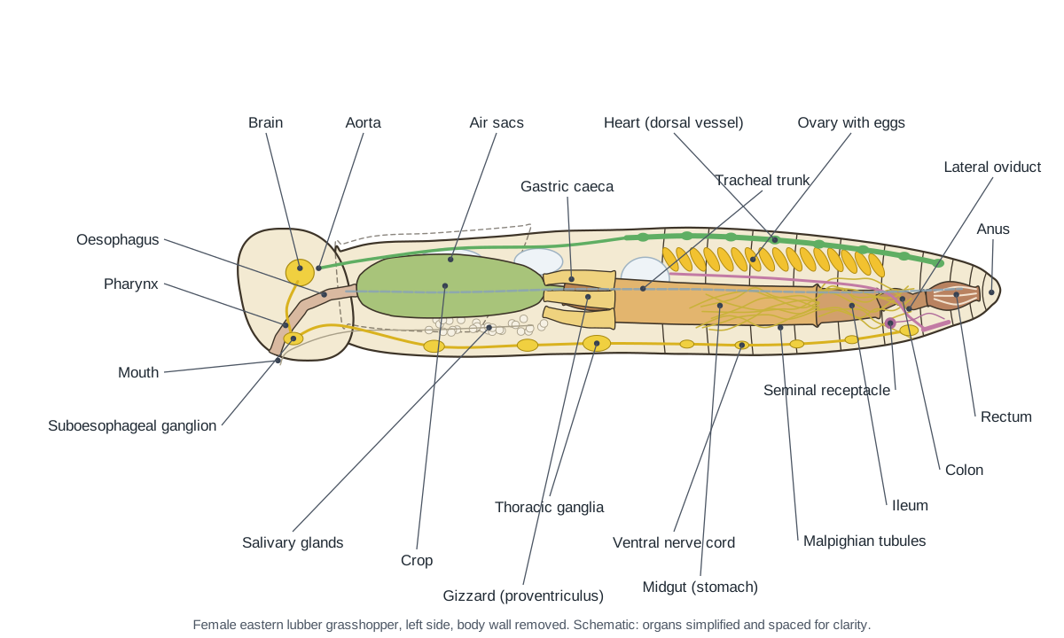
Inside a grasshopper, the gut runs the length of the body, the heart lies along the top of the abdomen, the gonads sit between the heart and the gut, the nerve cord runs along the floor, and air tubes (tracheae) and white air sacs fill the spaces. There is no lung, no liver and no kidney like ours: tracheae, the fat body and the Malpighian tubules do those jobs.
To see how these organs look in a real specimen from above, see the dorsal view in our grasshopper dissection guide.
Grasshopper digestive system diagram
Food travels through the grasshopper in this order: mouth → pharynx → oesophagus → crop → gizzard (proventriculus) → midgut with gastric caeca → ileum → colon → rectum → anus. The crop stores food, the gizzard grinds it, the midgut and caeca digest and absorb it, and the rectum takes back water.
- Salivary glands: Paired clusters of small white glands that make saliva. Produce saliva that moistens food. The two ducts join and open behind the hypopharynx.
- Mouth & pharynx: The mouth opening and the muscular tube behind it that swallows food. Pumps chewed food from the mouth into the oesophagus.
- Oesophagus: A narrow tube that carries food from the pharynx to the crop. Carries food to the crop. The brain lies above it and the suboesophageal ganglion below it.
- Crop: A large, thin-walled sac for storing food. Stores chewed leaves. Some digestion of carbohydrates starts here, using saliva and enzymes passed forward from the midgut.
- Gizzard (proventriculus): A short, muscular, narrower section after the crop. Grinds food with tooth-like ridges (denticles) on its inner lining and controls passage into the midgut.
- Gastric caeca: Six finger-like pouches around the front end of the midgut. Secrete digestive enzymes and absorb nutrients, adding surface area to the midgut.
- Midgut (stomach): The middle part of the gut, where most digestion and absorption happen. Secretes digestive enzymes and absorbs the digested food into the hemolymph.
- Malpighian tubules: Very many long, thread-like tubes attached to the gut where the midgut meets the hindgut. Remove nitrogen wastes and excess salts from the hemolymph and pass them into the hindgut. They do the job of kidneys.
- Ileum: The first, wide part of the hindgut. Moves waste back and starts absorbing water and salts.
- Colon: A short, narrow section of hindgut between the ileum and rectum. Carries waste to the rectum and absorbs some water.
- Rectum: The last, ridged chamber of the gut. Reabsorbs water and salts from waste, so the insect excretes dry pellets and saves water.
- Anus: The exit of the gut. Releases dry fecal pellets (frass).
Grasshopper circulatory system diagram
A grasshopper has an open circulatory system. A single tube, the dorsal vessel, pumps hemolymph forward: its abdominal part is the heart, which takes in hemolymph through paired slits (ostia), and its front part is the aorta, which empties into the head. The hemolymph then seeps back through the body cavity (hemocoel), bathing the organs.
- Heart (dorsal vessel): A slender, transparent tube along the top of the abdomen. Pumps hemolymph forward into the aorta. Hemolymph re-enters it through paired slits (ostia) in each segment.
- Aorta: The front part of the dorsal vessel, a simple tube without ostia. Carries hemolymph from the heart to the head, where it empties near the brain.
- Alary muscles: Fan-shaped pairs of fine muscles that attach the heart to the sides of each segment. Pull the heart open between beats so hemolymph flows in through the ostia.
Hemolymph carries food, hormones, wastes and immune cells, but not oxygen. That is why it is clear, greenish or yellowish rather than red.
Grasshopper respiratory system diagram
Grasshoppers breathe through ten pairs of spiracles that open into a network of tracheae. The tracheae branch into microscopic tracheoles that deliver oxygen straight to the cells, and large air sacs act as bellows when the abdomen pumps.
- Spiracles: Small openings along the sides of the body that let air into the tracheae. Let air in and out; tiny valves close them to save water.
- Tracheae: A network of silvery air tubes that carries oxygen to every tissue. Deliver oxygen directly to cells and carry carbon dioxide away. The finest branches (tracheoles) reach individual cells.
- Air sacs: Large, thin-walled, white balloons of the tracheal system. Store air and act like bellows when the abdomen pumps, moving air through the tracheae.
Grasshopper nervous system diagram
The grasshopper nervous system is a brain in the head above the gut and a ventral nerve cord with a chain of ganglia along the belly. The brain links to the suboesophageal ganglion by connectives that ring the oesophagus.
- Brain: The main nerve centre, in the head above the oesophagus. Processes signals from the eyes, ocelli and antennae and coordinates behaviour.
- Optic lobes: Large side lobes of the brain that connect to the compound eyes. Process visual information from the thousands of ommatidia.
- Circumoesophageal connectives: A pair of nerve cords that loop around the oesophagus. Connect the brain above the gut to the suboesophageal ganglion below it.
- Suboesophageal ganglion: A ganglion in the lower head, below the oesophagus. Controls the mandibles, maxillae, labium, salivary glands and neck muscles.
- Thoracic ganglia: Three large ganglia, one in each thoracic segment. Control the legs and wings of their segment, so walking and jumping are partly run locally.
- Abdominal ganglia: Five small ganglia along the abdominal nerve cord. Control abdominal breathing movements, the gut and, in the last one, the genitalia and cerci.
- Ventral nerve cord: A double nerve cord along the floor of the body, linking the ganglia. Carries signals between the brain and the segmental ganglia.
Male vs female grasshopper anatomy
You can sex a grasshopper from the outside. A female has an ovipositor of two pairs of short, hard, pointed valves at the abdomen tip, and is usually bigger. A male has a rounded, upturned tip formed by the boat-shaped subgenital plate. Use the Female/Male switch on the model to compare them.
| Female | Male | |
|---|---|---|
| Abdomen tip | Dorsal and ventral ovipositor valves, egg guide | Rounded, upturned subgenital plate |
| Gonads | Two large yellow ovaries of many ovarioles, full of eggs | Two testes fused into one mass over the gut |
| Ducts | Lateral oviducts → vagina → gonopore on sternite 8 | Vasa deferentia → ejaculatory duct → phallus |
| Storage and glands | Seminal receptacle (stores sperm) | Seminal vesicles, many accessory glands |
| Size (lubber) | About 5–7 cm, sometimes larger | About 4.3–5.6 cm |
- Ovaries: Two large, bright-yellow organs packed with developing eggs. Produce eggs. Each ovary is made of many egg tubes (ovarioles).
- Lateral oviducts: A tube from each ovary that carries eggs backwards. Carry eggs from the ovaries to the vagina.
- Vagina (common oviduct): The single tube formed where the two oviducts join. Carries eggs to the gonopore on the genital plate (sternite 8).
- Seminal receptacle: A small sac with a coiled duct that stores sperm after mating. Stores sperm and releases it onto the eggs as they are laid.
- Testes: Two testes fused into what looks like a single mass on top of the gut. Produce sperm in many small tubes called follicles.
- Vasa deferentia: A pair of white sperm ducts from the testes. Carry sperm from the testes towards the ejaculatory duct.
- Seminal vesicles: Small bright-white sacs that store mature sperm. Store sperm before mating.
- Accessory glands: A tuft of many blind-ending tubules near the base of the ejaculatory duct. Make seminal fluid and the spermatophore, a protein package that delivers sperm to the female.
- Ejaculatory duct: The single duct that carries sperm to the phallus. Carries sperm and seminal fluid out through the phallus.
- Phallus: The male copulatory organ, folded inside the subgenital plate. Transfers the spermatophore into the female during mating.
Grasshopper life cycle diagram: incomplete metamorphosis
Grasshoppers have incomplete metamorphosis: egg → nymph → adult, with no larva and no pupa. A nymph looks like a small adult without wings or working reproductive organs, and grows wing pads that get bigger at each moult.

In the eastern lubber, a female digs a hole with her ovipositor and lays a pod of 25–50 eggs 3–5 cm deep in the soil; she makes only one to three pods. The eggs spend 6–8 months in the soil over winter and hatch in spring. The nymphs are almost completely black with a yellow, orange or red stripe, and they normally go through five instars (occasionally six) before becoming adults. There is one generation a year.
Grasshopper parts labeled: every structure and its function
This is the text version of the 3D model: every labeled structure of the lubber grasshopper, grouped by body system, with what it does.
Exoskeleton & sense organs
- Head capsule (Epicranium, cranium; occiput and postgena at the back)
- The hard, one-piece box of exoskeleton that forms the head. Protects the brain and holds the eyes, antennae and mouthparts. Its inner ridges anchor the powerful jaw muscles.
- Vertex
- The top of the head between and behind the compound eyes. Roofs over the brain. Its pointed front tip, between the antennae, is called the fastigium.
- Frons (Forehead, face)
- The flat front face of the head, below the antennae. Forms the face and carries the median ocellus on its central ridge.
- Gena (Cheek (plural genae))
- The side of the head below and behind each compound eye: the grasshopper's cheek. Protects the side of the head and covers the attachment of the large mandible muscles.
- Clypeus
- A broad plate at the bottom of the face, between the frons and the labrum. Supports the labrum (upper lip) and anchors muscles of the pharynx.
- Compound eyes
- Two large oval eyes, each made of thousands of tiny units called ommatidia. Form an image and detect movement over a very wide field of view.
- Ocelli (Simple eyes (3))
- Three tiny simple eyes: one median ocellus on the face and two lateral ocelli. Sense light intensity and changes in brightness rather than forming detailed images.
- Antennae (Feelers: scape, pedicel and flagellum)
- One pair of jointed, thread-like feelers on the front of the head. Smell and touch: they carry many sense organs that detect odours, food plants and contact.
- Pronotum (prothorax) (Prothoracic shield)
- The large saddle-shaped plate that covers the top and sides of the first thoracic segment. Shields the prothorax, its muscles and the first thoracic spiracle. The forelegs attach below it.
- Prosternal spine (Prosternal tubercle)
- A short peg on the underside of the prothorax, between the front legs. An identification feature: it marks the spur-throated grasshoppers and lubbers.
- Mesothorax
- The middle thoracic segment, which carries the middle legs and the forewings. Anchors the middle legs and forewings and houses their muscles.
- Metathorax
- The last thoracic segment, which carries the jumping hind legs and the hindwings. Anchors the hind legs and hindwings and houses the largest leg muscles of the thorax.
- Abdomen: tergites (11 abdominal segments, dorsal plates)
- The dorsal plates of the 11-segmented abdomen. Protect the heart, gut and gonads. Thin pleural membranes between tergites and sternites let the abdomen pump air and stretch as eggs grow.
- Abdomen: sternites (Ventral plates)
- The ventral plates of the abdominal segments. Protect the nerve cord and gut from below. The last sternites form the subgenital plate (male) or genital plate and egg guide (female).
- Tympanum (Tympanic membrane, tympanal (hearing) organ)
- A thin oval eardrum on each side of the first abdominal segment. Vibrates in response to sound. Sense cells attached to its inner side send the signal to the nerve cord.
- Epiproct (Supra-anal plate (11th tergite))
- A triangular plate that covers the anus from above. Protects the anus. It is the tergite of the reduced 11th segment.
- Paraprocts
- A pair of small plates beside and below the anus. Close the anus from the sides; together with the epiproct they form the last (11th) segment.
- Cerci (Singular cercus)
- A pair of short, pointed sensory appendages at the tip of the abdomen. Touch and air-current receptors. In males of many species their shape helps identify the species.
- Ovipositor: dorsal valves (Upper valves (third valvulae)) · females only
- The upper pair of short, curved, hard digging valves at the tip of a female's abdomen. Dig into soil by opening and closing against the ventral valves, so the female can lay eggs below ground.
- Ovipositor: ventral valves (Lower valves (first valvulae)) · females only
- The lower pair of large, hard digging valves of the ovipositor. Work with the dorsal valves to drill a hole in the soil for the egg pod.
- Genital plate & egg guide (Sternite 8, female subgenital plate) · females only
- The female's eighth sternite, with a small triangular egg guide at its rear edge. Guides each egg from the genital opening (gonopore) into the ovipositor.
- Subgenital plate (Male ninth sternite) · males only
- A large, upturned, boat-shaped plate that forms the rounded tip of a male's abdomen. Forms a cup that encloses and protects the male genitalia (phallus).
Mouthparts
- Labrum (Upper lip)
- A broad, movable flap that forms the front wall of the mouth. Holds food in place between the jaws while they chew.
- Mandibles (Jaws)
- A pair of hard, dark jaws with cutting edges and grinding surfaces. Cut and grind leaves. They move from side to side, not up and down.
- Maxillae (Second jaws: cardo, stipes, lacinia, galea)
- A pair of jointed mouthparts behind the mandibles that handle food. Hold and push food towards the mandibles. Their lacinia and galea manipulate the food.
- Maxillary palps
- Long, five-segmented sensory feelers on the maxillae. Taste and touch food before it is eaten.
- Labium (Lower lip)
- The lower lip, formed from a pair of appendages fused together on the midline. Closes the back of the mouth and pushes food forward.
- Labial palps
- A pair of short, three-segmented sensory feelers on the labium. Taste and touch food.
- Hypopharynx (Tongue)
- A soft, tongue-like lobe in the middle of the mouthparts. Mixes food with saliva; the salivary duct opens just behind it.
Legs & wings
- Coxae (Singular coxa)
- The short first segment of each leg, joined to the thorax. Swing the leg forwards and backwards.
- Trochanters
- A small second leg segment between the coxa and the femur. Acts as a hinge that lets the femur move up and down.
- Femora (fore and middle legs) (Singular femur, the 'thigh')
- The long third segment of the front and middle walking legs. Contain the muscles that move the tibia for walking and climbing.
- Hind femur (Jumping 'thigh', metathoracic femur)
- The enlarged, club-shaped femur of the hind (jumping) leg. Holds the large extensor muscle that powers the jump. The leg is a saltatorial (jumping) leg.
- Tibiae (fore and middle legs) (Singular tibia, the 'shin')
- The slender fourth segment of the walking legs. Lever the tarsus forward for walking and holding food.
- Hind tibia (With tibial spines and spurs)
- The long tibia of the jumping leg, armed with two rows of spines. Kicks out to launch the jump. Its spines grip the ground and make defensive kicks painful.
- Tarsi (Feet (tarsomeres))
- The jointed foot of each leg, made of short segments called tarsomeres. Grip the surface when walking and climbing.
- Tarsal pads (Pulvilli, euplantulae)
- Soft friction pads on the underside of the tarsus. Give grip on smooth leaves and stems.
- Tarsal claws (Pretarsal claws, ungues)
- A pair of hooked claws at the tip of every leg. Hook into rough surfaces such as bark and leaf edges.
- Arolium
- A soft pad between the two claws. Sticks to smooth surfaces where claws cannot grip.
- Forewings (tegmina) (Singular tegmen)
- Narrow, leathery forewings that cover the hindwings at rest. Protect the delicate hindwings. In flying grasshoppers they also add some lift.
- Hindwings
- Thin, membranous wings that fold like a fan under the forewings. The flight wings of grasshoppers that fly. In the lubber they are too short for flight and are flashed in a warning display.
Muscles
- Extensor tibiae muscle
- The huge muscle in the hind femur that straightens the leg to jump. Contracts slowly while the flexor holds the leg folded, storing energy; the stored energy is then released at once to kick the tibia out.
- Flexor tibiae muscle
- The smaller muscle in the hind femur that folds the tibia. Folds the tibia and holds it locked while the extensor builds up force before a jump.
- Thoracic (leg and wing) muscles
- Large bundles of muscle that fill much of the thorax. Move the legs and wings. In flying grasshoppers they power flight.
Digestive
- Mouth & pharynx
- The mouth opening and the muscular tube behind it that swallows food. Pumps chewed food from the mouth into the oesophagus.
- Oesophagus (Esophagus)
- A narrow tube that carries food from the pharynx to the crop. Carries food to the crop. The brain lies above it and the suboesophageal ganglion below it.
- Crop
- A large, thin-walled sac for storing food. Stores chewed leaves. Some digestion of carbohydrates starts here, using saliva and enzymes passed forward from the midgut.
- Gizzard (proventriculus)
- A short, muscular, narrower section after the crop. Grinds food with tooth-like ridges (denticles) on its inner lining and controls passage into the midgut.
- Gastric caeca (Digestive caeca, gastric pouches (singular caecum))
- Six finger-like pouches around the front end of the midgut. Secrete digestive enzymes and absorb nutrients, adding surface area to the midgut.
- Midgut (stomach) (Ventriculus, mesenteron)
- The middle part of the gut, where most digestion and absorption happen. Secretes digestive enzymes and absorbs the digested food into the hemolymph.
- Ileum (Anterior intestine)
- The first, wide part of the hindgut. Moves waste back and starts absorbing water and salts.
- Colon
- A short, narrow section of hindgut between the ileum and rectum. Carries waste to the rectum and absorbs some water.
- Rectum (With 6 rectal pads (rectal papillae))
- The last, ridged chamber of the gut. Reabsorbs water and salts from waste, so the insect excretes dry pellets and saves water.
- Anus
- The exit of the gut. Releases dry fecal pellets (frass).
- Salivary glands
- Paired clusters of small white glands that make saliva. Produce saliva that moistens food. The two ducts join and open behind the hypopharynx.
Excretory
- Malpighian tubules
- Very many long, thread-like tubes attached to the gut where the midgut meets the hindgut. Remove nitrogen wastes and excess salts from the hemolymph and pass them into the hindgut. They do the job of kidneys.
Circulatory
- Heart (dorsal vessel)
- A slender, transparent tube along the top of the abdomen. Pumps hemolymph forward into the aorta. Hemolymph re-enters it through paired slits (ostia) in each segment.
- Aorta
- The front part of the dorsal vessel, a simple tube without ostia. Carries hemolymph from the heart to the head, where it empties near the brain.
- Alary muscles (Wing-shaped heart muscles)
- Fan-shaped pairs of fine muscles that attach the heart to the sides of each segment. Pull the heart open between beats so hemolymph flows in through the ostia.
Respiratory
- Spiracles (Breathing pores (10 pairs))
- Small openings along the sides of the body that let air into the tracheae. Let air in and out; tiny valves close them to save water.
- Tracheae (Air tubes; ends: tracheoles)
- A network of silvery air tubes that carries oxygen to every tissue. Deliver oxygen directly to cells and carry carbon dioxide away. The finest branches (tracheoles) reach individual cells.
- Air sacs
- Large, thin-walled, white balloons of the tracheal system. Store air and act like bellows when the abdomen pumps, moving air through the tracheae.
Nervous
- Brain (Supraoesophageal ganglion, cerebral ganglion)
- The main nerve centre, in the head above the oesophagus. Processes signals from the eyes, ocelli and antennae and coordinates behaviour.
- Optic lobes
- Large side lobes of the brain that connect to the compound eyes. Process visual information from the thousands of ommatidia.
- Circumoesophageal connectives
- A pair of nerve cords that loop around the oesophagus. Connect the brain above the gut to the suboesophageal ganglion below it.
- Suboesophageal ganglion
- A ganglion in the lower head, below the oesophagus. Controls the mandibles, maxillae, labium, salivary glands and neck muscles.
- Thoracic ganglia
- Three large ganglia, one in each thoracic segment. Control the legs and wings of their segment, so walking and jumping are partly run locally.
- Abdominal ganglia
- Five small ganglia along the abdominal nerve cord. Control abdominal breathing movements, the gut and, in the last one, the genitalia and cerci.
- Ventral nerve cord
- A double nerve cord along the floor of the body, linking the ganglia. Carries signals between the brain and the segmental ganglia.
Reproductive
- Ovaries (With ovarioles and eggs) · females only
- Two large, bright-yellow organs packed with developing eggs. Produce eggs. Each ovary is made of many egg tubes (ovarioles).
- Lateral oviducts · females only
- A tube from each ovary that carries eggs backwards. Carry eggs from the ovaries to the vagina.
- Vagina (common oviduct) · females only
- The single tube formed where the two oviducts join. Carries eggs to the gonopore on the genital plate (sternite 8).
- Seminal receptacle (Spermatheca) · females only
- A small sac with a coiled duct that stores sperm after mating. Stores sperm and releases it onto the eggs as they are laid.
- Testes · males only
- Two testes fused into what looks like a single mass on top of the gut. Produce sperm in many small tubes called follicles.
- Vasa deferentia (Sperm ducts (singular vas deferens)) · males only
- A pair of white sperm ducts from the testes. Carry sperm from the testes towards the ejaculatory duct.
- Seminal vesicles · males only
- Small bright-white sacs that store mature sperm. Store sperm before mating.
- Accessory glands · males only
- A tuft of many blind-ending tubules near the base of the ejaculatory duct. Make seminal fluid and the spermatophore, a protein package that delivers sperm to the female.
- Ejaculatory duct · males only
- The single duct that carries sperm to the phallus. Carries sperm and seminal fluid out through the phallus.
- Phallus (Penis, aedeagus, phallic complex) · males only
- The male copulatory organ, folded inside the subgenital plate. Transfers the spermatophore into the female during mating.
Fat body
- Fat body
- Loose, creamy-white lobes of tissue spread through the body cavity. Stores fat, protein and sugar, makes blood proteins and egg-yolk proteins, and processes nitrogen wastes: it works rather like a liver.
Why the lubber grasshopper is the reference species
This page describes the eastern lubber grasshopper, Romalea microptera, because it is the grasshopper most biology classes dissect. It is large, slow and easy to handle: adult males are about 4.3–5.6 cm long and females usually 5–7 cm, occasionally up to 9 cm. Its range is the southeastern United States, from North Carolina to Florida and west to central Texas. Adults are usually yellow or tawny with black markings, and a mostly black form occurs in northern Florida. (Capinera, J. L. & Scherer, C. W. Eastern lubber grasshopper, Romalea microptera. UF/IFAS EDIS publication IN132.)
Two features make the lubber different from the grasshoppers most people see in fields. First, it is flightless: its forewings reach only two-thirds to three-quarters of the way along the abdomen, and its hindwings are too short to give lift. The species name microptera means 'small wings'. Second, it relies on chemical defence instead of escape. When disturbed it spreads its rose-pink hindwings, hisses, and releases a foul-smelling froth from its spiracles. (Capinera, J. L. & Scherer, C. W. Eastern lubber grasshopper, Romalea microptera. UF/IFAS EDIS publication IN132.)
Many textbooks instead draw the differential grasshopper, Melanoplus differentialis, or a generic Melanoplus. Melanoplus differentialis is smaller (males about 28–37 mm, females 34–50 mm), brown to greenish, with yellowish hind tibiae bearing black spines and black chevrons on the hind femur. It flies, and its wings reach the end of the abdomen. Its internal organs are arranged in the same way as the lubber's, so everything on this page applies to it except body size, wing length and colour. (Wikipedia. Melanoplus differentialis.)
The exoskeleton: sclerites, sutures and membranes
A grasshopper has no internal skeleton. Its body is enclosed in a cuticle exoskeleton made of hard plates (sclerites) joined by thin, flexible membranes. On the head the plates are fused into a single capsule; its named regions (vertex, frons, gena, clypeus and occiput) are separated by grooves called sutures. The vertex is the top of the head, the frons the face, the clypeus the plate below it, the genae the cheeks and the occiput a small plate at the back. (NC State University, General Entomology: the insect head.)
On the abdomen each segment has a dorsal plate (tergum or tergite) and a ventral plate (sternum or sternite), joined at the sides by a soft pleural membrane. This is why a female's abdomen can swell as her ovaries fill with eggs, and why a grasshopper can pump air by squeezing its abdomen. Insects descend from ancestors with eleven abdominal segments; the grasshopper keeps all eleven, although the last ones are small and modified. (NC State University, General Entomology: the abdomen.)
How the hind leg works as a catapult
Grasshoppers jump with a catapult mechanism, not by kicking hard and fast with the muscle alone. Inside each hind femur are two muscles that pull on tendons attached to the tibia on either side of the knee joint: the large extensor tibiae, which straightens the leg, and the smaller flexor tibiae, which folds it. The flexor tendon runs over a small bump inside the femur, which helps it hold the leg folded while the extensor contracts. (Heitler, W. J. How grasshoppers jump: the leg. University of St Andrews.)
Measurements in the desert locust show how much energy the leg stores. The extensor muscle produces more than 15 newtons of force and takes about 0.3 seconds to reach it. The energy is stored in stiff, spring-like cuticle at the knee (the semilunar processes) and in the extensor tendon: about 7 millijoules per leg. Released at once, it pushes the insect off the ground in 25–30 milliseconds at a speed of about 3.2 metres per second. (Bennet-Clark, H. C. (1975). The energetics of the jump of the locust Schistocerca gregaria. Journal of Experimental Biology 63, 53–83.)
A desert locust about 5 cm long can make a standing long jump of up to a metre. Lubbers are heavier and jump much less, walking or crawling most of the time, but their legs are built on the same plan. The spines on the hind tibia are not only for grip: kicking is a defence, and the spines make a kick more effective. (Heitler, W. J. How grasshoppers jump. University of St Andrews.)
How grasshopper digestion works
The grasshopper gut is a single tube in three parts. The foregut (mouth, pharynx, oesophagus, crop and proventriculus) and the hindgut (ileum, colon and rectum) are lined with cuticle; the midgut is not. In Romalea six hollow gastric caeca open from the front of the midgut, and the midgut and caeca secrete most of the enzymes and absorb most of the nutrients. A transparent peritrophic membrane wraps the food inside the gut. (Fox, R. (2006). Romalea microptera, eastern lubber grasshopper. Invertebrate Anatomy OnLine, Lander University.)
The crop stores food, and some digestion starts there with salivary enzymes and enzymes passed forward from the midgut. The proventriculus grinds food with tooth-like denticles, much like a bird's gizzard, and a valve at its rear controls passage into the midgut. In the hindgut, six rectal pads reabsorb water and can remove more than 90% of the water from a fecal pellet. That saves water, which matters for an insect that lives in dry, open habitats. (NC State University, General Entomology: digestive system.)
Waste removal is linked to the gut. The Malpighian tubules arise where the midgut meets the hindgut, take up nitrogen wastes from the hemolymph and pass them into the hindgut as uric acid. Fox notes that in Romalea the tubules are very numerous and very long, and that the fat body also helps convert nitrogen wastes to uric acid. (Fox, R. (2006). Romalea microptera, eastern lubber grasshopper. Invertebrate Anatomy OnLine, Lander University.)
Open circulation and the tracheal system
A grasshopper's blood, called hemolymph, does not stay in vessels. The dorsal vessel is the only major vessel: its abdominal part, the heart, has segmental chambers with valves (ostia) and alary muscles, and its front part, the aorta, carries hemolymph to the head, where it pours out near the brain. From there it flows back through the body cavity. Hemolymph is about 90% watery plasma, usually clear but sometimes greenish or yellowish, and in most insects it has no haemoglobin. (NC State University, General Entomology: circulatory system.)
Oxygen arrives by a separate route: the tracheal system. Each spiracle opens into tubes stiffened by spiral bands (taenidia) that branch into ever finer tracheoles, where gases are exchanged with the cells. Air sacs, thin parts of the system without taenidia, store air. Large insects ventilate by opening and closing the spiracles while abdominal muscles change body volume, which moves air along the main trunks. (NC State University, General Entomology: respiratory system.)
In a lubber dissection the air sacs are large, white and easy to see under the tympana and in the thorax. Fox counts ten pairs of spiracles, two thoracic and eight abdominal, and notes that the first abdominal spiracle sits on the front edge of the tympanum and opens into an air sac on its inner surface. (Fox, R. (2006). Romalea microptera, eastern lubber grasshopper. Invertebrate Anatomy OnLine, Lander University.)
Brain, ventral nerve cord and ganglia
The insect brain is a complex of three fused pairs of ganglia. The protocerebrum serves the compound eyes and ocelli, the deutocerebrum the antennae, and the tritocerebrum the labrum and the nerves of the gut. Paired connectives loop around the oesophagus to the suboesophageal ganglion, which controls the mandibles, maxillae, labium, hypopharynx, salivary glands and neck muscles. From there a double ventral nerve cord runs back through the body, with a ganglion in each segment, like the rungs of a ladder. (NC State University, General Entomology: nervous system.)
In Romalea Fox counts three large thoracic ganglia and five smaller abdominal ganglia. Each thoracic ganglion runs the legs (and wings) of its segment. In a dissection the cord is easiest to see after the gut is removed, as a white band just inside the ventral diaphragm; covering the specimen with 40% isopropyl alcohol turns it opaque white. (Fox, R. (2006). Romalea microptera, eastern lubber grasshopper. Invertebrate Anatomy OnLine, Lander University.)
Find each of these systems in a real specimen with the grasshopper dissection guide.
Lubber, differential grasshopper and cricket compared
Grasshoppers share one body plan, but size, wings and ears differ between species and between grasshoppers and their cricket relatives.
| Feature | Eastern lubber (Romalea) | Differential grasshopper (Melanoplus) | Field cricket |
|---|---|---|---|
| Adult length | Males ~4.3–5.6 cm, females 5–7 cm | Males 2.8–3.7 cm, females 3.4–5 cm | Varies by species |
| Wings | Short; cannot fly | Full length; flies | Leathery forewings used to sing |
| Antennae | Short, stout | Short, stout | Long and thin |
| Ears | Tympanum on abdominal segment 1 | Tympanum on abdominal segment 1 | Tympana on front tibiae |
| Ovipositor | Short digging valves | Short digging valves | Long, needle-like |
For an overview of the three body parts that all insects share, with a kid-friendly worksheet, see insect body parts.
Frequently asked questions
What are the three main body parts of a grasshopper?
The head, thorax and abdomen. The head carries the eyes, antennae and chewing mouthparts; the thorax carries all six legs and the four wings; the abdomen has 11 segments and holds most of the digestive, excretory and reproductive organs.
How many eyes does a grasshopper have?
Five: two large compound eyes on the sides of the head and three small simple eyes (ocelli), one in the middle of the face and one beside each antenna.
Where are a grasshopper's ears?
On the abdomen. A grasshopper hears with a tympanum, a thin eardrum on each side of the first abdominal segment, just behind the thorax. Crickets and katydids have their ears on the front legs instead.
How does a grasshopper breathe?
Through ten pairs of spiracles, small openings along the thorax (2 pairs) and abdomen (8 pairs). They lead into a branching network of air tubes (tracheae) and air sacs that delivers oxygen straight to the cells. The blood does not carry oxygen.
Does a grasshopper have a heart?
Yes. It has a long, tube-shaped heart (the dorsal vessel) along the top of the abdomen. It pumps clear-to-greenish hemolymph forward through the aorta to the head, and the hemolymph then flows back through the open body cavity.
What does a grasshopper's crop do?
The crop is a large, thin-walled sac in the thorax that stores chewed food. Some carbohydrate digestion starts there. Food then passes through the muscular gizzard into the midgut, where most digestion and absorption happen.
How can you tell a male grasshopper from a female?
Look at the tip of the abdomen. Females end in two pairs of short, pointed, hard ovipositor valves for digging egg holes. Males have a rounded, upturned tip formed by the boat-shaped subgenital plate. Females are also usually larger.
What is the lubber grasshopper used for in biology class?
The eastern lubber, Romalea microptera, is the standard dissection grasshopper because it is large (adults about 5–7 cm), easy to collect in the southeastern US, and has typical grasshopper anatomy.
Do grasshoppers go through complete metamorphosis?
No. Grasshoppers have incomplete (simple) metamorphosis: egg, nymph, adult. There is no larva or pupa. The nymphs look like small wingless adults and grow wing pads over about five moults.
References
- Fox, R. (2006). Romalea microptera, eastern lubber grasshopper. Invertebrate Anatomy OnLine, Lander University
- Capinera, J. L. & Scherer, C. W. Eastern lubber grasshopper, Romalea microptera. UF/IFAS EDIS publication IN132
- NC State University, General Entomology: the insect head
- NC State University, General Entomology: mouthparts
- NC State University, General Entomology: the thorax
- NC State University, General Entomology: legs
- NC State University, General Entomology: wings
- NC State University, General Entomology: the abdomen
- NC State University, General Entomology: digestive system
- NC State University, General Entomology: circulatory system
- NC State University, General Entomology: respiratory system
- NC State University, General Entomology: nervous system
- NC State University, General Entomology: male reproductive system
- Bennet-Clark, H. C. (1975). The energetics of the jump of the locust Schistocerca gregaria. Journal of Experimental Biology 63, 53–83
- Heitler, W. J. How grasshoppers jump: the leg. University of St Andrews
- BIODIDAC (University of Ottawa). Romalea microptera, lateral thorax showing walking legs and tympanum
- Wikipedia. Melanoplus differentialis
- Wikipedia. Cricket (insect)
- Wikipedia. Hemimetabolism
More interactive anatomy models
- Insect body partsHead, thorax and abdomen: the three body parts every insect shares.
- Grasshopper dissectionGuided grasshopper dissection: crop, gastric caeca, heart and tracheae.
- Butterfly anatomyScaled wings, proboscis, compound eyes and internal organs of a butterfly.
- Ant anatomyAnt body parts: elbowed antennae, mandibles, petiole and stinger.
- Honeybee anatomyWorker bee anatomy: pollen baskets, honey stomach, sting and wax glands.
- Human anatomy1,000+ bones, muscles, organs, vessels and nerves from real anatomical data.