

Turtle skeleton at a glance
A turtle skeleton is a normal reptile skeleton turned inside out around a bony shell. The shell is not a separate case: its top, the carapace, is built from the turtle's own ribs and trunk vertebrae fused with plates of bone that form in the skin, and its bottom, the plastron, is joined to it at the sides by a bony bridge. The shoulder and hip girdles sit inside the rib cage, which no other animal does. In front of the shell are a toothless skull with a horny beak and eight neck vertebrae; behind it are the sacrum and a short tail.
This page uses the red-eared slider as the reference turtle. It is a hidden-necked pond turtle (suborder Cryptodira, family Emydidae) with a low, oval carapace that reaches about 18 cm in males and 25 cm in females, webbed feet and a flat plastron. Its skeleton is typical of hard-shelled turtles, so the same bones and names apply to painted turtles, box turtles and sea turtles; the sections further down list where those differ. (Animal Diversity Web: Trachemys scripta (pond slider).)
| Region | Main bones (slider) | Count |
|---|---|---|
| Skull | Premaxillae, maxillae, prefrontals, frontals, parietals, postorbitals, jugals, quadratojugals, squamosals, quadrates, braincase, palate; dentary and post-dentary bones of the lower jaw; hyoid | No teeth; keratin beak |
| Neck | Cervical vertebrae, atlas to C8 | 8 |
| Carapace | Nuchal, neurals, pleurals (with ribs), peripherals, suprapygal(s), pygal | About 50 bones |
| Trunk | Trunk (dorsal) vertebrae fused to the carapace, with their ribs | 10 |
| Plastron | Epiplastra, entoplastron, hyoplastra, hypoplastra, xiphiplastra; bridge | 9 bones |
| Girdles | Scapula with acromion, coracoid; ilium, pubis, ischium | Paired, inside the shell |
| Limbs | Humerus, radius, ulna, carpals, metacarpals, phalanges; femur, tibia, fibula, tarsals, metatarsals, phalanges | 5 digits per foot |
| Hips and tail | Sacral vertebrae; caudal vertebrae | 2 (sometimes 3); variable |
Directions used here: anterior is towards the head, posterior towards the tail, dorsal towards the carapace and ventral towards the plastron. "Left" and "right" are the turtle's own.
Turtle skeleton labeled diagram
The labeled turtle skeleton below shows the slider from the left with the side of the shell cut away, so you can see the bones that are normally hidden: the backbone and ribs running along the inside of the carapace, and the shoulder and hip girdles tucked under them.
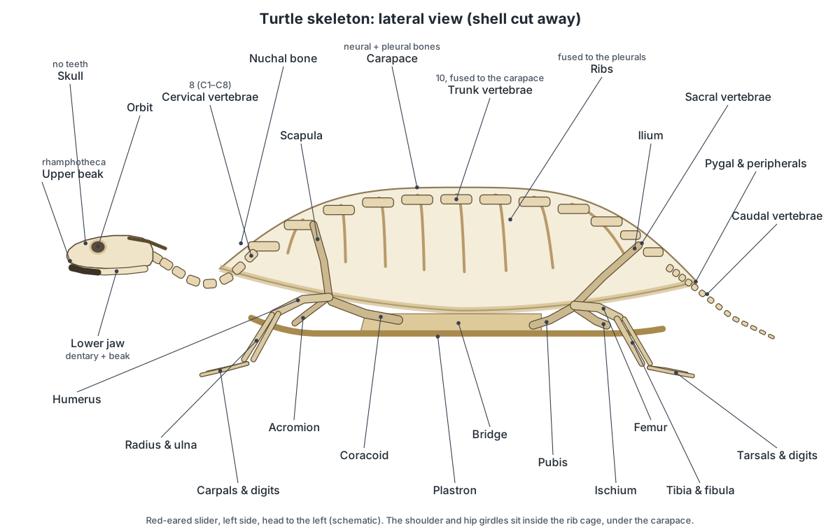
The bones a lab practical usually asks you to find on a turtle skeleton are:
- Nuchal bone: The single, broad bone at the front midline of the carapace, above the neck.
- Neural bones: Usually eight small, hexagonal plates down the midline of the carapace, fused to the vertebrae below.
- Pleural bones: Eight pairs of broad plates on the sides of the carapace, each a rib fused with a sheet of dermal bone.
- Peripheral bones: A ring of narrow bones (usually 11 on each side) that forms the rim of the carapace.
- Pygal bone: The single bone at the rear midline of the carapace rim, above the tail.
- Cervical vertebrae: Eight neck vertebrae, from the atlas behind the skull to the eighth, which meets the shell.
- Trunk vertebrae: Ten trunk vertebrae fused to the underside of the carapace midline.
- Ribs: Broad ribs that run outward under the carapace and are fused into the pleural bones.
- Scapula: A rod-like shoulder blade that rises from the shoulder joint to the underside of the carapace.
- Acromion process: A long process that runs from the scapula inward along the front of the shell.
- Coracoid: A flat, backward-pointing bone that completes the shoulder girdle.
- Ilium: The hip bone that rises from the hip socket to the sacral vertebrae.
- Pubis: The front, lower hip bone, with a forward-pointing process.
- Ischium: The rear, lower hip bone.
- Humerus: The curved upper-arm bone, whose head sits in the shoulder socket inside the shell.
- Femur: The thigh bone, from the hip socket inside the shell to the knee.
Is a turtle's shell part of its skeleton?
Yes. A turtle's shell is part of its skeleton, and most of it is bone. The carapace contains the turtle's ten trunk vertebrae and their ribs. Each rib is fused into a broad pleural (costal) plate, and the arch on top of each vertebra is fused to a neural plate on the midline. The spinal cord runs in the narrow space between the neural bones and the vertebral bodies, right under the top of the shell. Only the outer layer, the scutes, is skin: thin plates of keratin, the protein in your fingernails. (Wyneken (2001), The Anatomy of Sea Turtles, NOAA Tech. Memo. NMFS-SEFSC-470.)
The plastron is part of the skeleton too, although it comes from different bones. The front pair, the epiplastra, are thought to be the clavicles (collar bones), and the single entoplastron is thought to be the interclavicle. The other plastron bones are thought to come from gastralia, the belly ribs that many reptiles have in the skin of the abdomen. (Wikipedia: turtle shell (bone and scute nomenclature).)
So the common question "what type of skeleton does a turtle have?" has a simple answer: a turtle has an internal bony skeleton (an endoskeleton) like every other vertebrate. It is not an exoskeleton like a crab's, even though the shell is on the outside, because it grows from the turtle's own ribs, spine and dermal bone and is covered by living skin.
Turtle shell anatomy: carapace, plastron and scutes
A turtle shell has two layers in two parts. The two parts are the domed carapace on top and the flatter plastron underneath, joined at the sides by the bridge. The two layers are bone on the inside and keratin scutes on the outside. The pattern of bones and the pattern of scutes are different, and their seams are offset, like the joints in a brick wall.
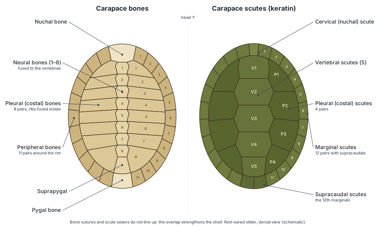
Carapace bones. On the midline, from front to back, are the broad nuchal over the neck, usually eight neurals, one or two suprapygals and the pygal over the tail. On each side are eight pleurals, and around the rim is a ring of peripherals, usually eleven on each side. Extra or missing neurals are fairly common, and many side-necked turtles have neurals that are reduced or hidden under the pleurals. (Wikipedia: turtle shell (bone and scute nomenclature).)
Carapace scutes. A slider has a small cervical (nuchal) scute at the front, five vertebral scutes down the middle, four pairs of pleural (costal) scutes on the sides and twelve pairs of marginal scutes around the edge, the last pair of which are called the supracaudals. Sea turtle guides call the pleurals "lateral scutes" and also count inframarginal scutes along the bridge. (Wyneken (2001), The Anatomy of Sea Turtles, NOAA Tech. Memo. NMFS-SEFSC-470.)
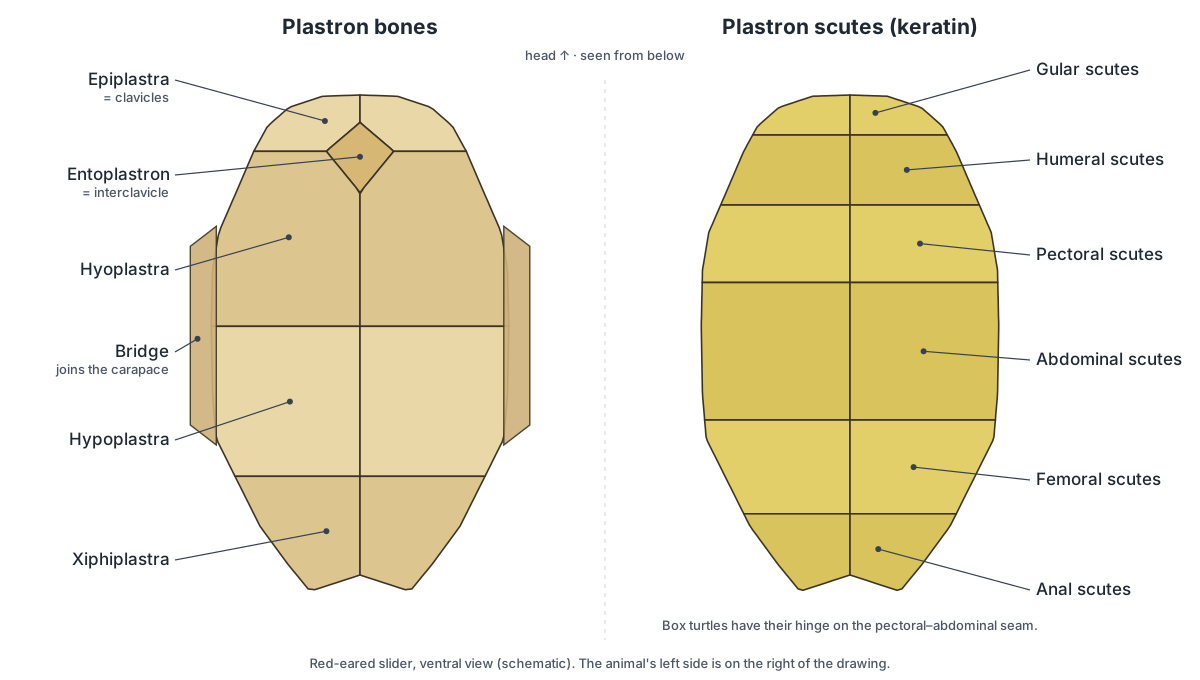
Plastron. The plastron has nine bones: paired epiplastra, the unpaired entoplastron, and paired hyoplastra, hypoplastra and xiphiplastra. The hyo- and hypoplastra extend up the sides as the bridge. On top of them lie twelve scutes in six pairs, named from front to back gular, humeral, pectoral, abdominal, femoral and anal. Side-necked turtles have a thirteenth, the intergular, between the gulars. (Wikipedia: Pleurodira (side-necked turtles).)
| Bone layer | Scute layer | |
|---|---|---|
| Made of | Bone (ribs, vertebrae, dermal bone) | Keratin (epidermis) |
| Carapace midline | Nuchal, 8 neurals, suprapygal(s), pygal | Cervical, 5 vertebrals, supracaudals |
| Carapace sides | 8 pairs of pleurals | 4 pairs of pleurals (costals) |
| Carapace rim | About 11 pairs of peripherals | 12 pairs of marginals |
| Plastron | 9 bones | 12 scutes (13 in side-necked turtles) |
| Grows by | Adding bone at the sutures | Adding keratin layers at the base |
| Living tissue? | Yes: blood vessels and nerves | Dead keratin over living skin |
Can a turtle live without its shell?
No. A turtle cannot leave its shell or live without it, because the shell contains its backbone and rib cage. Removing the shell would mean removing the spine. Cartoons of turtles climbing out of their shells, and searches for a "turtle skeleton without shell", imagine an animal that does not exist: there is no naked turtle body inside.
What a turtle can lose are its scutes. Aquatic turtles shed the outer layer of each scute from time to time, and a shell injury can strip scutes and expose bone. A cracked shell is a broken bone and needs a vet. If you look at a turtle skeleton in a museum with the plastron removed, you are seeing a preparation in which the bridge has been cut, not a turtle that ever lived that way.
The softshells and the leatherback sea turtle come closest to "shell-less". They have lost their scutes and much of the bone around the edge of the carapace, and are covered by leathery skin instead. Even so, their spine and ribs still form the core of the shell. (Wikipedia: Trionychidae (softshell turtles).)
Turtle spine and ribs: the axial skeleton
The axial skeleton of a turtle is the skull, the vertebral column and the ribs. The column has four regions. All turtles have eight cervical (neck) vertebrae. Behind them come ten trunk (dorsal, or thoracic) vertebrae, fused to the carapace, then two or three sacral vertebrae whose side processes support the hips, and a series of caudal vertebrae, 12 or more in sea turtles. Each trunk vertebra carries a pair of ribs, and each rib head sits at the junction of two vertebral bodies. (Wyneken (2001), The Anatomy of Sea Turtles, NOAA Tech. Memo. NMFS-SEFSC-470.)
Because the trunk vertebrae are fused into the shell, a turtle's back cannot bend at all. All its flexibility is in the neck and tail. In sea turtles the eighth neck vertebra is also fused to the carapace, so only seven move; in pond turtles the last neck vertebra meets the first trunk vertebra under the nuchal bone. The first and tenth ribs are small and only partly join the shell.
Turtle ribs are unusual in the direction they grow. In other reptiles the ribs curve down around the body cavity. In a turtle embryo the ribs stop short and grow outward and forward instead, inside a fold of the body wall called the carapacial ridge, which becomes the edge of the shell. Bone then spreads from each rib to make the pleural plates. (The Scientist: embryonic twist yields turtle shell.)
Shoulder and hip girdles inside the rib cage
A turtle's shoulder blades and hip bones are inside its rib cage, under the carapace. This is the most famous oddity of the turtle skeleton. In you, the shoulder blade lies on the outside of the ribs; in a turtle, the ribs have grown out over it, so the whole girdle hangs inside the shell. (National Geographic on Nagashima et al. (2009, Science): how the turtle got its shell.)
The shoulder girdle of each side has three prongs (it is triradiate). The rod-like scapula runs up from the shoulder joint and attaches to the carapace near the first trunk vertebra. The acromion process runs inward from the scapula and is tied by ligaments to the entoplastron. The flat coracoid points back over the plastron. The shoulder socket (glenoid) is formed by the scapula and coracoid together. (Wyneken (2001), The Anatomy of Sea Turtles, NOAA Tech. Memo. NMFS-SEFSC-470.)
The pelvis has the usual three bones on each side: the ilium rises to the sacral vertebrae and carapace, and the pubis and ischium form a floor over the plastron, with a large opening, the thyroid fenestra, between them. All three meet at the hip socket (acetabulum). In hidden-necked turtles like the slider the pelvis is tied to the shell only by ligaments; in side-necked turtles it is sutured firmly to both carapace and plastron. (Wikipedia: turtle (anatomy and evolution).)
Turtle head skeleton: skull, beak and neck
A turtle skull is solid and box-like, with large eye sockets, no teeth and a sharp keratin beak. The beak is called the rhamphotheca: an upper sheath over the premaxillae and maxillae and a lower sheath over the dentaries. It grows continuously, like a fingernail, and its shape matches the diet. In sliders it has a sharp cutting edge; green sea turtles have finely serrated jaws for cropping seagrass; loggerheads have broad, powerful crushing jaws for hard-shelled prey. (Wyneken (2001), The Anatomy of Sea Turtles, NOAA Tech. Memo. NMFS-SEFSC-470.)
The skull roof is made of paired prefrontals, frontals, parietals, postorbitals and squamosals. The cheek has the jugal and quadratojugal, and at the back the quadrate carries the jaw joint and supports the eardrum. A blade-like supraoccipital crest sticks out backward from the braincase, anchoring the jaw muscles. The lower jaw is a composite of the dentary, angular, surangular, prearticular, splenial and articular bones. Under the jaws hangs the hyoid apparatus, which supports the tongue and glottis but is usually lost from museum skeletons. (Wyneken (2001), The Anatomy of Sea Turtles, NOAA Tech. Memo. NMFS-SEFSC-470.)
Turtle skull openings: anapsid or diapsid?
A living turtle's skull has no true temporal openings (fenestrae) behind the eyes. For more than a century this 'anapsid' skull was read as a sign that turtles were a very ancient branch of reptiles that split off before the diapsids, the group with two openings on each side that includes lizards, snakes, crocodiles and birds. The skull is still often taught that way. (Bever et al. (2015), Evolutionary origin of the turtle skull, Nature.)
Genomic studies contradict that view: they place turtles inside the diapsid radiation, usually closest to the archosaurs (crocodilians and birds). If that is right, the closed skull of turtles is a secondary condition, built by closing over openings that their ancestors had. The fossils that tested this were found in 2015. Bever and colleagues used CT scans to show that Eunotosaurus, a 260-million-year-old stem turtle from the Karoo Basin of South Africa, had a lower temporal opening and a small upper one that was being covered over by an expanded supratemporal bone. They described it as 'a diapsid reptile in the process of becoming secondarily anapsid'. (Bever et al. (2015), Evolutionary origin of the turtle skull, Nature.)
The same year, Pappochelys, a 240-million-year-old stem turtle from Germany, was described with two pairs of temporal openings, a full diapsid skull. Together the two fossils support the idea that turtles began as diapsids and lost the openings later. The debate is not completely closed, because some researchers still question whether Eunotosaurus belongs on the turtle line, but the diapsid-origin view now has both genetic and fossil support. (Wikipedia: Pappochelys (Schoch & Sues 2015, Nature 523:584).)
Do not confuse true fenestrae with emarginations. Many modern turtles, including the red-eared slider, have a deep notch cut into the back of the skull roof (temporal emargination) and often another from below the cheek. These notches give the jaw-closing muscles room to bulge, but they are open edges, not holes ringed by bone. Their jaw muscles also run over a bony pulley: the otic capsule in hidden-necked turtles and the pterygoid in side-necked turtles. (Wikipedia: turtle (anatomy and evolution).)
Neck retraction: hidden-necked vs side-necked turtles
Turtles pull their heads in in one of two ways, and this splits living turtles into two suborders. Hidden-necked turtles (Cryptodira), including the slider, snapping turtles, box turtles, tortoises, softshells and sea turtles, bend the neck into a vertical S-shape and draw the head straight back. Double joints between the sixth, seventh and eighth neck vertebrae let the neck fold in that one plane. (Wikipedia: Cryptodira (hidden-necked turtles).)
Side-necked turtles (Pleurodira), such as the mata mata and the African helmeted turtles, fold the neck sideways and tuck the head under the front edge of the carapace beside a front leg. Today they live only in fresh water in South America, Africa, Madagascar and Australasia. (Wikipedia: Pleurodira (side-necked turtles).)
| Feature | Hidden-necked (Cryptodira) | Side-necked (Pleurodira) |
|---|---|---|
| Neck retraction | Vertical S-bend, head straight back | Sideways fold under the shell edge |
| Pelvis | Linked to the shell by ligaments | Sutured to carapace and plastron |
| Plastral scutes | 12 | 13 (with an intergular) |
| Jaw-muscle pulley | Otic capsule | Pterygoid |
| Examples | Sliders, snappers, box turtles, tortoises, sea turtles | Mata mata, side-necked turtles |
Sea turtles are cryptodires that have mostly lost the ability to retract the head, and snapping turtles cannot hide fully because their plastron is too small. (Wikipedia: turtle (anatomy and evolution).)
Turtle limbs: forelimb and hindlimb bones
Turtle legs have the standard four-limbed vertebrate bones, but the upper bones start inside the shell. The humerus runs from the shoulder joint under the carapace out through the front opening to the elbow; the forearm has a radius and ulna; the wrist has several carpals (radiale, intermedium, ulnare, centrale, pisiform and distal carpals); and each hand has five metacarpals and their phalanges. The hind leg has a femur, a tibia and fibula, a short ankle (calcaneum, astragalus and distal tarsals), five metatarsals and the toe phalanges. (Wyneken (2001), The Anatomy of Sea Turtles, NOAA Tech. Memo. NMFS-SEFSC-470.)
The elbow and knee bend so that the lower leg can swing inward when the limb is pulled into the shell. In a slider the feet are broad and webbed and carry long claws; males have very long front claws used in courtship. Tortoises have short, stumpy, unwebbed feet, and sea turtles have flippers. (Animal Diversity Web: Trachemys scripta (pond slider).)
How do turtles breathe with fused ribs?
Turtles breathe with their abdominal (flank) muscles, not their ribs. In lizards, snakes and mammals the ribs swing outwards to enlarge the chest and suck air in. A turtle cannot do this: its ribs are locked into the carapace, and the small intercostal muscles that move ribs in other reptiles are lost while the embryo develops. Instead, two pairs of antagonistic muscles do the work. (Lyson et al. (2014), Origin of the unique ventilatory apparatus of turtles, Nature Communications 5:5211.)
To breathe out, the transversus abdominis contracts. It is a sling-shaped muscle that runs from the front and back of the shell around the belly organs, so when it tightens it presses the viscera up against the lungs, which lie on top of everything under the carapace. To breathe in, the obliquus abdominis contracts. At rest it bulges like a dome in the soft skin between the hind leg and the bridge; when it flattens it pulls the gut and its lining back and down, the pressure in the lungs falls and air flows in. You can see this as a rhythmic pumping of the skin in front of the hind legs of a resting turtle. (Lyson et al. (2014), Origin of the unique ventilatory apparatus of turtles, Nature Communications 5:5211.)
The lungs are large, spongy and elastic, and they are attached directly to the underside of the carapace and the vertebral column. Because they sit dorsally, above the liver, stomach and intestines, they also help an aquatic turtle float level in the water. Movements of the limbs and neck in and out of the shell change the space inside the body cavity, so they add to breathing, especially when the turtle is active. (Wyneken (2001), The Anatomy of Sea Turtles, NOAA Tech. Memo. NMFS-SEFSC-470.)
Some aquatic turtles have an extra trick. They can take up dissolved oxygen across the lining of the cloaca and its sacs, which helps during long dives or when they spend the winter under ice. This cloacal respiration supplements the lungs; it does not replace them. (Wikipedia: turtle (anatomy and evolution).)
The fossil record suggests the abdominal breathing system evolved before the shell. The 260-million-year-old stem turtle Eunotosaurus already had a rigid, broad-ribbed trunk, no sign of intercostal muscles and bony attachment marks for the main breathing-out muscle. Lyson and colleagues concluded that the ribs and the abdominal muscles divided their jobs, the broad ribs stiffening the trunk and the muscles taking over breathing, about 50 million years before a fully bony shell appeared. (Lyson et al. (2014), Origin of the unique ventilatory apparatus of turtles, Nature Communications 5:5211.)
Turtle anatomy under the shell: where the organs are
Under the carapace, the organs are stacked in layers. The two big lungs lie on top, pressed against the underside of the carapace and the backbone. Below them, the large, dark liver spreads across the body cavity just above the plastron, with the three-chambered heart on the midline between its lobes, close behind the shoulder girdles. The stomach curves along the left side, and the coiled intestines fill the back half, leading to the cloaca at the base of the tail. The kidneys lie behind the lungs against the carapace, and the gonads sit near the base of the lungs. Unlike snakes, turtles have a urinary bladder on the midline in front of the pelvis; urine reaches it from the cloaca, not directly from the ureters. (Wyneken (2001), The Anatomy of Sea Turtles, NOAA Tech. Memo. NMFS-SEFSC-470.)
In the 3D model, hide the "Carapace bones" system to see the lungs and ribs from above, or hide "Plastron & bridge" to see the heart, liver and gut from below, which is how turtles are opened in a veterinary or museum dissection.
- Lungs: Two large, spongy lungs pressed against the underside of the carapace.
- Trachea & bronchi: The windpipe running down the neck, splitting into two unbranched bronchi that enter the lungs.
- Heart: A three-chambered heart (two atria, one ventricle) on the midline, just above the plastron.
- Liver: A large, dark, two-lobed liver lying across the body cavity just above the plastron.
- Stomach: A J-shaped muscular stomach on the turtle's left side.
- Intestines: The coiled small intestine and the shorter, wider large intestine (colon).
- Kidneys and ureters: Two flattened, lobed kidneys pressed against the inside of the carapace at the back of the body cavity, each drained by a ureter.
- Urinary bladder: A thin-walled sac that stores urine; turtles have one, unlike snakes.
- Cloaca: The shared chamber at the end of the gut, urinary and reproductive tracts.
Sea turtle skeleton: green, loggerhead and leatherback
A sea turtle skeleton has the same bones as a pond turtle's, reshaped for swimming. The shell is flatter and more streamlined, the head cannot be pulled in, and the front legs are long, stiff flippers. The flipper blade is formed by widening and flattening the wrist bones and by greatly lengthening the metacarpals and phalanges. The humerus is short and flat with a large medial process for the flight-like swimming muscles, and in adults the radius and ulna are bound together by fibrous tissue. Most hard-shelled species have two claws on each flipper. (Wyneken (2001), The Anatomy of Sea Turtles, NOAA Tech. Memo. NMFS-SEFSC-470.)
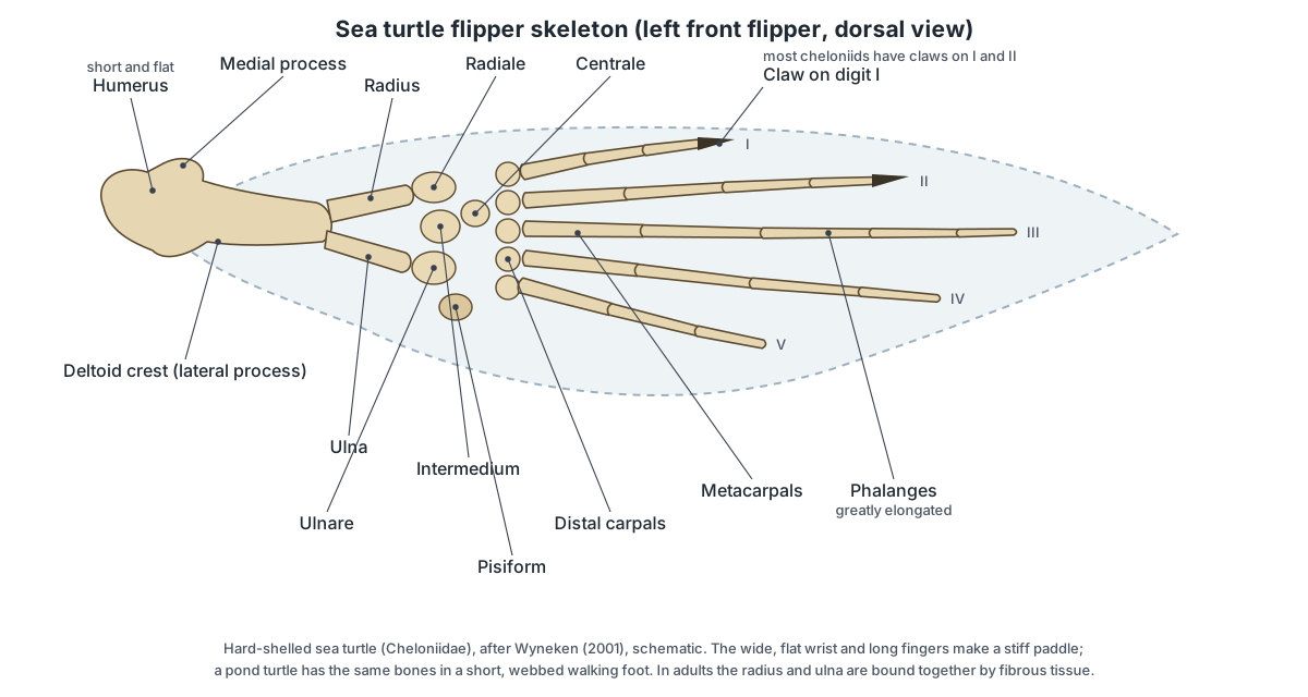
In young sea turtles the carapace is still open: the ribs reach out towards the peripherals with gaps, called fontanelles, between them. These close with bone in some adult ridleys and loggerheads but stay open towards the back in green turtles and hawksbills. The shape of the entoplastron also differs between species: arrow-shaped in green turtles, cross-shaped in loggerheads and T-shaped in hawksbills. (Wyneken (2001), The Anatomy of Sea Turtles, NOAA Tech. Memo. NMFS-SEFSC-470.)
The leatherback is the exception. It has no scutes and no hard shell. Its carapace is smooth, black, rubbery skin with seven ridges running its length, lying over a mosaic of small bony plates (dermal ossicles), a layer of oily connective tissue and fat, and the ribs and vertebrae. Its bony shell is reduced to an expanded nuchal, the ribs and the vertebrae, its plastron is only a ring of slender bones with no entoplastron, and the bones of its long flippers keep thick cartilage at their ends. Adults are typically 1.5 to 1.8 m long and weigh 340 to 450 kg, and they dive deeper than any other turtle. (NOAA Fisheries: leatherback turtle.)
| Species | Shell | Skull and jaws | Adult size |
|---|---|---|---|
| Green turtle (Chelonia mydas) | Smooth; 5 vertebral and 4 pairs of lateral scutes | Small head; serrated beak for seagrass and algae | 0.9–1.2 m, about 135–160 kg |
| Loggerhead (Caretta caretta) | Heart-shaped, reddish-brown; usually 5 pairs of lateral scutes | Very large head and crushing jaws | 0.75–1.05 m, 90–160 kg |
| Leatherback (Dermochelys coriacea) | No scutes; leathery skin with 7 ridges over dermal ossicles | Minimal keratin covering on the jaws | 1.5–1.8 m, 340–450 kg |
Sizes from NOAA Fisheries species pages, converted from feet and pounds and rounded. (NOAA Fisheries: green turtle.) (NOAA Fisheries: loggerhead turtle.)
Snapping turtle skeleton
A snapping turtle skeleton is built like a slider's but with a big head, a long neck and tail. The common snapping turtle (Chelydra serpentina) has a very small plastron that leaves most of its limbs exposed, so it cannot close up inside its shell; it relies on its powerful beak-like jaws and a neck that can reach back across the carapace to bite. Its tail is nearly as long as its shell and has saw-toothed keels along the top. (Animal Diversity Web: Chelydra serpentina (common snapping turtle).)
The alligator snapping turtle (Macrochelys temminckii) is much larger, commonly with a carapace of 35 to 80 cm and a weight of 8 to 80 kg. Its carapace carries three ridges of large, pointed scutes that flatten with age. Inside its mouth a worm-shaped pink appendage on the tongue lures fish within reach of its jaws. (Wikipedia: alligator snapping turtle.)
Box turtle skeleton: the hinged plastron
A box turtle can shut itself in completely because its plastron has a hinge. In the eastern box turtle (Terrapene carolina) a well-developed hinge runs across the plastron along the seam between the pectoral and abdominal scutes, dividing it into two movable lobes. The front lobe closes over the head and forelegs, the rear lobe over the hind legs and tail. (Illinois Natural History Survey: Terrapene carolina.)
To make this work the box turtle's plastron is "bridgeless": it has no rigid bony bridge locking it to the carapace, and the high, domed carapace leaves room for the folded limbs. Males have a concave rear plastral lobe and, usually, red eyes; females have a flat lobe and brown eyes. Like tortoises, box turtles keep their old scute layers, so growth rings build up on the shell. (Animal Diversity Web: Terrapene carolina (eastern box turtle).)
Softshell and mata mata skeletons
Softshell turtles (family Trionychidae) have a light, flexible shell with no horny scutes. A solid layer of bone lies under the middle of the carapace, but the edges have no bone and are soft, leathery and flexible. Some species have plastral bones that are not attached to the bones of the carapace. They also have a long, soft, snorkel-like snout. Despite the name, their shell is still built on their ribs and spine. (Wikipedia: Trionychidae (softshell turtles).)
The mata mata (Chelus fimbriatus) of South America is a side-necked turtle with a flat, triangular head, a long fringed neck, a snorkel-tipped snout and a rough, knobby carapace up to about 45 cm long. It cannot chew. It lies still on the bottom, then thrusts its head out and opens its mouth wide to suck in fish. (Wikipedia: mata mata.)
Tortoise vs turtle skeleton
A tortoise skeleton and a turtle skeleton have the same plan, because tortoises are turtles: the family Testudinidae is one branch of the hidden-necked turtles. Both have a shell of fused ribs and vertebrae, girdles inside the rib cage, eight neck vertebrae, a toothless beak and ten trunk vertebrae. The differences are in shape. In everyday North American English, "turtle" covers the whole order and "tortoise" means a land-living testudinid. (Wikipedia: turtle (anatomy and evolution).)
| Feature | Pond turtle (slider) | Tortoise (e.g. gopher tortoise) |
|---|---|---|
| Carapace | Low, streamlined oval | Usually high and domed, heavy |
| Feet | Long toes, webbed, long claws | Short, flattened, stump-like hind feet; no webbing |
| Forelimbs | Paddles for swimming | Shovel-like, for digging |
| Scutes | Outer layers shed | Kept, building growth rings |
| Head retraction | Straight back (cryptodire) | Straight back (cryptodire) |
| Swimming | Strong swimmer | Cannot swim, though it can float |
Gopher tortoise details from Animal Diversity Web; males of that species also have longer gular projections on the front of the plastron. (Animal Diversity Web: Gopherus polyphemus (gopher tortoise).) (Wikipedia: tortoise.)
Turtle shell growth and scute shedding
A turtle's shell grows with the turtle, from the bones and the scutes at the same time. The bones grow at their sutures, the seams where neighbouring plates meet. In a hatchling the carapace is mostly ribs and vertebrae; sheets of dermal bone then spread out along each rib until the pleural plates meet each other and the peripherals. Spaces left between rib tips and the rim are called fontanelles, and in some species they stay open for life. (Wyneken (2001), The Anatomy of Sea Turtles, NOAA Tech. Memo. NMFS-SEFSC-470.)
The keratin scutes grow in two ways: they widen at their margins and they thicken. Each growth season adds a new layer of keratin at the base of every scute. Terrestrial tortoises and box turtles keep all the layers, so their scutes build up raised rings (annuli). Aquatic turtles such as sliders shed the outer layer of individual scutes as thin, clear flakes while the new layer underneath takes over. (Wikipedia: turtle shell (bone and scute nomenclature).)
Counting annuli is only a rough guide to age. Growth depends on food and water, a tortoise can add more than one ring in a season, and old rings wear smooth. In aquatic turtles that shed, the rings are largely lost. (Wikipedia: tortoise.)
Normal shedding comes off in whole, thin, translucent pieces shaped like the scute, with healthy hard scute underneath. Soft spots, pits, smells, or bone showing through are not normal shedding and need a reptile vet. Because the shell is living bone with nerves and blood vessels just under the keratin, a turtle can feel touch and pain through it.
Evolution of the turtle shell: from Eunotosaurus to Archelon
The turtle shell evolved in steps over tens of millions of years, and the ribs came first. Eunotosaurus, about 260 million years old, had very broad trunk ribs that made its body stiff, but no shell. It was a land animal, which moves the ecological setting of the origin of turtles onto land rather than into water. (Bever et al. (2015), Evolutionary origin of the turtle skull, Nature.)
Pappochelys, from the Middle Triassic of Germany about 240 million years ago, adds the next clue. It was small, up to about 20 cm long with a tail making up half of that, and had broad, blade-like ribs and a belly covered with tightly packed, partly fused gastralia (belly ribs). Gastralia are thought to be the source of most of the plastron bones. (Wikipedia: Pappochelys (Schoch & Sues 2015, Nature 523:584).)
Odontochelys semitestacea, about 220 million years old, from Guizhou in southwest China, had a complete bony plastron but no bony carapace, only broadened ribs. It still had teeth in both jaws. The fossil showed that the plastron evolved before the carapace. (Wikipedia: Odontochelys (Li et al. 2008, Nature 456:497).)
Odontochelys also shows the shoulder blade still in front of the rib cage, as in other reptiles. Studies of turtle embryos by Nagashima and colleagues in 2009 explain how modern turtles moved it inside. The ribs stop growing downwards around the body and instead grow outwards and forwards, and the body wall folds inwards along the carapacial ridge (the future edge of the shell). The ribs then cover the shoulder blade, which ends up inside the rib cage. The muscles linking scapula and ribs keep their old connections and simply twist into the new arrangement. (National Geographic on Nagashima et al. (2009, Science): how the turtle got its shell.)
The fully shelled Proganochelys of the Late Triassic of Germany and Thailand had a complete carapace and plastron. It could not pull its head into the shell; it had a long neck and a long, spiked tail ending in a club. The oldest known side-necked and hidden-necked turtles, the two living groups that can pull the head in, date from the Late Jurassic. (Wikipedia: turtle shell (bone and scute nomenclature).)
The largest turtle that ever lived was Archelon, a sea turtle of the Late Cretaceous Western Interior Seaway of North America, about 80 to 74 million years ago. The biggest specimen, nicknamed Brigitta and now in the Natural History Museum Vienna, is about 4.6 m long and 4 m across the flippers. Its carapace had thin bony plates and was probably leathery, like a leatherback's, but Archelon belongs to an extinct group, the protostegids, that is not closely related to living leatherbacks; the leathery shells evolved separately. (Wikipedia: Archelon.)
Turtle skeleton parts: complete list
Every bone, scute and organ in the 3D model, grouped by system. Click any name in the viewer's list to jump to it in 3D.
Scutes & skin (keratin)
- Cervical scute (Nuchal scute, precentral scute)
- The small single scute at the front midline of the carapace, above the neck. Covers and protects the front rim of the shell over the neck opening.
- Vertebral scutes (Central scutes)
- Five broad keratin plates running in a row down the middle of the carapace. Form the protective, waterproof surface over the backbone and neural bones.
- Pleural scutes (Costal or lateral scutes)
- Four pairs of large keratin plates on the sides of the carapace, between the vertebrals and marginals. Cover the flanks of the carapace.
- Marginal scutes
- The ring of small scutes around the rim of the carapace: 12 on each side, counting the supracaudals. Protect the shell edge; the rear marginals of sliders are slightly serrated.
- Supracaudal scutes (12th marginals, postcentral scutes)
- The last pair of marginal scutes, meeting at the midline above the tail. Cover the rear rim of the shell above the tail opening.
- Gular scutes
- The front pair of plastron scutes, under the throat. Protect the front lobe of the plastron under the neck.
- Humeral scutes
- The second pair of plastron scutes, under the front legs (humerus). Cover the front lobe, which shields the head and forelimbs when they are pulled in.
- Pectoral scutes
- The third pair of plastron scutes, at chest level. Cover the chest region of the plastron.
- Abdominal scutes
- The fourth and usually largest pair of plastron scutes, at the bridge. Cover the belly and the central plastron.
- Femoral scutes
- The fifth pair of plastron scutes, in front of the hind legs (femur). Cover the rear lobe of the plastron.
- Anal scutes
- The last pair of plastron scutes, at the rear notch near the tail. Cover the tail end of the plastron; the anal notch between them leaves room for the tail.
- Claws
- Keratin claws on the tips of the digits. Grip the bottom and banks, tear food and dig nests.
- Skin (Scaly skin of the head, neck, limbs and tail)
- Soft, scaly, olive-green skin with yellow stripes and the red patch behind each eye that names the red-eared slider. Protects the soft parts; the webbed skin between the toes makes the feet into paddles.
Carapace bones
- Nuchal bone
- The single, broad bone at the front midline of the carapace, above the neck. Forms the front of the shell and anchors the neck muscles; the last neck vertebra connects to the shell beneath it.
- Neural bones (Neurals, vertebral plates)
- Usually eight small, hexagonal plates down the midline of the carapace, fused to the vertebrae below. Form the roof of the trunk vertebrae inside the shell; the spinal cord runs in the space between the neural bones and the vertebral bodies.
- Pleural bones (Costal plates, costals)
- Eight pairs of broad plates on the sides of the carapace, each a rib fused with a sheet of dermal bone. Make up most of the domed carapace and enclose the lungs and other organs.
- Peripheral bones (Peripherals)
- A ring of narrow bones (usually 11 on each side) that forms the rim of the carapace. Form the shell edge; the middle peripherals join the bridge to the plastron.
- Suprapygal bone (Suprapygals (one or two, by species))
- A midline bone (or pair of bones) between the last neural and the pygal. Closes the back of the carapace between the last pleurals.
- Pygal bone
- The single bone at the rear midline of the carapace rim, above the tail. Forms the rear edge of the shell over the tail.
Plastron & bridge
- Epiplastra (Epiplastron (singular))
- The front pair of plastron bones. Form the front edge of the plastron under the neck.
- Entoplastron
- The single, unpaired, diamond- or T-shaped bone in the front of the plastron. Ties the front plastron bones together; the acromion processes of the shoulder girdles attach to it by ligaments.
- Hyoplastra (Hyoplastron (singular))
- The large second pair of plastron bones, from behind the entoplastron to the middle of the bridge. Form much of the plastron and the front strut of the bridge (axillary buttress) that joins the carapace.
- Hypoplastra (Hypoplastron (singular))
- The large third pair of plastron bones, from the middle of the bridge back to the hind legs. Form the rear strut of the bridge (inguinal buttress) and the central plastron.
- Xiphiplastra (Xiphiplastron (singular))
- The rear pair of plastron bones, with the anal notch between them. Form the rear tip of the plastron in front of the tail.
- Bridge (Axillary and inguinal buttresses)
- The bony pillars on each side that join the plastron to the carapace, between the front and hind legs. Locks top and bottom shell into one rigid box.
Skull & jaws
- Premaxillae
- The small paired bones at the tip of the upper jaw, below the nostril opening. Form the front of the upper jaw that carries the horny beak.
- Maxillae
- The main bones of the upper jaw on each side, below the orbit. Carry the cutting edge of the upper beak and form part of the palate.
- Prefrontals
- Paired bones roofing the snout in front of the eyes. Form the front of the skull roof and the front rim of each orbit.
- Frontals
- Paired roofing bones between the eyes. Roof the front of the braincase and the upper rims of the orbits.
- Parietals
- The paired bones forming the roof of the braincase behind the eyes. Cover the brain and anchor the large jaw-closing muscles.
- Postorbitals
- Bones forming the back rim of each orbit. Close the rear of the orbit and brace the skull roof.
- Jugals (Cheek bones)
- Small cheek bones below and behind each orbit. Form part of the cheek and the lower edge of the orbit.
- Quadratojugals
- Cheek bones in front of the ear region. Link the cheek to the quadrate, which carries the jaw joint.
- Squamosals
- Bones at the upper back corners of the skull. Form the rear corner of the skull and anchor neck and jaw muscles.
- Quadrates (With the middle ear (tympanic) cavity)
- The bone on each side that carries the jaw joint and surrounds the middle ear. The lower jaw hinges on it; its wide, rounded rim supports the eardrum.
- Supraoccipital crest
- A blade-like crest projecting backwards from the top of the braincase. Anchors the large jaw-closing (adductor) muscles.
- Braincase & occipital condyle (Basioccipital, exoccipitals, prootic, opisthotic)
- The bones enclosing the brain and inner ear, ending in the single knob that joins the neck. Protects the brain and inner ear; the occipital condyle articulates with the atlas (first neck vertebra).
- Palate (Vomer, palatines, pterygoids)
- The bones forming the roof of the mouth. Separate the mouth from the nasal passage and anchor jaw muscles.
- Orbit & scleral ring (Scleral ossicles)
- The large eye socket and the ring of small bony plates that supports the eyeball. Houses and protects the eye; the scleral ossicles keep its shape.
- Hyoid apparatus (Hyoid body and horns)
- A bony body and paired horns that hang between and behind the lower jaws. Supports the tongue and glottis and anchors throat muscles used in feeding and breathing.
- Dentaries
- The front bones of the lower jaw, joined at the chin. Carry the lower beak and the bite force.
- Post-dentary bones (Surangular, angular, articular, prearticular, coronoid, splenial)
- The smaller bones at the back of each lower jaw. Form the jaw joint and the attachment for the jaw muscles.
- Upper rhamphotheca (Upper beak)
- The horny keratin sheath covering the upper jaw bones. Works as the cutting and crushing surface instead of teeth.
- Lower rhamphotheca (Lower beak)
- The keratin sheath covering the front of the dentaries. Bites against the upper beak to slice plants and prey.
Vertebrae & ribs
- Cervical vertebrae (Neck vertebrae C1–C8)
- Eight neck vertebrae, from the atlas behind the skull to the eighth, which meets the shell. Let the head reach, strike and fold back into the shell.
- Trunk vertebrae (Dorsal or thoracic vertebrae (10))
- Ten trunk vertebrae fused to the underside of the carapace midline. Carry the ribs and spinal cord; they cannot bend, because they are part of the shell.
- Ribs (Dorsal ribs fused to the pleurals)
- Broad ribs that run outward under the carapace and are fused into the pleural bones. Form the frame of the carapace; because they cannot move, turtles breathe with abdominal muscles instead.
- Sacral vertebrae
- Two (sometimes three) vertebrae whose rib-like side processes support the hips. Connect the backbone to the pelvis through the ilia.
- Caudal vertebrae (Tail vertebrae)
- A tapering series of small tail vertebrae. Support the tail, which steers in water and, in males, positions the cloaca during mating.
Shoulder & hip girdles
- Scapula (Shoulder blade (dorsal process))
- A rod-like shoulder blade that rises from the shoulder joint to the underside of the carapace. Braces the shoulder joint against the shell.
- Acromion process
- A long process that runs from the scapula inward along the front of the shell. Ties the shoulder girdle to the plastron (entoplastron) by ligaments and anchors limb muscles.
- Coracoid (Procoracoid in some texts)
- A flat, backward-pointing bone that completes the shoulder girdle. Forms part of the shoulder socket (glenoid) and anchors the muscles that pull the forelimb back.
- Ilium
- The hip bone that rises from the hip socket to the sacral vertebrae. Connects the pelvis to the backbone and, by ligaments, to the carapace.
- Pubis
- The front, lower hip bone, with a forward-pointing process. Forms the front of the pelvis and anchors belly and thigh muscles.
- Ischium
- The rear, lower hip bone. Forms the back of the pelvis; the large opening between pubis and ischium is the thyroid fenestra.
Forelimb
- Humerus
- The curved upper-arm bone, whose head sits in the shoulder socket inside the shell. Moves the front leg forward and back for walking and swimming.
- Radius
- One of the two forearm bones, on the thumb side. Supports the forearm and wrist.
- Ulna
- The second forearm bone, on the little-finger side. Supports the forearm and forms the elbow joint with the humerus.
- Carpals (Wrist bones: radiale, intermedium, ulnare, centrale, pisiform, distal carpals)
- A cluster of small wrist bones. Make the wrist flexible.
- Metacarpals
- Five slender bones of the palm. Support the webbed front foot.
- Phalanges of the front foot (Finger bones)
- The bones of the five fingers. Spread the web for swimming and carry the claws.
Hindlimb
- Femur
- The thigh bone, from the hip socket inside the shell to the knee. Moves the hind leg for walking, swimming and digging nests.
- Tibia
- The larger shin bone. Carries the weight of the hind leg.
- Fibula
- The slender outer bone of the lower leg. Supports the outer side of the ankle.
- Tarsals (Astragalus, calcaneum, distal tarsals)
- The short ankle, made of a few large and several small bones. Form the ankle joint.
- Metatarsals
- Five bones of the sole of the hind foot. Support the broad, webbed hind foot, the slider's main paddle.
- Phalanges of the hind foot (Toe bones)
- The bones of the toes. Spread the web for swimming and carry the claws used for digging.
Organs under the shell
- Lungs
- Two large, spongy lungs pressed against the underside of the carapace. Breathe air. They fill as the flank muscles pull the organs away and empty as other muscles squeeze the organs up against them.
- Trachea & bronchi (Windpipe)
- The windpipe running down the neck, splitting into two unbranched bronchi that enter the lungs. Carries air between the glottis and the lungs, even when the neck is folded back into the shell.
- Heart
- A three-chambered heart (two atria, one ventricle) on the midline, just above the plastron. Pumps blood to the lungs and body; it can shunt blood past the lungs while the turtle holds its breath underwater.
- Liver
- A large, dark, two-lobed liver lying across the body cavity just above the plastron. Processes nutrients, stores energy and makes bile.
- Stomach
- A J-shaped muscular stomach on the turtle's left side. Begins digesting food swallowed in chunks (turtles cannot chew).
- Intestines
- The coiled small intestine and the shorter, wider large intestine (colon). Digest and absorb food and reabsorb water.
- Cloaca
- The shared chamber at the end of the gut, urinary and reproductive tracts. Releases faeces, urine, eggs or sperm.
- Kidneys and ureters (Metanephric kidneys)
- Two flattened, lobed kidneys pressed against the inside of the carapace at the back of the body cavity, each drained by a ureter. Filter the blood and make urine, which runs down the ureters to the cloaca.
- Urinary bladder
- A thin-walled sac that stores urine; turtles have one, unlike snakes. Stores urine and lets water be reabsorbed from it.
Frequently asked questions
Is a turtle's shell part of its skeleton?
Yes. The bony shell is part of the skeleton. The carapace (top shell) is built from the ribs and the trunk vertebrae fused with plates of dermal bone, and the plastron (bottom shell) includes bones thought to be the clavicles and interclavicle. Only the outer keratin scutes are skin rather than bone.
Can a turtle come out of its shell?
No. A turtle cannot leave its shell, because its backbone and ribs are fused into the carapace. A turtle without a shell would be a turtle without a spine and rib cage. Cartoons of turtles stepping out of their shells are fiction; a shell injury is a bone injury and needs a vet.
How many bones are in a turtle's shell?
About 50 to 60. A typical hard-shelled turtle has a nuchal, eight neurals, eight pairs of pleurals, about eleven pairs of peripherals, one or two suprapygals and a pygal in the carapace, plus nine plastron bones. Counts vary between species and individuals.
How do turtles breathe if their ribs are fused to the shell?
With muscles in the flanks instead of the ribs. One pair of abdominal muscles squeezes the organs up against the lungs to breathe out; another pair flattens the soft skin near the hind legs to pull the organs down and breathe in. Leg and neck movements add to this.
Do turtles have teeth?
No living turtle has teeth. Their jaws are covered by a sharp keratin beak called the rhamphotheca. The earliest stem turtles, such as Odontochelys from about 220 million years ago, still had teeth.
How many neck bones does a turtle have?
Eight cervical vertebrae, in all turtles. Hidden-necked turtles fold them into a vertical S to pull the head straight back; side-necked turtles bend the neck sideways under the front edge of the shell.
What is the difference between scutes and shell bones?
Scutes are the outer keratin plates you see on a living turtle; the bones lie underneath. They are separate layers with different patterns: the seams between scutes do not line up with the sutures between bones, which strengthens the shell.
What is the difference between a turtle and a tortoise skeleton?
The plan is the same: a shell of fused ribs and vertebrae, girdles inside the rib cage and eight neck vertebrae. Tortoises usually have a higher, heavier domed shell, short stumpy feet with no webbing and shovel-like forelimbs for digging; pond turtles have a flatter, streamlined shell and long, webbed toes.
Does a leatherback sea turtle have a shell?
It has a reduced one. Its carapace is a mosaic of small bony plates (dermal ossicles) under thick, oily, leathery skin with seven ridges, and its bony shell is reduced to the ribs, vertebrae and an expanded nuchal bone. It has no scutes and no entoplastron.
References
- Wyneken, J. (2001). The Anatomy of Sea Turtles. U.S. Department of Commerce, NOAA Technical Memorandum NMFS-SEFSC-470, 172 pp.
- Habova, M., Pyszko, M., Horak, O., Cermakova, E. & Paral, V. (2022). Differences in the anatomy of the lower respiratory tract in selected species of the order Testudines. Veterinarni Medicina 67. doi:10.17221/64/2021-VETMED
- Lyson, T. R., Schachner, E. R., Botha-Brink, J., Scheyer, T. M., Lambertz, M., Bever, G. S., Rubidge, B. S. & de Queiroz, K. (2014). Origin of the unique ventilatory apparatus of turtles. Nature Communications 5, 5211. doi:10.1038/ncomms6211
- Bever, G. S., Lyson, T. R., Field, D. J. & Bhullar, B.-A. S. (2015). Evolutionary origin of the turtle skull. Nature 525, 239–242. doi:10.1038/nature14900
- Nagashima, H. et al. (2009). Evolution of the turtle body plan by the folding and creation of new muscle connections. Science 325, 193–196 (summary: National Geographic).
- The Scientist (2009). Embryonic twist yields turtle shell.
- NOAA Fisheries. Leatherback turtle (Dermochelys coriacea).
- NOAA Fisheries. Green turtle (Chelonia mydas).
- NOAA Fisheries. Loggerhead turtle (Caretta caretta).
- Animal Diversity Web, University of Michigan. Trachemys scripta (pond slider).
- Animal Diversity Web. Chelydra serpentina (snapping turtle).
- Animal Diversity Web. Terrapene carolina (eastern box turtle).
- Illinois Natural History Survey, Herpetology. Terrapene carolina.
- Animal Diversity Web. Gopherus polyphemus (gopher tortoise).
- Wikipedia. Turtle shell: bone and scute nomenclature (pointer to Pritchard 1988; Zangerl 1969; Li et al. 2008).
- Wikipedia. Odontochelys, Pappochelys and Archelon (pointers to Li et al. 2008, Nature 456:497; Schoch & Sues 2015, Nature 523:584).
More interactive anatomy models
- Snake anatomyElongated organs, one working lung, a flexible jaw and the snake heart.
- Snake skeletonHundreds of vertebrae and ribs, a kinetic skull and vestigial hip bones.
- Lizard anatomySkull, scales, three-chambered heart and tail autotomy of a lizard.
- Human anatomy1,000+ bones, muscles, organs, vessels and nerves from real anatomical data.
- Octopus anatomyThree hearts, a ring-shaped brain, eight arms and a beak, in 3D.
- Squid anatomyPen, eight arms, two tentacles, giant axons and three hearts of the longfin squid.