

Rabbit anatomy at a glance
A rabbit is a small, plant-eating mammal built for spotting predators and running from them. Its key anatomical features are long ears that also cool the body, eyes on the sides of the head with an almost 360° view, teeth that never stop growing, a light skeleton driven by powerful hind legs, and a huge caecum in which bacteria ferment fibre. Rabbits re-eat soft droppings from the caecum, a habit called caecotrophy.
Rabbits are lagomorphs (order Lagomorpha), not rodents. The order also contains hares and pikas. This page describes the domestic rabbit, which descends from the European rabbit, Oryctolagus cuniculus. The reference animal is the New Zealand White, an albino breed of about 4–6 kg used in research and in many school and university labs. Wild European rabbits are smaller, about 1.5–2.5 kg and 38–50 cm long (Animal Diversity Web), and breeds range from about 1.5 kg (Dutch) to 8–9 kg (Flemish Giant) (Brewer, 2006). Their anatomy is the same; only size and proportions change.
Terms: cranial means towards the head, caudal towards the tail, dorsal the back and ventral the belly. A male rabbit is a buck, a female a doe and a baby a kit (MSD Veterinary Manual).
Rabbit anatomy diagram: external anatomy labeled
The external anatomy of a rabbit includes the long ears (pinnae), large side-facing eyes, whiskers, a twitching nose above a cleft upper lip, short forelegs with five toes, long hind feet with four toes and furred soles, and a short upturned tail called the scut. Scent glands sit under the chin, beside the genitals (inguinal glands) and around the anus. Does have 8–10 nipples and often a dewlap under the chin.
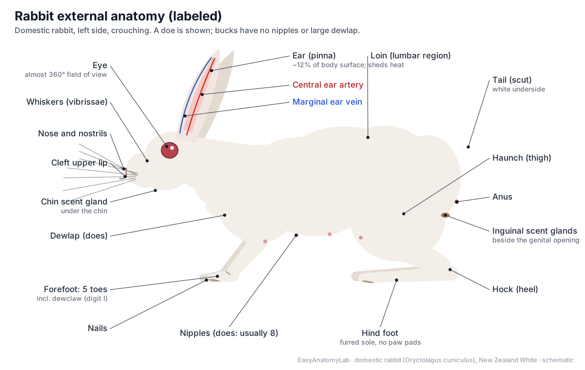
- Ear (pinna): Funnels sound into the ear canal and, through its dense blood vessels, sheds body heat (thermoregulation).
- Eye: Gives a field of view of almost 360° with a narrow overlap in front, to spot predators.
- Whiskers: Sense nearby objects in the dark and in the blind spot just under the nose.
- Nose and nostrils: Take in air and odours. Rabbits are obligate nose breathers.
- Cleft upper lip: Lets the lips part around the incisors. Folds of lip skin can meet behind the incisors so the rabbit gnaws with its mouth cavity closed.
- Dewlap: Stores fat. Pregnant does pull fur from the dewlap, chest and belly to line the nest.
- Forefoot (front paw): Supports the front of the body, digs, and holds food. It does most of the steering when hopping.
- Hind foot: Takes the push of each hop and supports most of the weight when the rabbit sits.
- Tail (scut): Signals alarm: a fleeing rabbit lifts its tail and flashes the white underside.
Rabbits have three groups of scent glands: the chin glands (between the halves of the lower jaw), the paired inguinal glands in hairless pockets beside the genital opening, and the anal glands, which scent the droppings. Rabbits rub their chins on objects ("chinning") to mark territory.
Internal anatomy of a rabbit
Inside a rabbit, a small chest holds the heart, two lungs and a thymus that stays large in adults. Behind the diaphragm, the abdomen is filled mostly by the gut: a small stomach on the left, the coiled small intestine, and the enormous caecum, which takes up much of the lower abdomen. The liver lies against the diaphragm, the kidneys lie on the roof of the abdomen, and the bladder and reproductive organs sit at the back, towards the pelvis.
Fade the skin in the 3D model to about 10% and hide the Muscular and Skeletal systems to see this layout. Switch between doe and buck to compare the reproductive organs.
Rabbit anatomy labeled: every part and its function
This is the text version of the 3D model: every labeled structure of the rabbit, grouped by body system, with what it does.
Skin & external
- Skin and fur (Integument, pelage)
- Thin, loose skin covered by dense fur; New Zealand Whites are albino, with white fur and pink eyes. Insulates the body, protects it, senses touch and carries the scent glands.
- Ear (pinna) (Auricle, external ear)
- The long, upright, cartilage-supported ear flap, about 10–12 cm long in a New Zealand White. Funnels sound into the ear canal and, through its dense blood vessels, sheds body heat (thermoregulation).
- Whiskers (Vibrissae, tactile hairs)
- Long, stiff sensory hairs on the muzzle and above the eyes. Sense nearby objects in the dark and in the blind spot just under the nose.
- Nose and nostrils (Nares, rhinarium)
- Two slit-like nostrils that the rabbit twitches open and closed many times a minute. Take in air and odours. Rabbits are obligate nose breathers.
- Cleft upper lip (Split lip, philtrum)
- The upper lip is split down the middle, exposing the incisors. Lets the lips part around the incisors. Folds of lip skin can meet behind the incisors so the rabbit gnaws with its mouth cavity closed.
- Dewlap (Throat fold) · females only
- A fold of skin and fat under the chin and throat, most developed in mature does. Stores fat. Pregnant does pull fur from the dewlap, chest and belly to line the nest.
- Forefoot (front paw) (Manus)
- The short front foot, with five digits: four toes and a short inner first digit (the dewclaw). Supports the front of the body, digs, and holds food. It does most of the steering when hopping.
- Hind foot (Pes, furred plantar surface)
- The long hind foot, with four toes and a thickly furred sole instead of pads. Takes the push of each hop and supports most of the weight when the rabbit sits.
- Nails (claws) (Ungues)
- Long, slightly curved nails, one on each toe: 18 in all (5 + 5 front, 4 + 4 hind). Grip the ground and dig burrows.
- Tail (scut) (Cauda)
- A short, upturned tail covered in fluffy fur, white on the underside. Signals alarm: a fleeing rabbit lifts its tail and flashes the white underside.
- Chin scent gland (Submandibular gland (cutaneous))
- Scent glands in the skin between the halves of the lower jaw. Marks objects with scent when the rabbit rubs its chin on them ("chinning").
- Inguinal scent glands (Inguinal pouches)
- Paired, hairless skin pockets beside the genitals that collect a waxy, strong-smelling secretion. Produce scent used in social and sexual signalling.
- Nipples (Papillae mammae) · females only
- Usually 8 nipples (4 pairs), sometimes 10, in two rows on the chest and belly. Deliver milk to the kits. The mammary glands lie under the skin around them.
- Scrotal sacs (Scrotum) · males only
- Two hairless sacs that hold the testes, placed in front of (cranial to) the penis. Keep the testes cooler than the body core, which sperm production needs.
- Vulva (External genital opening) · females only
- The slit-shaped genital opening of the doe, in front of the anus. Opening of the vestibule, into which both the vagina and the urethra empty.
- Penis and prepuce (External genitalia of the buck) · males only
- A short, backward-pointing penis in a fold of skin (the prepuce), with no bone inside. Carries urine and semen through the urethra; it points backward at rest.
- Anus (Anal opening)
- The opening of the rectum, under the tail. Passes hard droppings, and caecotropes, which the rabbit eats straight from the anus.
Skeletal
- Nasal bones (Os nasale)
- Long, paired bones forming the roof of the muzzle. Roof over the nasal cavity, which holds the scroll-like turbinates.
- Premaxilla (Incisive bone)
- The bone at the front of the upper jaw that holds the four upper incisors. Anchors both pairs of upper incisors, the large ones and the peg teeth.
- Maxilla (fenestrated) (Upper jaw bone)
- The main bone of the upper jaw. Its side wall is a lattice of holes: the fenestrated maxilla. Holds the upper cheek teeth and forms the side of the snout.
- Frontal bones (Os frontale)
- Paired bones of the forehead, with a wing-like supraorbital process over each eye. Roof the front of the braincase and the top of the orbits.
- Parietal and interparietal bones (Ossa parietalia, os interparietale)
- The roof of the braincase behind the frontal bones, with a small midline interparietal bone at the back. Protect the cerebral hemispheres.
- Occipital bone (Os occipitale)
- The back of the skull, around the foramen magnum. Its two occipital condyles articulate with the atlas; the spinal cord leaves the skull through the foramen magnum.
- Zygomatic arch (Cheekbone)
- The bony bar below each orbit, formed by the zygomatic bone and processes of the maxilla and squamosal. Anchors the large masseter muscle, the main chewing muscle.
- Tympanic bulla (Bulla tympanica)
- A rounded bony capsule on the underside of the skull that encloses the middle ear. Houses the middle-ear cavity and its three ossicles, and leads to the external ear canal.
- Mandible (Lower jaw)
- The two halves of the lower jaw, joined at the chin, carrying the lower incisors and cheek teeth. Moves sideways and up for grinding; its tall ramus gives leverage to the masseter.
- Cervical vertebrae (7) (C1–C7, atlas and axis)
- The seven neck vertebrae, as in almost all mammals; the first two are the atlas and axis. Support and move the head.
- Thoracic vertebrae (12) (T1–T12)
- Usually twelve vertebrae, one for each pair of ribs; some rabbits have 13. Carry the ribs and the tall spines that anchor back and neck muscles.
- Lumbar vertebrae (7) (L1–L7)
- Seven long, strong vertebrae in the lower back, with large transverse processes. Anchor the powerful back muscles that extend the spine during a hop.
- Sacrum (4 fused) (Os sacrum)
- Four sacral vertebrae fused into one bone. Joins the spine to the pelvis so the hind-limb thrust reaches the body.
- Caudal vertebrae (15–16) (Coccygeal vertebrae)
- About 15–16 small tail vertebrae that shrink to simple rods at the tip. Support and move the tail.
- Ribs (12 pairs) (Costae)
- Twelve pairs: 7 true ribs reach the sternum; 5 false ribs, of which the last 3 float. Protect the heart and lungs and move air during breathing.
- Sternum (Breastbone)
- Six bony segments (sternebrae): the manubrium, four body segments and the long xiphoid process. Joins the true ribs together at the front.
- Scapula (Shoulder blade)
- A flat, triangular bone with a tall spine and a long, hooked acromion. Attaches the foreleg to the trunk through muscle and forms the shoulder joint.
- Clavicle (small) (Collarbone)
- A slender, curved rod of bone that does not reach both ends of the gap it spans. Gives some attachment to shoulder muscles; it is too small to brace the shoulder.
- Humerus (Upper arm bone)
- The bone of the upper foreleg, between the shoulder and the elbow. Lever for the shoulder and elbow muscles.
- Radius and ulna (Forearm bones)
- The two forearm bones; the ulna forms the point of the elbow (olecranon). Support the front of the body when it lands after a hop.
- Carpal bones (9) (Wrist)
- Nine small wrist bones in two rows. Form the flexible wrist.
- Metacarpals and phalanges (Bones of the forefoot)
- Five metacarpals and the toe bones of five digits. Form the short front foot and toes.
- Pelvis (os coxae) (Hip bones: ilium, ischium, pubis)
- Each hip bone is made of the fused ilium, ischium and pubis, enclosing a large obturator foramen. Forms the hip socket (acetabulum) and passes the thrust of the hind legs to the spine.
- Femur (Thigh bone)
- The long thigh bone, with a large third trochanter on its outer side. Lever for the powerful thigh muscles.
- Patella (Kneecap)
- A small bone in the tendon of the quadriceps muscle, at the front of the knee. Increases the leverage of the quadriceps when it straightens the knee.
- Tibia and fibula (Shin bones)
- The long tibia and the thin fibula, which fuses with the tibia over its lower half. A long lower leg adds speed to every jump.
- Calcaneus (hock) (Heel bone, tuber calcanei)
- The heel bone, which projects behind the ankle as the point of the hock. Lever arm for the calf muscles that extend the foot in each hop.
- Tarsal bones (6) (Ankle bones)
- Six ankle bones, including the talus and calcaneus. Form the hock joint between the lower leg and the long foot.
- Metatarsals and phalanges (Bones of the hind foot)
- Four long working metatarsals (II–V) and the bones of four toes. Make the hind foot a long lever for jumping.
Teeth
- Upper incisors (First maxillary incisors (I1))
- Two long, curved, chisel-edged front teeth that grow about 2 mm a week. Cut and gnaw food.
- Peg teeth (Second maxillary incisors (I2))
- Two small, tube-like teeth just behind the upper incisors: the lagomorph trait that rodents lack. Form a stop that the lower incisors close against, between the first and second upper incisors.
- Lower incisors (Mandibular incisors)
- One pair of long, curved lower front teeth. Work against the upper incisors and peg teeth to cut food.
- Cheek teeth (premolars and molars) (Premolars 3/2, molars 3/3)
- Six upper and five lower grinding teeth on each side, all open-rooted and ever-growing. Grind grass and hay with a sideways chewing motion.
- Diastema (Toothless gap)
- The long gap with no teeth between the incisors and the cheek teeth, where canines would be. Lets the cheeks fold in so the rabbit can gnaw without swallowing debris.
Muscular
- Masseter (Main chewing muscle)
- A large, thick muscle on the cheek, much bigger than the temporal muscle. Closes the jaw and drives the side-to-side grinding stroke.
- Back muscles (longissimus) (Epaxial muscles, erector spinae)
- Thick bands of muscle on each side of the spine, largest in the loins. Extend (straighten) the spine to add power to each jump.
- Abdominal wall muscles (External and internal oblique, transversus, rectus abdominis)
- Thin muscle sheets that form the walls of the large abdomen. Hold the large gut in place and help with breathing out, defecation and giving birth.
- Triceps brachii (Extensor of the elbow)
- The large muscle at the back of the upper foreleg. Straightens the elbow and takes the landing shock on the forelegs.
- Gluteal muscles (Glutaeus superficialis and medius)
- Muscles of the rump over the hip. Extend the hip, driving the thigh backward at take-off.
- Biceps femoris (Hamstring (lateral))
- A broad muscle on the outside of the thigh. Extends the hip and helps flex the knee.
- Semitendinosus and semimembranosus (Hamstrings (medial))
- Long muscles at the back of the thigh. Extend the hip and flex the knee.
- Quadriceps femoris (Rectus femoris and vasti)
- The big muscle mass at the front of the thigh. Straightens the knee in the hop.
- Gastrocnemius and Achilles tendon (Calf muscle, tendo calcaneus)
- The calf muscle, whose strong tendon attaches to the point of the hock. Extends the foot, giving the final push of each hop.
- Tibialis cranialis (Tibialis anterior)
- The muscle at the front of the shin. Flexes the hock, lifting the foot.
Nervous & senses
- Eye (Globe, bulbus oculi)
- A large, bulging eye on the side of the head; pink-red in albino New Zealand Whites. Gives a field of view of almost 360° with a narrow overlap in front, to spot predators.
- Harderian gland (Gland of the third eyelid)
- A large gland behind the eye with a small white lobe and a large pink lobe. Lubricates the eye and third eyelid with an oily secretion.
- Olfactory bulbs (Bulbi olfactorii)
- Two large knobs at the front of the brain that receive smell signals. Process smell, a key sense for rabbits.
- Cerebrum (Cerebral hemispheres)
- The two cerebral hemispheres, with an almost smooth surface. Senses, memory and voluntary movement.
- Cerebellum (Little brain)
- A folded part of the brain behind the cerebrum. Coordinates balance and movement, including fast zig-zag running.
- Brainstem (Medulla oblongata and pons)
- The stalk joining the brain to the spinal cord. Controls breathing, heart rate and reflexes.
- Spinal cord (Medulla spinalis)
- The cord of nerve tissue inside the vertebral canal. Carries signals between the brain and body.
- Sciatic nerve (Nervus ischiadicus)
- The thickest nerve, running down the back of the thigh. Controls the muscles of the hind leg and carries sensation from it.
- Ear canal and inner ear (External acoustic meatus, cochlea)
- The tube at the base of each pinna leading to the eardrum, the middle ear and the cochlea. Carries sound to the eardrum; the inner ear also senses balance.
Respiratory
- Trachea (Windpipe)
- The tube of cartilage rings carrying air from the larynx to the lungs. Conducts air.
- Right lung (Pulmo dexter)
- The larger lung, with four lobes: cranial, middle, caudal and accessory. Gas exchange.
- Left lung (Pulmo sinister)
- The smaller lung: a small cranial lobe (sometimes divided) and a caudal lobe. Gas exchange.
- Diaphragm (Thoracic diaphragm)
- The dome of muscle and tendon that separates the small chest from the large abdomen. Main breathing muscle: it contracts to draw air into the lungs.
Cardiovascular
- Heart (Cor)
- A small four-chambered heart, about 0.3% of body weight. Pumps blood through the lungs and the body.
- Aorta (Main artery)
- The main artery, arching over the heart and running back under the spine. Carries oxygen-rich blood to the body.
- Caudal vena cava (Posterior vena cava)
- The large vein returning blood from the abdomen and hind legs to the heart. Returns oxygen-poor blood to the right atrium.
- Central ear artery (Central auricular artery)
- The artery running up the middle of each pinna. Supplies the pinna; its flow changes to release or keep body heat.
- Marginal ear vein (Lateral auricular vein)
- The vein running along the outer edge of each pinna. Drains blood from the pinna.
- Jugular vein and carotid artery (Vessels of the neck)
- The large paired vessels of the neck. Carry blood to (carotid) and from (jugular) the head.
Digestive
- Tongue (Lingua)
- A long tongue with a raised hump at the back. Moves food back to the cheek teeth and into the throat.
- Salivary glands (Parotid, mandibular, sublingual, zygomatic)
- Four pairs of glands that moisten food. Produce saliva, which contains some starch-digesting amylase.
- Oesophagus (Esophagus, gullet)
- The muscular tube from the throat to the stomach. Carries swallowed food to the stomach.
- Stomach (Gaster)
- A thin-walled, simple stomach that is never empty; it holds about 15% of the gut volume. Begins digestion with strong acid (pH 1–2).
- Duodenum (First part of the small intestine)
- A long U-shaped loop leaving the stomach. Receives bile and pancreatic juice for digestion.
- Jejunum and ileum (Small intestine)
- The coiled main part of the small intestine; only about 12% of the gut volume. Absorbs sugars, fats and amino acids.
- Sacculus rotundus (Ileocaecal tonsil)
- A round, thick-walled swelling at the end of the ileum, packed with lymphoid tissue. Lymphoid (immune) tissue that samples gut contents; also marks the ileocaecal junction.
- Caecum (Cecum)
- The enormous, thin-walled fermentation chamber: about 40% of the gut and 10 times the stomach's capacity. Bacteria ferment fibre here into volatile fatty acids, vitamins and protein.
- Vermiform appendix (Processus vermiformis)
- A narrow, thick-walled tube about 10–13 cm long at the blind tip of the caecum. Lymphoid tissue for immunity; it may also secrete alkaline fluid that buffers caecal acids.
- Proximal colon (Ascending colon with haustra)
- The first part of the colon, with pouches (haustra) and bands that sort particles by size. Separates gut contents: small particles and fluid go back to the caecum; large fibre particles move on to form hard droppings.
- Fusus coli (Colonic pacemaker)
- A short, thick muscular segment of colon, 5–8 cm long, between the proximal and distal colon. Acts as the pacemaker for colonic movement and switches between making hard droppings and caecotropes.
- Distal colon and rectum (Descending colon, rectum)
- The final, narrow part of the large intestine, where droppings are shaped. Absorbs water and forms hard round droppings or soft caecotropes.
- Liver (Hepar)
- A dark red organ with left lateral, left medial, right, quadrate and caudate lobes. Makes bile, stores nutrients and processes absorbed food and toxins.
- Gallbladder (Vesica fellea)
- A small green sac on the liver that stores bile. Stores and releases bile into the duodenum through the bile duct.
- Pancreas (Diffuse pancreas)
- A loose, pinkish, lacy gland scattered through the mesentery of the duodenal loop. Makes digestive enzymes and the hormones insulin and glucagon.
Urinary
- Kidneys (Renes)
- Two bean-shaped kidneys, each with a single renal papilla (unipapillate). Filter the blood and excrete wastes and excess calcium.
- Ureters (Ureteres)
- Thin tubes carrying urine from each kidney to the bladder. Transport urine.
- Urinary bladder (Vesica urinaria)
- A thin-walled sac that often contains cloudy urine from calcium carbonate crystals. Stores urine.
Reproductive
- Testes (Testicles) · males only
- Two oval testes in the scrotal sacs; they can be pulled back into the abdomen. Make sperm and testosterone.
- Ductus deferens (Vas deferens) · males only
- The tube carrying sperm from each epididymis to the urethra. Transports sperm during ejaculation.
- Accessory sex glands (Vesicular gland, prostate, bulbourethral glands) · males only
- Glands around the neck of the bladder and urethra that add fluid to semen. Produce seminal fluid.
- Ovaries (Ovaria) · females only
- Two small, elongated ovaries behind the kidneys. Produce eggs and hormones. The doe ovulates only after mating (induced ovulation).
- Uterine tubes (Oviducts, Fallopian tubes) · females only
- Fine coiled tubes from each ovary to its uterus. Catch the eggs; fertilisation happens here.
- Duplex uterus (Two separate uteri (uterine horns)) · females only
- Two completely separate uteri, each with its own cervix: a duplex uterus with no uterine body. Each uterus carries its own row of embryos for the 31–32 day pregnancy.
- Two cervices (Cervix uteri (paired)) · females only
- Each uterus ends in its own cervix, and both open side by side into the single vagina. Close each uterus during pregnancy.
- Vagina and vestibule (Vagina, urogenital sinus) · females only
- A long, wide, thin-walled vagina; the urethra opens into its far part, the vestibule. Birth canal and copulatory organ; the vestibule also carries urine.
Lymphoid
- Thymus (Thymus gland)
- A pale, lobed gland in front of the heart that stays large in adult rabbits. Trains T-lymphocytes for the immune system.
- Spleen (Lien)
- A small, long, dark red organ along the stomach. Filters blood and stores lymphocytes.
Rabbit skeleton and skeletal anatomy
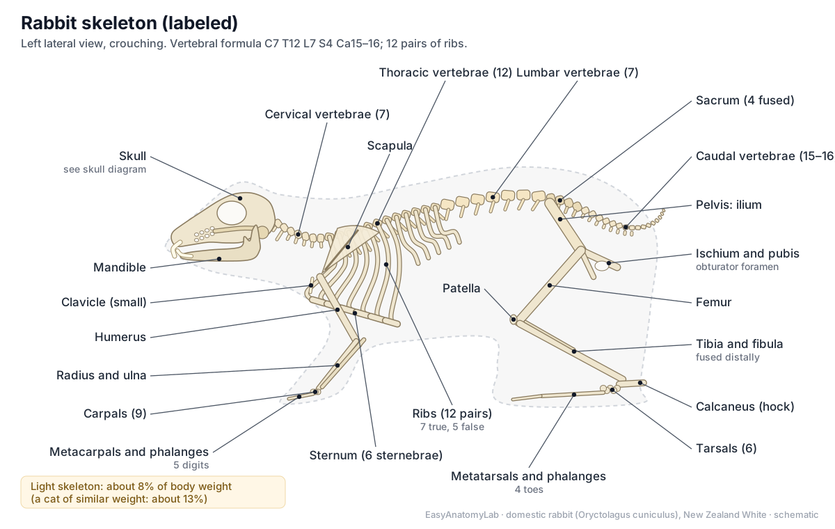
The rabbit skeleton is light and fragile: it makes up only about 8% of the body weight, compared with about 13% in a cat of similar size (Brewer, 2006). The vertebral column has 7 cervical, 12 thoracic, 7 lumbar, 4 fused sacral and about 15–16 caudal vertebrae. There are 12 pairs of ribs, a six-part sternum, a small clavicle and long hind-limb bones.
| Region | Rabbit | Cat | Human |
|---|---|---|---|
| Cervical vertebrae | 7 | 7 | 7 |
| Thoracic vertebrae | 12 (sometimes 13) | 13 | 12 |
| Lumbar vertebrae | 7 | 7 | 5 |
| Sacral vertebrae (fused) | 4 | 3 | 5 |
| Caudal vertebrae | 15–16 | about 20–23 | 3–5 (coccyx) |
| Pairs of ribs | 12 | 13 | 12 |
| Clavicle | Small, not reaching both ends | Small, free-floating | Full collarbone |
| Skeleton as % of body weight | about 8% | about 13% | about 15% |
Rabbit counts: Bensley (1918) and Brewer (2006). Cat and human counts are standard textbook values given for comparison; the human skeleton percentage is an approximate textbook figure.
- Scapula: Attaches the foreleg to the trunk through muscle and forms the shoulder joint.
- Clavicle (small): Gives some attachment to shoulder muscles; it is too small to brace the shoulder.
- Pelvis (os coxae): Forms the hip socket (acetabulum) and passes the thrust of the hind legs to the spine.
- Femur: Lever for the powerful thigh muscles.
- Tibia and fibula: A long lower leg adds speed to every jump.
- Calcaneus (hock): Lever arm for the calf muscles that extend the foot in each hop.
The forelimb has the scapula, humerus, radius and ulna, nine carpal bones, five metacarpals and five digits. The hindlimb has the femur, patella, a tibia and fibula fused over the lower half, six tarsal bones, four working metatarsals (the first is a vestige) and four toes with three phalanges each (Bensley, 1918). The long femur, tibia and foot make a long lever system for jumping.
Rabbit skull and teeth: dental anatomy
A rabbit has 28 teeth, with the dental formula I 2/1, C 0/0, P 3/2, M 3/3. In the upper jaw there are two large incisors with two small peg teeth directly behind them, no canines, a long toothless gap called the diastema, and six cheek teeth on each side. The lower jaw has one pair of incisors and five cheek teeth on each side. All of the teeth are open-rooted and grow throughout life (elodont) (Verstraete & Osofsky, 2005).
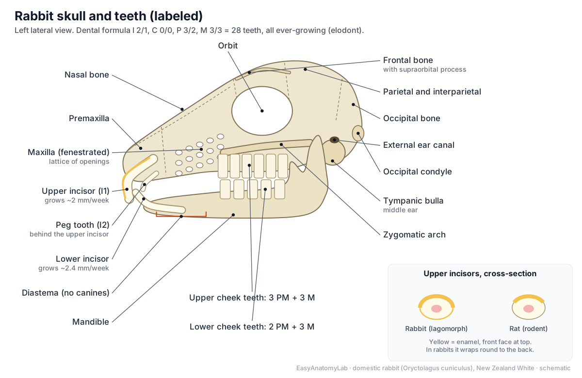
- Upper incisors: Cut and gnaw food.
- Peg teeth: Form a stop that the lower incisors close against, between the first and second upper incisors.
- Lower incisors: Work against the upper incisors and peg teeth to cut food.
- Cheek teeth (premolars and molars): Grind grass and hay with a sideways chewing motion.
- Diastema: Lets the cheeks fold in so the rabbit can gnaw without swallowing debris.
- Maxilla (fenestrated): Holds the upper cheek teeth and forms the side of the snout.
- Mandible: Moves sideways and up for grinding; its tall ramus gives leverage to the masseter.
The skull is light. The side wall of the maxilla is a lattice of holes (the fenestrated maxilla), the orbits are very large, and the tympanic bullae that house the middle ears are prominent. The lower jaw is narrower than the upper, so the rabbit chews on one side at a time with a sideways grinding stroke, powered mainly by the large masseter muscle.
Rabbit digestive anatomy and caecotrophy
The rabbit digestive system runs from the mouth and oesophagus to a simple stomach, a long duodenum, the jejunum and ileum, and then the sacculus rotundus, where the small intestine joins an enormous caecum ending in a vermiform appendix. The colon leaves the caecum next to the sacculus rotundus and passes through the fusus coli to the rectum and anus. The caecum holds about 40% of the gut's volume (Brewer, 2006).
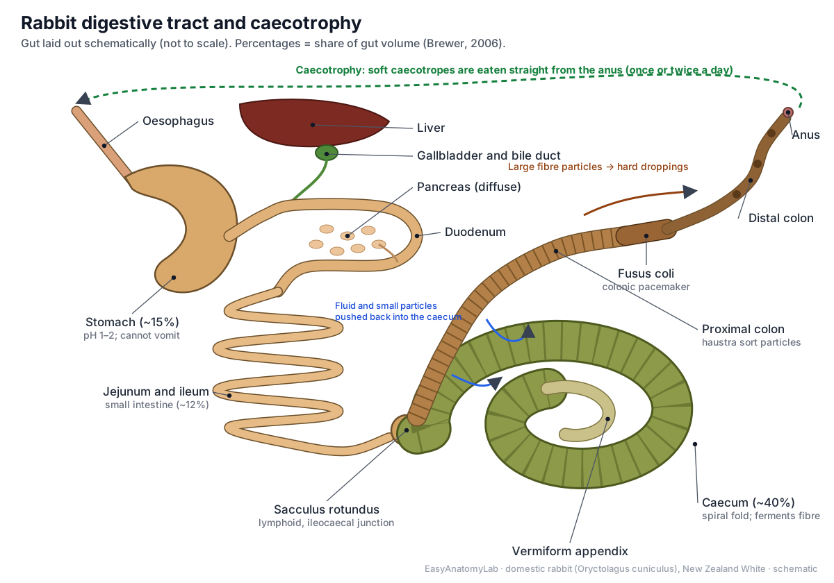
- Stomach: Begins digestion with strong acid (pH 1–2).
- Jejunum and ileum: Absorbs sugars, fats and amino acids.
- Sacculus rotundus: Lymphoid (immune) tissue that samples gut contents; also marks the ileocaecal junction.
- Caecum: Bacteria ferment fibre here into volatile fatty acids, vitamins and protein.
- Vermiform appendix: Lymphoid tissue for immunity; it may also secrete alkaline fluid that buffers caecal acids.
- Proximal colon: Separates gut contents: small particles and fluid go back to the caecum; large fibre particles move on to form hard droppings.
- Fusus coli: Acts as the pacemaker for colonic movement and switches between making hard droppings and caecotropes.
- Distal colon and rectum: Absorbs water and forms hard round droppings or soft caecotropes.
- Liver: Makes bile, stores nutrients and processes absorbed food and toxins.
- Pancreas: Makes digestive enzymes and the hormones insulin and glucagon.
Rabbits make two kinds of droppings. Hard droppings are dry, round pellets of indigestible fibre. Caecotropes (soft or night droppings) are clusters of soft, shiny pellets made of caecal contents rich in bacteria, protein and B vitamins. The rabbit eats the caecotropes directly from the anus, which is called caecotrophy. The liver has left lateral, left medial, right, quadrate and caudate lobes, and a small gallbladder (Stamatova-Yovcheva et al., 2021).
Rabbit foot, paw and leg anatomy
Rabbits have no paw pads. The soles of all four feet are covered in thick, coarse fur that cushions and grips (Antinoff, 2009). Each forefoot has five toes, including a short dewclaw (digit I) higher on the inner side; each hind foot has four toes. All toes end in long nails.
The hind leg is folded in a Z shape when the rabbit sits: the thigh points forward, the lower leg points back to the hock, and the long hind foot lies flat on the ground. When resting, a rabbit takes its weight on the whole sole from the hock to the toes. That is why worn fur on the hock leads to sore hocks (ulcerative pododermatitis), especially on wire or hard floors (Rosell & de la Fuente, 2013).
| Forefoot (front paw) | Hind foot | |
|---|---|---|
| Digits | 5 (incl. dewclaw) | 4 |
| Wrist / ankle bones | 9 carpals | 6 tarsals, incl. calcaneus (hock) |
| Long bones | 5 metacarpals | 4 working metatarsals + 1 vestige |
| Pads | None: furred sole | None: furred sole |
| Main job | Support, digging, steering | Thrust for hopping |
- Gluteal muscles: Extend the hip, driving the thigh backward at take-off.
- Quadriceps femoris: Straightens the knee in the hop.
- Biceps femoris: Extends the hip and helps flex the knee.
- Gastrocnemius and Achilles tendon: Extends the foot, giving the final push of each hop.
Rabbit tail anatomy
A rabbit's tail, the scut, is short and carried curled up against the rump. Inside are about 15–16 small caudal vertebrae that shrink to simple rods towards the tip (Bensley, 1918). The fur is fluffy and, in wild-coloured rabbits, white underneath: a fleeing rabbit lifts its tail and flashes the white underside, which is thought to warn other rabbits. Like all lagomorphs, rabbits have only a short or rudimentary tail (Animal Diversity Web, Lagomorpha).
Rabbit ear anatomy and thermoregulation
Each rabbit ear has a tall pinna of elastic cartilage covered by thin skin, an ear canal and, deeper in the skull, a middle ear inside the tympanic bulla and the inner ear. The two pinnae make up about 12% of the body surface (Brewer, 2006). They funnel sound and can turn independently towards it.
The ears are also the rabbit's radiator. Rabbits cannot sweat, and panting is not efficient for them, so they control heat loss by changing blood flow through the central ear artery and the marginal ear veins. Arteries and veins run side by side, which allows counter-current heat exchange (Brewer, 2006). Hot rabbits have warm, flushed ears; cold rabbits have cool ears. The same vessels are used by vets for blood samples and injections.
- Ear (pinna): Funnels sound into the ear canal and, through its dense blood vessels, sheds body heat (thermoregulation).
- Central ear artery: Supplies the pinna; its flow changes to release or keep body heat.
- Marginal ear vein: Drains blood from the pinna.
- Ear canal and inner ear: Carries sound to the eardrum; the inner ear also senses balance.
- Tympanic bulla: Houses the middle-ear cavity and its three ossicles, and leads to the external ear canal.
Rabbit eye anatomy
Rabbit eyes are large and set high on the sides of the head, giving a field of view of almost 360°, with a binocular overlap of only about 10–35° in front and a blind spot just below the nose (Peiffer et al., 1994). The retina is mostly rods, which suits low light, and there is no reflective tapetum. Rabbits have a third eyelid, a large Harderian gland and only one tear drainage opening (lacrimal punctum), in the lower lid. New Zealand Whites are albino, so their irises lack pigment and the eyes look pink or red.
Rabbit muscle anatomy
Most of a rabbit's muscle mass is in the hindquarters and back. The gluteal muscles and hamstrings (biceps femoris, semitendinosus and semimembranosus) extend the hip, the quadriceps straightens the knee, and the gastrocnemius pulls on the Achilles tendon to extend the foot. Long back muscles (longissimus) straighten the spine in each bound. On the head, the large masseter powers chewing.
- Back muscles (longissimus): Extend (straighten) the spine to add power to each jump.
- Gluteal muscles: Extend the hip, driving the thigh backward at take-off.
- Biceps femoris: Extends the hip and helps flex the knee.
- Semitendinosus and semimembranosus: Extend the hip and flex the knee.
- Quadriceps femoris: Straightens the knee in the hop.
- Gastrocnemius and Achilles tendon: Extends the foot, giving the final push of each hop.
- Tibialis cranialis: Flexes the hock, lifting the foot.
- Triceps brachii: Straightens the elbow and takes the landing shock on the forelegs.
- Masseter: Closes the jaw and drives the side-to-side grinding stroke.
- Abdominal wall muscles: Hold the large gut in place and help with breathing out, defecation and giving birth.
The strength of these muscles compared with the light, thin-walled bones is why careless handling can break a rabbit's back (Antinoff, 2009).
Heart, lungs and thymus
The rabbit's chest is small compared with its abdomen (Antinoff, 2009). The right lung has four lobes (cranial, middle, caudal and accessory) and the left lung is smaller. The heart is small, about 0.3% of body weight, and its right atrioventricular valve has only two cusps instead of the three of a typical "tricuspid" valve (Bensley, 1918; Brewer, 2006). Unlike most adult mammals, rabbits keep a large thymus in front of the heart throughout life.
- Heart: Pumps blood through the lungs and the body.
- Right lung: Gas exchange.
- Left lung: Gas exchange.
- Thymus: Trains T-lymphocytes for the immune system.
- Diaphragm: Main breathing muscle: it contracts to draw air into the lungs.
Kidneys, bladder and cloudy urine
Rabbits handle calcium differently from most mammals: they absorb most of the calcium in their food and excrete the excess in urine. On a high-calcium diet, about 60% of calcium leaves in the urine, compared with under 2% in rats (Brewer, 2006). As a result, normal rabbit urine is often cloudy with calcium carbonate crystals, and it can range from yellow to orange or red-brown (Antinoff, 2009). Each kidney has a single renal papilla.
Male vs female rabbit anatomy: how to sex a rabbit
To tell a male rabbit from a female, support the rabbit on its back and press gently on each side of the genital opening. A buck shows a round, tube-like penis tip with a circular opening; a doe shows a slit-shaped vulva that points towards the anus. The distance between the genitals and the anus is similar in both sexes, so it does not help (Antinoff, 2009).
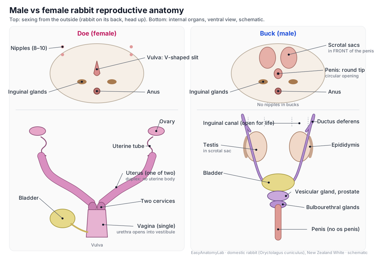
| Buck (male) | Doe (female) | |
|---|---|---|
| Genital opening | Round tip with a circular opening | Slit, pointing towards the anus |
| Scrotal sacs | Two hairless sacs in front of the penis | None |
| Nipples | None | Usually 8 (4 pairs), sometimes 10 |
| Dewlap | Usually small or absent | Often large in mature does |
| Gonads | Testes; inguinal canals open for life | Two ovaries; ovulation induced by mating |
| Internal tract | Epididymis, ductus deferens, accessory glands, penis without a bone | Two uteri, two cervices, one vagina |
Sexing very young kits is difficult and often wrong; it becomes easier as the rabbits grow, and a buck's testes become obvious once they descend into the scrotal sacs.
Rabbit reproductive anatomy
The female rabbit has a duplex uterus: two completely separate uteri, each with its own cervix, opening side by side into one vagina, with no uterine body (Brewer, 2006). The urethra opens into the far end of the vagina, the vestibule. Does are induced ovulators: eggs are released about 10 hours after mating rather than on a regular cycle.
- Ovaries: Produce eggs and hormones. The doe ovulates only after mating (induced ovulation).
- Uterine tubes: Catch the eggs; fertilisation happens here.
- Duplex uterus: Each uterus carries its own row of embryos for the 31–32 day pregnancy.
- Two cervices: Close each uterus during pregnancy.
- Vagina and vestibule: Birth canal and copulatory organ; the vestibule also carries urine.
In the buck, the testes lie in scrotal sacs in front of the penis, an arrangement rabbits share with marsupials (Animal Diversity Web, Lagomorpha). The inguinal canals stay open, so the testes can be drawn back into the abdomen. Sperm pass from each epididymis through a ductus deferens to the urethra, where the vesicular gland, prostate and bulbourethral glands add fluid. The penis has no bone (baculum); rodents have one but lagomorphs do not.
- Testes: Make sperm and testosterone.
- Ductus deferens: Transports sperm during ejaculation.
- Accessory sex glands: Produce seminal fluid.
- Penis and prepuce: Carries urine and semen through the urethra; it points backward at rest.
Pregnant rabbit anatomy
A pregnant doe carries her embryos in a row in each of her two uteri, so the two sides of a litter develop separately. Pregnancy usually lasts 31–32 days (Brewer, 2006); Animal Diversity Web gives about 30 days with a range of 30–37. Average litters are about 5–6 kits, with a range of 1 to 14. Kits are born naked, blind and helpless (altricial) in a fur-lined nest, and the doe nurses them only briefly once or twice a day. A New Zealand White kit weighs about 65 g at birth and opens its eyes at about 10 days (Brewer, 2006).
Because mating triggers ovulation, a sterile mating can cause a false pregnancy (pseudopregnancy) that lasts about 15–16 days (Brewer, 2006).
Rabbit vs rodent: lagomorph differences
Rabbits look like large rodents and both groups gnaw with ever-growing incisors, but they belong to different orders. The quickest way to tell them apart is the teeth: a rabbit has four upper incisors (two large and two peg teeth), a rodent has two.
| Feature | Rabbit (lagomorph) | Rat or mouse (rodent) |
|---|---|---|
| Upper incisors | 4: two large + two peg teeth | 2 |
| Incisor enamel | Wraps round to the back of the tooth | Front face only |
| Cheek teeth | 6 upper, 5 lower per side, ever-growing | 3 molars per side in rats and mice, rooted |
| Chewing | Side to side, one side at a time | Mainly forward and back |
| Maxilla | Fenestrated (lattice of holes) | Solid |
| Scrotum | In front of the penis | Behind the penis |
| Penile bone | Absent | Present (baculum) |
| Tail | Short scut | Usually long |
| Caecotrophy | Yes, from a huge caecum | Many rodents also re-eat droppings |
Sources: Animal Diversity Web (Lagomorpha); Verstraete & Osofsky (2005); Wikipedia's summary of the baculum. Rodent cheek-tooth and chewing details are general textbook features of rats and mice.
Rabbit vs hare and jackrabbit anatomy
Hares (genus Lepus) are close relatives of rabbits in the same family, Leporidae. Jackrabbits are hares, not rabbits. Hares are generally larger, with longer ears, bigger eyes and longer, more powerful hind legs, built for running in the open rather than hiding in burrows (Animal Diversity Web).
| European rabbit | Hares and jackrabbits | |
|---|---|---|
| Newborn young | Altricial: naked, blind, helpless | Precocial: furred, eyes open, active |
| Nest | Fur-lined nest, often in a burrow | Shallow scrape (form) above ground |
| Ears and hind legs | Shorter | Longer and more powerful |
| Chromosomes | 44 in O. cuniculus (rabbits range 38–52) | 48 in all hares |
| Interparietal bone | Present | Absent (Bensley, 1918) |
Young, nests and chromosomes: Wikipedia's hare article, which cites Robinson & Skinner (1983) and Smith et al. (2018). The rabbit chromosome number (2n = 44) is the standard karyotype of O. cuniculus.
How caecotrophy works, step by step
Caecotrophy is the habit of eating the soft droppings (caecotropes) that come from the caecum. It starts in the proximal colon. Sacculations in its wall, the haustra, hold back small particles and fluid, while the larger particles collect in the middle of the gut. Wave-like haustral contractions push the small particles back into the caecum, and segmental contractions push the large fibre particles on towards the anus. The large particles become the familiar hard, round droppings. (Varga (2014), Digestive disorders, Textbook of Rabbit Medicine, 2nd ed. (PMC).)
The small particles stay in the caecum, where bacteria ferment them. A few hours later the process switches. Varga describes caecotroph production starting about 4 hours after the last meal; most rabbits pass soft faeces in the morning and evening, and these pass through the colon 1.5–2.5 times faster than hard faeces. The switch is controlled by the fusus coli, a thick muscular part of the colon that acts as its pacemaker. Because the autonomic nervous system and the hormone aldosterone control the fusus coli, stress can upset the whole cycle and lead to gut stasis. (Varga (2014), Digestive disorders, Textbook of Rabbit Medicine, 2nd ed. (PMC).)
The caecotropes are a real food. Brewer summarises studies in which coprophagy (eating droppings) supplied about 83% more niacin, 100% more riboflavin, 165% more pantothenic acid and 42% more vitamin B12 than in rabbits prevented from doing it, as well as extra protein. Rabbits take the caecotropes straight from the anus, so owners rarely see them; a pile of uneaten soft droppings is a sign that something is wrong. (Brewer (2006), Biology of the rabbit, JAALAS 45(1): 8–24 (PMC).)
Rabbit gut proportions in numbers
The rabbit's gut is built around the caecum. In Brewer's figures, the stomach makes up about 15% of the volume of the gastrointestinal tract, the small intestine about 12% (versus about 22% in the dog, pig and horse), and the caecum about 40%, with roughly ten times the capacity of the stomach. The colon of a New Zealand White is about 1 m long and the fusus coli about 5–8 cm. (Brewer (2006), Biology of the rabbit, JAALAS 45(1): 8–24 (PMC).)
Bensley's laboratory manual traces the same tract in a dissected rabbit. The small intestine joins the large intestine through a rounded swelling, the sacculus rotundus, whose wall has a fine hexagonal pattern from its many lymph follicles. The caecum has three limbs and a spiral constriction that marks an internal spiral fold, and it ends in a narrow, thick-walled vermiform appendix about five inches (13 cm) long. The colon leaves the caecum beside the sacculus rotundus and has strongly sacculated walls. (Bensley (1918), Practical Anatomy of the Rabbit, 2nd ed., University of Toronto Press (archive.org).)
Two more facts explain common clinical problems. The stomach is very acidic (pH about 1–2) and rabbits cannot vomit, so swallowed hair and food cannot be brought back up. The pancreas is diffuse, scattered through the mesentery of the duodenal loop, and its duct opens far along the duodenum from the bile duct. (Varga (2014), Digestive disorders, Textbook of Rabbit Medicine, 2nd ed. (PMC).)
Rabbit teeth in detail: why they never stop growing
All of a rabbit's permanent teeth are elodont: continuously growing and open-rooted. Some authors use the term aradicular hypsodont, meaning that the teeth have a long crown, keep erupting and never form closed roots. The dental formula of the permanent teeth is I 2/1, C 0/0, P 3/2, M 3/3, which gives 28 teeth. Rabbits are diphyodont: the milk first incisors are usually shed around birth, and the milk second incisors and premolars fall out within a month. (Verstraete & Osofsky (2005), Dentistry in pet rabbits, Compendium (UC Davis).)
The upper first incisors grow about 2 mm per week and the lower incisors about 2.4 mm per week. Their enamel is thicker on the front (facial) side, so the back wears faster and keeps a chisel edge. The lower incisors close between the upper first incisors and the peg teeth. Behind a long toothless gap, the premolars and molars work as one grinding unit with transverse enamel folds. The upper tooth rows are wider apart than the lower (anisognathism), the occlusal plane is tilted about 10°, and the jaw joint allows wide side-to-side movement but little forward-and-back movement. (Verstraete & Osofsky (2005), Dentistry in pet rabbits, Compendium (UC Davis).)
Because the teeth never stop growing, they must be worn down by chewing. Incisor malocclusion is common in pet rabbits. In young rabbits it is usually inherited and caused by a short upper jaw (maxillary brachygnathia). In older rabbits it usually accompanies problems of the cheek teeth, such as sharp spikes and overgrown roots, so a rabbit with crooked incisors needs a full mouth examination. (Verstraete & Osofsky (2005), Dentistry in pet rabbits, Compendium (UC Davis).)
A light skeleton and powerful muscles: handling rabbits safely
The rabbit skeleton is relatively fragile. Brewer gives it as about 8% of body weight, compared with about 13% for a cat of similar weight. Antinoff likewise gives 8% and adds that the bones have thin cortices and shatter easily. Students sometimes quote 7–8%; the exact figure depends on the animal and the method, but the contrast with the cat holds. (Brewer (2006), Biology of the rabbit, JAALAS 45(1): 8–24 (PMC).)
The muscles that drive the hind legs, by contrast, are strong. A long spine with powerful back muscles is prone to luxation or fracture when a rabbit kicks or struggles while its hindquarters are not supported. This is why a rabbit is always lifted with one hand under the chest and the other supporting the rump, and never by the ears. (Antinoff (2009), Practical anatomy and physical examination: ferrets, rabbits, rodents (dvm360 proceedings).)
Bensley's count of the vertebral column is 7 cervical, 12 thoracic, 7 lumbar, 4 sacral and 14–16 caudal vertebrae; in his detailed description he counts sixteen caudal vertebrae. There are twelve pairs of ribs: seven true ribs attached to the sternum and five false ribs. The sternum has six segments, and the clavicle is a slender rod that is imperfectly developed. Brewer gives the same formula and notes that some rabbits have 13 thoracic vertebrae. (Bensley (1918), Practical Anatomy of the Rabbit, 2nd ed., University of Toronto Press (archive.org).)
Inside the rabbit eye
Rabbit eyes are large and set high on the sides of the head. With eye and head movements, a rabbit has a field of view of almost 360°, but the binocular field, where both eyes see the same thing, is only about 10° to 35° wide. Most photoreceptors are rods, and there is no tapetum lucidum, the reflective layer that makes cat and dog eyes shine at night. The retina is merangiotic: its blood vessels run in a horizontal band from the optic disc instead of covering the whole retina. (Peiffer, Pohm-Thorsen & Corcoran (1994), Models in ophthalmology and vision research, in The Biology of the Laboratory Rabbit (PMC).)
Several features matter in practice. Rabbits have only one lacrimal punctum, in the lower eyelid, and the nasolacrimal duct follows a winding route through the lacrimal and maxillary bones, close to the roots of the cheek teeth, so dental disease often causes runny eyes. The third eyelid covers only about two-thirds of the cornea. Behind it lies the Harderian gland, with a small white and a large pink lobe, which is larger in males and during the breeding season. (Bayón del Río (2009), Exotic ophthalmology II, WSAVA World Congress (VIN).)
Why rabbits get sore hocks
Rabbits have no paw pads. Instead, thick fur covers the soles to protect them. When the fur wears away, the skin over the bones is pressed between the bone and the floor, and painful ulcers can form: ulcerative pododermatitis, or sore hocks. (Antinoff (2009), Practical anatomy and physical examination: ferrets, rabbits, rodents (dvm360 proceedings).)
On commercial farms, sore hocks are found mainly on the plantar surface (sole) of the hind feet and sometimes on the palmar surface of the forefeet. In a survey of breeding farms in Spain and Portugal, the mean prevalence was 7.5% in does and 5.2% in bucks, and farms that gave the rabbits plastic footrests had about a third of the prevalence of farms without them (4.9% versus 13.7%). The authors link the lesions to the environment, especially wire mesh floors. (Rosell & de la Fuente (2013), Assessing ulcerative pododermatitis of breeding rabbits, Animals 3: 318–326 (PMC).)
Rabbit anatomy worksheet: test yourself
Use the 3D model and diagrams to answer these.
- What is the dental formula of a rabbit, and how many teeth is that?
- Which small teeth tell you a skull belongs to a lagomorph and not a rodent?
- Name the organ where fibre is fermented, and the spiral structure inside it.
- What is the fusus coli, and what does it control?
- How many lumbar vertebrae does a rabbit have? How many pairs of ribs?
- Why do rabbits get sore hocks?
- Give two external features that distinguish a buck from a doe.
- What is unusual about the rabbit uterus?
Answer key
- I 2/1, C 0/0, P 3/2, M 3/3 = 28 teeth.
- The peg teeth (second upper incisors) behind the large upper incisors.
- The caecum; its spiral fold (spiral valve).
- A thick muscular segment of the colon that acts as its pacemaker and switches between hard droppings and caecotropes.
- Seven lumbar vertebrae; twelve pairs of ribs.
- They have no paw pads, so when the fur on the sole wears away, the skin over the hock is pressed against hard floors and ulcerates.
- Bucks have scrotal sacs in front of the penis, a round penis tip and no nipples; does have a slit-shaped vulva and 8–10 nipples.
- It is duplex: two separate uteri, each with its own cervix, opening into one vagina.
Frequently asked questions
How many teeth does a rabbit have?
An adult rabbit has 28 teeth: dental formula I 2/1, C 0/0, P 3/2, M 3/3. That is four upper incisors (two large ones and two small peg teeth behind them), two lower incisors, and six upper and five lower cheek teeth on each side. All of them grow throughout life.
Do rabbits have paw pads?
No. Unlike cats and dogs, rabbits have no paw pads. The soles of their feet are covered with thick fur, which cushions them. If this fur wears thin on hard or wire floors, painful sores called sore hocks (ulcerative pododermatitis) can develop.
How can you tell a male rabbit from a female?
Gently press the skin beside the genital opening with the rabbit supported on its back. A buck shows a round tube with a circular opening (the penis), and a doe shows a slit (the vulva). Adult bucks also have two hairless scrotal sacs in front of the penis, and bucks have no nipples.
What are caecotropes and why do rabbits eat their poop?
Caecotropes (night droppings) are soft, mucus-coated pellets made from the contents of the caecum, where bacteria ferment fibre. The rabbit eats them directly from the anus, usually once or twice a day, to absorb the protein and B vitamins the bacteria produced. Hard round droppings are not normally eaten.
Is a rabbit a rodent?
No. Rabbits are lagomorphs (order Lagomorpha), with hares and pikas. Unlike rodents, they have a second pair of small upper incisors (peg teeth), enamel all around their incisors, a scrotum in front of the penis, and no penile bone.
Why are rabbit bones so fragile?
A rabbit's skeleton makes up only about 7–8% of its body weight, compared with about 13% in a cat of similar size, and its bones have thin walls. The very strong back muscles can therefore break the spine if a rabbit kicks while its hindquarters are not supported.
Why do rabbits have such big ears?
Rabbit ears are used for hearing and for cooling. The two ears make up about 12% of the body's surface, and they are full of blood vessels. Because rabbits cannot sweat or pant well, widening the ear vessels is their main way to lose heat.
Can rabbits see behind them?
Almost. Their eyes sit high on the sides of the head, giving a field of view of almost 360°. The price is a narrow zone of binocular vision in front and a small blind spot just in front of and below the nose, which the whiskers and lips cover.
Why do rabbits have two uteri?
Female rabbits have a duplex uterus: two completely separate uteri, each with its own cervix opening into one vagina. Each side carries its own embryos. Does are also induced ovulators, releasing eggs about 10 hours after mating.
References
- Bensley, B. A. (1918). Practical Anatomy of the Rabbit: An Elementary Laboratory Textbook in Mammalian Anatomy, 2nd ed. University of Toronto Press. Internet Archive.
- Brewer, N. R. (2006). Biology of the rabbit. Journal of the American Association for Laboratory Animal Science 45(1), 8–24.
- Verstraete, F. J. M. & Osofsky, A. (2005). Dentistry in pet rabbits. Compendium on Continuing Education for the Practicing Veterinarian 27(9), 671–684.
- Antinoff, N. (2009). Practical anatomy and physical examination: ferrets, rabbits, rodents, and other selected species (Proceedings). dvm360.
- Peiffer, R. L., Pohm-Thorsen, L. & Corcoran, K. (1994). Models in ophthalmology and vision research. In Manning, Ringler & Newcomer (eds), The Biology of the Laboratory Rabbit, 2nd ed. Academic Press.
- Bayón del Río, A. (2009). Exotic ophthalmology II. World Small Animal Veterinary Association World Congress Proceedings. VIN.
- Varga, M. (2014). Digestive disorders. In Textbook of Rabbit Medicine, 2nd ed. Butterworth-Heinemann/Elsevier.
- Stamatova-Yovcheva, K., Dimitrov, R., Dilek, Ö. G. & Yovchev, D. (2021). Sagittal anatomic investigation of the rabbit liver. Veterinary Journal of Mehmet Akif Ersoy University 6(1).
- Rosell, J. M. & de la Fuente, L. F. (2013). Assessing ulcerative pododermatitis of breeding rabbits. Animals 3(2), 318–326.
- Animal Diversity Web, University of Michigan. Oryctolagus cuniculus, European rabbit.
- Animal Diversity Web, University of Michigan. Lagomorpha: pikas, rabbits and hares.
- MSD Veterinary Manual. Description and physical characteristics of rabbits.
- Wikipedia. Hare (overview of rabbit–hare differences, with primary references: Robinson & Skinner 1983; Smith et al. 2018).
- Wikipedia. Baculum (citing Feldhamer et al., Mammalogy: Adaptation, Diversity, Ecology).
Studying another lagomorph or a rodent? Compare the rat dissection guide for the rodent pattern of teeth, caecum and reproductive organs.
More interactive anatomy models
- Human anatomy1,000+ bones, muscles, organs, vessels and nerves from real anatomical data.
- Octopus anatomyThree hearts, a ring-shaped brain, eight arms and a beak, in 3D.
- Squid anatomyPen, eight arms, two tentacles, giant axons and three hearts of the longfin squid.
- Squid dissectionStep-by-step guided squid dissection with every structure labeled.
- Fish anatomyExternal and internal anatomy of a bony fish: fins, gills, swim bladder, heart and brain.
- Perch dissectionGuided yellow perch (fish) dissection, from scales to the swim bladder.