

Lizard anatomy at a glance
A lizard is a scaly reptile with, in most species, four sprawling legs, a long tail, movable eyelids and external ear openings. Its body has three regions: the head, the trunk (which holds the body cavity) and the tail, which starts at the vent. Inside, it has a three-chambered heart, two simple lungs, kidneys that excrete uric acid, a cloaca shared by the gut, urinary and reproductive tracts, and, in males, a pair of hemipenes.
Lizards are not one natural group. There are more than 7,000 species, and snakes evolved from within them, so "lizard" is a paraphyletic name for all squamates (scaled reptiles) that are not snakes or worm lizards. Limbs have been lost or reduced more than two dozen times in different lizard lineages.
This page uses the green anole, Anolis carolinensis, as the reference lizard. It is a small, tree-living iguanian lizard from the south-eastern United States, widely kept in schools and used as a laboratory model; in 2011 it became the first non-bird reptile to have its genome sequenced. Adult males are 12.5–20 cm long including the tail. Where other lizards differ, such as bearded dragons, monitors and geckos, the text says so.
Directions: anterior means towards the snout, posterior towards the tail, dorsal is the back and ventral the belly. Left and right are always the lizard's own. Body length is given as snout–vent length (SVL) because so many lizards have broken or regrown tails.
External anatomy of a lizard: labeled diagram
The external anatomy of a lizard includes the snout with two nostrils, two eyes with movable eyelids, the parietal (third) eye on top of the head, an ear opening with the eardrum on each side, the mouth with its lip scales, four limbs with five clawed digits each, the vent (cloacal opening) and the tail. Male anoles add a large throat fan (dewlap) and enlarged scales behind the vent.
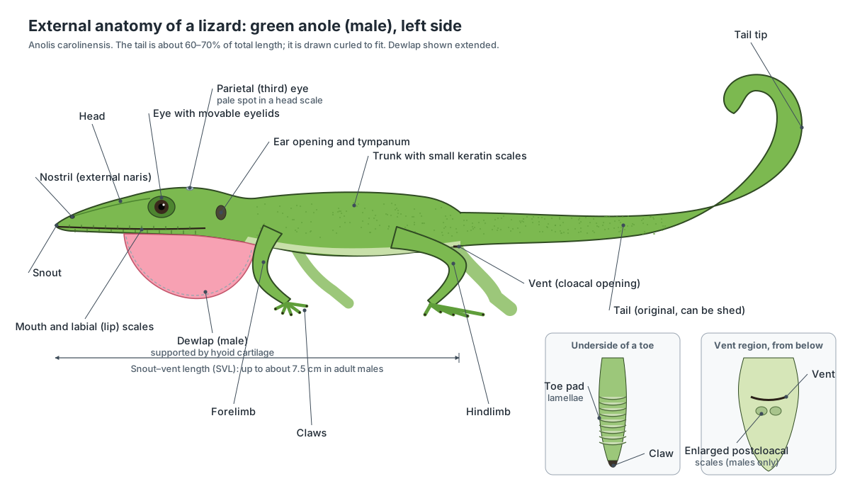
- Dewlap: A visual signal used in courtship, in territorial contests between males and to deter predators, together with head-bobs and push-ups.
- Ear opening and tympanum: Sound vibrates the eardrum, which passes the vibration through a single middle-ear bone (columella) to the inner ear.
- Eyelids: Close to protect and clean the eye. A thin third eyelid (nictitating membrane) can also sweep across it.
- Toe pads: Each lamella is covered with microscopic hair-like setae that grip surfaces, so anoles can climb smooth leaves and walls.
- Vent: The single external opening of the cloaca, through which faeces, urine (as uric acid), eggs and sperm leave the body, and through which the hemipenes are everted.
The skin is covered by small overlapping scales made of keratin. Unlike fish scales, these are thickened folds of the skin itself, and they are shed regularly, in patches in lizards rather than in one piece as in snakes. Some lizards, such as skinks and monitors, have small bony plates (osteoderms) under the scales; anoles do not.
Lizard internal anatomy: organs and where they are
Inside a lizard, the heart sits in the chest between the shoulders; the paired, sac-like lungs fill the upper part of the front half of the body cavity; the large, two-lobed liver lies on the floor of the cavity behind the heart; the tubular stomach runs along the left side; and the small intestine coils behind it. At the back of the cavity are the fat bodies, the gonads, the kidneys inside the pelvis, the urinary bladder and the cloaca. Lizards have no diaphragm, so the chest and abdomen form one body cavity (coelom).
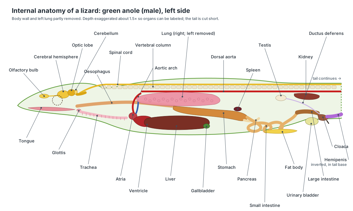
| System | Main organs in the green anole |
|---|---|
| Digestive | Tongue, oesophagus, stomach, small intestine, large intestine, liver, gallbladder, pancreas, cloaca |
| Respiratory | Nostrils, glottis and larynx, trachea, two bronchi, two sac-like lungs |
| Circulatory | Three-chambered heart, pulmonary artery, left and right aortic arches, dorsal aorta, postcaval vein, spleen |
| Excretory | Two compact kidneys at the back of the body cavity, urinary bladder, cloaca |
| Reproductive (male) | Two testes, sperm ducts, two hemipenes in the tail base |
| Reproductive (female) | Two ovaries, two oviducts, one egg at a time |
| Nervous and sensory | Brain, spinal cord, eyes, parietal eye, ears, vomeronasal organ |
Internal vs external lizard anatomy
External anatomy is everything you can see on the outside of the lizard without cutting: scales, eyes, ears, nostrils, dewlap, limbs, toe pads, claws, vent and tail. Internal anatomy covers the organs and skeleton inside. The two are linked: the dewlap is moved by the hyoid bones inside the throat, the ear opening leads to a single middle-ear bone, the parietal eye sits over a hole in the skull, and the swollen tail base of a male hides the hemipenes. Use the skin slider in the 3D model to move between the two views.
Lizard anatomy labeled: every part and its function
This is the text version of the 3D model: every labeled structure of the green anole, grouped by body system, with what it does.
External
- Head (Cephalic region, snout)
- The long, wedge-shaped head of the anole, from the pointed snout back to the ear openings. Holds the brain, eyes, nostrils, ears, jaws and tongue. The narrow snout lets the anole strike at insects among leaves and twigs.
- Trunk and scales (Body, body wall, integument)
- The slender body between the shoulders and the hips, covered in small overlapping keratin scales. Protects the organs, holds the lungs and gut, and anchors the limb girdles. The scaly skin limits water loss.
- Tail (Caudal region (original tail))
- The long, tapering tail behind the vent: about 60–70% of the total length in a green anole. Balance during climbing and jumping, fat storage, and defence: it can be shed (autotomy) and later regrown.
- Dewlap (Throat fan, gular fan) · males only
- A fan of pink-red skin under the throat that the male anole extends during displays. A visual signal used in courtship, in territorial contests between males and to deter predators, together with head-bobs and push-ups.
- Nostrils (External nares)
- A pair of small openings near the tip of the snout. Air enters here for breathing and smelling. They lead through the nasal cavity to openings (choanae) in the roof of the mouth.
- Mouth and labial scales (Oral opening, jaws, lips)
- The long mouth slit running back along each side of the head, edged by rows of lip (labial) scales. Catches and holds prey. Lizards do not chew; they crush prey a few times and swallow it whole.
- Eyelids (Upper and lower palpebrae)
- Movable upper and lower eyelids around each eye; the lower lid is the larger and more mobile. Close to protect and clean the eye. A thin third eyelid (nictitating membrane) can also sweep across it.
- Ear opening and tympanum (External auditory meatus, eardrum)
- A small oval opening behind the jaw on each side of the head, with the eardrum (tympanic membrane) visible inside. Sound vibrates the eardrum, which passes the vibration through a single middle-ear bone (columella) to the inner ear.
- Forelimbs (Front legs, arms)
- The two front legs, each with an upper arm, forearm and a hand with five clawed fingers. Climbing, gripping branches and, together with the hindlimbs, walking with the sprawling, side-to-side gait typical of lizards.
- Hindlimbs (Back legs)
- The two hind legs, larger than the forelimbs, each with a thigh, shank and a long foot with five clawed toes. Main source of thrust for running, climbing and jumping.
- Toe pads (Subdigital lamellae, adhesive pads)
- Widened pads under the middle of each finger and toe, made of a row of overlapping flaps (lamellae). Each lamella is covered with microscopic hair-like setae that grip surfaces, so anoles can climb smooth leaves and walls.
- Claws (Ungues)
- A small curved keratin claw at the tip of every finger and toe. Grip rough bark and soil where adhesive pads work less well.
- Vent (Cloacal opening)
- A transverse (side-to-side) slit on the underside at the base of the tail. The single external opening of the cloaca, through which faeces, urine (as uric acid), eggs and sperm leave the body, and through which the hemipenes are everted.
- Postcloacal scales (Enlarged postanal scales) · males only
- A pair of enlarged scales just behind the vent in male anoles. A reliable way to sex anoles, together with the larger dewlap and the swollen tail base of males.
Skeletal
- Premaxilla
- The single bone at the very tip of the upper jaw. Carries the front upper teeth and forms the front edge of the nostrils.
- Maxilla (Upper jaw bone)
- The main tooth-bearing bone of the upper jaw, one on each side. Carries most of the upper teeth and forms the side wall of the snout.
- Nasal bones
- A pair of thin bones roofing the snout. Roof over the nasal cavity, which holds the olfactory epithelium.
- Frontal bone
- The bone roofing the skull between the eyes. Protects the front of the brain and the olfactory tracts and forms the upper rim of each orbit.
- Parietal bone and parietal foramen
- The bone roofing the back of the skull, pierced by a small hole (parietal foramen) for the parietal eye. Protects the brain, anchors jaw-closing muscles along its edges, and holds the parietal foramen, under which the parietal (third) eye sits.
- Postorbital, jugal and squamosal (Postorbital bar and upper temporal arch)
- The slender bones that frame the back of the orbit and the upper temporal opening. Form the rear rim of the orbit and the upper temporal arch, and give attachment to the jaw-closing muscles.
- Quadrate
- A column-shaped bone at the back of the skull on which the lower jaw hinges. Forms the upper half of the jaw joint. In lizards it can swing at its top (streptostyly), which changes the gape and the position of the lower jaw.
- Palate: pterygoid, palatine and vomer
- The paired bones forming the roof of the mouth. Separate the mouth from the nasal passages and braincase and link the snout to the quadrate, so the palate moves when the skull flexes.
- Braincase (Neurocranium: occipital, prootic and sphenoid bones)
- The bony box around the brain and inner ears, at the back of the skull. Protects the brain and inner ears and joins the skull to the first vertebra through a single occipital condyle.
- Scleral ring (Scleral ossicles)
- A ring of small overlapping bony plates in the wall of each eyeball. Supports the eyeball and gives attachment for the muscles that change its shape for focusing.
- Dentary (Lower jaw (tooth-bearing bone))
- The long tooth-bearing bone forming the front of each half of the lower jaw. Carries all the lower teeth.
- Postdentary bones (Surangular, angular, splenial, coronoid, articular)
- The smaller bones behind and inside the dentary that make up the rear of each lower jaw. The coronoid anchors the jaw-closing muscles; the articular forms the lower half of the jaw joint with the quadrate; the others brace the jaw.
- Teeth (Pleurodont dentition)
- Rows of small, pointed teeth fixed to the inner face of the jaw bones (pleurodont), replaced throughout life. Grip and pierce insects and other small prey. Lizards do not chew.
- Hyoid apparatus (Hyobranchial skeleton, ceratobranchials)
- A Y-shaped frame of bone and cartilage in the floor of the throat, with long horns that stiffen the dewlap. Supports and moves the tongue during feeding and drinking, and in anoles extends the dewlap.
- Cervical vertebrae (Neck vertebrae, atlas and axis)
- The vertebrae of the neck, starting with the ring-shaped atlas and the axis. Support and turn the head. The atlas rotates on the single occipital condyle of the skull.
- Dorsal (trunk) vertebrae (Thoracolumbar vertebrae)
- The vertebrae of the trunk, nearly all of them carrying ribs. Form the backbone that supports the trunk and bends from side to side during walking.
- Sacral vertebrae (Sacrum)
- Two vertebrae with broad side processes that join the backbone to the hip bones. Transfer the thrust of the hindlimbs to the backbone.
- Caudal vertebrae (Tail vertebrae, chevrons (haemal arches))
- The long series of tail vertebrae, getting smaller towards the tip, with chevron bones hanging below. Support the tail and anchor the tail muscles. The chevrons (haemal arches) protect the tail artery and vein.
- Autotomy fracture planes (Intravertebral autotomy septa)
- Pre-formed planes of weakness across the middle of each breakable tail vertebra, lined up with breaks in the muscle, fat and skin. Let the lizard shed its tail at a chosen point when grabbed, then seal the wound with little blood loss.
- Ribs
- Curved bones on the trunk vertebrae that surround the body cavity. Support the body wall and, moved by the intercostal muscles, ventilate the lungs. Lizards have no diaphragm.
- Sternum (Breastbone, xiphisternum)
- A diamond-shaped plate of cartilage (partly calcified) on the underside of the chest. Joins the front ribs and the coracoids, closing the rib cage below.
- Pectoral girdle (Scapulocoracoid, clavicle, interclavicle)
- The shoulder skeleton: a scapula and coracoid on each side, plus the clavicles and a T-shaped interclavicle on the chest. Attaches the forelimbs to the body. The humerus joins it at the shoulder socket (glenoid).
- Humerus (Upper arm bone)
- The single bone of the upper arm. Swings forward and back in a near-horizontal plane in the sprawling lizard gait.
- Radius and ulna (Forearm bones)
- The two forearm bones. Support the forearm and let the hand rotate slightly.
- Hand bones (Carpals, metacarpals, phalanges (manus))
- The wrist bones (carpals), five metacarpals and the finger bones (phalanges). Support the fingers and the toe pads.
- Pelvic girdle (Pelvis: ilium, ischium, pubis)
- The hip bones: on each side an ilium joined to the sacrum, and a pubis and ischium meeting their partners below. Attaches the hindlimbs to the backbone. The femur joins it at the hip socket (acetabulum).
- Femur (Thigh bone)
- The single, strong bone of the thigh. Pulled backwards by the caudofemoralis muscle from the tail, it gives the main thrust in running.
- Tibia and fibula (Shank bones)
- The two bones of the lower leg. Support the shank. The ankle joint of lizards lies between two rows of tarsals (mesotarsal joint).
- Foot bones (Tarsals, metatarsals, phalanges (pes))
- The ankle bones, five metatarsals and the toe bones. Form the long, flexible foot used for pushing off.
Muscular
- Jaw-closing muscles (Adductor mandibulae)
- The muscles in the temporal region that close the mouth. Bite down on prey.
- Depressor mandibulae
- The muscle behind the jaw joint that opens the mouth. Opens the mouth by pulling up the back end of the lower jaw.
- Ceratohyoid muscles (Dewlap-extending muscles)
- Paired throat muscles at the front of the hyoid apparatus. Swing the second ceratobranchials forward to extend the dewlap.
- Epaxial muscles (Back muscles)
- The long muscles along the top of the backbone. Bend the body sideways and stiffen it during locomotion.
- Body wall muscles (Hypaxial, intercostal and rectus abdominis muscles)
- The thin sheets of muscle in the sides and belly, including the intercostals between the ribs. Move the ribs to breathe and support the abdomen.
- Caudofemoralis
- A large muscle running from the tail vertebrae to the femur. Pulls the thigh backwards: the main power stroke of running.
- Tail muscles (Caudal myomeres)
- Segmented muscle blocks along the tail, each fitting into cone-shaped pockets of the next. Move the tail and, at autotomy, contract to tear the tail apart at a fracture plane.
Nervous
- Olfactory bulbs
- The paired front parts of the brain that receive nerves from the nose. Process smell. A separate accessory olfactory bulb receives the vomeronasal nerve.
- Cerebral hemispheres (Telencephalon)
- The paired forebrain hemispheres. Integrate senses, learning and behaviour such as courtship and territorial display.
- Optic lobes (Optic tectum (midbrain))
- Two large rounded lobes on the top of the midbrain. Process vision and guide prey-catching strikes; very large in sight-hunting lizards like anoles.
- Cerebellum
- A small plate of brain tissue behind the optic lobes. Coordinates movement and balance.
- Medulla oblongata (Brainstem)
- The back of the brain where it narrows into the spinal cord. Controls breathing and heart rate and relays signals between the brain and the body.
- Spinal cord
- The cord of nerve tissue inside the vertebral column. Carries signals between the brain and the body and controls simple reflexes such as the writhing of a shed tail.
Sensory
- Eye
- A large eye on each side of the head, with a round pupil and good colour vision. Vision is the anole's main sense for finding prey, mates and rivals.
- Parietal eye (Third eye, pineal eye)
- A tiny light-sensitive organ on the top of the head, visible as a pale spot in the centre of one head scale. Senses light and dark (not images). It helps regulate daily rhythms, hormones and basking.
- Middle and inner ear (Columella, cochlear duct, semicircular canals)
- A single rod-like middle-ear bone (columella) linking the eardrum to the fluid-filled inner ear. The columella carries sound to the cochlear duct; the semicircular canals sense balance and head movement.
- Vomeronasal organ (Jacobson's organ)
- A pair of small chemical-sensing pouches above the roof of the mouth. Detects scent particles carried in by the tongue, used to recognise prey, mates and rivals.
Digestive & fat storage
- Tongue
- A thick, fleshy tongue with a slightly notched tip, attached to the floor of the mouth. Catches small prey with its sticky surface (lingual prehension), moves food to the throat and carries scent particles to the vomeronasal organ.
- Oesophagus (Esophagus, gullet)
- The muscular tube from the throat to the stomach. Carries swallowed prey to the stomach; its folded wall stretches to take whole insects.
- Stomach
- A tubular, muscular stomach on the left side of the body cavity. Stores and begins to digest prey with acid and enzymes.
- Small intestine (Duodenum and ileum)
- The coiled tube after the stomach where most digestion and absorption take place. Digests food with enzymes from the pancreas and bile from the liver, then absorbs the nutrients.
- Large intestine (Colon, rectum)
- The short, wide final section of the gut. Absorbs water and forms faeces.
- Liver
- The largest internal organ: a dark red-brown, two-lobed gland in the front of the abdomen. Makes bile, stores energy as glycogen and fat, processes nutrients and, in females, makes yolk proteins (vitellogenin).
- Gallbladder
- A small, greenish sac on the liver. Stores bile and releases it into the duodenum.
- Pancreas
- A pale, thin gland in the loop between the stomach and duodenum. Releases digestive enzymes into the duodenum and hormones (insulin, glucagon) into the blood.
- Abdominal fat bodies (Fat pads)
- A pair of yellow fat pads on the floor of the rear abdomen. Energy store for hibernation and for making yolk and eggs; they shrink during the breeding season.
- Cloaca (Coprodeum, urodeum, proctodeum)
- The shared chamber at the end of the gut that receives faeces, urine and eggs or sperm. Its three parts handle faeces (coprodeum), urine and reproductive products (urodeum), and form the exit (proctodeum). It also reabsorbs water.
Respiratory
- Glottis and larynx
- The slit-like opening to the windpipe, on the floor of the mouth behind the tongue. Opens to let air in and out and closes when the lizard swallows.
- Trachea (Windpipe)
- The windpipe, a tube stiffened by cartilage rings. Carries air from the glottis to the bronchi.
- Bronchi
- The two short branches of the trachea, one to each lung. Carry air into the lungs.
- Lungs
- A pair of simple, sac-like lungs with a honeycomb of small chambers (faveoli) in their walls. Gas exchange: oxygen enters the blood and carbon dioxide leaves through the thin faveolar walls.
Circulatory
- Sinus venosus
- A thin-walled chamber that collects oxygen-poor blood from the body before the right atrium. Receives the large veins and passes blood into the right atrium.
- Right atrium
- The upper right chamber, receiving oxygen-poor blood from the body. Pumps oxygen-poor blood into the ventricle (cavum venosum).
- Left atrium
- The upper left chamber, receiving oxygen-rich blood from the lungs. Pumps oxygen-rich blood into the left part of the ventricle (cavum arteriosum).
- Ventricle (Cavum arteriosum, cavum venosum, cavum pulmonale)
- The single, partly divided ventricle of the three-chambered heart. Pumps blood to the lungs and to the body at the same time. Ridges inside it keep most of the oxygen-rich and oxygen-poor blood apart.
- Pulmonary artery
- The vessel carrying oxygen-poor blood from the ventricle to the lungs. Sends blood to the lungs to pick up oxygen.
- Left and right aortic arches (Systemic arches)
- Two arteries leaving the heart that curve up around the oesophagus and join behind it as the dorsal aorta. Carry mainly oxygen-rich blood to the head and body. In lizards the carotid arteries to the head branch from the right arch.
- Dorsal aorta
- The main artery running back along the roof of the body cavity, under the backbone. Distributes blood to the gut, kidneys, gonads, hindlimbs and tail.
- Postcaval vein (Posterior vena cava)
- The large vein returning blood from the kidneys and liver to the heart. Returns oxygen-poor blood from the back of the body.
- Spleen
- A small, dark red organ of the immune and blood systems. Filters the blood, removes old red blood cells and holds immune cells.
Excretory
- Kidneys
- A pair of compact, lobed kidneys at the very back of the body cavity. Filter the blood and remove nitrogen waste, mostly as uric acid.
- Urinary bladder
- A thin-walled sac opening into the cloaca. Stores urine and lets water be reabsorbed back into the body.
Reproductive
- Testes · males only
- A pair of oval, cream-coloured testes; the right one sits a little in front of the left. Produce sperm and testosterone.
- Epididymis and ductus deferens (Vas deferens) · males only
- The coiled sperm ducts leading from each testis to the cloaca. Store and carry sperm.
- Hemipenes · males only
- A pair of copulatory organs, kept turned inside out in pockets in the base of the tail. One is everted through the vent during mating. Sperm runs along a groove on its surface (sulcus spermaticus).
- Ovaries · females only
- A pair of ovaries with developing yolky follicles. Produce eggs and the hormones oestrogen and progesterone.
- Oviducts · females only
- A pair of long ducts from the body cavity to the cloaca. Take up the ovulated egg, add albumen and the soft leathery shell, and hold the egg until it is laid.
- Shelled egg · females only
- A single soft-shelled egg in one oviduct, as is typical of a green anole. Carries the embryo and its yolk. The egg is laid in moist soil or leaf litter.
Examining the anatomy of lizards via an anole: worksheet answers
Worksheets built around the green anole usually ask students to measure the lizard, find the external features, explain colour change and the dewlap, and compare the anole with other reptiles. Typical answers: an adult male is about 12.5–20 cm long in total but at most about 7.5 cm from snout to vent, so the tail makes up roughly 60–70% of its length. Males are about 15% larger than females and weigh only 3–7 g. (Wikipedia: Carolina anole.)
How does an anole change colour? It has three layers of pigment cells in the skin: yellow xanthophores on top, blue-reflecting cells below them and brown-black melanophores at the bottom. When the melanophores spread their pigment upward, the green is masked and the lizard turns brown. The change follows stress, activity, temperature and social signals; the evidence does not support the popular idea that anoles match their background. Darker browns and black often signal a cold or stressed animal. (Animal Diversity Web: Anolis carolinensis.)
How do you tell the sexes apart? Males have a large pink-red dewlap, about three times the size of the female's, and can raise a crest along the neck and back when displaying. Females have a small, pale dewlap or none at all, and usually a pale stripe down the middle of the back. Males also have a pair of enlarged scales just behind the vent, a less obvious but reliable difference. (Wikipedia: Dactyloidae (anoles).)
How does an anole climb glass? Under each finger and toe is a pad made of a row of overlapping flaps (lamellae) covered in microscopic hair-like setae; the number of lamellae varies between species and with body size. They grip smooth surfaces, although less strongly than a gecko's toe pads. Anoles that live high in the canopy tend to have better-developed pads than those living near the ground. (Wikipedia: Dactyloidae (anoles).)
How do anoles reproduce? Breeding runs roughly from April to August. A female matures one egg at a time, alternating between her two ovaries, and lays a single egg (sometimes two) about every two weeks, around 10 or more in a season. Eggs are about 12.5 × 9.3 mm, are buried in moist soil or leaf litter, and hatch after 30–45 days at 27–29 °C. Females can store sperm for up to eight months. (Wikipedia: Carolina anole.)
Lizard skeleton and bones
A lizard skeleton has a kinetic skull, a long backbone of procoelous vertebrae (ball-and-socket joints) divided into cervical, dorsal, two sacral and many caudal vertebrae, ribs on almost every trunk vertebra, a sternum, pectoral and pelvic girdles, and four limbs with five digits each. The tail vertebrae of most lizards carry autotomy fracture planes.
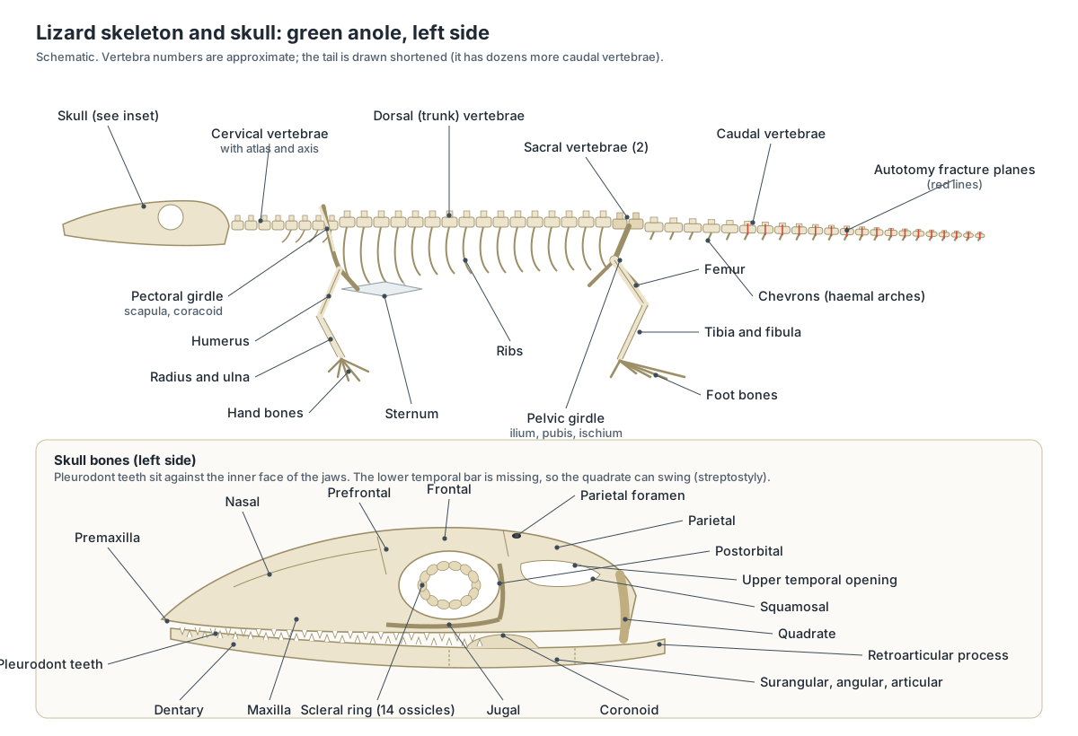
- Cervical vertebrae: Support and turn the head. The atlas rotates on the single occipital condyle of the skull.
- Dorsal (trunk) vertebrae: Form the backbone that supports the trunk and bends from side to side during walking.
- Sacral vertebrae: Transfer the thrust of the hindlimbs to the backbone.
- Caudal vertebrae: Support the tail and anchor the tail muscles. The chevrons (haemal arches) protect the tail artery and vein.
- Ribs: Support the body wall and, moved by the intercostal muscles, ventilate the lungs. Lizards have no diaphragm.
- Sternum: Joins the front ribs and the coracoids, closing the rib cage below.
- Pectoral girdle: Attaches the forelimbs to the body. The humerus joins it at the shoulder socket (glenoid).
- Pelvic girdle: Attaches the hindlimbs to the backbone. The femur joins it at the hip socket (acetabulum).
Limb bones follow the standard four-legged plan: humerus, radius and ulna, carpals, metacarpals and phalanges in the forelimb; femur, tibia and fibula, tarsals, metatarsals and phalanges in the hindlimb. Two features are typical of lizards: the ankle bends between two rows of tarsal bones (a mesotarsal joint), and the fifth metatarsal is hooked, which sets the fifth toe apart.
Lizard skull anatomy
A lizard skull is a light, open framework with large orbits and an upper temporal opening on each side, but no lower temporal bar. Losing that bar frees the quadrate bone, on which the lower jaw hinges, to swing forward and back (streptostyly). Many lizards can also flex the skull at the joint between the frontal and parietal bones (mesokinesis) or slide the outer skull against the braincase (metakinesis). Chameleons, agamids such as bearded dragons and phrynosomatids such as horned lizards have relatively rigid, akinetic skulls, and the purpose of skull kinesis in lizards is still debated.
- Premaxilla: Carries the front upper teeth and forms the front edge of the nostrils.
- Maxilla: Carries most of the upper teeth and forms the side wall of the snout.
- Nasal bones: Roof over the nasal cavity, which holds the olfactory epithelium.
- Frontal bone: Protects the front of the brain and the olfactory tracts and forms the upper rim of each orbit.
- Parietal bone and parietal foramen: Protects the brain, anchors jaw-closing muscles along its edges, and holds the parietal foramen, under which the parietal (third) eye sits.
- Postorbital, jugal and squamosal: Form the rear rim of the orbit and the upper temporal arch, and give attachment to the jaw-closing muscles.
- Quadrate: Forms the upper half of the jaw joint. In lizards it can swing at its top (streptostyly), which changes the gape and the position of the lower jaw.
- Palate: pterygoid, palatine and vomer: Separate the mouth from the nasal passages and braincase and link the snout to the quadrate, so the palate moves when the skull flexes.
- Braincase: Protects the brain and inner ears and joins the skull to the first vertebra through a single occipital condyle.
- Scleral ring: Supports the eyeball and gives attachment for the muscles that change its shape for focusing.
- Dentary: Carries all the lower teeth.
- Postdentary bones: The coronoid anchors the jaw-closing muscles; the articular forms the lower half of the jaw joint with the quadrate; the others brace the jaw.
Lizard mouth and teeth
A lizard's mouth runs back almost to the ear. Its teeth are simple, pointed and usually all alike; they grip and pierce prey rather than chew it, and lizards swallow food whole or after a few bites. In the floor of the mouth sit the fleshy tongue and, behind it, the slit-like glottis that leads to the windpipe. In the roof are the internal nostrils (choanae) and the openings of the vomeronasal organs.
| Tooth type | How teeth attach | Replaced? | Lizards |
|---|---|---|---|
| Pleurodont | To the inner face of a high outer jaw wall | Yes, throughout life | Anoles, iguanas, geckos, skinks, monitors and most others |
| Acrodont | Apparently on the crest of the jaw | No (except a few front teeth) | Agamids such as bearded dragons, and chameleons |
| Thecodont (for comparison) | In deep sockets | Yes | Not found in lizards; crocodilians and mammals |
Pleurodont vs acrodont teeth: anoles and bearded dragons
Most lizards and snakes, including anoles and iguanas, have pleurodont teeth: each tooth is fused on one side to the inner (lingual) face of a high outer jaw wall. This leaves space beside the tooth for a replacement to develop, so pleurodont lizards replace their teeth throughout life. (Haridy (2018): tooth implantation in Pogona vitticeps, PeerJ.)
Agamids (such as the bearded dragon) and chameleons form the group Acrodonta. Most of their teeth appear fused to the top of the jaw (acrodont) and are not replaced; the tuatara is the only other living reptile like this. A bearded dragon also has a few pointed pleurodont teeth at the front of the jaw, which are replaced. Histology of Pogona vitticeps suggests its acrodont teeth start out pleurodont and only look acrodont after bone remodelling and wear, so the jaw edge itself becomes part of the cutting surface in old animals. (Haridy (2018): tooth implantation in Pogona vitticeps, PeerJ.)
Lizard tongue
Lizard tongues come in two main kinds. Iguanian lizards, including anoles, bearded dragons and horned lizards, have a thick, fleshy tongue with a slightly notched tip that they use to pick up prey (lingual prehension). Many other lizards, especially monitors and tegus, have long, deeply forked tongues used mainly for smelling: they flick the tongue to pick up scent particles and deliver them to the vomeronasal organ in the roof of the mouth. Chameleons shoot out an extremely long, sticky tongue, and geckos use the tongue to wipe their eye spectacles clean.
The tongue sits on the hyoid apparatus, the same set of bones and cartilages that, in anoles, extends the dewlap. A monitor's forked tongue lets it compare scent from the left and right tips, like smelling in stereo.
Lizard eye anatomy
Most lizards have large eyes with movable upper and lower eyelids and a thin third eyelid (nictitating membrane). The wall of each eyeball is stiffened by a scleral ring, typically of 14 small overlapping bones, which supports the eye and anchors the muscles used for focusing. Anoles hunt by sight and are thought to see ultraviolet light, which may make dewlap colours stand out.
Geckos and some skinks are different: their eyelids are fused into a transparent, fixed scale (spectacle), as in snakes. Chameleons can turn each eye independently. On top of the head, many lizards also have a light-sensing parietal eye.
- Eye: Vision is the anole's main sense for finding prey, mates and rivals.
- Eyelids: Close to protect and clean the eye. A thin third eyelid (nictitating membrane) can also sweep across it.
- Scleral ring: Supports the eyeball and gives attachment for the muscles that change its shape for focusing.
- Parietal eye: Senses light and dark (not images). It helps regulate daily rhythms, hormones and basking.
The parietal eye: what the lizard's third eye does
The parietal eye sits in a hole in the skull roof, the parietal (or pineal) foramen, and is always covered by skin. It develops together with the pineal organ from the roof of the forebrain and is part of the same epithalamic complex. It occurs in the tuatara, most lizards, frogs and some fishes, but not in snakes, crocodilians, turtles, birds or mammals. (Wikipedia: parietal eye.)
The parietal eye has only a simple lens and retina and cannot form images. In bearded dragons it appears to be involved in sensing magnetic fields: dragons reacted to a weak low-frequency electromagnetic field, the reaction disappeared when the third eye was covered with a small aluminium cap, and it only occurred when the light reaching the eye had a wavelength shorter than 580 nm. (Nishimura (2020): the parietal eye of Pogona vitticeps, Animals.)
Lizard ear anatomy
Lizards have no outer ear flap, but most have a visible ear opening on each side of the head, behind the jaw, with the eardrum (tympanic membrane) just inside. Sound vibrates the eardrum, which moves a single slender middle-ear bone, the columella, against the inner ear. The inner ear holds the cochlear duct for hearing and the semicircular canals for balance. In the green anole, the ear opening is a small oval behind the corner of the mouth.
Some lizards have lost the ear opening: earless lizards (Holbrookia) have it covered by scales, and many burrowing lizards have it reduced. Snakes have no ear opening, eardrum or middle-ear cavity at all, which is one of the clearest ways to tell a legless lizard from a snake.
Lizard heart anatomy: three chambers
A lizard heart has three chambers: two atria and one ventricle. The ventricle is not a single open space: two incomplete partitions, the vertical septum and the muscular ridge, divide it into three linked compartments, the cavum arteriosum, cavum venosum and cavum pulmonale. A thin-walled sinus venosus collects blood from the body before the right atrium.
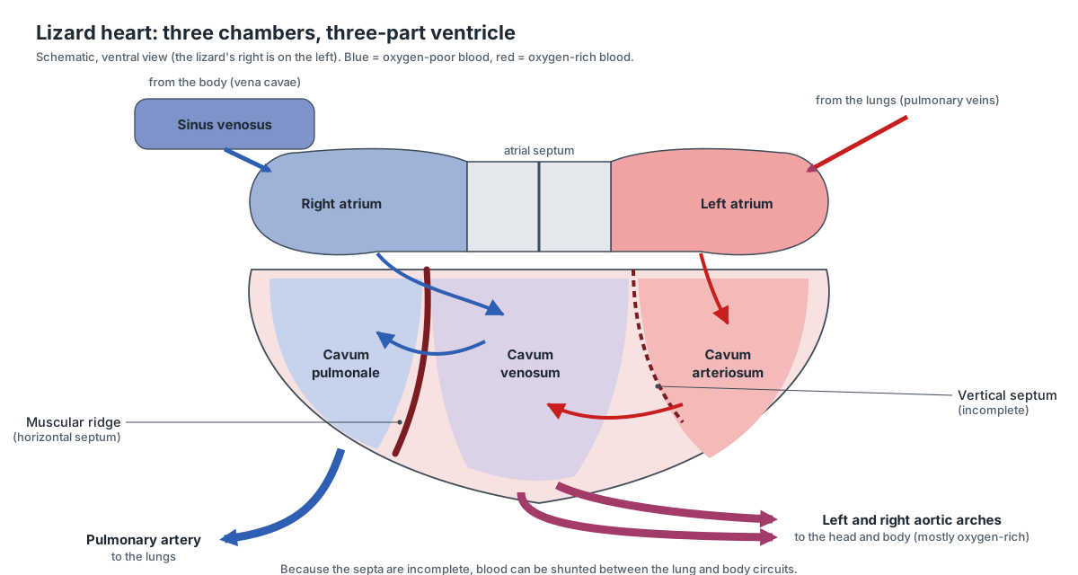
| Compartment | Receives blood from | Sends blood to |
|---|---|---|
| Cavum arteriosum (left) | Left atrium (oxygen-rich, from the lungs) | Across the cavum venosum to the aortic arches |
| Cavum venosum (middle) | Right atrium (oxygen-poor, from the body) | Left and right aortic arches; passes right-atrial blood on to the cavum pulmonale |
| Cavum pulmonale (right) | Right atrium, via the cavum venosum | Pulmonary artery to the lungs |
Because the partitions are incomplete, a lizard can send some blood past the lungs (a right-to-left shunt), for example while it holds its breath. Monitor lizards have the most complete division and can keep a high-pressure body circuit and a low-pressure lung circuit, like a mammal.
Inside the three-chambered lizard heart
A lizard heart has a sinus venosus, two fully separated atria and one ventricle. Blood flows in through three systemic veins to the right atrium and through one or two pulmonary veins to the left atrium, and leaves through three arteries: the left aorta, the right aorta and a single pulmonary artery. (Jensen et al. (2013): development of the hearts of lizards and snakes, PLoS ONE.)
The ventricle is divided by two incomplete partitions into three compartments. From the animal's left to right they are the cavum arteriosum, the cavum venosum and the cavum pulmonale. A sheet of trabeculated muscle, the vertical septum, partly separates the cavum arteriosum from the cavum venosum, and a spiralling muscular ridge (also called the horizontal septum) partly separates the cavum venosum from the cavum pulmonale. (Jensen et al. (2013): development of the hearts of lizards and snakes, PLoS ONE.)
During filling, oxygen-rich blood from the left atrium enters the cavum arteriosum, while oxygen-poor blood from the right atrium enters the cavum venosum and cavum pulmonale. When the ventricle contracts, the right-atrial blood is sent mainly to the pulmonary artery and the left-atrial blood to the two aortae. Mixing is small, but because resistance in the lung circuit is often higher, some blood can bypass the lungs (a right-to-left shunt), especially in resting or diving animals. (Jensen et al. (2013): development of the hearts of lizards and snakes, PLoS ONE.)
Monitor lizards are the exception among lizards: during contraction their ventricle works almost like two separate pumps, so they can keep a high blood pressure in the body and a low pressure in the lungs, much like mammals. This goes with the high metabolic rates and endurance of monitors. (Wikipedia: monitor lizard.)
Lizard lungs and breathing
Lizards breathe air through the nostrils, glottis and trachea into two lungs. Anole lungs are simple sacs whose inner walls form a honeycomb of small chambers (faveoli). Monitors and iguanas have larger, multi-chambered lungs, and in savannah monitors and green iguanas air moves one way through the lung, as in birds. With no diaphragm, lizards ventilate by moving the ribs with the body wall muscles. Because the trunk bends sideways when they run, they find it hard to breathe while running fast (Carrier's constraint).
Lizard brain anatomy
A lizard brain is small and elongated. From front to back it has the olfactory bulbs (smell), the paired cerebral hemispheres, the optic lobes of the midbrain (vision), a small cerebellum (coordination) and the medulla oblongata, which continues as the spinal cord. The pineal organ and parietal eye grow from the roof of the forebrain.
- Olfactory bulbs: Process smell. A separate accessory olfactory bulb receives the vomeronasal nerve.
- Cerebral hemispheres: Integrate senses, learning and behaviour such as courtship and territorial display.
- Optic lobes: Process vision and guide prey-catching strikes; very large in sight-hunting lizards like anoles.
- Cerebellum: Coordinates movement and balance.
- Medulla oblongata: Controls breathing and heart rate and relays signals between the brain and the body.
- Spinal cord: Carries signals between the brain and the body and controls simple reflexes such as the writhing of a shed tail.
The popular phrase "lizard brain" for the instinctive parts of the human brain is a simplification; real lizard brains have well-developed forebrains that support learning, social behaviour and complex displays.
Lizard cloaca and excretion
The cloaca is the chamber at the end of the gut into which the intestine, the urinary ducts and the reproductive ducts all open. It has three parts: the coprodeum (faeces), the urodeum (urine, eggs or sperm) and the proctodeum, which opens to the outside through the transverse vent. Lizard kidneys lack the loop of Henle and cannot make concentrated urine; instead, nitrogen leaves mainly as solid uric acid, the white part of lizard droppings, and water is reabsorbed in the cloaca and the urinary bladder. Most lizards have a bladder; snakes and crocodilians do not.
- Kidneys: Filter the blood and remove nitrogen waste, mostly as uric acid.
- Urinary bladder: Stores urine and lets water be reabsorbed back into the body.
- Cloaca: Its three parts handle faeces (coprodeum), urine and reproductive products (urodeum), and form the exit (proctodeum). It also reabsorbs water.
- Large intestine: Absorbs water and forms faeces.
Male vs female lizard anatomy: reproductive system
Male and female lizards share paired gonads at the back of the body cavity, but their ducts and external features differ. Male lizards have two hemipenes, copulatory organs that lie inside out in pockets in the base of the tail and are everted through the vent to mate. Only one is used at a time, and sperm runs along a groove on its surface. Females have two ovaries and two oviducts that add albumen and the shell to each egg.
| Male green anole | Female green anole | |
|---|---|---|
| Size | About 15% larger, larger head | Smaller |
| Dewlap | Large, pink-red, used in displays | Small and pale, or absent |
| Back | Can raise a crest along the neck and back | Usually a pale stripe down the middle of the back |
| Vent region | Enlarged postcloacal scales; wide tail base | Small, even scales; narrower tail base |
| Gonads | Two testes; sperm ducts to the cloaca | Two ovaries, maturing one egg at a time, alternately |
| Copulatory organs | Two hemipenes in the tail base | None (a small hemiclitoris in some squamates) |
| Eggs | — | One (sometimes two) soft-shelled egg about every two weeks in the breeding season |
Other lizards differ. Male bearded dragons have larger femoral and precloacal pores (waxy scent glands under the thighs and in front of the vent), a darker beard and visible hemipenal bulges. About 20% of lizard species give birth to live young, including blue-tongued skinks, and a few whiptail lizards reproduce by parthenogenesis, with all-female populations. Switch the 3D model between male and female to compare.
Lizard tail anatomy: autotomy and regeneration
A lizard tail contains a long series of caudal vertebrae with chevron bones below them, segmented muscles, fat stores, the caudal artery and vein, and the end of the spinal cord. In anoles, geckos, skinks and many other lizards, most tail vertebrae are crossed by a pre-formed fracture plane, so the tail can be shed when a predator grabs it (caudal autotomy). The shed tail wriggles for minutes and distracts the predator while the lizard escapes.
- Caudal vertebrae: Support the tail and anchor the tail muscles. The chevrons (haemal arches) protect the tail artery and vein.
- Autotomy fracture planes: Let the lizard shed its tail at a chosen point when grabbed, then seal the wound with little blood loss.
- Tail muscles: Move the tail and, at autotomy, contract to tear the tail apart at a fracture plane.
- Caudofemoralis: Pulls the thigh backwards: the main power stroke of running.
Losing the tail has costs: fat reserves, balance and sometimes social rank. The regrown tail is supported by a cartilage tube rather than vertebrae, is usually shorter and differently coloured, and cannot break at the same place again.
How tail autotomy works, and what grows back
Lizards shed the tail at pre-formed planes of weakness. In most species the plane runs straight across the middle of a tail vertebra (intravertebral autotomy); breaking between two vertebrae (intervertebral autotomy) is less common. The break follows the boundary between muscle segments, so the tail separates cleanly. The lizard helps by contracting its tail muscles, and the break usually happens just in front of where the tail was grabbed, or a few segments further forward. (Sanggaard et al. (2012): structural features of lizard tail autotomy, PLoS ONE.)
Studies of the Tokay gecko found that the muscle fibres at a fracture plane end in finger-like, interlocking tips that are held to the connective tissue septum by adhesion; no fibres or tendons cross the plane. During autotomy the tips change shape, which suggests the lizard can release the adhesion quickly. This design lets the tail stay strong in daily life but break easily on demand. (Sanggaard et al. (2012): structural features of lizard tail autotomy, PLoS ONE.)
Not every tail vertebra can break. In the skink Egernia kingii, adults have 5–6 vertebrae at the base of the tail with no fracture plane, followed by 39–44 that can break. This keeps the base of the tail, where large leg muscles attach, intact. In some other lizards the fracture planes fuse with age, so old adults lose the ability. (Barr et al. (2022): caudal autotomy fracture planes in Egernia kingii, Scientific Reports.)
Agamid lizards vary a great deal. Some can shed and regrow the tail, some can shed it but not regrow it, and some cannot shed it at all. Museum specimens of bearded dragons (Pogona vitticeps and P. barbata) examined in one review all had intact tails, and agamids that run on their hind legs tend to keep their tails, which they need for balance. (Ananjeva et al. (2021): autotomy of agamid lizards, Journal of Developmental Biology.)
The new tail of a green anole is not a copy of the old one. A wound skin forms over the stump within about 10 days, and a tube of ependymal cells grows out from the spinal cord. By 15–20 days a central cartilage tube and new muscle start to form, and outgrowth largely stops after about 60 days. The regenerated tail has a single unsegmented cartilage tube instead of vertebrae, unsegmented muscle bundles and no segmental nerves or dorsal root ganglia. That is why a regrown tail is stiffer and cannot be shed again at the same place. (Hutchins et al. (2014): tail regeneration in Anolis carolinensis, PLoS ONE.)
Lizard foot and leg anatomy
Lizard legs stick out sideways from the body (sprawling posture), with the upper arm and thigh held nearly horizontal and the forearm and shank pointing down. Each foot has five clawed digits. The usual phalangeal formula is 2-3-4-5-3 in the hand and 2-3-4-5-4 in the foot, so the fourth toe is the longest. Anoles, like many geckos, add adhesive toe pads under the middle of each digit.
- Forelimbs: Climbing, gripping branches and, together with the hindlimbs, walking with the sprawling, side-to-side gait typical of lizards.
- Hindlimbs: Main source of thrust for running, climbing and jumping.
- Humerus: Swings forward and back in a near-horizontal plane in the sprawling lizard gait.
- Radius and ulna: Support the forearm and let the hand rotate slightly.
- Hand bones: Support the fingers and the toe pads.
- Femur: Pulled backwards by the caudofemoralis muscle from the tail, it gives the main thrust in running.
- Tibia and fibula: Support the shank. The ankle joint of lizards lies between two rows of tarsals (mesotarsal joint).
- Foot bones: Form the long, flexible foot used for pushing off.
- Claws: Grip rough bark and soil where adhesive pads work less well.
Leg length tracks habitat. Among Caribbean anoles, species that perch on broad tree trunks have long legs for running and jumping, while those living on thin twigs have short legs for careful clambering.
Lizard muscle anatomy
The main muscles of a lizard are the epaxial muscles along the back, the hypaxial and intercostal muscles of the body wall, the jaw muscles, the limb muscles and the segmented muscles of the tail. The largest single limb muscle is the caudofemoralis, which runs from the tail base to the femur and pulls the thigh back during running, so the base of the tail is part of the locomotor system as well as a balance organ.
- Epaxial muscles: Bend the body sideways and stiffen it during locomotion.
- Body wall muscles: Move the ribs to breathe and support the abdomen.
- Jaw-closing muscles: Bite down on prey.
- Depressor mandibulae: Opens the mouth by pulling up the back end of the lower jaw.
- Ceratohyoid muscles: Swing the second ceratobranchials forward to extend the dewlap.
Bearded dragon anatomy vs the green anole
The central bearded dragon (Pogona vitticeps) is an agamid from inland Australia and the most popular pet lizard. It reaches about 60 cm, more than half of it tail. Its organ systems match the anole's, but several features differ:
- A spiny throat pouch (beard) that inflates and darkens in threat and courtship displays; it is darkest in breeding males.
- Mostly acrodont teeth that are not replaced, with a few pleurodont front teeth that are.
- Femoral pores under the thighs, more pronounced in males, and no adhesive toe pads.
- A well-developed parietal eye, used in experiments on light-dependent magnetic sensing.
- Sex is set by ZW chromosomes, but incubation above about 32 °C can turn genetic males into functional females.
- Bearded dragons rarely, if ever, shed their tails.
Monitor lizard anatomy
Monitor lizards (Varanus), which include the Bengal monitor and the Komodo dragon, are active predators with the highest standard metabolic rates of any living reptile. Living species range from about 20 cm to over 3 m long. Their anatomy reflects this lifestyle:
- A long, deeply forked tongue that carries scent to the vomeronasal organ.
- A heart whose ventricle is functionally divided during contraction, giving mammal-like pressure differences between the body and lung circuits.
- Large, multi-chambered lungs with one-way airflow in the savannah monitor.
- Venom glands in the lower jaw in at least some species.
- No femoral pores: they are absent in all monitors, as in skinks and chameleons.
Gecko feet: how adhesive toe pads stick
Many geckos and almost all anoles have toe pads: rows of widened scales under the digits that bear microscopic hair-like filaments (setae). Only the setae with flattened, spatula-shaped tips are true adhesive setae. Toe pads have evolved independently many times within geckos and separately in anoles, so the two are a textbook case of convergent evolution. (Russell, Delannoy & Garner (2026): toe pads of Gekko gecko, Journal of Anatomy.)
The numbers are large. In the Tokay gecko (Gekko gecko), the toe pads cover about 400 mm² in total, a single front toe carries about 262,000 setal stalks and a single hind toe about 353,000, and each seta branches into roughly 250–560 spatula tips. Earlier experiments cited in this study (Autumn et al., 2002) showed that these tips stick by weak molecular attraction (van der Waals forces), so the foot grips dry glass without any glue. (Russell, Delannoy & Garner (2026): toe pads of Gekko gecko, Journal of Anatomy.)
Frilled lizard and blue-tongued skink anatomy
The frill of the frill-necked lizard (Chlamydosaurus kingii) is a large flap of skin around the neck, supported by long rod-like hyoid bones and linked to each side of the head by Grey's cartilage. When the lizard opens its mouth, the hyoid rods and lower jaw spread the frill into a disc about 30 cm across; at rest it folds back over the shoulders like a cape. It is the same hyoid apparatus that stiffens an anole's dewlap. Frilled lizards reach about 90 cm in total length and can run on their hind legs. (Wikipedia: frill-necked lizard.)
Blue-tongued skinks (Tiliqua) have long, heavy bodies, short legs and a broad blue tongue. Threatened, the skink opens its mouth and shows the tongue as a bluff warning. All blue-tongued skinks give birth to live young: the eggs are kept and hatch inside the mother, with litters ranging from about 1–4 young in some species to 5–24 in others. (Wikipedia: blue-tongued skink.)
Basilisk and flying lizard (Draco): built to run on water and glide
The common basilisk (Basiliscus basiliscus) runs across water on its hind legs. Its hind feet are large, and its toes carry fringes of scales that fold flat on land and spread on water, increasing the foot's area. Each step has three phases: a slap that pushes water down and traps an air pocket around the foot, a stroke that pushes the lizard forward, and a recovery in which the foot is pulled out before the air pocket collapses. Young, light basilisks can run much farther on water than adults. (Wikipedia: common basilisk.)
Flying lizards of the genus Draco glide between trees on a membrane (patagium) along each side of the body, supported by five to seven greatly lengthened thoracic ribs. The ribs are spread by specialised iliocostalis and intercostal muscles. Field observations of Draco dussumieri showed that in flight the lizard grabs the front edge of the membrane with its hands, forming a composite wing; the arms then steer, and are free again for climbing after landing. (Dehling (2017): how lizards fly, PLoS ONE.)
Horned lizard anatomy: horns, blood-squirting and ant eating
Horned lizards (Phrynosoma) have flattened, round, toad-like bodies and a crown of true horns, which have a bony core, unlike the spiny scales on their back and sides. Several species can squirt blood from the corners of the eyes: they restrict blood flow leaving the head, the pressure rises, and small vessels around the eyelids burst, spraying blood up to about 1.5 m. The blood tastes foul to dogs, coyotes and cats but has no effect on birds. At least eight species are known to do it. (Wikipedia: horned lizard.)
Many horned lizards eat mainly harvester ants, which bite and sting. The Texas horned lizard has a factor in its blood plasma that neutralises the ants' venom, catches each ant with a flick of the tongue, and coats it in large amounts of mucus in the throat and oesophagus, so swallowed ants cannot sting. Like other iguanians, horned lizards catch food with a sticky tongue rather than with the jaws. (Fertschai et al. (2021): prey capture in the Texas horned lizard, Biology Open.)
Legless lizard anatomy: glass lizards
Legless lizards look like snakes but keep lizard anatomy. Glass lizards (Ophisaurus) of North America, such as the eastern glass lizard, have no legs, yet they have movable eyelids, external ear openings, a groove along each side of the body, rigid jaws and a very long tail that breaks easily, hence the name "glass snake". The eastern glass lizard grows to 46–108 cm, but its head and body are at most about 30 cm; the rest is tail.
Lizard vs snake anatomy
Snakes are highly modified lizards, but the two groups can be told apart by several features. These work even for legless lizards.
| Feature | Lizards (incl. legless lizards) | Snakes |
|---|---|---|
| Eyelids | Movable eyelids in most (not most geckos) | None; a fixed clear spectacle |
| Ear opening and eardrum | Usually present | Absent |
| Lower jaw | Two halves firmly joined at the chin | Halves joined by a stretchy ligament, move separately |
| Tongue | Fleshy and notched, or forked in monitors and tegus | Always long and deeply forked |
| Tail | Often long (over half the length in many species); often autotomic | Usually short relative to body |
| Lungs | Two working lungs | Left lung reduced or absent in most |
| Urinary bladder | Present in most | Absent |
| Scleral ring and parietal eye | Present in most | Absent |
| Belly scales | Small, similar to other scales in most | Usually one row of wide belly plates (ventral scutes) |
| Shedding | In patches | Usually in one piece |
| Shared features | Hemipenes, transverse vent, movable quadrate, vomeronasal organ | Same |
For the snake side of the comparison, see the snake anatomy and snake skeleton pages.
Why the tuatara is not a lizard
The tuatara (Sphenodon punctatus) of New Zealand looks like a large lizard, but it is the only living member of a separate order, Rhynchocephalia. Its closest living relatives are the squamates (lizards and snakes), and it has kept many features that lizards lost. In 1867 Albert Günther noted these features and proposed the separate order. (Wikipedia: tuatara.)
Anatomical differences students can look for: the tuatara has a complete lower temporal bar, so its skull is rigid and its quadrate cannot swing; its teeth are acrodont, with a second row on the palate so the lower teeth fit between two upper rows; it has no ear opening or eardrum; its ribs carry hooked uncinate processes and it has well-developed belly ribs (gastralia); and the male has no copulatory organ, whereas male lizards have paired hemipenes. Its parietal eye is the most developed of any living animal, but it is only visible in hatchlings. (Wikipedia: tuatara.)
Lizard vs frog anatomy
A comparison often set in class: frogs are amphibians and lizards are reptiles. A lizard has dry, scaly skin, breathes only with lungs, lays shelled eggs on land (or bears live young), has claws, a long tail and a single middle-ear bone behind a recessed eardrum. A frog has moist, glandular skin that also exchanges gases, lays jelly-coated eggs in water that hatch into tadpoles, has no claws or tail as an adult, and pushes air into its lungs by throat pumping. Both have a three-chambered heart and a cloaca.
Lizard anatomy worksheet: test yourself
Use the 3D model and the diagrams above. Answers are in brackets.
- Which external features show that a green anole is male? (Large dewlap, enlarged postcloacal scales, wide tail base.)
- What supports the dewlap? (The second ceratobranchials of the hyoid apparatus.)
- How many chambers does the lizard heart have, and what are the three parts of the ventricle? (Three; cavum arteriosum, cavum venosum, cavum pulmonale.)
- Where does a lizard tail break during autotomy? (At fracture planes across the caudal vertebrae.)
- What supports a regenerated tail? (A cartilage tube instead of vertebrae.)
- Name two features that separate a glass lizard from a snake. (Movable eyelids, ear openings, rigid jaw, long breakable tail.)
- What does the parietal eye detect? (Light levels, not images.)
- Why do lizards produce white droppings? (Nitrogen is excreted as uric acid.)
Frequently asked questions
How many chambers does a lizard heart have?
Three: two atria and one ventricle. The ventricle is partly divided by ridges into three compartments (cavum arteriosum, cavum venosum and cavum pulmonale), which keeps most oxygen-rich and oxygen-poor blood apart but lets lizards shunt blood when needed. Monitor lizards have the most completely divided ventricle.
Why do lizards drop their tails?
To escape predators. Most lizards can break the tail at pre-formed fracture planes that cross the tail vertebrae. The shed tail keeps wriggling and distracts the predator. The stump heals and a new tail grows, but it is supported by a cartilage tube instead of vertebrae and cannot be shed again at the same place.
Do lizards have ears?
Yes. Lizards have no outer ear flaps, but most have an ear opening on each side of the head with the eardrum just inside. A single middle-ear bone, the columella, carries sound to the inner ear. Snakes have no ear opening or eardrum.
What is the third eye on a lizard?
It is the parietal eye, a small light-sensitive organ on the top of the head, under a translucent scale and over a hole in the skull roof (parietal foramen). It cannot form images but senses light levels, helping to control daily rhythms, hormones and basking.
Do lizards have teeth?
Yes. Most lizards, including anoles and iguanas, have pleurodont teeth fixed to the inner side of the jaw and replaced throughout life. Agamids such as bearded dragons, and chameleons, have mostly acrodont teeth fused to the top of the jaw that are not replaced.
How can you tell a male lizard from a female?
In green anoles, males are larger, have a large pink-red dewlap, a wider tail base (holding the two hemipenes) and a pair of enlarged scales behind the vent. Females have a small or no dewlap and often a pale stripe down the back. In bearded dragons, males have larger femoral and precloacal pores, a darker beard and hemipenal bulges.
What is the difference between a legless lizard and a snake?
Legless lizards such as glass lizards have movable eyelids, external ear openings, a groove along each side of the body, a rigid jaw and a very long tail that breaks easily. Snakes have no eyelids or ear openings, flexible jaws, a deeply forked tongue and a short tail.
Is a tuatara a lizard?
No. The tuatara of New Zealand looks like a lizard but is the only living member of a separate order, Rhynchocephalia, the sister group of lizards and snakes. It has a complete lower temporal bar, acrodont teeth with two upper rows, no ear opening and no copulatory organ in males.
What does a lizard's cloaca do?
The cloaca is the shared chamber at the end of the digestive, urinary and reproductive tracts. Faeces, uric acid (the white part of lizard droppings), eggs and sperm all leave through it, and it reabsorbs water. It opens to the outside through the vent.
References
- Jensen, B., van den Berg, G., van den Doel, R., Oostra, R.-J., Wang, T. & Moorman, A. F. M. (2013). Development of the hearts of lizards and snakes and perspectives to cardiac evolution. PLoS ONE 8, e63651.
- Haridy, Y. (2018). Histological analysis of post-eruption tooth wear adaptations, and ontogenetic changes in tooth implantation in the acrodontan squamate Pogona vitticeps. PeerJ 6, e5923.
- Hutchins, E. D. et al. (2014). Transcriptomic analysis of tail regeneration in the lizard Anolis carolinensis reveals activation of conserved vertebrate developmental and repair mechanisms. PLoS ONE 9, e105004.
- Sanggaard, K. W. et al. (2012). Unique structural features facilitate lizard tail autotomy. PLoS ONE 7, e51803.
- Barr, J. I., Boisvert, C. A., Trinajstic, K. & Bateman, P. W. (2022). Ontogeny and caudal autotomy fracture planes in a large scincid lizard, Egernia kingii. Scientific Reports 12, 6793.
- Ananjeva, N. B., Gordeev, D. A. & Korost, D. V. (2021). The review of the autotomy of agamid lizards with considerations about the types of autotomy and regeneration. Journal of Developmental Biology 9, 32.
- Lailvaux, S. P., Leifer, J., Kircher, B. K. & Johnson, M. A. (2015). The incredible shrinking dewlap: signal size, skin elasticity, and mechanical design in the green anole lizard (Anolis carolinensis). Ecology and Evolution 5, 4400–4409.
- Russell, A. P., Delannoy, S. M. & Garner, A. M. (2026). Integrative architecture and structural boundaries of the subdigital adhesive toe pads of Gekko gecko. Journal of Anatomy.
- Dehling, J. M. (2017). How lizards fly: a novel type of wing in animals. PLoS ONE 12, e0189573.
- Fertschai, I., Sherbrooke, W. C., Ott, M. & Chagnaud, B. P. (2021). Avoiding being stung or bitten: prey capture behaviors of the ant-eating Texas horned lizard (Phrynosoma cornutum). Biology Open 10, bio058453.
- Nishimura, T. (2020). The parietal eye of lizards (Pogona vitticeps) needs light at a wavelength lower than 580 nm to activate light-dependent magnetoreception. Animals 10, 489.
- Animal Diversity Web, University of Michigan Museum of Zoology: Anolis carolinensis (green anole).
- Wikipedia: Carolina anole; Dactyloidae; Reptile; Scleral ring; Cranial kinesis; Hemipenis (overview articles used for general facts, each checked against the cited primary sources where possible).
- Wikipedia: Horned lizard; Monitor lizard; Tuatara; Eastern glass lizard; Central bearded dragon; Frill-necked lizard; Blue-tongued skink; Common basilisk (species comparison sections).
More interactive anatomy models
- Snake anatomyElongated organs, one working lung, a flexible jaw and the snake heart.
- Snake skeletonHundreds of vertebrae and ribs, a kinetic skull and vestigial hip bones.
- Turtle skeletonHow ribs and spine fuse into the shell: carapace, plastron and girdles.
- Human anatomy1,000+ bones, muscles, organs, vessels and nerves from real anatomical data.
- Octopus anatomyThree hearts, a ring-shaped brain, eight arms and a beak, in 3D.
- Squid anatomyPen, eight arms, two tentacles, giant axons and three hearts of the longfin squid.