

The cow digestive system at a glance
The cow digestive system is a ruminant digestive system: food is fermented by microbes in a huge forestomach before the cow digests it with its own enzymes. It runs from the mouth through the oesophagus to a stomach with four compartments (the rumen, reticulum, omasum and abomasum), then the small intestine (duodenum, jejunum and ileum), the large intestine (caecum, spiral colon, descending colon and rectum) and the anus. The liver and pancreas add bile and enzymes to the small intestine.
This page describes an adult Holstein, the black-and-white dairy breed. Beef breeds, bulls and steers have the same organs; their size changes with body size and diet. Directions are the cow's own: cranial is towards the head, caudal towards the tail, dorsal the back and ventral the belly.
The big idea is simple. Cows eat grass and hay, which are mostly cellulose. Neither cows nor any other vertebrates make an enzyme that can break cellulose down. Rumen microbes can. So the cow keeps a 150-litre fermentation vat full of bacteria, protozoa and fungi, feeds it chewed plants and saliva, and lives on the acids the microbes release and on the microbes themselves.
Do cows have four stomachs?
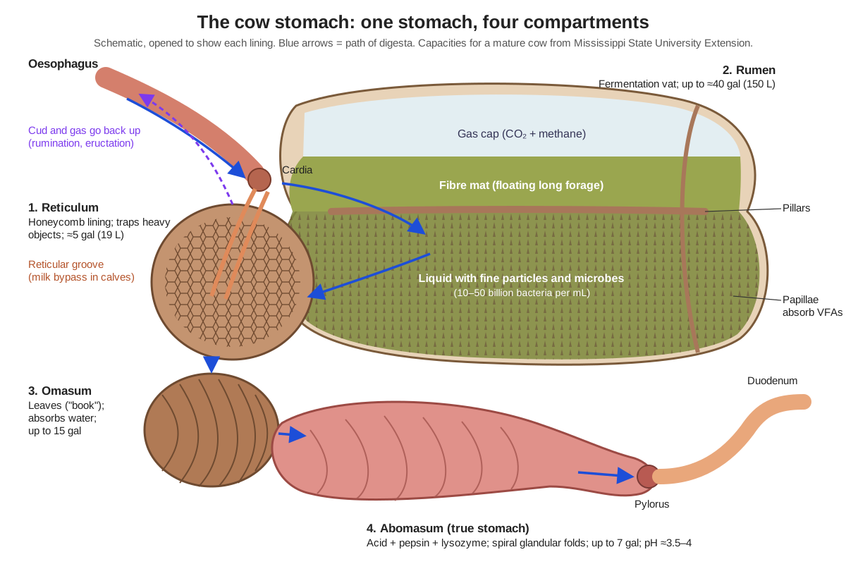
No. A cow has one stomach divided into four compartments. People often say "four stomachs" because the compartments look so different. The first three, the rumen, reticulum and omasum, are called the forestomachs. They are lined with a skin-like (stratified squamous) epithelium and have no digestive glands. Only the fourth compartment, the abomasum, has glands that secrete acid and enzymes, so it is called the true stomach. It works much like a human stomach.
Together, the four compartments fill almost three-quarters of the abdominal cavity. In a mature cow, Mississippi State University Extension gives capacities of up to about 40 gallons (≈150 L) for the rumen, about 5 gallons for the reticulum, up to 15 gallons for the omasum and up to 7 gallons for the abomasum. The rumen and reticulum make up about 84% of the stomach's volume, the omasum about 12% and the abomasum about 4%. Other sources give rumen volumes from about 100 to 200 litres, because cows differ in size and diet.
Cow digestive system diagram: the left side
Seen from the left, the cow digestive system is mostly rumen. The rumen fills almost the whole left half of the abdomen, from the diaphragm back to the pelvis. That is why the left side is the standard view for a cow digestive system diagram, and why vets listen for rumen contractions in the hollow of the left flank (the left paralumbar fossa). The spleen lies flat on the left side of the rumen.
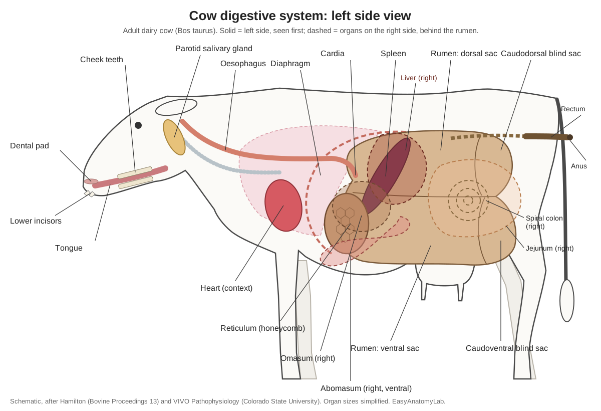
Almost everything else is pushed to the right. A bovine surgery guide describes the omasum lying right of the midline opposite ribs 7 to 11, the liver and gallbladder entirely on the right inside the rib cage, the abomasum on the floor of the right lower abdomen with its pylorus just below the gallbladder, and the intestines filling the right flank. Even the left kidney is pushed across: it hangs against the right side of the rumen. In the 3D model, turn the cow around to see the right-side organs, or switch off the "Rumen, reticulum & omasum" chip to see behind the rumen.
Parts of the cow digestive system in order
Food passes through the cow digestive system in this order:
- Mouth: lips, tongue, lower incisors and the toothless dental pad (cows have no upper incisors), cheek teeth, and salivary glands such as the parotid.
- Oesophagus: carries food down, and cud and gas back up.
- Reticulum and rumen (the reticulorumen): food lands in both; this is the fermentation vat. Cud returns from here to the mouth for rumination.
- Omasum: absorbs water and filters particles.
- Abomasum: the true stomach, with acid and enzymes.
- Small intestine: duodenum (with bile from the liver and juice from the pancreas), jejunum and ileum.
- Large intestine: caecum, ascending colon (including the spiral colon), transverse and descending colon.
- Rectum and anus.
Textbooks usually list the stomach as "rumen, reticulum, omasum, abomasum". Strictly, swallowed food enters the rumen and reticulum together, because they form one chamber divided only by a low fold.
The four compartments and what each one does
Each compartment has its own lining and job. The rumen ferments, the reticulum sorts, the omasum absorbs water, and the abomasum digests.
| Compartment | Nickname and lining | Main function | Mature cow size |
|---|---|---|---|
| Rumen | Paunch; papillae | Microbial fermentation of fibre and starch; absorbs volatile fatty acids | Up to ≈40 gal (≈150 L); largest |
| Reticulum | Honeycomb, hardware stomach | Sorts particles; traps heavy objects; starts each contraction cycle and regurgitation | ≈5 gal (≈19 L) |
| Omasum | Manyplies, the book; leaves (laminae) | Absorbs water, minerals and remaining fatty acids; holds back large particles | Up to 15 gal (source-dependent) |
| Abomasum | True stomach; glandular folds | Acid (pH ≈3.5–4) and pepsin digest protein; lysozyme breaks down rumen bacteria | Up to 7 gal |
Sizes: Mississippi State University Extension P2503. The omasal leaves make up about one-third of the surface area of the forestomachs (VIVO, Colorado State University).
How the cow digestive system works, step by step
A cow digests food in two stages: first microbes ferment it in the rumen, then the cow's own enzymes digest what is left, including the microbes, in the abomasum and small intestine.
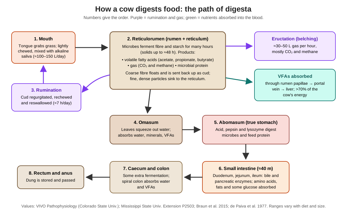
- Ingestion. The cow wraps its tongue around grass and tears it off against the dental pad with its lower incisors. Grazing cattle take 25,000 to more than 40,000 bites a day. Food is chewed only briefly and mixed with saliva before it is swallowed.
- Sorting in the reticulorumen. Swallowed food settles into layers. Gas rises to the top, fresh long forage floats in a fibre mat, and dense grain and well-soaked fibre sink into the liquid below and into the reticulum. Contractions 1 to 3 times a minute keep everything mixing.
- Rumination. Coarse fibre is brought back up to the mouth as cud, chewed again and reswallowed. Dairy cows spend about seven hours a day ruminating, and the extra chewing produces extra saliva.
- Microbial fermentation. Bacteria, protozoa and fungi break down cellulose, starch and protein. Their main products are the volatile fatty acids acetate, propionate and butyrate, plus carbon dioxide and methane, and new microbial cells.
- Absorption from the rumen. The fatty acids pass through the rumen papillae into the blood and go to the liver. They supply more than 70% of the cow's energy.
- Methane and eructation. The cow belches 30–50 litres of gas an hour. Almost all of it leaves through the mouth and nose.
- Passage. Only small, dense particles and fluid can leave the reticulum through the reticulo-omasal orifice. The omasum absorbs water and minerals.
- Enzymatic digestion. In the abomasum, hydrochloric acid and pepsin start digesting feed protein and the masses of microbes washed out of the rumen. In the duodenum, bile and pancreatic enzymes continue the job.
- Absorption in the small intestine. Amino acids, fats and the small amount of glucose that escaped the rumen are absorbed along about 40 metres of small intestine.
- Large intestine and excretion. The caecum and colon ferment a little more fibre and absorb water and fatty acids. Dung leaves through the rectum and anus.
How long it takes depends on the feed. Solid fibre can stay in the rumen for up to about 48 hours, and marker studies in dairy cows give total-tract mean retention times of about 37 to 47 hours.
Rumen microbes: the cow's fermentation partners
A cow cannot digest cellulose by itself. No vertebrate is known to make an enzyme that breaks the beta-1,4 bonds between the glucose units of cellulose, but many microbes do. The rumen gives those microbes a warm, oxygen-free, well-mixed home with a steady supply of chewed plants. In return the microbes break down cellulose and hemicellulose, keep the sugars for their own growth, and release the waste products the cow lives on. (Bowen, Fermentation Chemistry for Herbivores, VIVO Pathophysiology.)
The numbers are huge. Each millilitre of rumen contents holds roughly 10 to 50 billion bacteria, about a million protozoa and variable numbers of yeasts and fungi. Almost all of them are anaerobes. Different groups specialise: some digest cellulose, others starch, protein, sugars or acids, and some make methane. Ciliate protozoa also eat bacteria, which may stop the bacteria from overgrowing. (Bowen, Fermentation Microbiology and Ecology, VIVO Pathophysiology.)
The mix of microbes depends on the diet. Fibre-digesting bacteria decline when a cow eats a lot of grain, and rumen pH, normally between 6 and 7, falls when large amounts of rapidly fermented carbohydrate arrive. Around pH 5.5 the protozoa drop sharply because they cannot tolerate the acid. This is why dairy rations are changed gradually: the microbes, not just the cow, have to adapt. (Bowen, Fermentation Microbiology and Ecology, VIVO Pathophysiology.)
Volatile fatty acids: how fermentation feeds the cow
Fermentation turns plant sugars mainly into three volatile fatty acids (VFAs): acetic, propionic and butyric acid, plus carbon dioxide and methane. On a high-fibre diet their molar ratio is roughly 70:20:10. Acetate is always the largest share. Much of the dietary protein that enters the rumen is also broken down and ends up as VFAs. (Bowen, Fermentation Chemistry for Herbivores, VIVO Pathophysiology.)
VFAs provide more than 70% of a ruminant's energy. Almost all of them are absorbed straight through the rumen wall, helped by the papillae, and carried by the portal vein to the liver. Each acid has its own job. Acetate is burned for energy throughout the body and is the main building block for milk fat. Propionate is taken up by the liver and turned into glucose, which matters because almost no glucose reaches the small intestine of a ruminant. Butyrate is converted in the rumen wall into beta-hydroxybutyrate, a ketone body used as fuel. (Bowen, Nutrient Absorption and Utilization in Ruminants, VIVO Pathophysiology.)
The microbes provide more than energy. As they multiply, they flow on to the abomasum and small intestine, where the cow digests them as high-quality protein. They can build protein from non-protein nitrogen such as urea, which the cow recycles into the rumen through its saliva, and they make all the B vitamins. A horse ferments in its hindgut, behind the small intestine, so it cannot digest most of its microbial protein. A cow can. (Bowen, Fermentation Chemistry for Herbivores, VIVO Pathophysiology.)
Saliva, rumination and cud chewing
Ruminants make enormous amounts of saliva. Colorado State's VIVO pathophysiology text gives published estimates of 100 to 150 litres a day for adult cows. Saliva supplies fluid for the fermentation vat and, because it is rich in bicarbonate, it buffers the acids the microbes make. (Bowen, Rumen Physiology and Rumination, VIVO Pathophysiology.)
Estimates vary with the cow and how they were measured. A dairy-nutrition review gives 130 to over 300 litres a day for lactating dairy cows, with about 3.5 kg of sodium bicarbonate secreted in that saliva. Mississippi State Extension gives 'up to 50 quarts' (about 47 litres) for a mature cow. All agree that output depends on how long the cow chews, so long fibre in the diet means more saliva and a better-buffered rumen. (West, Buffers: What and When to Use, Western Canadian Dairy Seminar 1998.)
Rumination is regurgitation, rechewing and reswallowing. A special reticular contraction, with the lower oesophageal sphincter relaxed, pushes a bolus into the oesophagus. Reverse peristalsis carries it to the mouth, the tongue squeezes out the fluid to be swallowed, and the solid cud is chewed again. Cows ruminate mostly while resting, often lying down. (Bowen, Rumen Physiology and Rumination, VIVO Pathophysiology.)
In a study of 300 healthy lactating cows (Brown Swiss, Holstein-Friesian and Swiss Fleckvieh) fitted with noseband pressure sensors, cows ate for about 265 minutes a day and ruminated for about 441 minutes. They brought up about 578 cuds a day and chewed each one about 55 times. That is more than seven hours of cud chewing every day. (Braun, Zürcher & Hässig (2015), Eating and rumination behaviour in 300 cows of three breeds, BMC Veterinary Research 11:231.)
Gas, eructation and motility
The reticulum and rumen contract in a set cycle 1 to 3 times a minute, faster while the cow eats. Primary contractions start in the reticulum and sweep caudally around the rumen, mixing the contents. Secondary contractions involve only part of the rumen and are linked to belching. The brainstem coordinates these movements through the vagus nerve. (Bowen, Rumen Physiology and Rumination, VIVO Pathophysiology.)
An adult cow produces about 30 to 50 litres of gas an hour. The contents separate by density: gas rises to the top, freshly eaten forage floats in a middle mat, and dense grain and soaked fibre sink. Contractions move the gas bubble forward to the cardia so the cow can belch almost every secondary contraction. Most belched gas is first breathed into the lungs and then breathed out. Anything that stops belching is dangerous: the swelling rumen soon presses on the diaphragm, and bloated animals die of asphyxia. (Bowen, Rumen Physiology and Rumination, VIVO Pathophysiology.)
Small and large intestine of a cow
A cow's small intestine is about 40 metres long. In a study of 40 adult European-breed cattle the small intestine averaged 39.5 m, the caecum 0.58 m and the colon and rectum 7.1 m, with no difference between bulls and cows. Mississippi State Extension describes it as a tube "up to 150 feet" long holding about 20 gallons, with a caecum about 3 feet long.
The duodenum leaves the pylorus, rises to the liver, then runs back along the right flank almost to the hip before turning forwards again. The jejunum hangs in a garland of coils on the edge of the mesentery, wrapped around the spiral colon. The spiral colon is part of the ascending colon coiled into a flat disc: two turns spiral inwards, it doubles back at the centre, and two turns spiral outwards. Pigs also have a spiral colon, but theirs is cone-shaped. The caecum is a blind pouch whose tip points back towards the pelvis. It ferments some fibre, but much less than a horse's caecum.
Ruminant vs monogastric vs hindgut fermenter
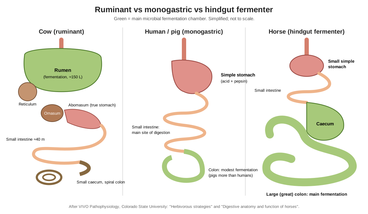
A cow (ruminant) ferments food before its true stomach, a human or pig (monogastric) digests most food with its own enzymes in a simple stomach and small intestine, and a horse (hindgut fermenter) ferments fibre after its small intestine, in a huge caecum and colon.
| Feature | Cow (ruminant) | Human / pig (monogastric) | Horse (hindgut fermenter) |
|---|---|---|---|
| Stomach | One stomach, four compartments | Simple, one compartment | Simple, one compartment |
| Main fermentation site | Rumen and reticulum, before the true stomach | Colon (small role) | Caecum and large colon, after the small intestine |
| Digests cellulose well? | Yes | Hardly | Yes |
| Uses microbial protein? | Yes: microbes are digested in the abomasum and small intestine | Not significantly | No: microbes are lost in the dung |
| Uses dietary sugar and starch directly? | Little: most is fermented first | Yes | Yes, in the small intestine |
| Main energy source | Volatile fatty acids | Glucose | Glucose and volatile fatty acids |
| Chews cud? | Yes | No | No |
| Gallbladder | Yes | Yes | No |
Cow vs human digestive system. Both have a mouth, oesophagus, glandular stomach, small and large intestine, liver and pancreas. The difference is the forestomach. A human swallows food straight into an acid stomach; a cow first ferments it for a day or two. Cows also lack upper incisors, chew cud, make far more saliva and get most of their glucose from the liver rather than from the gut.
Cow vs horse digestive system. Both are herbivores that rely on microbes, but the horse puts its fermentation vat behind the small intestine. The result: a horse can absorb starch as glucose in its small intestine, but loses the protein in its gut microbes; a cow ferments almost all starch first but digests its microbes. Horses also have no gallbladder.
Calf digestive system: the reticular groove and a big abomasum
A newborn calf works like a monogastric animal. At birth the abomasum is the largest compartment, more than half of the stomach area, while the reticulorumen is about 35% and the omasum 14%. As the calf starts eating starter feed and forage, the reticulorumen grows quickly: about 55% at one month, 65% at two months, and roughly 62% of the area in the adult, with the abomasum shrinking in relative terms. Calves are functionally non-ruminants until about 2 to 3 months of age. (Parish, Rivera & Boland, Understanding the Ruminant Animal Digestive System, Mississippi State University Extension P2503.)
Milk bypasses the undeveloped rumen. When a calf suckles, muscular folds on the wall of the reticulum close reflexly into a channel, the oesophageal or reticular groove, which runs from the cardia towards the omasum. Milk is shunted past the rumen to the abomasum, where it is curdled by rennin and digested by the calf's own enzymes. If milk is put into an immature rumen it basically rots rather than ferments. (Bowen, Rumen Physiology and Rumination, VIVO Pathophysiology.)
Bloat
Bloat (ruminal tympany) is rumen over-distension with gas the cow cannot belch. In frothy bloat the normal fermentation gas is trapped in a stable foam; it is linked to lush legume pasture such as alfalfa and clover and to fine, high-grain feedlot diets. In free-gas bloat the gas separates but cannot escape, often because something blocks the oesophagus. The left flank swells first and, in severe cases, the left paralumbar fossa can rise above the backbone. Merck notes that death rates in cattle on bloat-prone pasture can reach 20% without control measures, so farmers use antifoaming agents such as poloxalene in high-risk periods. (Merck Veterinary Manual: Bloat in Ruminants.)
Displaced abomasum (LDA and RDA)
A displaced abomasum is a common problem of high-producing dairy cows. The abomasum normally lies on the abdominal floor, but if it fills with gas and loses tone it can float up, usually on the left between the rumen and the body wall (left displaced abomasum, LDA). Left displacements outnumber right ones by about 30 to 1, and about 80–85% happen within a month after calving. Risk factors include low feed intake, low blood calcium, ketosis and high-grain, low-fibre rations. (Merck Veterinary Manual: Left or Right Displaced Abomasum and Abomasal Volvulus in Cattle.)
The key sign is a 'ping', a high-pitched ring heard with a stethoscope while flicking the abdomen. For an LDA it is usually heard between ribs 9 and 13 on the left. A right displacement can twist (abomasal volvulus), which is an emergency that needs surgery. (Merck Veterinary Manual: Left or Right Displaced Abomasum and Abomasal Volvulus in Cattle.)
Hardware disease (traumatic reticuloperitonitis)
Cattle eat quickly and do not sort their feed, so they sometimes swallow nails or wire, for example from cut tyres used to hold down silage covers. Heavy objects fall into the reticulum, and its contractions can push a sharp object through the wall, letting bacteria into the abdomen. Affected cows suddenly eat less and give less milk, have a mild fever and stand with an arched back. Because only the diaphragm separates the reticulum from the heart, the wire can even reach the pericardium. Merck recommends giving every animal a magnet by mouth at about one year of age; it stays in the reticulum and holds iron objects. (Merck Veterinary Manual: Traumatic Reticuloperitonitis in Cattle.)
Ruminal acidosis (grain overload)
Grain ferments much faster than grass. If a cow that is not adapted to grain eats a large amount, the rumen fills with volatile fatty acids and lactic acid, the pH drops below 5.5 and lactic-acid bacteria take over. The acid damages the rumen lining; bacteria that leak through can cause liver abscesses, and the condition has been linked to laminitis. Subacute acidosis, with repeated drops to pH 5.2–5.6, lowers milk yield and body condition. The main prevention is to change to high-grain rations slowly and keep enough long fibre in the diet. (Bowen, Ruminal Acidosis (Grain Overload), VIVO Pathophysiology.)
Cow methane and the climate: what the numbers say
Methane is a by-product of rumen fermentation: methanogenic microbes use hydrogen released during fermentation. Typical rumen gas is about 66% carbon dioxide and 27% methane. Using US EPA inventory methods, Kansas State University reported a national enteric emission factor of 143 kg of methane per head per year for dairy cows (2012 data), compared with 95 kg for beef cows. In that inventory, enteric fermentation produced about 2.2% of total human-caused US greenhouse gas emissions, with beef cattle responsible for 71% and dairy cattle for 25% of the enteric total. (Liu, How Much Gas Do Beef or Dairy Cattle Produce? Kansas State University MF3185 (2015).)
On a per-day scale, a review in the Journal of Animal Science estimated that ruminant livestock produce 250 to 500 litres of methane per day. The amount depends on feed intake, the type of carbohydrate, feed processing and additives such as fats and ionophores. Methane is also lost feed energy, so reducing it can improve efficiency as well as emissions. (Johnson & Johnson (1995), Methane emissions from cattle, Journal of Animal Science 73.)
Globally, Colorado State's VIVO text cites Food and Agriculture Organization data putting ruminants at roughly 20% of global methane emissions, or about 3–5% of total greenhouse gas production. It also notes that estimates are hard to pin down, so treat any single figure as approximate and check its year and method. (Bowen, Rumen Physiology and Rumination, VIVO Pathophysiology.)
Male cow digestive system: bulls and steers
A male cow's digestive system is the same as a female's. Strictly, "cow" means an adult female; the male is a bull, or a steer if castrated. Bulls, steers, cows and heifers all have the same mouth, four-compartment stomach, intestines, liver and pancreas, in the same positions. A study of intestinal length found no significant difference between 20 bulls and 20 cows.
What differs is the reproductive tract and the udder, and the effects of size and diet. A large bull has larger compartments than a small heifer, and a feedlot steer on a high-grain diet has different rumen microbes and a lower rumen pH than a grazing cow. Diseases differ too: displaced abomasum is mostly a disease of dairy cows after calving, while feedlot bloat and grain overload are more common in fattening steers.
Cow digestive system labeled: every part and its function
Every structure in the 3D model, grouped by system. Click any name in the viewer list to zoom to it.
External
- Hide (Skin and coat)
- The skin and hair covering the body, black and white in a Holstein. Protects the body and holds in heat. On the left flank, the hollow behind the last rib (the left paralumbar fossa) lies right over the rumen, so it is where vets feel rumen movements and where bloat shows first.
- Muzzle (Planum nasolabiale, nose pad)
- The bare, moist pad of skin around the nostrils and upper lip. Senses feed by touch and smell. Cattle graze by wrapping the tongue around grass, so the lips and muzzle steer the feed in.
- Eye
- The cow's eyes sit on the sides of the head. A wide field of view lets a grazing cow watch for danger while its head is down.
- Udder (Mammary gland)
- Four mammary glands (quarters), each with its own teat, between the hind legs. Makes milk. Milk sugar (lactose) is built from glucose that the liver makes mostly from propionate, a product of rumen fermentation.
- Hooves (claws) (Cloven hooves)
- Each foot ends in two weight-bearing claws: cattle are even-toed ungulates. Carry the cow's weight. Acidosis from too much grain is linked to laminitis, inflammation inside the hoof.
Skeleton (context)
- Skull
- The bones of the head, including the upper jaw (maxilla) that carries the upper cheek teeth. Protects the brain and anchors the large chewing muscles that grind cud for hours a day.
- Mandible (Lower jaw)
- The lower jaw, which carries the lower incisors and lower cheek teeth. Moves sideways during chewing, so the upper and lower cheek teeth grind plants like millstones.
- Cervical vertebrae (Neck bones (7))
- The seven neck vertebrae, as in almost all mammals. Support the head. The oesophagus runs below them, on the left of the trachea in the middle of the neck.
- Thoracic vertebrae (13)
- Thirteen vertebrae, one for each pair of ribs. Carry the ribs. Their tall spines form the withers.
- Lumbar vertebrae (6)
- Six lower-back vertebrae with long, flat transverse processes. Their transverse processes form the upper edge of the paralumbar fossa. The right kidney lies under the first to fourth lumbar vertebrae.
- Sacrum
- Five fused vertebrae joined to the pelvis. Transfers weight from the hind legs to the spine and roofs the pelvic canal, through which the rectum runs.
- Caudal vertebrae (Tail vertebrae)
- The tail bones, about 18 to 20 in cattle. Support the tail, which hangs over the anus and swats flies.
- Ribs (13 pairs)
- Thirteen pairs of ribs; ribs 1–8 join the sternum, ribs 9–13 form the costal arch. Protect the heart and lungs, and also the reticulum, liver, omasum and front of the rumen, which sit inside the rib cage because the diaphragm bulges far forwards.
- Sternum (Breastbone)
- The breastbone along the floor of the chest. Anchors the sternal ribs. The reticulum lies just above and behind the xiphoid.
- Scapula (Shoulder blade)
- The flat shoulder blade on the side of the chest. Attaches the foreleg to the trunk with muscle only (cattle have no collarbone).
- Forelimb bones (Humerus, radius & ulna, carpus, cannon bone, phalanges)
- The bones of the front legs, ending in two digits. Carry a little over half the cow's weight.
- Pelvis (Hip bones: ilium, ischium, pubis)
- The hip bones, with the hooks (tuber coxae) and pins (tuber ischii) you can see on a cow. Joins the hind legs to the spine and surrounds the pelvic canal. The blind end of the caecum points towards the pelvic inlet.
- Hindlimb bones (Femur, patella, tibia, tarsus, cannon bone, phalanges)
- The bones of the hind legs, ending in two digits. Push the cow forward and carry the udder region.
Mouth & salivary glands
- Dental pad (Pulvinus dentalis)
- A tough pad of gum where upper incisors would be. Cattle have no upper incisors. The lower incisors press grass against it; the tongue and a jerk of the head then tear the grass off.
- Lower incisors (8 front teeth, including the incisor-shaped canines)
- Eight spade-shaped front teeth in the lower jaw. Grip grass against the dental pad. Their eruption is used to estimate a cow's age.
- Cheek teeth (Premolars and molars, 24)
- Three premolars and three molars on each side of each jaw: 24 grinding teeth. Grind feed during eating and again during rumination. Their crests of hard enamel wear into ridges that shred fibre.
- Tongue
- A long, rough, muscular tongue that wraps around grass. Grabs and pulls feed into the mouth, mixes it with saliva, and squeezes fluid out of each regurgitated cud before rechewing.
- Hard palate
- The bony roof of the mouth, covered by ridged mucosa. The tongue presses feed against its ridges while mixing it with saliva.
- Parotid salivary gland
- A large, serous salivary gland below the ear, along the back edge of the jaw. Secretes a watery, alkaline saliva rich in bicarbonate and phosphate, which buffers the acids made in the rumen.
- Mandibular salivary gland
- In cattle the largest salivary gland: a lobed gland under the angle of the jaw. Adds a more mucous saliva. Mucin in saliva helps stop the stable foam that causes frothy bloat.
- Pharynx (Throat)
- The shared passage behind the mouth, where food and air cross. Moves each swallowed bolus into the oesophagus, and receives regurgitated cud on its way back to the mouth.
Rumen, reticulum & omasum
- Oesophagus (Esophagus, gullet)
- The muscular tube from the throat to the stomach. In a ruminant it works in both directions. Carries swallowed feed to the rumen and reticulum, carries cud back up to the mouth by reverse peristalsis, and carries belched gas out.
- Cardia (Cardiac orifice)
- The opening where the oesophagus enters the stomach. Admits swallowed feed and lets cud and gas leave. It must be uncovered by liquid for the cow to belch.
- Reticular groove (Oesophageal groove)
- Two muscular lips on the wall of the reticulum, from the cardia to the omasum. In a suckling calf the lips close reflexly into a tube, so milk bypasses the rumen and goes straight to the omasum and abomasum.
- Reticulum (Honeycomb, 'hardware stomach')
- The smallest and most cranial forestomach compartment, against the diaphragm. Works as one unit with the rumen (the reticulorumen). It sorts digesta: heavy items sink into it, and fine particles are pushed on to the omasum. Its contraction starts each mixing cycle and each regurgitation of cud.
- Reticular honeycomb (Reticular mucosa)
- The lining of the reticulum, folded into six-sided cells like a honeycomb. Traps heavy particles and helps hold back coarse fibre. Small papillae stud the floor of each cell.
- Rumen: dorsal sac
- The upper half of the rumen, the largest stomach compartment, filling the left side of the abdomen. Holds the gas cap and the floating fibre mat. Microbes here ferment fibre into volatile fatty acids, which are absorbed through the wall.
- Rumen: ventral sac
- The lower half of the rumen, which reaches across the midline onto the right side of the abdominal floor. Holds the liquid layer with fine particles, where much of the digestion of small particles and microbes happens.
- Caudodorsal blind sac
- The rounded caudal end of the dorsal sac. Part of the rumen's mixing chamber. Separated from the rest of the dorsal sac by a coronary pillar and groove.
- Caudoventral blind sac
- The rounded caudal end of the ventral sac, which reaches further back than the dorsal one. Part of the rumen's mixing chamber; contractions here push digesta forwards again.
- Ruminal pillars (Pilae ruminis)
- Thick muscular folds inside the rumen wall that divide it into sacs. Their rhythmic contractions churn and lift the contents, so the rumen mixes like a slow washing machine.
- Ruminal papillae
- Millions of finger- and leaf-shaped projections lining the rumen. Increase the surface area for absorbing volatile fatty acids, which pass into the portal vein and go to the liver.
- Rumen gas cap
- The layer of fermentation gas at the top of the rumen. Holds carbon dioxide and methane until the cow belches it out (eructation).
- Fibre mat (Rumen raft)
- A floating layer of long, recently eaten forage. Traps fibre so microbes have time to ferment it, and its scratchy feel triggers rumination.
- Rumen liquid and fine particles
- The bottom layer: fluid, fine and dense particles, and huge numbers of microbes. Carries small particles and microbes out of the reticulum to the omasum. Fluid flows through much faster than solids.
- Reticulo-omasal orifice
- The opening from the reticulum into the omasum. Lets only small, dense particles and fluid leave the reticulorumen; long fibre is held back for more chewing and fermentation.
- Omasum (Manyplies, 'the book')
- A firm, round compartment packed with leaves, between the reticulum and abomasum. Absorbs water, minerals and remaining volatile fatty acids, and grinds and filters particles before they enter the abomasum.
- Omasal laminae (Omasal leaves)
- Broad leaves of different lengths that fill the omasum like the pages of a book. Squeeze and dry the digesta; packed with fine ingesta, they hold back particles while fluid passes along the omasal canal.
Abomasum (true stomach)
- Abomasum: fundus
- The cranial, bag-like start of the abomasum, the cow's true glandular stomach. Glands here secrete hydrochloric acid and pepsinogen, so protein digestion by the cow's own enzymes starts here.
- Abomasum: body
- The long middle part of the abomasum, lined with spiral folds of glandular mucosa. Digests microbial and feed protein with acid and pepsin. Its spiral folds increase the secreting surface.
- Pyloric part (Pyloric antrum)
- The narrower, muscular end of the abomasum. Grinds and mixes the acid contents and meters them into the duodenum.
- Pylorus (Pyloric sphincter)
- The ring of muscle at the exit of the abomasum. Controls emptying of the abomasum into the duodenum.
Small intestine
- Duodenum
- The first part of the small intestine, where bile and pancreatic juice arrive. Neutralises acid from the abomasum and starts digestion of protein, fat and the little starch that escapes the rumen.
- Jejunum
- The longest part of the small intestine, hanging in coils on the edge of the mesentery. Digests and absorbs amino acids from microbial protein, fats and some glucose.
- Ileum
- The short last part of the small intestine. Absorbs remaining nutrients and bile salts before digesta enters the large intestine.
Large intestine
- Caecum (Cecum)
- A blind-ended pouch at the start of the large intestine. A second, much smaller fermentation chamber for fibre that escaped the rumen.
- Proximal loop of the ascending colon
- The S-shaped first part of the colon, between the caecum and the spiral loop. Carries digesta from the caecum into the spiral colon and absorbs water and fatty acids.
- Spiral colon (Spiral loop of the ascending colon, ansa spiralis)
- A flat coil of colon: two turns spiral in, a central flexure, then two turns spiral out. Absorbs water, electrolytes and volatile fatty acids and starts forming dung.
- Transverse and descending colon
- The last part of the colon, running back along the roof of the abdomen. Absorbs the last water and moves dung to the rectum.
- Rectum
- The final, wider part of the large intestine inside the pelvic canal. Stores dung until defecation. Vets reach through the rectum to feel the uterus, kidneys and rumen.
- Anus
- The exit of the digestive tract, under the tail. Releases dung. A dairy cow passes dung many times a day.
Liver & pancreas
- Liver
- The large, dark red gland that lies entirely on the right side of the cow, against the diaphragm. Turns propionate from the rumen into glucose (gluconeogenesis), makes bile, and processes everything absorbed from the stomach and gut via the portal vein.
- Gallbladder
- A pear-shaped sac that stores bile. Stores bile and releases it into the duodenum to emulsify fats.
- Bile duct
- The duct that carries bile from the liver and gallbladder to the duodenum. Delivers bile to the duodenum.
- Pancreas
- A pale gland in the mesentery of the duodenum. Secretes bicarbonate and enzymes (proteases, lipase, a little amylase) into the duodenum, and insulin into the blood.
Other organs (context)
- Spleen
- A flat, elongated organ attached to the left side of the rumen. Filters blood and stores red blood cells; it is part of the immune system, not the digestive tract.
- Heart
- The heart lies in the chest just in front of the diaphragm. Pumps blood. A wire that pierces the reticulum can reach the heart sac, because only the diaphragm separates them.
- Lungs
- The lungs fill the chest above and around the heart. Gas exchange. Most belched rumen gas is first breathed into the lungs and then breathed out (VIVO).
- Trachea (Windpipe)
- The windpipe, ventral to the oesophagus in the neck. Carries air to the lungs. Shown so you can tell it from the oesophagus.
- Diaphragm
- The dome-shaped muscle between chest and abdomen. It slopes steeply in cattle. Breathing muscle. Because it bulges so far forwards, the reticulum, liver and much of the stomach sit inside the rib cage.
- Left kidney
- A lobed kidney pushed across to the right of the midline by the rumen. Filters blood. Its unusual position is a direct result of the size of the rumen.
- Right kidney
- A lobed kidney under the lumbar vertebrae on the right. Filters blood. Cattle kidneys are divided into many small lobes (reniculi).
Cow digestive system fun facts
- A cow has no top front teeth: just a hard dental pad.
- The rumen of a mature cow holds about as much as a large bathtub, up to roughly 150 litres.
- Each millilitre of rumen fluid holds 10–50 billion bacteria, more than the number of people on Earth.
- A dairy cow spends more than seven hours a day chewing cud and brings up nearly 600 cuds a day.
- A cow can make 100–150 litres of saliva a day, and lactating dairy cows may make even more.
- Belched gas moves up the oesophagus at up to about 2 metres per second.
- Farmers feed cows a magnet to stop swallowed nails piercing the reticulum.
- A calf drinks milk straight past its rumen through the reticular groove.
- The rumen is so big that it pushes the left kidney over to the right side of the body.
Cow digestive system worksheet with answer key
Use the 3D model and diagrams to answer, then check your answers below.
- How many stomachs does a cow have?
- Which compartment is the 'true stomach', and why?
- On which side of the cow is the rumen?
- Which compartment has a honeycomb lining and traps nails and wire?
- Which compartment is called 'the book' and what does it do?
- Name the three main volatile fatty acids made in the rumen.
- Why does a cow chew cud?
- What does the reticular (oesophageal) groove do in a calf?
- Which compartment is the largest in a newborn calf?
- How does most methane leave a cow?
- Why can a horse not use its microbial protein as well as a cow can?
- Put these in the order food reaches them: abomasum, mouth, omasum, small intestine, reticulorumen, large intestine.
Answer key
- One stomach with four compartments: rumen, reticulum, omasum and abomasum.
- The abomasum, because it is the only one with glands that secrete acid and digestive enzymes, like a human stomach.
- The left side. It fills almost all of the left half of the abdomen.
- The reticulum.
- The omasum. Its leaves absorb water and minerals and hold back particles before the abomasum.
- Acetic acid (acetate), propionic acid (propionate) and butyric acid (butyrate).
- To break fibre into smaller pieces for the microbes and to make more saliva, which buffers rumen acid.
- It closes into a tube when the calf suckles, so milk bypasses the rumen and goes to the omasum and abomasum.
- The abomasum (more than half of the stomach area at birth).
- By belching (eructation) through the mouth and nostrils.
- A horse ferments in its caecum and colon, after the small intestine, so the microbes are not digested and absorbed. A cow's microbes pass through the abomasum and small intestine.
- Mouth → reticulorumen → omasum → abomasum → small intestine → large intestine.
Studying eyes too? The cow eye is a classic dissection specimen: see the cow eye dissection guide.
Frequently asked questions
Do cows have four stomachs?
Not quite. A cow has one stomach with four compartments: the rumen, reticulum, omasum and abomasum. The first three (the forestomachs) ferment and sort food with the help of microbes. Only the abomasum makes acid and digestive enzymes, so it is called the true stomach.
What are the four parts of a cow's stomach in order?
Food passes from the oesophagus into the reticulum and rumen (which work as one chamber, the reticulorumen), then the omasum, then the abomasum. The usual list is rumen, reticulum, omasum, abomasum, but swallowed food actually lands in both the rumen and reticulum first.
Which side of the cow is the rumen on?
The left. The rumen fills almost the whole left half of the abdomen, from the diaphragm to the pelvis. That is why vets listen and feel for rumen movements in the left flank, and why bloat swells the left side first.
How big is a cow's rumen?
In a mature cow the rumen holds about 40 gallons, or roughly 150 litres (Mississippi State University Extension). Published values range from about 100 to 200 litres depending on the cow's size and diet. The rumen and reticulum together make up about 84% of the stomach's volume.
Why do cows chew cud?
Chewing cud (rumination) breaks long plant fibres into smaller pieces so rumen microbes can ferment them, and it makes the cow produce a lot of saliva, which buffers rumen acid. In one study of 300 dairy cows, rumination took about 441 minutes a day, about 578 cuds, each chewed about 55 times (Braun et al. 2015).
How long does it take a cow to digest food?
Fibre can stay in the rumen for up to about 48 hours. Measured total-tract mean retention times in dairy cows are commonly around 35 to 50 hours, and they vary with the diet and the marker used to measure them.
Does a male cow have the same digestive system as a female?
Yes. Bulls (intact males), steers (castrated males), cows and heifers all have the same four-compartment stomach and intestines. Sex changes the reproductive organs and the udder, not the digestive tract. Diet, age and size change how big the compartments are.
How is a cow's digestive system different from a human's?
A human has a simple stomach and digests food mainly with its own enzymes. A cow ferments food first in the rumen, reticulum and omasum, using microbes that can break down cellulose, and only then digests it in the abomasum and small intestine. Cows get most of their energy from volatile fatty acids made by microbes, not from glucose absorbed from the gut.
Why do cows burp methane?
Methane-producing microbes (methanogenic archaea) in the rumen use hydrogen released during fermentation to make methane. The cow gets rid of it, together with carbon dioxide, by belching (eructation), which happens with almost every secondary contraction of the rumen. So nearly all cow methane leaves through the mouth and nostrils, not the rear end.
What is hardware disease in cows?
Hardware disease (traumatic reticuloperitonitis) happens when a cow swallows a nail or piece of wire. It drops into the reticulum, and the reticulum's contractions can push it through the wall, causing peritonitis. Farmers give cattle a magnet at about a year old to hold metal in the reticulum (Merck Veterinary Manual).
References
- Parish, Rivera & Boland, Understanding the Ruminant Animal Digestive System, Mississippi State University Extension P2503
- Bowen, Digestive Anatomy in Ruminants, VIVO Pathophysiology, Colorado State University
- Bowen, Rumen Physiology and Rumination, VIVO Pathophysiology
- Bowen, Fermentation Microbiology and Ecology, VIVO Pathophysiology
- Bowen, Fermentation Chemistry for Herbivores, VIVO Pathophysiology
- Bowen, Nutrient Absorption and Utilization in Ruminants, VIVO Pathophysiology
- Bowen, Digestive Anatomy and Function of Horses, VIVO Pathophysiology
- Bowen, Herbivorous Strategies, VIVO Pathophysiology
- Bowen, Ruminal Acidosis (Grain Overload), VIVO Pathophysiology
- Hamilton, Laparotomy (bovine abdominal anatomy), The Bovine Proceedings 13
- Merck Veterinary Manual: Left or Right Displaced Abomasum and Abomasal Volvulus in Cattle
- Merck Veterinary Manual: Traumatic Reticuloperitonitis in Cattle
- Merck Veterinary Manual: Bloat in Ruminants
- West, Buffers: What and When to Use, Western Canadian Dairy Seminar 1998
- Liu, How Much Gas Do Beef or Dairy Cattle Produce? Kansas State University MF3185 (2015)
- Johnson & Johnson (1995), Methane emissions from cattle, Journal of Animal Science 73
- de Paiva, Borelli & Peduti Neto (1977), Total intestinal length in European-breed cattle, Rev. Fac. Med. Vet. Zootec. USP 14
- Braun, Zürcher & Hässig (2015), Eating and rumination behaviour in 300 cows of three breeds, BMC Veterinary Research 11:231
- Lee & Hristov (2014), Comparison of 3 solid digesta passage markers in dairy cows, Journal of Dairy Science 97
- FutureBeef (Queensland Government and partners): Ageing cattle by their teeth
More interactive anatomy models
- Human anatomy1,000+ bones, muscles, organs, vessels and nerves from real anatomical data.
- Octopus anatomyThree hearts, a ring-shaped brain, eight arms and a beak, in 3D.
- Squid anatomyPen, eight arms, two tentacles, giant axons and three hearts of the longfin squid.
- Squid dissectionStep-by-step guided squid dissection with every structure labeled.
- Fish anatomyExternal and internal anatomy of a bony fish: fins, gills, swim bladder, heart and brain.
- Perch dissectionGuided yellow perch (fish) dissection, from scales to the swim bladder.