

Cat skeleton at a glance
A cat's skeleton is built for a hunter that stalks, sprints, climbs and pounces. Compared with a human it has a much longer back (more thoracic and lumbar vertebrae), a long tail, shoulders that are held to the body only by muscle, and limbs that stand on the toes. The table gives the regions in order from head to tail.
| Region | Bones | Number in the cat | Human for comparison |
|---|---|---|---|
| Skull | Cranium, face, mandible | About 29 bones, many fused in adults; 30 teeth | 22 bones; 32 teeth |
| Neck | Cervical vertebrae (atlas, axis, C3–C7) | 7 | 7 |
| Chest | Thoracic vertebrae, ribs, sternum | 13 vertebrae, 13 pairs of ribs, 8 sternebrae | 12 vertebrae, 12 pairs of ribs |
| Lower back | Lumbar vertebrae | 7 | 5 |
| Pelvis | Sacrum (fused) and two hip bones | 3 sacral vertebrae fused; each hip bone = ilium + ischium + pubis | 5 sacral vertebrae fused |
| Tail | Caudal vertebrae | About 22–23 (range about 18–24) | 3–5 fused coccygeal |
| Each forelimb | Clavicle, scapula, humerus, radius, ulna, carpals, metacarpals, phalanges | 1 + 1 + 1 + 1 + 1 + 7 + 5 + 14 = 31 | 32 per arm |
| Each hindlimb | Hip bone, femur, patella, tibia, fibula, tarsals, metatarsals, phalanges | 1 + 1 + 1 + 1 + 1 + 7 + 5 + 12 = 29 | 31 per leg |
Vertebral counts and limb bones are from Reighard and Jennings' Anatomy of the Cat (1901), the classic reference for laboratory cats. The metatarsal count of five includes the tiny remnant of the first metatarsal; the cat has only four working hind toes.
Cat skeleton labeled diagram
In a lateral (side) view of a cat skeleton, label the skull and mandible, the seven neck vertebrae starting with the atlas, the scapula over the front of the rib cage, the 13 thoracic vertebrae with their ribs, the seven long lumbar vertebrae, the sacrum, the pelvis and the tail. In each limb, work from the body outwards.
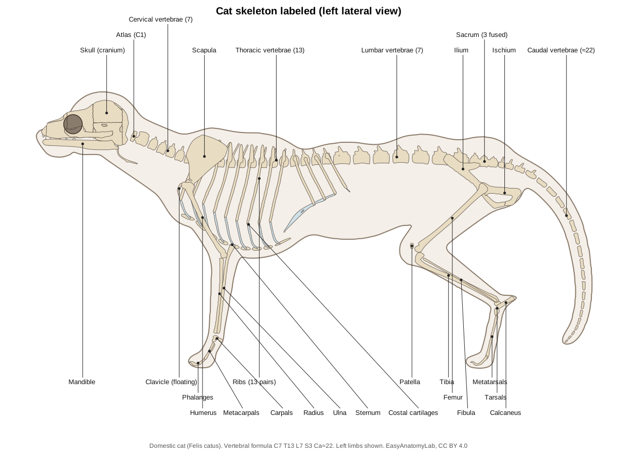
A useful way to read a cat skeleton is by the joints. The forelimb has the shoulder (scapula–humerus), elbow (humerus–radius and ulna, with the olecranon as the point of the elbow), carpus or wrist, and the toe joints. The hindlimb has the hip (femur in the acetabulum), the stifle or knee (femur–tibia, with the patella in front), the hock or ankle (tibia–talus, with the calcaneus forming the heel) and the toe joints. Because the cat is digitigrade (it walks on its toes), what looks like a backward-bending "knee" on the hindleg is really the hock, the cat's ankle.
How many bones does a cat have?
A domestic cat has roughly 230 to 250 bones. There is no single correct number, because the total depends on what you count. The biggest variable is the tail: Reighard and Jennings, whose 1901 Anatomy of the Cat is still the standard reference for lab cats, counted 22 or 23 caudal vertebrae, while other sources give ranges from about 18 to 24. The rest of the vertebral formula is fixed: 7 cervical, 13 thoracic, 7 lumbar and 3 sacral vertebrae, the sacral ones fused into a single sacrum. (Reighard, J. & Jennings, H. S. (1901). Anatomy of the Cat. New York: Henry Holt (Cornell University Library copy, Internet Archive).)
If you add up a typical long-tailed adult the way most vet-anatomy courses do, you get about 242: 50 vertebrae (counting the sacrum as one bone and 22 tail vertebrae), 26 ribs, 8 sternal segments, 62 bones in the two forelimbs (each with a clavicle, scapula, humerus, radius, ulna, 7 carpals, 5 metacarpals and 14 phalanges), 58 in the two hindlimbs (each with a hip bone, femur, patella, tibia, fibula, 7 tarsals, 4 metatarsals plus the remnant of the first, and 12 phalanges), about 29 bones in the skull (the exact figure depends on whether paired and fused bones are counted separately) and 9 in the hyoid apparatus. The six tiny ear ossicles, the os penis of the male and the many small sesamoid bones behind the joints of the paws push the figure higher if you include them. (Reighard, J. & Jennings, H. S. (1901). Anatomy of the Cat. New York: Henry Holt (Cornell University Library copy, Internet Archive).)
Other things change the count in a real cat. Kittens have more separate pieces than adults: the occipital bone starts as four parts, the scapholunar bone of the wrist as three, and each hip bone as three (ilium, ischium and pubis). Polydactyl cats have extra toes, and tailless or short-tailed breeds such as the Manx have fewer caudal vertebrae. So when a quiz asks for one number, '230–250, about 244 in a typical long-tailed cat' is a fair answer; the important point is that the extra bones compared with a human (206) come mostly from the tail and the extra thoracic and lumbar vertebrae. (Reighard, J. & Jennings, H. S. (1901). Anatomy of the Cat. New York: Henry Holt (Cornell University Library copy, Internet Archive).)
Cat skeleton labeled: a region-by-region walk-through
The quickest way to label any cat skeleton diagram is to split it into six regions and name the bones of each from head to tail and from the body out to the toes. This is the order most vet-anatomy and zoology lab practicals follow.
- Head. Skull (cranium plus face), with the large orbit in the middle of each side, the zygomatic arch below it and the tympanic bulla underneath at the back. Below the skull is the mandible, and in the throat the small hyoid apparatus.
- Neck. Seven cervical vertebrae: the ring-shaped atlas with broad wings, the axis with its tall crest, and five more. They curve down from the back of the skull to the top of the chest.
- Chest (thorax). Thirteen thoracic vertebrae with tall spines over the shoulders, thirteen pairs of ribs, their costal cartilages and the sternum along the floor of the chest. The scapula lies flat on the side of the rib cage, with the tiny clavicle in front of the shoulder joint.
- Lower back and pelvis. Seven long lumbar vertebrae, the sacrum (three fused vertebrae) and the two hip bones, each made of the ilium, ischium and pubis meeting at the acetabulum.
- Tail. About 22 caudal vertebrae, getting smaller towards the tip.
- Limbs. Forelimb: scapula, humerus, radius and ulna, carpals, metacarpals and phalanges. Hindlimb: femur, patella, tibia and fibula, tarsals (with the calcaneus as the heel), metatarsals and phalanges.
Three things trip students up. First, the cat stands on its toes, so the joint that bends backwards halfway up the hindleg is the hock (ankle), not the knee; the knee is higher up, tucked against the belly. Second, the elbow sits close to the bottom of the chest, and the point of the elbow is the olecranon of the ulna. Third, the scapula is not attached to the spine by any bone, so in a mounted skeleton it is held on with wire.
How many bones: breakdown table
This table adds up the bones of a typical adult long-tailed cat, counting each fused bone (sacrum, hip bone) as one. Change the tail count and the total changes with it.
| Group | Bones | Count |
|---|---|---|
| Skull | Cranial and facial bones, mandible | ≈ 29 |
| Ear ossicles | Malleus, incus, stapes × 2 | 6 |
| Hyoid apparatus | Body + paired horns | 9 |
| Vertebrae | 7 cervical + 13 thoracic + 7 lumbar + sacrum (1) + ≈ 22 caudal | ≈ 50 |
| Ribs | 13 pairs | 26 |
| Sternum | Manubrium, body segments, xiphoid | 8 |
| Forelimbs | 2 × (clavicle, scapula, humerus, radius, ulna, 7 carpals, 5 metacarpals, 14 phalanges) | 62 |
| Hindlimbs | 2 × (hip bone, femur, patella, tibia, fibula, 7 tarsals, 5 metatarsals, 12 phalanges) | 58 |
| Total | Without sesamoids or os penis | ≈ 248 (≈ 242 without the ossicles) |
Add or remove one tail vertebra and the total moves by one; a Manx cat with no tail can have about 20 fewer bones than this. The small sesamoid bones behind the toe joints and the male's os penis add more. That is why books and websites give anything from about 230 to about 250: none of them is wrong, they simply count differently.
Cat skull and head skeleton
The cat skull is short and rounded, with a large braincase, huge orbits (eye sockets) and a short face. Its widest part is across the zygomatic arches, which flare out to make room for the powerful jaw-closing muscles. The orbit is open behind: the postorbital processes of the frontal and zygomatic bones point towards each other but are joined only by a ligament.
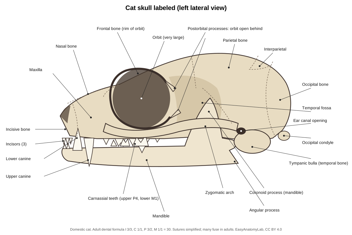
From front to back, the bones a lab expects you to name are the incisive bone (premaxilla, holding the upper incisors), the nasal bones on the bridge of the nose, the maxilla (upper canine and cheek teeth), the frontal bones between the eyes, the parietal bones over the brain, the small interparietal, and the occipital bone around the foramen magnum, whose two condyles rest on the atlas. On the side and underside are the temporal bone, with the large rounded tympanic bulla that houses the middle ear, and the zygomatic arch. The mandible has two halves joined at the chin by a fibrous, unfused joint.
Adult cats have 30 teeth: on each side, 3 incisors, 1 canine, 3 premolars and 1 molar above, and 3 incisors, 1 canine, 2 premolars and 1 molar below. Kittens have 26 milk teeth, which start to appear at about four weeks of age and are replaced by the permanent set by about six months. The largest cheek teeth, the upper fourth premolar and the lower first molar, are the carnassials: they slide past each other like scissor blades to slice meat. Cats have no flat grinding molars at all.
A small chain of bones, the hyoid apparatus, hangs from the skull near the tympanic bulla and slings the tongue and larynx. Its long cranial horns and short caudal horns are easy to lose when a skull is cleaned, so many museum cat skeletons lack them.
Cat spine: a flexible backbone
The cat's vertebral formula is C7 T13 L7 S3 Ca≈22. The neck has the usual seven mammalian vertebrae. The atlas (C1) is a ring with broad wings that you can feel behind the ears; the axis (C2) has a tall blade-like spinous process and a peg, the dens, around which the head turns.
Each of the 13 thoracic vertebrae carries a pair of ribs. Their spinous processes are tallest over the shoulders and slope backwards until the tenth, the anticlinal vertebra, which stands upright; those behind it slope forwards. This change of direction marks the point where the pull of the front and back muscles meets, and it is a landmark on radiographs.
The seven lumbar vertebrae are long and heavy, with long transverse processes that point forwards and downwards and anchor the big back muscles. When a cat gallops, the lumbar spine flexes and extends with each stride, swinging the hindlimbs further forward and back and lengthening the stride. A 2026 study found that the cat's thoracic spine is also unusually free to twist, much more so than its lumbar spine, which helps in the mid-air righting reflex. The three sacral vertebrae are fused into one sacrum that joins the pelvis.
Cat tail skeleton
A cat's tail contains about 22 or 23 caudal vertebrae. The first few look like small versions of the lumbar vertebrae, with arches over the spinal cord, short transverse processes and small haemal processes below. Further back the arches disappear and each vertebra becomes a simple rod with swollen ends, getting smaller towards the tip. The spinal cord itself ends well before the tail; only nerve roots and the caudal artery run on into it.
The tail is a balancing pole: a cat on a narrow fence swings it to the opposite side to keep its centre of mass over its feet. It is also a signal: held upright with a slight hook at the tip, it means a friendly greeting. Because the tail has so many bones, injuries such as a tail pulled or caught in a door can stretch the nerves at its base and affect bladder and bowel control, which is why vets take tail injuries seriously. The Manx cat carries a dominant gene that shortens the tail; fully tailless Manx cats can have spinal and sacral defects.
Rib cage and sternum
The cat has 13 pairs of ribs. The first nine pairs are true (sternal) ribs that reach the sternum on their own costal cartilages. Pairs 10 to 12 are false ribs: their cartilages unite with one another and usually with the ninth, forming the costal arch. The cartilages of the 13th pair end free (floating ribs). The sternum is a rod of separate bony segments (sternebrae): the manubrium in front, the body, and the xiphoid process with a broad cartilage plate at the back. The rib cage is narrow and deep, so the forelimbs can sit close together under the chest.
Floating clavicle and shoulder
The cat's clavicle is a slender, curved rod about 2–3 cm long that lies in front of the shoulder joint, buried where the clavotrapezius muscle meets the clavodeltoid. It has no bony joint with the sternum or with the scapula; Reighard and Jennings describe it as "imbedded in the muscles of the shoulder and connected by fibrous tissue to the apex of the acromion process". Because of this the whole shoulder blade can slide over the rib cage, attached only by muscles such as the serratus ventralis and rhomboideus.
A free-floating shoulder has three benefits. The scapula swings with each stride and so lengthens it; the muscles act as shock absorbers when the cat lands from a jump; and the shoulders can be pulled close together, so a cat can fit through any gap its head fits through. Find the clavicle in the model with the other systems hidden: it is the small, separate bone in front of each shoulder. It often falls off a cleaned skeleton and is missing from many museum mounts.
Forelimb bones
The scapula is broad and flat with a tall spine that ends in the acromion and a backward hook, the metacromion. The humerus runs down and back to the elbow. Just above its medial epicondyle is a hole, the supracondylar foramen, through which the median nerve and brachial artery pass; dogs do not have it, and vets fixing a broken cat humerus must avoid it.
The forearm has two separate bones. The ulna forms the point of the elbow (olecranon); the radius carries most of the weight and can rotate against the ulna, so a cat can turn its paw palm-up to scoop, bat or hold prey, far more than a dog can. The wrist has seven carpal bonesin two rows; the scapholunar is the fused scaphoid and lunate, and the accessory carpal (pisiform) sticks out behind the wrist, under a small carpal pad.
Cat paw skeleton and retractable claws
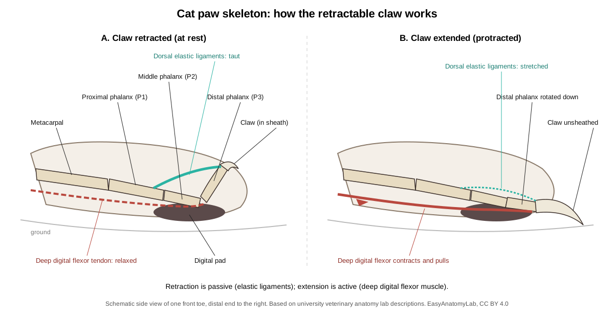
Each front paw has five toes: four weight-bearing toes and the dewclaw (digit I) on the inside, which has only two phalanges and does not touch the ground. Each of the other toes has three phalanges: proximal, middle and distal. The hind paws have four toes; the first metatarsal is only a tiny remnant. A normal cat therefore has 18 toes, and polydactyl cats have more.
Bones of the cat paw
| Part | Forepaw (front) | Hindpaw (back) |
|---|---|---|
| Wrist / ankle | 7 carpals in two rows: scapholunar, cuneiform, pisiform (accessory); trapezium, trapezoid, magnum, unciform | 7 tarsals: calcaneus, talus, central (navicular), cuboid and three cuneiforms |
| Palm / sole | 5 metacarpals (I short) | 4 metatarsals (II–V) + a tiny remnant of I |
| Toes | 5: dewclaw (2 phalanges) + 4 toes with 3 phalanges each = 14 | 4 toes with 3 phalanges each = 12 |
| Claws | 5, retractile | 4, retractile |
| Pads | 4 digital, 1 metacarpal, 1 carpal pad | 4 digital, 1 metatarsal pad |
Each toe is a chain of three joints. The joint between the metacarpal (or metatarsal) and the proximal phalanx carries the weight and sits over the big palm or sole pad. The middle phalanx is short and slightly twisted, so that the retracted distal phalanx can tuck up beside it rather than on top of it. Small sesamoid bones sit behind the main toe joints, in the tendons of the flexor muscles, to protect the tendons and improve their leverage.
The claw grows over the hooked ungual process of the distal phalanx. At rest, elastic ligaments on the top of each toe pull the distal phalanx up and back, so that it sits beside the middle phalanx and the claw is held in a fold of skin, off the ground. This is why cats walk silently and keep their claws sharp. To extend the claws, the cat contracts the deep digital flexor muscle of the forearm; its tendon, attached under the distal phalanx, rotates it down and forwards, stretching the elastic ligaments. When the muscle relaxes, the ligaments pull the claw back again with no effort. University veterinary anatomy labs describe the same arrangement in the dog, but the dog's ligaments are weaker and its claws stay out.
Declawing (onychectomy) removes the whole distal phalanx with the claw. It is an amputation of the last bone of each toe, which is why many veterinary associations oppose it except for medical reasons.
Pelvis and hindlimb bones
Each hip bone (os coxae) is made of three bones that fuse in the young cat: the long, narrow ilium in front, which joins the sacrum; the ischium behind, ending in the ischial tuberosity where the hamstrings attach; and the pubis below. All three meet in the acetabulum, the socket for the head of the femur, and the ischium and pubis surround a large opening, the obturator foramen.
The femur is long and straight. The patella sits in the quadriceps tendon at the front of the knee. The lower leg has a strong tibia and a thin fibula beside it. The ankle has seven tarsal bones, including the talus, which forms the hinge with the tibia, and the calcaneus, whose long backward projection is the point of the hock. The calf muscles pull on it through the calcaneal (Achilles) tendon. The long metatarsals and the hock held high off the ground make the hind foot a long lever: a cat can jump several times its own height.
Righting reflex: how a cat lands on its feet
The righting reflex is the automatic twist a falling cat makes to turn its feet towards the ground. The balance organs of the inner ear and the eyes detect which way is up, and the cat turns the front half of its body first, then the back half. Because it has nothing to push against in the air, it does this by changing its shape: bending at the middle and rotating the two halves against each other, so that total angular momentum stays zero. Kane and Scher modelled this in 1969 as two linked cylinders that bend but do not twist, a model funded by NASA because the same problem faces astronauts turning in free fall. (Kane, T. R. & Scher, M. P. (1969). A dynamical explanation of the falling cat phenomenon. NASA Technical Reports Server record.)
The skeleton makes the trick possible. A 2026 study of cat spines measured the thoracic region (the chest part of the backbone) as far more flexible in twisting than the lumbar region: it had a 'neutral zone' of about 47 degrees in which it turned almost freely, while the lumbar spine had none. Filming live cats showed the front of the trunk finishing its rotation before the back, which fits a flexible thorax and a stiffer lower back. The floating clavicle helps too, because the shoulders are not braced against the sternum. (Higurashi, Y. et al. (2026). Torsional flexibility of the thoracic spine is superior to that of the lumbar spine in cats. Anatomical Record (Europe PMC).)
Landing on the feet does not make falls safe. In the classic study of 'high-rise syndrome', vets at the Animal Medical Center in New York treated 132 cats that had fallen from buildings in five months. Ninety percent had chest injuries (mostly bruised lungs and air around the lungs), 57% facial injuries, 39% broken limbs and 17% a fractured hard palate, usually from hitting the chin. Ninety percent of the treated cats survived. Later reviews agree that limb fractures, facial injuries and chest trauma are the typical pattern, so screens on windows and balconies are the only real protection. (Whitney, W. O. & Mehlhaff, C. J. (1987). High-rise syndrome in cats. JAVMA 191, 1399–1403 (Europe PMC abstract).)
Cat skeleton front view and from above
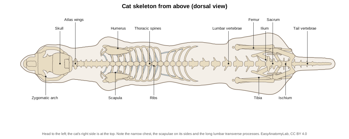
Seen from above, a cat skeleton is long and narrow. The rib cage is deep but slim, so the shoulder blades lie almost vertically against its sides, and the forelimbs hang close together under the chest. The lumbar vertebrae have long transverse processes that stick out sideways like oars, the ilia lie on either side of the sacrum, and the widest point of the whole skeleton is the skull at the zygomatic arches.
From the front you see the round braincase and the two large orbits that face forwards, giving the cat overlapping (binocular) vision for judging a pounce. Below them are the short muzzle and the long upper canines. The clavicles sit in front of the shoulder joints, and the narrow sternum lies between the forelimbs. A narrow chest lets a cat walk with its paws placed almost in one line, which is how it balances along the top of a fence.
The clavicle and the "cats fit through any gap" myth
A common saying is that a cat can squeeze through any gap its head fits through, because it has no collarbone. Both halves need care. Cats do have a clavicle, but it is small and does not link the shoulder to the sternum, so the shoulders can be drawn close together and the narrow chest can follow. That really does let cats pass through surprisingly small openings.
But the head is not a perfect gauge. The rib cage cannot shrink, the pelvis is a rigid ring of bone, and a fat cat carries a lot of soft tissue around its belly. A cat that gets its head through a gap can still get stuck at the chest or hips, and cats are regularly rescued from cat flaps, fences and pipes. Treat the saying as a rule of thumb, not anatomy.
Cat vs human skeleton
Cat and human skeletons have the same basic plan, because both are mammals, and many bones have the same names. The differences come from walking on four legs and on the toes.
| Feature | Cat | Human |
|---|---|---|
| Total bones | About 230–250 | 206 |
| Vertebrae | C7 T13 L7 S3 + ≈ 22 caudal | C7 T12 L5 S5 (fused) + 3–5 coccygeal |
| Ribs | 13 pairs (9 true, 3 false, 1 floating) | 12 pairs (7 true, 3 false, 2 floating) |
| Clavicle | Tiny, floating in muscle | Large, joins sternum and scapula |
| Stance | Digitigrade: walks on the toes | Plantigrade: walks on the whole foot |
| Hand / forepaw | 5 digits, thumb reduced to a dewclaw | 5 digits with an opposable thumb |
| Foot / hindpaw | 4 digits; first metatarsal a remnant | 5 toes |
| Skull | Large orbits, short face, open orbit behind | Huge braincase, flat face, closed orbit |
| Teeth | 30, blade-like carnassials | 32, flat grinding molars |
| Pelvis | Long and narrow, parallel to the spine | Short and bowl-shaped, carries the organs |
| Tail | Long, about 22 vertebrae | None (fused coccyx) |
The human count of 206 is for an adult; a newborn baby has nearly 300 separate pieces that fuse as it grows, just as a kitten has more separate bones than an adult cat.
Cat vs dog skeleton
Cats and dogs are both carnivores and share the same basic skeleton and the same vertebral formula, but the differences match their lifestyles: the cat is an ambush hunter and climber, the dog an endurance runner.
| Feature | Cat | Dog |
|---|---|---|
| Vertebrae | C7 T13 L7 S3, tail about 22 | C7 T13 L7 S3, tail about 20–23 (varies by breed) |
| Clavicle | Small floating bone in the shoulder muscles | A tiny remnant or none |
| Claws | Retractable, held up by elastic ligaments | Not retractable; always touch the ground |
| Skull | Short, rounded, large orbits; little variation between breeds except flat-faced breeds | Very variable, from short (pug) to long (greyhound) |
| Teeth | 30 (adult), 26 (kitten) | 42 (adult), 28 (puppy) |
| Humerus | Supracondylar foramen for the median nerve and brachial artery | No supracondylar foramen |
| Forearm | Radius rotates freely: paw can turn palm-up | Limited rotation: paws stay facing down |
| Dewclaws | On the forepaws; none on the hindpaws | On the forepaws; on the hindpaws only in some dogs and breeds |
Sources: Reighard and Jennings for the cat skeleton; the AO Foundation veterinary surgery reference for the humerus; Cornell Feline Health Center and the Veterinary Information Network for the dental formulas.
Sabre-toothed cat skeleton
The best-known sabre-toothed cat is Smilodon fatalis. The La Brea Tar Pits in Los Angeles have yielded bones of more than 2,000 individuals. It was about five feet (1.5 m) long and three feet tall and weighed roughly 350 to 620 pounds (160–280 kg), with upper canines that were long, flattened and serrated 'like steak knives'. Their shape made them easier to break than the canines of living big cats, and few teeth at La Brea were broken during life, so scientists think Smilodon used them to puncture prey rather than to grab and hold it. It went extinct about 11,000 years ago, at the end of the last ice age. (La Brea Tar Pits and Museum: Here, kitty, kitty (Smilodon fatalis).)
Smilodon was a member of the cat family (Felidae) but not an ancestor of the house cat. It belonged to an extinct branch, the sabre-toothed cats (subfamily Machairodontinae), while all living cats belong to the two surviving branches: Felinae, which includes the domestic cat, and Pantherinae, the roaring big cats. When you compare a Smilodon skeleton with a house cat's, look first at the skull: the huge upper canines and a jaw that could open far wider than any living cat's to clear them.
Articulated vs real cat skeletons
A cat skeleton you see in a museum or classroom is either a real skeleton that has been cleaned and articulated (joined together with wire, rods and glue), or a cast or plastic replica. In a real articulated skeleton the small pieces that are held only by soft tissue are often lost or wrongly placed during cleaning: the clavicles, the hyoid bones, the sesamoids, the os penis, the last tail vertebrae and sometimes the patella. The scapulae have to be wired on, because no bone holds them, and the pose is set by the preparator, so the angles of the joints may not match a living cat.
Replicas are cheaper and sturdier but are often simplified: tails with the wrong number of vertebrae, paws moulded as one piece, and no clavicle at all. If you are counting bones for a lab, check a real skeleton or a reference such as Reighard and Jennings rather than a model. The 3D cat on this page is also simplified (no sesamoids or ear ossicles, simplified joint surfaces), but it includes the floating clavicle and the full count of vertebrae and toe bones.
Cat skeleton structure: every bone in the model
This is the text version of the 3D model: every bone and bone group, grouped by system, with what it does. Click the same names in the viewer to find them.
Skeletal
- Incisive bone (Premaxilla)
- The paired bone at the very front of the upper jaw that holds the upper incisor teeth. Carries the six upper incisors and forms the front of the hard palate and nasal aperture.
- Nasal bones
- Two narrow bones that form the roof (bridge) of the nose. Roof over the nasal cavity, which holds the scroll-like turbinate bones that warm air and carry the smell receptors.
- Maxilla (Upper jaw bone)
- The large paired bone of the upper jaw and side of the face. Holds the upper canine and cheek teeth, forms most of the hard palate, and gives origin to whisker-pad and lip muscles.
- Zygomatic arch (Cheek bone (zygomatic bone + zygomatic process of the temporal bone))
- A strong, wide bony arch on each side of the skull, below the orbit. Anchors the masseter muscle and guards the eye from below. Its wide flare makes room for large jaw muscles, which give cats a strong bite at the canines.
- Frontal bones
- The paired bones of the forehead, between and above the orbits. Forms the inner wall and roof of each orbit and the front of the braincase; contains an air space (frontal sinus).
- Parietal bones
- The paired bones forming most of the rounded roof and sides of the braincase. Protect the brain and give origin to the temporalis muscle.
- Interparietal bone
- A small single bone in the midline between the two parietal bones and the occipital bone. Part of the braincase roof. In adult cats it fuses with its neighbours and is hard to see.
- Occipital bone
- The bone at the back and base of the skull that surrounds the foramen magnum. Its two occipital condyles articulate with the atlas (first vertebra), and the spinal cord passes through the foramen magnum.
- Temporal bone and tympanic bulla (Squamous, petrous and tympanic parts)
- The bone on the side and underside of the braincase that houses the ear, including the large rounded tympanic bulla. Holds the middle and inner ear, forms the socket for the jaw joint and carries the back root of the zygomatic arch.
- Mandible (Lower jaw (two halves joined at the chin))
- The lower jaw: two halves joined at the chin by a fibrous joint (mandibular symphysis). Holds the lower teeth and gives attachment to the masseter, temporalis and digastric muscles.
- Incisors
- Twelve tiny front teeth: three on each side, top and bottom. Nibble, groom fur and pull meat from bones.
- Canine teeth (Fangs)
- Four long, conical, slightly curved fangs. Grip and kill prey. Domestic cats usually kill small prey with a bite to the neck, driving the canines between the vertebrae.
- Premolars and molars (carnassials) (Cheek teeth)
- Blade-like cheek teeth: three premolars and one molar in each upper jaw, two premolars and one molar in each lower jaw. Slice meat like scissors. The largest pair, the carnassials (upper fourth premolar and lower first molar), shear past each other.
- Hyoid apparatus (Hyoid bone)
- A chain of small bones that slings the tongue and larynx from the skull. Supports the tongue and larynx and gives attachment to swallowing muscles.
- Atlas (C1) (First cervical vertebra)
- The ring-shaped first neck vertebra with broad, flat wings. The atlanto-occipital joint lets the cat nod its head up and down.
- Axis (C2) (Second cervical vertebra, epistropheus)
- The longest neck vertebra, with a tall blade-like spinous process and a peg (dens) that projects into the atlas. The atlas rotates around the dens, letting the cat turn its head from side to side.
- Cervical vertebrae C3–C7 (Neck vertebrae)
- The five remaining neck vertebrae; with the atlas and axis there are seven, as in almost all mammals. Support and move the neck. Their transverse processes are flattened and pierced by the transverse foramina for the vertebral artery.
- Thoracic vertebrae (13)
- Thirteen vertebrae, each bearing a pair of ribs; humans have twelve. Form the roof of the rib cage. The tall spinous processes of the first few form the withers and anchor shoulder and back muscles.
- Lumbar vertebrae (7)
- Seven large vertebrae of the lower back, with long transverse processes; humans have five. Carry the weight of the abdomen and anchor the powerful back and abdominal muscles. Flexing and extending this long lumbar region lengthens the cat's stride when it gallops or springs.
- Sacrum
- Three sacral vertebrae fused into a single bone. Joins the spine to the pelvis at the sacroiliac joints, so thrust from the hindlimbs reaches the body.
- Caudal vertebrae (tail) (Coccygeal vertebrae)
- The tail vertebrae: about 22 or 23 in a typical long-tailed cat, getting smaller and simpler towards the tip. Support the tail, which the cat uses for balance on narrow perches and for signalling (an upright tail is a friendly greeting).
- Ribs (13 pairs) (Costae)
- Thirteen pairs of curved bones forming the sides of the chest. Protect the heart and lungs and move during breathing.
- Costal cartilages
- Bars of flexible cartilage that continue the ribs ventrally. Let the chest wall flex during breathing and when the cat squeezes through gaps.
- Sternum (Breastbone: manubrium, body (sternebrae), xiphoid process)
- A rod of bony segments (sternebrae) along the floor of the chest. Anchors the costal cartilages and the pectoral muscles.
- Scapula (Shoulder blade)
- A broad, flat, triangular bone lying on the side of the chest, with a tall spine on its outer surface. The shoulder is attached to the body only by muscles, so the scapula slides freely, lengthening the stride and absorbing landing shock.
- Clavicle (floating collarbone) (Collarbone)
- A small, thin, curved rod of bone that forms no joint with any other bone. Gives attachment to the clavotrapezius and clavodeltoid muscles. Because it does not link the shoulder to the sternum, the forelimbs can move closer together and the shoulders can squeeze through narrow gaps.
- Humerus (Upper arm bone)
- The long bone of the upper arm, from the shoulder to the elbow. Lever for the shoulder and elbow muscles.
- Radius
- The forearm bone that carries most of the weight to the wrist. Its round head turns against the ulna, so cats can rotate the forearm and turn the paw over (supination), unlike dogs, which can do this only a little.
- Ulna
- The forearm bone that forms the point of the elbow. Its olecranon process is the lever of the triceps, which straightens the elbow.
- Carpal bones (7) (Wrist bones)
- Seven small bones in two rows: three in the proximal row and four in the distal row. Allow the wrist to flex and give spring to each step.
- Metacarpals (5) (Palm bones)
- Five bones of the palm; the first (for the dewclaw) is short. Lengthen the limb and act as springs in the digitigrade stance.
- Phalanges of the forepaw (Toe bones (proximal, middle and distal phalanges))
- Each of the four main front toes has three phalanges: proximal, middle and distal. The distal phalanx carries the claw. At rest it is pulled up beside the middle phalanx, keeping the claw off the ground.
- Dewclaw (digit I) (Pollex, first digit)
- The thumb-like first toe on the inside of each forepaw, with only two phalanges. Grips prey and helps when climbing.
- Ilium
- The long, narrow, blade-like front part of the hip bone (os coxae). Joins the hip bone to the sacrum (sacroiliac joint) and anchors the gluteal, sartorius and tensor fasciae latae muscles.
- Ischium
- The caudal part of the hip bone, ending in the ischial tuberosity. The ischial tuberosity anchors the hamstring muscles (biceps femoris, semitendinosus, semimembranosus).
- Pubis
- The ventral part of the hip bone that meets its partner at the pelvic symphysis. Closes the pelvic ring ventrally and anchors the gracilis and adductor muscles.
- Femur (Thigh bone)
- The long, straight thigh bone, from the hip to the knee (stifle). Lever for the powerful thigh muscles that drive jumping.
- Patella (Kneecap)
- A small sesamoid bone in the tendon of the quadriceps. Protects the tendon and increases the leverage of the quadriceps that straighten the knee.
- Tibia (Shin bone)
- The main bone of the lower leg (crus), slightly longer than the femur. Carries weight to the foot; its crest anchors the sartorius, gracilis and hamstrings.
- Fibula
- A thin bone lying alongside the tibia. Anchors lower-leg muscles; its lower end (lateral malleolus) steadies the ankle.
- Calcaneus (Heel bone, os calcis)
- The longest tarsal bone, projecting backwards to form the point of the hock. Lever for the calf muscles: the calcaneal (Achilles) tendon pulls on it to extend the hock and push the cat off the ground.
- Other tarsal bones (Talus, central (navicular), cuboid and three cuneiforms)
- Six small ankle bones besides the calcaneus: seven tarsals in all. The talus forms the pulley-like hinge of the ankle with the tibia; the others transfer weight to the metatarsals.
- Metatarsals
- Four long bones of the sole (II–V), plus a tiny remnant of the first. Form the long lever of the hind foot used in jumping.
- Phalanges of the hindpaw
- Each of the four hind toes has three phalanges. As in the forepaw, the distal phalanx carries a retractile claw.
- Os penis (Baculum) · males only
- A tiny bone in the tip of the penis. Stiffens the tip of the penis.
Cat skeleton quiz with answer key
Test yourself. Click a question to reveal the answer.
What is the vertebral formula of the cat?
C7 T13 L7 S3 Ca≈22: seven cervical, thirteen thoracic, seven lumbar, three fused sacral and about 22 caudal vertebrae.
Which is the anticlinal vertebra, and how do you recognise it?
The tenth thoracic vertebra. Its spinous process stands upright; those in front slope backwards and those behind slope forwards.
How many pairs of ribs does a cat have, and how many are true ribs?
Thirteen pairs. The first nine are true (sternal) ribs; pairs 10–12 are false ribs and the 13th pair floats free.
Where is the cat's clavicle, and why is it called floating?
In the muscles in front of the shoulder joint, where the clavotrapezius meets the clavodeltoid. It has no bony joint with the sternum or scapula.
Which three bones make up each hip bone?
The ilium, ischium and pubis, which meet in the acetabulum.
What passes through the supracondylar foramen of the humerus?
The median nerve and the brachial artery.
Which bone forms the point of the hock?
The calcaneus (heel bone), pulled by the calcaneal (Achilles) tendon.
How many toes does a normal cat have, and how many phalanges does the dewclaw have?
Eighteen toes: five on each forepaw and four on each hindpaw. The dewclaw (digit I) has two phalanges.
What retracts the claws, and what extends them?
Dorsal elastic ligaments retract them passively; contraction of the deep digital flexor muscle extends them.
What is the adult dental formula of the cat?
I 3/3, C 1/1, P 3/2, M 1/1 on each side = 30 teeth. The carnassials are the upper fourth premolar and lower first molar.
Why is the cat's orbit described as 'open'?
The postorbital processes of the frontal and zygomatic bones do not meet, so there is no complete bony bar behind the eye; only a ligament closes the gap.
Why do published bone counts for cats range from about 230 to 250?
Mainly because the number of tail vertebrae varies, and because sources differ on counting fused bones, ear ossicles, sesamoids and the os penis.
From the skeleton to the whole cat
The same 3D cat has every muscle, organ, vessel and nerve that a cat dissection lab asks for. Switch them on with the system buttons above, or follow the step-by-step cat dissection guide, which uses this model as a virtual dissection.
Frequently asked questions
How many bones does a cat have?
About 230 to 250, roughly 244 in a typical long-tailed adult. The count varies mainly with the number of tail vertebrae (about 22 or 23, but 18 to 24 in different counts), with age (kittens have more separate pieces) and with whether small bones such as the ear ossicles, sesamoids and os penis are included. A human has 206.
Do cats have a collarbone?
Yes, but it is tiny and 'floating'. The cat's clavicle is a slender curved rod buried in the shoulder muscles; it is not joined to the sternum or the scapula by a bony joint. This lets the shoulders move freely and squeeze close together.
How many vertebrae does a cat have?
About 52: 7 cervical, 13 thoracic, 7 lumbar, 3 sacral (fused into the sacrum) and about 22 caudal vertebrae in the tail. Humans have 7 cervical, 12 thoracic and 5 lumbar.
How many bones are in a cat's tail?
Usually about 22 or 23 caudal vertebrae, according to Reighard and Jennings' Anatomy of the Cat; other sources give 18 to 24. Manx and other short-tailed cats have fewer, sometimes none. Only the first few tail vertebrae have arches and processes; the rest are small rods.
Why is a cat's spine so flexible?
The cat has long lumbar vertebrae with elastic discs between them, a thoracic spine that twists unusually easily, and shoulders held on by muscles rather than a rigid collarbone. Together these let it arch, twist in mid-air and lengthen its stride when it runs.
What bones are in a cat's paw?
Each forepaw has 7 carpal bones, 5 metacarpals and 14 phalanges (three in each of the four main toes and two in the dewclaw). Each hindpaw has 7 tarsal bones, 4 metatarsals plus a tiny remnant of the first, and 12 phalanges in four toes. The claw grows on the distal phalanx.
Are cat claws bones?
No. The claw itself is a sheath of keratin, like a fingernail, but it is wrapped around a hooked bony core, the ungual process of the distal phalanx. That is why declawing is an amputation of the last toe bone.
What is the difference between a cat and a dog skeleton?
The biggest differences are the clavicle (a small floating bone in the cat, a tiny remnant or none in the dog), retractable claws in the cat, a shorter, rounder skull with 30 teeth instead of 42, a supracondylar foramen in the cat's humerus, and forearms that rotate more freely in cats. The vertebral formula (7 cervical, 13 thoracic, 7 lumbar, 3 sacral) is the same.
References
- Reighard, J. & Jennings, H. S. (1901). Anatomy of the Cat. New York: Henry Holt (Cornell University Library copy, Internet Archive)
- Cornell Feline Health Center: When kitty needs a dentist
- Catanzaro, T. E. Appendix U: Dental scoring (feline and canine dental formulas), VIN
- AO Foundation VET surgery reference: anatomical differences between dogs and cats (humerus)
- Kane, T. R. & Scher, M. P. (1969). A dynamical explanation of the falling cat phenomenon. NASA Technical Reports Server record
- Higurashi, Y. et al. (2026). Torsional flexibility of the thoracic spine is superior to that of the lumbar spine in cats. Anatomical Record (Europe PMC)
- Whitney, W. O. & Mehlhaff, C. J. (1987). High-rise syndrome in cats. JAVMA 191, 1399–1403 (Europe PMC abstract)
- Oxley, J. & Montrose, T. (2016). High-rise syndrome in cats. Vet Times
- La Brea Tar Pits and Museum: Here, kitty, kitty (Smilodon fatalis)
More interactive anatomy models
- Cat dissectionGuided cat dissection: muscles, digestive organs, vessels and reproductive system.
- Human anatomy1,000+ bones, muscles, organs, vessels and nerves from real anatomical data.
- Octopus anatomyThree hearts, a ring-shaped brain, eight arms and a beak, in 3D.
- Squid anatomyPen, eight arms, two tentacles, giant axons and three hearts of the longfin squid.
- Squid dissectionStep-by-step guided squid dissection with every structure labeled.
- Fish anatomyExternal and internal anatomy of a bony fish: fins, gills, swim bladder, heart and brain.