

Before you start: materials and safety
You need a preserved cat, a tray, basic dissecting tools and protective equipment. A double-injected cat (arteries filled with red latex, veins with blue) makes the blood vessels far easier to follow; triple-injected cats also have yellow latex in the hepatic portal system.
Materials
- Preserved cat, ideally double-injected (arteries filled with red latex, veins with blue) or triple-injected (hepatic portal vein yellow)
- Large dissecting tray, or a tray with a rack, and a plastic bag or bin with a lid for storing the specimen between labs
- Scalpel, fine and blunt-tipped scissors, two pairs of forceps, a blunt probe and a seeker
- Bone cutters or heavy scissors (for the rib cage and pelvic symphysis)
- String or rubber bands to tie the limbs open, and dissecting pins
- Nitrile gloves (thicker than latex if your preservative is formalin-based), safety glasses or goggles, a lab coat or apron
- Paper towels, a spray bottle of water or wetting solution to stop the tissues drying, and a labelled bag for the skin
- Your lab manual or this page's diagrams, and a pencil for the worksheet
Safety
- Work in a well-ventilated room. Preserved cats are packed in fixative (formalin or a non-formaldehyde preservative); rinse the cat under running water before you start and keep the bag closed when you are not working.
- Wear gloves and eye protection at all times. Fixative can splash when you cut through the body wall or squeeze the bag.
- Cut away from your hands and your lab partner. Lift each layer with forceps before you cut it, so the blade never goes deeper than you can see.
- Separate muscles with a blunt probe or your fingers along the natural seams of connective tissue; use the scalpel only to cut skin and fascia.
- Never eat, drink or touch your face in the lab. Wash your hands when you finish, even if you wore gloves.
- Put tissue scraps in the waste container your teacher provides, never in the sink, and sharps in the sharps box.
Pre-lab questions
- Define cranial, caudal, dorsal, ventral, medial, lateral, proximal and distal for a four-legged animal.
- What does 'double-injected' mean, and which vessels are filled with red and with blue latex?
- Why do you separate muscles with a probe instead of cutting them?
- Which muscle sticks to the inside of the skin of the trunk and is removed with it?
Words you will need
A cat stands on four legs, so lab manuals use veterinary direction terms instead of "up" and "front". Cranial means towards the head and caudal towards the tail. Dorsal is the back and ventral the belly. Medial is towards the midline and lateral away from it. On a limb, proximal is nearer the body and distal further away. A muscle's origin is its fixed attachment and its insertion the one it moves. To reflect a muscle is to cut it across its belly and fold the two halves back to see what lies underneath. Fascia is the sheet of connective tissue that wraps muscles; the peritoneum is the thin, shiny lining of the abdominal cavity and its organs.
Cat dissection labeled diagrams
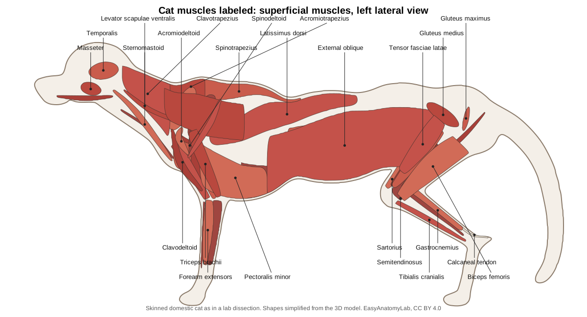
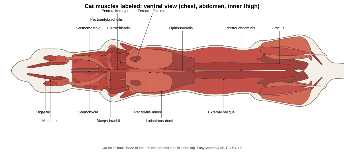
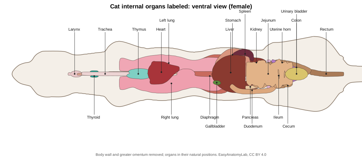
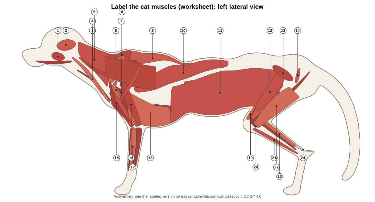
Use these diagrams to check your identifications. The lateral view shows the muscles you see on the side of a skinned cat; the ventral view shows the chest, abdomen and inner thigh with the cat on its back.
Cat dissection procedure
The order below follows standard cat dissection lab manuals. Each step matches a step of the 3D guide above, which hides the layers you would have removed and highlights the structures to find.
1. External anatomy and sexing the cat
- Rinse the cat and lay it on its back in the tray. Identify the head, neck, trunk (thorax and abdomen), tail and the four limbs.
- Find the pinnae, the eyes (look for the third eyelid in the inner corner), the vibrissae on the muzzle and above the eyes, and the nostrils on the hairless nose pad.
- Spread the toes of a forepaw: count five toes, including the dewclaw on the inside, and four on the hindpaw. Press on a toe pad to push the claw out of its sheath.
- Find the two rows of nipples on the chest and abdomen. Lift the tail and find the anus. Below it a female has a slit-like vulva; a male has the scrotum, with the prepuce of the penis just below it. Use the Female/Male switch on the model to compare.
Identify: Skin and fur, Pinna (ear flap), Eye, Vibrissae (whiskers), Paw pads, Claws, Dewclaw (digit I), Nipples (mammary papillae), Anus, Vulva, Scrotum.
2. Removing the skin
- With the cat on its back, lift the skin of the neck with forceps and make a small cut through the skin only, in the midventral line.
- Push a blunt scissor blade under the skin and cut forwards to the chin and backwards to the genital region, keeping the scissor tips pointing up.
- Cut around the neck, then down the inside of each limb to the wrist and ankle, and around the genitals and anus.
- Peel the skin back by separating it from the muscles with your fingers and a probe. A thin muscle, the cutaneous maximus, sticks to the inside of the skin over the trunk; remove it with the skin. The platysma does the same on the neck.
- Keep the skin in one piece: wrap the cat in it between labs to stop the muscles drying out. Clean away fat and fascia so each muscle's edges show.
Caution: On the neck the external jugular vein runs just under the skin. Cut shallowly so you do not destroy it.
Identify: Skin and fur, External jugular veins.
3. Superficial chest muscles (pectoral group)
- With the cat on its back, find the pectoantebrachialis: a narrow band about 1–2 cm wide from the manubrium to the forearm, the most superficial chest muscle.
- Under and caudal to it, the broad pectoralis major runs from the sternum to the humerus. Caudal to that is the larger, fan-shaped pectoralis minor.
- The most caudal band, the xiphihumeralis, starts at the xiphoid process and passes deep to the pectoralis minor to the humerus.
- Free the edges of each muscle with a probe. Cut and reflect (fold back) the pectoantebrachialis and pectoralis major only if your manual asks you to.
Identify: Pectoantebrachialis, Pectoralis major, Pectoralis minor, Xiphihumeralis, Sternomastoid, Sternohyoid.
4. Superficial back and shoulder muscles
- Turn the cat onto its belly. The trapezius group covers the neck and shoulders: the clavotrapezius from the back of the skull to the clavicle, the acromiotrapezius over the shoulder blades, and the triangular spinotrapezius behind it.
- Find the levator scapulae ventralis, a narrow ribbon that emerges between the clavotrapezius and the acromiotrapezius.
- Over the shoulder find the deltoid group: the clavodeltoid (continuing the clavotrapezius down the arm), the acromiodeltoid over the point of the shoulder, and the spinodeltoid behind it.
- The large latissimus dorsi covers the side of the chest behind the shoulder and runs to the humerus.
Identify: Clavotrapezius, Acromiotrapezius, Spinotrapezius, Levator scapulae ventralis, Clavodeltoid, Acromiodeltoid, Spinodeltoid, Latissimus dorsi.
5. Deep muscles of the shoulder and back
- Cut the acromiotrapezius and spinotrapezius across their middles and fold them back. (On the model, click a muscle with 'Focus selected' on to see it through the layers above.)
- On the scapula find the supraspinatus in front of the spine and the infraspinatus behind it. The thick teres major lies along the caudal border of the scapula.
- Lift the vertebral border of the scapula away from the body to see the rhomboideus attaching it to the spine, the splenius on the neck, and the fan-shaped serratus ventralis underneath.
- Reflect the pectoral muscles to see the long scalenus running forwards over the side of the chest to the neck, and the intercostal muscles between the ribs.
Identify: Supraspinatus, Infraspinatus, Teres major, Rhomboideus, Splenius, Serratus ventralis, Scalenus, Intercostal muscles, Scapula.
6. Muscles of the forelimb
- On the back of the arm, the large triceps brachii has a long head (from the scapula), a lateral head and a medial head; all insert on the olecranon.
- On the inner arm find the thin epitrochlearis over the triceps, and the biceps brachii on the front of the humerus.
- On the forearm, the very thin brachioradialis runs along the front. The extensors lie on the dorsal (front) side and the flexors on the palmar (back) side.
Identify: Triceps brachii, Biceps brachii, Epitrochlearis, Brachioradialis, Forearm extensors, Forearm flexors, Humerus.
7. Head and neck muscles and salivary glands
- On the side of the head find the thick masseter on the cheek and the temporalis above the zygomatic arch.
- Under the jaw find the digastric along each half of the mandible. Reflect it to see the thin, transverse mylohyoid forming the floor of the mouth.
- On the ventral neck the sternomastoid runs diagonally to the back of the skull and the narrow sternohyoid runs along the midline to the hyoid. Dorsal to the sternomastoid, the narrow cleidomastoid runs from the clavicle to the mastoid process under the clavotrapezius.
- Find the salivary glands: the parotid below the ear, the mandibular below it at the angle of the jaw, the small sublingual beside it and the molar gland near the corner of the mouth.
Identify: Masseter, Temporalis, Digastric, Mylohyoid, Sternomastoid, Cleidomastoid, Sternohyoid, Salivary glands.
8. Abdominal wall muscles
- Find the linea alba, the white line down the ventral midline. Either side of it lies the rectus abdominis.
- The external oblique is the outermost sheet over the side of the abdomen. Cut a small window through it, without going deeper, to see the internal oblique, whose fibres run the other way.
- Cut a smaller window through the internal oblique to see the transversus abdominis. The three sheets are very thin: work slowly.
Identify: External oblique, Internal oblique, Transversus abdominis, Rectus abdominis.
9. Lateral thigh and hip
- With the cat on its side, the biceps femoris covers most of the lateral thigh. The tensor fasciae latae is in front of it and the narrow semitendinosus behind it.
- Above them find the gluteus medius (the largest gluteal muscle in the cat) and the small gluteus maximus behind it.
- The slender caudofemoralis lies between the gluteus maximus and the biceps femoris; its thin tendon runs under the biceps to the patella.
- Cut and reflect the biceps femoris to find the vastus lateralis (part of the quadriceps), the thick white sciatic nerve and, beside the nerve, the thread-like tenuissimus muscle.
Identify: Biceps femoris, Tensor fasciae latae, Semitendinosus, Gluteus medius, Gluteus maximus, Caudofemoralis, Tenuissimus, Vastus lateralis.
10. Medial thigh and lower leg
- Turn the cat on its back. The medial thigh is covered by two broad, thin muscles: the sartorius in front and the gracilis behind.
- Cut and reflect them to find the semimembranosus, the adductor femoris, the thin adductor longus just in front of it and the rectus femoris. The femoral artery, vein and nerve run in the groove (femoral triangle) in front of the adductor longus.
- On the lower leg, the gastrocnemius forms the calf and ends in the calcaneal (Achilles) tendon on the heel. The soleus lies under its lateral head. The tibialis cranialis runs along the front of the tibia, with the extensor digitorum longus beside it and the fibularis (peroneus) muscles on the lateral side. The deep digital flexors lie against the back of the tibia under the calf muscles.
Identify: Sartorius, Gracilis, Semimembranosus, Adductor femoris, Adductor longus, Rectus femoris, Gastrocnemius, Soleus, Tibialis cranialis, Extensor digitorum longus, Fibularis (peroneus) muscles, Deep digital flexors of the hindlimb, Calcaneal tendon.
11. Opening the neck and thorax
- Separate the sternohyoids in the neck to expose the trachea, with the larynx at its cranial end and the two lobes of the thyroid gland on its sides.
- Cut through the chest muscles and the costal cartilages just to one side of the sternum, from the diaphragm to the first rib. Make cuts along the diaphragm on both sides and spread the rib cage open.
- Identify the lungs (right: cranial, middle, caudal and accessory lobes; left: cranial and caudal lobes), the heart in its pericardial sac, and the soft, pale thymus over the heart and trachea.
- Find the diaphragm, the dome of muscle behind the lungs, and the oesophagus dorsal to the trachea.
Caution: The cut edges of ribs are sharp. Cover them with paper towel when you work inside the thorax.
Identify: Trachea, Larynx, Thyroid gland, Thymus, Right lung, Left lung, Bronchi, Heart, Diaphragm, Oesophagus.
12. Heart and great vessels
- Open the pericardium. On the heart identify the right and left auricles (ear-like flaps of the atria) and the ventricles.
- Find the pulmonary trunk leaving the right ventricle (blue latex) and the aorta arching to the left (red latex).
- Follow the aortic arch: the first branch is the large brachiocephalic trunk, which gives both common carotid arteries and the right subclavian; the second is the left subclavian artery. There are only two branches, not three as in humans.
- Find the cranial vena cava entering the right atrium, formed by the two brachiocephalic veins, each of which receives an external jugular vein; the small internal jugular vein runs beside the common carotid artery and joins the external jugular near the first rib. Push the right lung aside to find the azygos vein joining the cranial vena cava.
- Follow each subclavian artery to the armpit (axillary artery) and down the arm (brachial artery).
Identify: Heart, Pulmonary trunk and arteries, Pulmonary veins, Aorta (ascending and aortic arch), Brachiocephalic trunk, Left subclavian artery, Common carotid arteries, Subclavian, axillary and brachial arteries, Cranial vena cava, Brachiocephalic veins, External jugular veins, Internal jugular veins, Azygos vein, Thoracic aorta.
13. Opening the abdomen: omentum, liver, stomach and spleen
- Cut along the linea alba from the diaphragm to the pubis, then make cuts sideways just behind the ribs and in front of the thighs. Fold the body-wall flaps back.
- The first thing you see is the greater omentum, a lacy, fatty apron over the intestines. Lift it forwards, or remove it if your teacher allows.
- Find the large, dark liver behind the diaphragm and count its lobes. Find the green gallbladder in a cleft of the right medial lobe.
- Lift the liver to see the stomach on the left, with the spleen along its greater curvature. The thin lesser omentum joins the stomach to the liver.
Identify: Greater omentum, Liver, Gallbladder, Stomach, Spleen, Lesser omentum, Diaphragm.
14. Following the digestive tract
- Feel the pyloric sphincter as a firm ring where the stomach meets the duodenum. Cut the stomach open lengthwise to see the rugae.
- Follow the U-shaped duodenum. The pancreas lies in its loop and along the stomach towards the spleen. Trace the common bile duct from the gallbladder to the duodenum.
- Lift the coils of the jejunum and ileum and hold them to the light to see the fan-shaped mesentery with its blood vessels.
- Find the ileocecal junction, the small blind cecum, then the colon running up the right side, across, and down the left side to the rectum in the pelvis.
Identify: Pyloric sphincter, Duodenum, Pancreas, Common bile duct, Jejunum, Ileum, Mesentery, Cecum, Colon, Rectum, Oesophagus.
15. Arteries and veins of the abdomen
- Push the intestines to the cat's left. The abdominal aorta (red) and caudal vena cava (blue) lie side by side on the dorsal body wall.
- From cranial to caudal, find the celiac trunk, the cranial mesenteric artery, the renal arteries and veins, and the small caudal mesenteric artery.
- Near the sacrum the aorta gives the two external iliac arteries and then the two internal iliac arteries; the cat has no common iliac arteries. The veins do form common iliac veins.
- Follow the external iliac artery and vein out to the thigh as the femoral artery and vein. If your cat is triple-injected, find the yellow hepatic portal vein entering the liver.
Identify: Abdominal aorta, Celiac trunk, Cranial mesenteric artery, Renal arteries, Renal veins, Caudal mesenteric artery, Caudal vena cava, Hepatic portal vein, External iliac and femoral arteries, Internal iliac arteries, Common iliac and femoral veins.
16. Urinary system and adrenal glands
- Find the kidneys against the dorsal body wall. They are retroperitoneal: peel the peritoneum off one to free it. Note the capsular veins on its surface.
- Find the small, pale adrenal gland medial to the cranial end of each kidney.
- Follow each ureter back to the urinary bladder in the caudal abdomen, and the urethra from the bladder into the pelvis.
- Cut one kidney lengthwise to see the cortex, medulla, renal pyramid and renal pelvis.
Identify: Kidneys, Adrenal glands, Ureters, Urinary bladder, Urethra.
17. Female reproductive system
- Switch the model to Female. Find the small, oval ovaries just caudal to the kidneys, each wrapped by a coiled uterine tube.
- Follow each long uterine horn back to where they join in the short uterine body, dorsal to the bladder.
- To follow the tract further, cut through the pelvic symphysis with bone cutters and spread the thighs. Find the vagina and the urogenital sinus, which opens at the vulva.
- Compare the mammary glands under the skin of the chest and abdomen.
Identify: Ovaries, Uterine tubes, Uterine horns, Uterine body and cervix, Vagina, Urogenital sinus, Mammary glands.
18. Male reproductive system
- Switch the model to Male. Cut open the scrotum to find the two testes, each with an epididymis along its border.
- Follow the ductus deferens in the spermatic cord through the inguinal canal into the abdomen, where it loops over the ureter.
- Cut through the pelvic symphysis. Find the prostate gland around the urethra behind the bladder, the two bulbourethral glands near the root of the penis, and the penis itself, which points backwards.
- Note what is missing: the cat has no seminal vesicles.
Identify: Scrotum, Testes, Epididymis, Ductus deferens and spermatic cord, Prostate gland, Bulbourethral glands, Penis, Os penis.
19. Nerves: brachial plexus and sciatic nerve
- On the medial side of the forelimb, clean the axilla to see the brachial plexus beside the axillary artery and vein.
- From cranial to caudal, find the musculocutaneous, radial, median and ulnar nerves. Follow the median nerve and brachial artery through the supracondylar foramen of the humerus.
- In the thigh, reflect the biceps femoris to find the sciatic nerve, the thickest nerve in the body.
- In the neck, find the vagus nerve bound to the sympathetic trunk beside the common carotid artery. In the thorax, find the thin phrenic nerves running over the heart to the diaphragm.
Identify: Brachial plexus, Musculocutaneous nerve, Radial nerve, Median nerve, Ulnar nerve, Sciatic nerve, Vagus nerve, Phrenic nerves, Spinal cord.
Cat muscles labeled: the muscles a lab asks for
Cat lab practicals usually test the superficial muscles of the chest, back, shoulder, arm, abdomen and thigh, plus a few deep ones. Several cat muscles are split into separate parts that form a single muscle in humans, and a few have no human equivalent at all. The table gives each muscle with its human counterpart.
| Cat muscle | Origin → insertion | Action | Human equivalent |
|---|---|---|---|
| Pectoantebrachialis | Manubrium → fascia of the forearm | Adducts forelimb | None |
| Pectoralis major | Cranial sternum → humerus | Adducts forelimb | Pectoralis major |
| Pectoralis minor | Body of sternum → proximal humerus | Adducts, retracts forelimb | Pectoralis minor (but larger than the major in the cat) |
| Xiphihumeralis | Xiphoid process → humerus | Adducts forelimb | None |
| Clavotrapezius | Lambdoidal crest, dorsal neck → clavicle | Draws shoulder forwards | Trapezius (cranial part) |
| Acromiotrapezius | Dorsal midline of neck and front thorax → acromion, scapular spine | Holds scapulae together | Trapezius (middle part) |
| Spinotrapezius | Thoracic spines → scapular spine | Pulls scapula back and up | Trapezius (caudal part) |
| Clavodeltoid | Clavicle → ulna | Flexes elbow | Deltoid (clavicular part) |
| Acromiodeltoid | Acromion → deltoid ridge of humerus | Flexes, rotates humerus | Deltoid (acromial part) |
| Spinodeltoid | Scapular spine → humerus | Flexes, rotates humerus | Deltoid (spinal part) |
| Latissimus dorsi | Thoracic and lumbar spines (fascia) → humerus | Pulls forelimb back | Latissimus dorsi |
| Levator scapulae ventralis | Occipital bone, atlas → metacromion | Pulls scapula forwards | None |
| Biceps brachii | Supraglenoid tubercle → radius | Flexes elbow | Biceps brachii (one head in the cat) |
| Triceps brachii | Scapula, humerus → olecranon | Extends elbow | Triceps brachii |
| Epitrochlearis | Latissimus dorsi → olecranon | Extends elbow | None |
| External oblique | Ribs, lumbodorsal fascia → linea alba, pubis | Compresses abdomen | External oblique |
| Rectus abdominis | Pubis → sternum, costal cartilages | Flexes trunk | Rectus abdominis |
| Sartorius | Ilium → patella, medial tibia | Adducts, rotates thigh; extends knee | Sartorius (broader in the cat) |
| Gracilis | Pelvic symphysis → medial tibia | Adducts thigh | Gracilis (much broader in the cat) |
| Biceps femoris | Ischial tuberosity → patella, tibia | Abducts thigh, flexes knee | Biceps femoris |
| Caudofemoralis | Caudal vertebrae → patella | Abducts thigh, extends knee | None |
| Gastrocnemius | Caudal femur → calcaneus | Extends hock (ankle) | Gastrocnemius |
Origins and insertions follow Reighard and Jennings (1901), with minor simplification. Many lab manuals use slightly different names, for example clavobrachialis for clavodeltoid, or tibialis anterior for tibialis cranialis. If your manual differs, use its names in the practical.
Cat digestive system
When you open the abdomen the first thing you see is the greater omentum, a lacy, fat-streaked apron hanging from the stomach over the intestines. Under it, the dark red-brown liver fills the space behind the diaphragm. It has five main lobes (left lateral, left medial, right medial, right lateral and caudate), and the green gallbladder sits in a cleft of the right medial lobe.
The J-shaped stomach lies on the left, behind the liver. Its outlet, the thick ring of the pyloric sphincter, opens into the duodenum, which makes a U-turn along the right side. The pale, lobulated pancreas lies in the loop of the duodenum and along the stomach towards the dark red, tongue-shaped spleen. The common bile duct and the pancreatic duct open together into the duodenum, about 3 cm from the pylorus. The lesser omentum is a thin sheet joining the liver to the stomach and duodenum.
The rest of the small intestine, the jejunum and ileum, is coiled in the ventral abdomen and hangs from the dorsal wall on the fan-shaped mesentery, which carries its blood vessels. The ileum opens into the large intestine at the ileocolic valve, next to a small blind pouch, the cecum. The cat has no appendix. The colon runs up the right side, across, and down the left side to the rectum and anus. Short intestines and a tiny cecum are typical of a strict carnivore, which needs little fermentation of plant food.
Cat respiratory system and heart
In the neck, the larynx (voice box, with the epiglottis at its front) sits at the top of the trachea, whose C-shaped rings are easy to feel. The two small lobes of the thyroid gland lie on its sides just behind the larynx. In the chest the trachea divides into two bronchi. The right lung has four lobes (cranial, middle, caudal and accessory) and the left lung two (a cranial lobe in two parts and a caudal lobe). In young cats a large, pale thymus covers the front of the heart.
The heart lies between the lungs, its apex pointing back, down and to the left. On its surface you can see the ear-like auricles of the two atria and the coronary vessels in the groove between the ventricles. The diaphragm separates the thorax from the abdomen; the thin phrenic nerves that control it run over the sides of the heart.
Cat veins and arteries
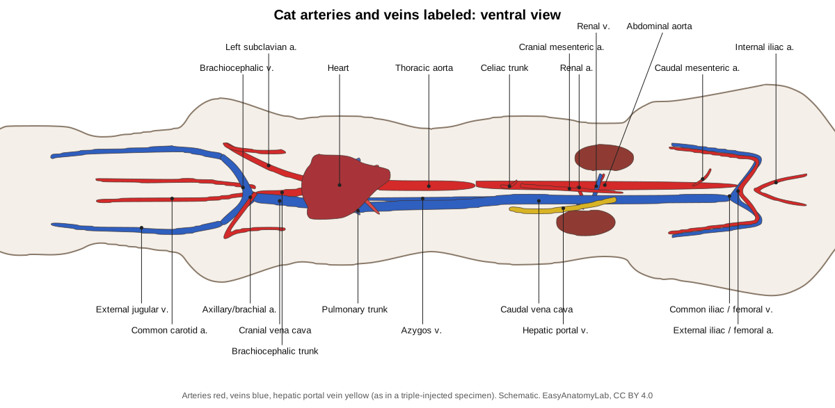
The cat's main blood vessels follow the human pattern with a few differences that lab practicals love to test. The most important: only two branches leave the aortic arch, and the cat has no common iliac arteries.
| Vessel | Cat | Human |
|---|---|---|
| Aortic arch branches | Brachiocephalic trunk, left subclavian (2) | Brachiocephalic, left common carotid, left subclavian (3) |
| Common carotid arteries | Both from the brachiocephalic trunk | Right from brachiocephalic, left from the arch |
| Jugular veins | External jugular much larger than internal | Internal jugular larger |
| Iliac arteries | Aorta gives external, then internal iliacs; no common iliacs | Aorta splits into two common iliacs |
| Iliac veins | Common iliac veins present | Common iliac veins present |
| Venae cavae | Cranial and caudal vena cava (precava, postcava) | Superior and inferior vena cava |
| Azygos vein | Right side, joins the cranial vena cava | Right side, joins the superior vena cava |
Follow the arteries from the heart: the aortic arch gives the brachiocephalic trunk (which divides into both common carotid arteries and the right subclavian) and the left subclavian. Each subclavian becomes the axillary artery in the armpit and the brachial artery in the arm, which passes with the median nerve through the supracondylar foramen of the humerus. The descending aorta gives intercostal arteries in the chest; in the abdomen it gives the celiac trunk (to the liver, stomach and spleen), the cranial mesenteric artery (small intestine), the paired adrenolumbar, renal and gonadal arteries, the caudal mesenteric artery (colon), and finally the external iliac arteries (which become the femoral arteries of the thighs) and the internal iliac arteries. A small median sacral artery continues into the tail.
Veins run with the arteries and are named the same way, with three exceptions to look for: the large external jugular vein just under the skin of the neck, the azygos vein on the right of the thoracic vertebrae, and the hepatic portal vein, which carries blood from the stomach, intestines, pancreas and spleen to the liver rather than to the heart.
Cat urinary system
The two bean-shaped kidneys lie against the dorsal body wall either side of the lumbar vertebrae, behind the peritoneum (retroperitoneal); the right one is a little further forward. Cat kidneys have conspicuous capsular veins running over their surface. A small, pale adrenal gland lies medial to the cranial end of each, separate from the kidney. Thin ureters run back to the urinary bladder, which empties through the urethra. Cut a kidney lengthwise to see the cortex, medulla, renal pyramid and renal pelvis.
Male vs female: cat reproductive system
Sex your cat before you start: a male has a scrotum below the anus; a female has a slit-like vulva.
- Female: small oval ovaries just behind the kidneys, coiled uterine tubes, and a Y-shaped (bicornuate) uterus with two long uterine horns and a short body above the bladder. The vagina joins the urethra in a shared urogenital sinus that opens at the vulva. Mammary glands lie in two rows under the skin. A pregnant cat shows swellings along the horns, one for each kitten.
- Male: testes in the scrotum, each with an epididymis; the ductus deferens runs in the spermatic cord through the inguinal canal and loops over the ureter to the urethra. The accessory glands are the prostate and a pair of bulbourethral glands. The cat has no seminal vesicles. The short penis points backwards, has small spines on the glans and contains a tiny bone, the os penis.
Cut through the pelvic symphysis to follow the tract into the pelvis. If your partner has a cat of the opposite sex, swap and identify each other's structures.
Where do dissection cats come from?
Most cats used in US anatomy classes are bought already preserved from biological supply companies, and the largest suppliers say the animals come from animal shelters that had already euthanised them. In 2013 a product safety manager at Carolina Biological Supply told a high-school newspaper that the company gets its cats from government-operated or regulated humane shelters, that the cats are euthanised at the shelter under the shelter's own policies, and that the company does not handle live animals. These statements come from the supplier itself; the supplier lists are confidential, so they cannot be checked independently. (Cruz, S. & Hubeny, I. (2013). Digging into cat dissections. The Torch (Glenbrook North High School), quoting Carolina Biological Supply.)
The trade has been controversial. In 1990 and 1991 animal-protection groups filmed dealers killing cats at a supplier's premises, and the company ended its practice of letting federally licensed dealers kill cats on its property; it said the dealers were subcontractors. Animal-rights groups have also claimed that some specimen cats were strays or lost pets. Those claims come from advocacy organisations and investigations of individual dealers, not from systematic data, so it is fair to say that the sourcing of every individual specimen is not transparent. (Education Week (1991). Groups' charges thrust specimen supplier into spotlight.)
Ask your teacher where your school's cats come from: suppliers usually state the source on their product pages or will answer on request. Many schools now use other specimens, such as fetal pigs that are by-products of the pork industry, or a virtual dissection for students who object.
Opting out: does a virtual cat dissection work as well?
Biology teachers' organisations support real dissection but also support student choice. The National Association of Biology Teachers' 2019 statement calls well-crafted dissections 'a total sensory experience', warns that software alone may shortchange students, and asks teachers to respect students' right to make informed decisions about taking part and to offer meaningful alternatives where they can. (National Association of Biology Teachers: The use of animals in biology education (position statement, 2019).)
Controlled studies suggest alternatives teach the anatomy just as well. A 2007 systematic review found 17 controlled studies (including 3 with high-school biology students and 6 with undergraduates); in all 17, students taught with an alternative did as well as or better than students who dissected. (Patronek, G. J. & Rauch, A. (2007). Systematic review of comparative studies examining alternatives to the harmful use of animals in biomedical education. JAVMA 230, 37–43 (Europe PMC).)
A larger 2021 review of 50 studies from 1968 to 2020 found that humane methods such as models, videos, computer simulations and ethically sourced cadavers gave learning outcomes that were superior in 30%, equivalent in 60% and inferior in 10% of comparisons with traditional animal use. (Zemanova, M. A. & Knight, A. (2021). The educational efficacy of humane teaching methods: a systematic review of the evidence. Animals 11, 114 (Europe PMC).)
Cat dissection lab questions and answer key
Answer these during or after the dissection. Click a question to reveal the answer.
Name the three muscles of the trapezius group in the cat and give the insertion of each.
Clavotrapezius (inserts on the clavicle), acromiotrapezius (acromion and spine of the scapula) and spinotrapezius (tuberosity of the scapular spine). In humans these three are a single trapezius muscle.
Which chest muscles does the cat have that a human does not?
The pectoantebrachialis and the xiphihumeralis. The cat's pectoralis minor is also larger than its pectoralis major, the reverse of the human pattern.
What is the most superficial muscle of the cat's chest, and where does it insert?
The pectoantebrachialis, a narrow band from the manubrium of the sternum to the fascia of the forearm.
How many branches leave the aortic arch in the cat, and what are they?
Two: the brachiocephalic trunk and then the left subclavian artery. The brachiocephalic trunk gives off both common carotid arteries and continues as the right subclavian. Humans have three branches (brachiocephalic, left common carotid, left subclavian).
Which vein of the neck is larger in the cat: the external or the internal jugular?
The external jugular vein. It lies just under the skin over the sternomastoid; the internal jugular is small and runs beside the common carotid artery.
Where does the azygos vein drain, and on which side does it lie?
It runs along the right side of the thoracic vertebrae, draining the intercostal spaces, and empties into the cranial vena cava just in front of the root of the right lung.
List the unpaired branches of the abdominal aorta from cranial to caudal.
Celiac trunk, cranial mesenteric artery, caudal mesenteric artery (with the median sacral artery continuing into the tail). The paired branches between them include the adrenolumbar, renal, gonadal and iliolumbar arteries.
Does the cat have common iliac arteries? Common iliac veins?
No common iliac arteries: the aorta gives the external iliacs and then the internal iliacs directly. The veins do form common iliac veins, which join to form the caudal vena cava.
What are the greater omentum, lesser omentum and mesentery?
All are double folds of peritoneum. The greater omentum hangs from the greater curvature of the stomach over the intestines and stores fat; the lesser omentum joins the liver to the lesser curvature of the stomach and duodenum; the mesentery suspends the small intestine from the dorsal body wall and carries its blood vessels.
Where is the pancreas, and what does it secrete?
It has two limbs: one in the U of the duodenum and one along the greater curvature of the stomach towards the spleen. It secretes digestive enzymes into the duodenum and the hormones insulin and glucagon into the blood.
How many lobes do the cat's lungs have?
The right lung has four (cranial, middle, caudal and accessory); the left has two distinct lobes, cranial (partly divided into two parts) and caudal. That gives six or seven, depending on whether the two parts of the left cranial lobe are counted separately.
Why are the kidneys described as retroperitoneal?
They lie against the dorsal body wall, behind (outside) the parietal peritoneum, instead of hanging inside the peritoneal cavity on a mesentery.
What shape is the cat's uterus, and why does that suit a cat?
Bicornuate: two long uterine horns join in a short uterine body. Cats have litters, and the kittens develop spaced out along both horns.
Which accessory sex glands does a male cat have?
A prostate gland and a pair of bulbourethral (Cowper's) glands. Cats have no seminal vesicles.
Which nerve and artery pass through the supracondylar foramen of the cat's humerus?
The median nerve and the brachial artery. Humans and dogs do not normally have this foramen.
Name the four large nerves that leave the brachial plexus, from cranial to caudal.
Musculocutaneous, radial, median and ulnar nerves. (The brachial plexus is formed from spinal nerves C6, C7, C8 and T1, with a small part of C5.)
Which thigh muscle covers the sciatic nerve?
The biceps femoris. Cut and reflect it to see the nerve running down the back of the thigh.
Which muscle of the hindlimb has no human equivalent?
The caudofemoralis, which runs from the caudal (tail) vertebrae to the patella. (Others with no exact human match include the levator scapulae ventralis, pectoantebrachialis, xiphihumeralis and epitrochlearis of the forelimb.)
Virtual cat dissection
The 3D viewer at the top of the page is a free virtual cat dissection. Each of the 19 steps sets the skin, hides the layers you would have removed and lists the structures to find; click any of them for its location, function and facts. With "Focus selected" on, the other parts fade so you can see deep muscles under the superficial ones. Students who opt out of a physical dissection can work through the same steps, label the worksheet and answer the same lab questions. For the bones under the muscles, see the cat skeleton page, which uses the same model.
Every structure in the cat model
The text version of the 3D model, grouped by system. Bones are listed on the cat skeleton page.
Skin & external
- Skin and fur (Integument)
- The furred skin that covers the whole body; it is loosely attached to the muscles underneath. Protects the body, keeps it warm, senses touch and carries scent glands.
- Pinna (ear flap) (Auricle, external ear)
- The upright, triangular flap of the external ear, supported by elastic cartilage. Collects sound and funnels it into the ear canal. Many small ear muscles turn each pinna independently towards a sound.
- Eye (Eyeball, bulbus oculi)
- A very large eyeball for the size of the head, set facing forwards. Vision, especially in dim light: a reflective layer behind the retina (tapetum lucidum) bounces light back through it and makes the eyes shine at night.
- Vibrissae (whiskers) (Sinus hairs, mystacial whiskers)
- Long, stiff touch hairs in rows on the upper lip, with smaller tufts above the eyes and on the chin. Each whisker sits in a follicle surrounded by a blood sinus and many nerve endings, so the slightest bend is felt. Cats use them to judge gaps and to sense prey at close range.
- Paw pads (Digital, metacarpal/metatarsal and carpal pads)
- Thick, hairless cushions of skin and fat under the toes and the sole. Absorb shock and give grip. The cat walks on its toes (digitigrade stance), so its weight rests on these pads.
- Claws (Ungues)
- Curved, sharp, keratin sheaths that cover the bony ungual process of each distal phalanx. Climbing, holding prey and fighting. At rest they are pulled back (retracted) into a skin sheath so they stay sharp and do not touch the ground.
- Nipples (mammary papillae) (Teats)
- Small projections of skin in two rows along the chest and belly. In females each nipple carries the openings of the milk ducts of one mammary gland. Males have small, rudimentary nipples.
- Scrotum (Scrotal sac) · males only
- A furred sac of skin holding the two testes, just below the anus. Keeps the testes slightly cooler than the body, which sperm production needs.
- Vulva (External genitalia, urogenital opening) · females only
- The vertical, slit-like external opening of the urogenital sinus. External opening of the reproductive and urinary tracts.
Muscular
- Masseter
- A thick, powerful jaw-closing muscle on the cheek. Closes the jaw (elevates the mandible).
- Temporalis
- The largest jaw-closing muscle, fan-shaped on the side of the skull. Closes the jaw and holds it shut on struggling prey.
- Digastric
- A strap muscle under the lower jaw. Opens the jaw (depresses the mandible).
- Mylohyoid
- A thin sheet of transverse muscle fibres that forms the floor of the mouth. Raises the floor of the mouth and the tongue during swallowing.
- Sternomastoid (Sternocleidomastoid (human))
- A strap muscle running diagonally up the side of the neck. Turns and flexes the head and neck.
- Cleidomastoid (Clavicular part of the human sternocleidomastoid)
- A narrow, flat neck muscle running from the skull to the clavicle. Pulls the clavicle forwards when the head is fixed; turns the head and lowers the snout when the clavicle is fixed.
- Sternohyoid
- A narrow strap muscle along the ventral midline of the neck. Pulls the hyoid and tongue back during swallowing.
- Clavotrapezius (Cranial part of the human trapezius (with clavodeltoid = brachiocephalicus))
- A broad, flat muscle covering the side and back of the neck. Draws the clavicle and the front of the shoulder forwards and upwards.
- Acromiotrapezius (Middle part of the human trapezius)
- A thin, square muscle over the shoulders. Holds the two scapulae together and pulls them towards the midline.
- Spinotrapezius (Caudal part of the human trapezius)
- A flat triangular muscle behind the acromiotrapezius. Pulls the scapula backwards and upwards.
- Clavodeltoid (Clavobrachialis, cleidobrachialis)
- A band of muscle down the front of the shoulder and arm. Flexes the elbow and pulls the forelimb forwards.
- Acromiodeltoid
- A short, thick muscle over the point of the shoulder. Flexes and rotates the humerus outwards (lateral rotation).
- Spinodeltoid
- A flat band behind the acromiodeltoid. Flexes the shoulder and rotates the humerus, together with the acromiodeltoid.
- Latissimus dorsi
- A large, flat, triangular muscle covering the side of the chest behind the shoulder. Pulls the forelimb backwards and upwards: the power stroke of each stride.
- Levator scapulae ventralis
- A narrow strap from the head to the shoulder blade; humans have no equivalent. Pulls the scapula forwards.
- Rhomboideus (Rhomboideus major, minor and capitis)
- Deep muscles that tie the dorsal border of the scapula to the spine. Pulls the scapula towards the spine and holds it against the body.
- Splenius
- A broad, thin, deep muscle of the back of the neck. Raises the head and turns it to the side.
- Supraspinatus
- A thick muscle filling the fossa in front of the scapular spine. Extends the shoulder and steadies the joint.
- Infraspinatus
- A muscle filling the fossa behind the scapular spine. Rotates the humerus outwards and stabilises the shoulder.
- Teres major
- A thick muscle along the caudal (axillary) border of the scapula. Pulls the humerus backwards and rotates it inwards.
- Serratus ventralis (Serratus anterior (human))
- A large fan of muscle slips between the scapula and the rib cage. Slings the trunk between the forelimbs, carrying the weight of the chest.
- Scalenus (Scalene muscles (scalenus medius, posterior and anterior))
- A long, complex muscle running from the ribs forwards along the side of the chest to the neck. Bends the neck to the side and down; with the neck fixed it draws the ribs forwards, helping breathing.
- Intercostal muscles (External and internal intercostals)
- Two thin layers of muscle filling the spaces between the ribs. Move the ribs during breathing and stiffen the chest wall.
- Pectoantebrachialis
- The most superficial chest muscle: a narrow band from the sternum to the forearm. Humans have no equivalent. Pulls the forelimb towards the chest (adduction).
- Pectoralis major
- A broad chest muscle from the sternum to the humerus. Adducts the forelimb and pulls it backwards.
- Pectoralis minor
- A large fan-shaped chest muscle caudal to the pectoralis major. Adducts the forelimb and pulls it backwards.
- Xiphihumeralis
- The most caudal chest muscle, a thin band from the xiphoid process. Adducts the forelimb.
- Biceps brachii
- A spindle-shaped muscle on the front of the upper arm. Flexes (bends) the elbow.
- Triceps brachii
- The large muscle mass on the back of the upper arm, with long, lateral and medial heads. Extends (straightens) the elbow; the long head also flexes the shoulder.
- Epitrochlearis (Extensor antibrachii longus)
- A thin, flat muscle on the inner side of the upper arm. Helps extend the elbow.
- Brachioradialis (Supinator longus)
- A very thin ribbon of muscle on the front of the forearm. Turns the paw palm-up (supination).
- Forearm extensors (Extensor carpi radialis, extensor digitorum communis and lateralis, extensor carpi ulnaris)
- The group of muscles on the dorsal (front) side of the forearm. Extend the wrist and toes.
- Forearm flexors (Flexor carpi radialis and ulnaris, flexor digitorum superficialis and profundus, palmaris longus)
- The group of muscles on the palmar (back) side of the forearm. Flex the wrist and toes. The deep digital flexor (flexor digitorum profundus) pulls the distal phalanges down to unsheathe the claws.
- External oblique (Obliquus externus abdominis)
- The outermost, thin sheet of the abdominal wall. Compresses the abdomen and bends the trunk.
- Internal oblique
- The middle sheet of the abdominal wall. Compresses the abdomen.
- Transversus abdominis
- The innermost sheet of the abdominal wall. Compresses the abdomen.
- Rectus abdominis
- A long, straight band either side of the ventral midline. Flexes the trunk and supports the abdominal organs.
- Sartorius
- A wide, flat strap across the front (cranial) and inner side of the thigh. Adducts and rotates the thigh and extends the knee.
- Gracilis
- A broad, thin muscle covering the caudal half of the medial thigh. Adducts the thigh and pulls it backwards.
- Tensor fasciae latae
- A triangular muscle on the front of the lateral thigh. Tightens the fascia lata, extends the knee and flexes the thigh.
- Gluteus medius
- The largest gluteal muscle in the cat, over the ilium. Abducts and extends the thigh.
- Gluteus maximus
- A small, thin muscle over the hip. Abducts the thigh.
- Caudofemoralis
- A slender muscle from the tail to the knee. Humans have no equivalent. Abducts the thigh and helps extend the knee.
- Tenuissimus (Abductor cruris caudalis)
- A very slender ribbon of muscle, only 3–4 mm wide, under the biceps femoris. Helps the biceps femoris abduct the thigh and flex the knee.
- Biceps femoris
- A very large, wide muscle covering most of the lateral thigh. Abducts the thigh, extends the hip and flexes the knee (hamstring muscle).
- Semitendinosus
- A narrow hamstring muscle on the back (caudal edge) of the thigh. Flexes the knee and extends the hip.
- Semimembranosus
- A thick hamstring muscle on the medial side of the back of the thigh. Extends the hip and flexes the knee.
- Adductor femoris (Adductor magnus et brevis)
- A large adductor muscle of the medial thigh. Adducts the thigh (pulls it towards the midline).
- Adductor longus
- A thin, narrow adductor muscle of the medial thigh. Adducts the thigh (pulls it towards the midline).
- Vastus lateralis
- The largest part of the quadriceps, on the lateral thigh. Extends the knee.
- Rectus femoris
- The spindle-shaped part of the quadriceps on the front of the thigh. Extends the knee and flexes the hip.
- Gastrocnemius
- The large calf muscle on the back of the lower leg. Extends the hock (ankle) and flexes the knee: the push-off for running and jumping.
- Soleus
- A flat calf muscle under the lateral head of the gastrocnemius. Extends the hock.
- Tibialis cranialis (Tibialis anterior (human))
- The muscle on the front of the lower leg, beside the tibial crest. Flexes the hock (lifts the foot).
- Extensor digitorum longus
- A spindle-shaped muscle on the craniolateral side of the lower leg. Extends (straightens) the four hind toes and flexes the hock.
- Fibularis (peroneus) muscles (Peroneus longus, brevis and tertius)
- A group of slender muscles on the lateral side of the lower leg. Flex the hock and turn the foot outwards (eversion); the peroneus tertius also extends the fifth toe.
- Deep digital flexors of the hindlimb (Flexor digitorum longus and flexor hallucis longus)
- Deep muscles on the caudal (plantar) side of the lower leg that flex the toes. Flex the toes and pull the claws out (protract them), like the deep digital flexor of the forelimb.
- Calcaneal tendon (Achilles tendon, common calcanean tendon)
- The strong cord of tendon at the back of the hock. Transmits the pull of the calf muscles to the heel bone.
Nervous
- Brain
- The brain, with large olfactory bulbs, folded cerebral hemispheres and a cerebellum behind them. Controls behaviour, the senses, balance and movement.
- Spinal cord
- The cord of nerve tissue inside the vertebral canal. Carries signals between the brain and body and runs reflexes.
- Brachial plexus
- A network of large nerves in the armpit that supplies the forelimb. Sorts the nerve fibres from the spinal cord into the named nerves of the forelimb.
- Radial nerve
- The largest nerve of the brachial plexus. Supplies the triceps and the extensors of the wrist and toes.
- Median nerve
- A nerve running down the medial arm with the brachial artery. Supplies most of the forearm flexors and the paw.
- Ulnar nerve
- A nerve passing behind the medial epicondyle of the humerus. Supplies the flexors on the ulnar side and the paw.
- Musculocutaneous nerve
- A smaller nerve to the front of the arm. Supplies the biceps and brachialis (elbow flexors).
- Sciatic nerve
- The largest nerve of the body, a thick white cord in the back of the thigh. Supplies the hamstrings and everything below the knee.
- Vagus nerve (Cranial nerve X (vagosympathetic trunk in the neck))
- A long cranial nerve running down the neck beside the common carotid artery and on into the thorax and abdomen. Parasympathetic control of the heart, lungs and digestive tract.
- Phrenic nerves
- Thin nerves running over the sides of the heart to the diaphragm. Control the diaphragm, and so breathing.
Respiratory
- Larynx (Voice box (with epiglottis))
- A box of cartilages at the top of the trachea; the epiglottis is the flap at its front. Closes the airway during swallowing (epiglottis) and produces the voice. Purring is made by rapid twitching of the laryngeal muscles.
- Trachea (Windpipe)
- The air tube from the larynx to the chest, stiffened by C-shaped rings of cartilage. Conducts air to the lungs; the rings keep it open.
- Bronchi (Primary bronchi)
- The two branches of the trachea, one to each lung. Carry air into each lung, where they branch into smaller and smaller airways.
- Right lung
- The larger lung, with four lobes: cranial, middle, caudal and accessory (mediastinal). Gas exchange: oxygen into the blood, carbon dioxide out.
- Left lung
- The smaller lung, with a cranial lobe (two parts) and a caudal lobe. Gas exchange.
- Diaphragm
- The dome-shaped sheet of muscle and tendon that separates the thoracic and abdominal cavities. The main breathing muscle: contracting flattens the dome and draws air into the lungs.
Cardiovascular
- Heart (Cor: right and left atria and ventricles)
- A four-chambered muscular pump in a sac (pericardium) in the middle of the chest. The right side pumps blood to the lungs; the left side pumps it to the body.
- Pulmonary trunk and arteries
- The large vessel leaving the right ventricle that divides into right and left pulmonary arteries. Carries oxygen-poor blood to the lungs.
- Pulmonary veins
- Short veins from the lungs to the left atrium. Return oxygen-rich blood from the lungs.
- Aorta (ascending and aortic arch)
- The main artery leaving the left ventricle, curving to the left and back over the heart. Distributes oxygen-rich blood to the whole body.
- Brachiocephalic trunk (Innominate artery)
- The first, large branch of the aortic arch. Gives off the left and right common carotid arteries and continues as the right subclavian artery.
- Left subclavian artery
- The second branch of the aortic arch. Supplies the left forelimb, the neck (vertebral artery) and the chest wall (internal thoracic artery).
- Common carotid arteries
- Paired arteries running up the neck beside the trachea. Supply the head and brain.
- Subclavian, axillary and brachial arteries
- The continuous artery of each forelimb, named by region. Supply the shoulder, arm and forearm.
- Thoracic aorta (Descending aorta (thoracic part))
- The aorta running back along the vertebral column in the chest. Gives intercostal arteries to the chest wall and small branches to the bronchi and oesophagus.
- Abdominal aorta
- The aorta in the abdomen, lying on the lumbar vertebrae. Supplies the abdominal organs, body wall and hindlimbs.
- Celiac trunk (Coeliac artery)
- The first unpaired branch of the abdominal aorta. Divides into the hepatic, left gastric and splenic arteries for the liver, stomach, spleen and pancreas.
- Cranial mesenteric artery (Superior mesenteric artery (human))
- A large unpaired artery that fans out in the mesentery. Supplies the small intestine, pancreas, cecum and first part of the colon.
- Renal arteries
- Paired arteries from the aorta to the kidneys. Bring blood to the kidneys for filtering.
- Caudal mesenteric artery (Inferior mesenteric artery (human))
- A small unpaired artery from the caudal abdominal aorta. Supplies the descending colon and rectum.
- External iliac and femoral arteries
- The main artery of each hindlimb. Supplies the hindlimb.
- Internal iliac arteries (Hypogastric arteries)
- Paired arteries to the pelvis, branching from the aorta just behind the external iliacs. Supply the pelvic organs, rump and tail muscles.
- Cranial vena cava (Precava, superior vena cava)
- The large vein returning blood from the head, neck and forelimbs. Drains the front half of the body into the heart.
- Caudal vena cava (Postcava, inferior vena cava)
- The large vein returning blood from the abdomen and hindlimbs. Drains the back half of the body into the heart.
- Brachiocephalic veins (Innominate veins)
- Short, wide veins that join to form the cranial vena cava. Each collects the external jugular, internal jugular and subclavian veins of its side.
- External jugular veins
- Large superficial veins on each side of the neck. Drain the face and head.
- Internal jugular veins
- Small, deep veins of the neck that drain the brain. Drain blood from the brain and deep structures of the head and neck.
- Azygos vein
- A single vein running along the right side of the thoracic vertebrae. Drains the intercostal spaces and the dorsal wall of the trunk.
- Hepatic portal vein
- The vein carrying blood from the gut and spleen to the liver. Brings absorbed nutrients to the liver to be processed before they reach the rest of the body.
- Renal veins
- Paired veins from the kidneys to the caudal vena cava. Return filtered blood from the kidneys.
- Common iliac and femoral veins
- The veins of the hindlimbs and pelvis. Return blood from the hindlimbs and pelvis to the caudal vena cava.
Digestive
- Tongue
- A muscular tongue covered with backward-pointing, hook-like papillae. Grooms the fur, laps water and rasps meat from bones.
- Salivary glands (Parotid, mandibular, sublingual and molar glands)
- Glands that make saliva: the cat has parotid, mandibular, sublingual and molar glands (plus a small zygomatic gland). Moisten food for swallowing.
- Oesophagus (Esophagus)
- The muscular tube from the pharynx to the stomach. Moves swallowed food to the stomach by waves of muscle contraction.
- Stomach (Cardiac, fundic, body and pyloric regions)
- A J-shaped muscular sac on the left side of the cranial abdomen. Stores food and begins protein digestion with acid and enzymes.
- Pyloric sphincter (Pylorus)
- A thick ring of smooth muscle at the outlet of the stomach. Controls how fast stomach contents enter the small intestine.
- Duodenum
- The first part of the small intestine, forming a U-shaped loop. Receives bile and pancreatic juice and continues digestion.
- Jejunum
- The long, coiled middle part of the small intestine. Digestion and absorption of nutrients through its velvety villi.
- Ileum
- The last part of the small intestine. Absorbs nutrients and bile salts.
- Cecum (Caecum)
- A small, blind pouch at the start of the large intestine. Minor in carnivores; some fermentation by bacteria.
- Colon (Ascending, transverse and descending colon)
- The main part of the large intestine: short ascending and transverse parts and a longer descending colon. Absorbs water and forms faeces.
- Rectum
- The last straight part of the large intestine. Stores faeces before they are expelled.
- Anus
- The opening of the digestive tract under the base of the tail. Expels faeces; controlled by the anal sphincter muscles.
- Liver (Hepar: right lateral, right medial, left lateral, left medial and caudate lobes)
- The largest organ in the abdomen: dark red-brown, with five main lobes. Makes bile, stores glycogen, processes nutrients from the portal vein and removes toxins.
- Gallbladder
- A small greenish sac that stores bile. Stores and concentrates bile and releases it into the duodenum after a meal.
- Common bile duct (Ductus choledochus (cystic + hepatic ducts))
- The duct that carries bile from the liver and gallbladder to the duodenum. Delivers bile, which breaks fat into small droplets for digestion.
- Pancreas
- A pale, lobulated gland with two limbs: one in the loop of the duodenum and one along the stomach towards the spleen. Releases digestive enzymes into the duodenum and the hormones insulin and glucagon into the blood.
- Spleen
- A long, flat, dark red organ along the stomach. Filters the blood, removes old red blood cells and stores blood; part of the immune (lymphoid) system.
- Greater omentum
- A lacy, fat-streaked double sheet of peritoneum hanging like an apron over the intestines. Stores fat, protects the gut and walls off infections.
- Lesser omentum
- A thin sheet of peritoneum between the liver and the stomach. Holds the stomach close to the liver and carries the bile duct and portal vein in its free edge.
- Mesentery
- The fan-shaped double sheet of peritoneum that suspends the small intestine from the dorsal body wall. Holds the intestines in place and carries their blood vessels, nerves and lymph vessels.
Urinary
- Kidneys
- Two bean-shaped organs high in the back of the abdomen. Filter the blood to make urine, and control water and salt balance and blood pressure.
- Ureters
- Thin tubes from each kidney to the urinary bladder. Carry urine to the bladder.
- Urinary bladder
- A pear-shaped muscular sac. Stores urine.
- Urethra
- The tube that carries urine out of the bladder. Drains urine; in males it also carries semen.
Reproductive
- Testes · males only
- Two oval organs in the scrotum. Produce sperm and the hormone testosterone.
- Epididymis · males only
- A coiled duct on the surface of each testis. Stores sperm while they mature.
- Ductus deferens and spermatic cord (Vas deferens) · males only
- The sperm duct, running from the epididymis inside the spermatic cord into the abdomen. Carries sperm to the urethra.
- Prostate gland · males only
- A small gland around the urethra just behind the bladder. Adds fluid to the semen.
- Bulbourethral glands (Cowper's glands) · males only
- Two pea-sized glands at the root of the penis. Secrete fluid into the urethra before mating.
- Penis · males only
- A short penis that points backwards (caudally) at rest. Copulation and urination.
- Ovaries · females only
- Two small, oval organs just behind the kidneys. Produce eggs and the hormones oestrogen and progesterone.
- Uterine tubes (Oviducts, fallopian tubes) · females only
- Small, coiled tubes curving around each ovary. Catch the eggs and carry them to the uterus; fertilisation happens here.
- Uterine horns (Cornua uteri) · females only
- The two long branches of the Y-shaped (bicornuate) uterus. The kittens develop in the horns, spaced along both of them.
- Uterine body and cervix · females only
- The short, single part of the uterus where the horns join, ending at the cervix. Connects the horns to the vagina; the cervix closes it during pregnancy.
- Vagina · females only
- The muscular tube from the cervix to the urogenital sinus. Receives the penis and forms the birth canal.
- Urogenital sinus (Vestibule) · females only
- The shared chamber that receives both the vagina and the urethra. Common passage for urine and for mating and birth.
- Mammary glands · females only
- Two long rows of milk glands under the skin of the chest and belly. Produce milk for the kittens.
Endocrine & lymphoid
- Thyroid gland
- Two small, elongated lobes on the sides of the trachea. Makes thyroid hormones that set the metabolic rate.
- Thymus
- A soft, pale, lobed organ in the front of the chest, large in kittens and small in adults. Trains T lymphocytes of the immune system.
- Adrenal glands (Suprarenal bodies)
- Two small, pale, oval glands. The cortex makes cortisol and aldosterone; the medulla makes adrenaline.
Frequently asked questions
Why do anatomy classes dissect cats?
The cat is a mammal with the same organ systems and almost the same muscles as a human, it is large enough to dissect in detail, and preserved specimens are available from supply companies. Human anatomy and physiology lab manuals with cat dissections compare each cat structure with its human equivalent.
Where do cats used for dissection come from?
Most are bought preserved from biological supply companies, which say they obtain cats from animal shelters that had already euthanised them. The supplier lists are confidential, and animal-protection groups have documented problems with some dealers in the past, so ask your school or supplier if you want to know the source of a specific specimen.
Are cats killed for dissection?
Supply companies say the cats are not killed for dissection but are euthanised by shelters under their own policies and then sold rather than cremated or sent to landfill. That is the companies' own account. Critics argue that buying shelter animals still supports the trade; this is a matter of ethics on which people disagree.
What is the first cut in a cat dissection?
A shallow midventral incision through the skin only, starting at the neck and running back to the genital region, followed by cuts around the neck and down each limb so the skin can be peeled off in one piece.
What muscle is attached to the skin of the cat?
The cutaneous maximus, a thin sheet over the trunk that twitches the skin. It comes away with the skin during skinning. The platysma does the same on the neck.
How is the cat's aortic arch different from a human's?
The cat's arch has only two branches, the brachiocephalic trunk and the left subclavian artery; both common carotids come from the brachiocephalic trunk. The human arch has three: brachiocephalic, left common carotid and left subclavian.
Is there a virtual cat dissection?
Yes. The 3D model on this page follows the same 19 steps as a real cat dissection; each step removes the right layers and lists the structures to find, so students who opt out of a physical dissection can complete the same lab and answer the same questions.
What is the greater omentum in a cat?
A lacy, fatty double sheet of peritoneum that hangs from the greater curvature of the stomach and covers the intestines like an apron. It is the first structure you see when you open the abdomen.
References
- Reighard, J. & Jennings, H. S. (1901). Anatomy of the Cat. New York: Henry Holt (Cornell University Library copy, Internet Archive)
- Biology Corner: Cat dissection student guide
- AO Foundation VET surgery reference: anatomical differences between dogs and cats (humerus)
- Catanzaro, T. E. Appendix U: Dental scoring (feline and canine dental formulas), VIN
- National Association of Biology Teachers: The use of animals in biology education (position statement, 2019)
- Patronek, G. J. & Rauch, A. (2007). Systematic review of comparative studies examining alternatives to the harmful use of animals in biomedical education. JAVMA 230, 37–43 (Europe PMC)
- Zemanova, M. A. & Knight, A. (2021). The educational efficacy of humane teaching methods: a systematic review of the evidence. Animals 11, 114 (Europe PMC)
- Education Week (1991). Groups' charges thrust specimen supplier into spotlight
- Cruz, S. & Hubeny, I. (2013). Digging into cat dissections. The Torch (Glenbrook North High School), quoting Carolina Biological Supply
More interactive anatomy models
- Cat skeletonThe feline skeleton bone by bone: floating clavicle, flexible spine and tail.
- Human anatomy1,000+ bones, muscles, organs, vessels and nerves from real anatomical data.
- Octopus anatomyThree hearts, a ring-shaped brain, eight arms and a beak, in 3D.
- Squid anatomyPen, eight arms, two tentacles, giant axons and three hearts of the longfin squid.
- Squid dissectionStep-by-step guided squid dissection with every structure labeled.
- Fish anatomyExternal and internal anatomy of a bony fish: fins, gills, swim bladder, heart and brain.