

Rat dissection at a glance
A rat dissection is the standard introduction to mammal anatomy because the rat has every organ system of a human in a body small enough for one lab period. You will open the body in this order: external anatomy and sexing, skin, abdominal cavity, digestive organs, thoracic cavity and heart, kidneys, reproductive organs and, optionally, the brain. Expect to spend two to three lab periods if you do all of it.
Directions are given for the rat, not for you. Cranial means towards the head, caudal towards the tail, dorsal the back and ventral the belly. When the rat lies on its back, its right side is on your left. Most lab diagrams, including the ones below, show this ventral view.
Keep a few headline facts in mind, because they come up in almost every rat dissection quiz: rats have no gallbladder; their stomach has two regions separated by a limiting ridge; the caecum is huge; the left lung has one lobe and the right lung four; the right kidney sits further forward than the left; and females have separate urethral and vaginal openings.
Before you start: materials and safety
You need a preserved rat, a dissecting tray, a few sharp tools and protective equipment. A double-injected rat (red latex in the arteries, blue in the veins) makes the blood vessels much easier to follow.
Materials
- Preserved rat (Rattus norvegicus), often double-injected: arteries red latex, veins blue latex
- Dissecting tray with wax, or a cutting board, and 6–8 dissecting pins or string to tie the legs
- Sharp scissors (one blunt-tipped blade is ideal), fine forceps, a blunt probe and a scalpel
- Bone cutters or strong scissors for the ribs and pelvis; a single-edge razor blade for the brain
- Hand lens or dissecting microscope (teeth, pancreas, oviducts)
- Nitrile gloves, safety glasses, apron or lab coat
- Paper towels, wash bottle, a ruler, and a labelled bag or container to store the specimen
Safety
- Rinse the rat under running water before you start: preserved specimens are stored in fixative. Work in a ventilated room.
- Wear gloves and eye protection. Cut away from your fingers and your partner, and keep scalpels in the tray when not in use.
- Lift the skin and body wall with forceps before cutting, so the scissors never plunge into the organs underneath.
- No eating or drinking in the lab. Wash your hands when you finish.
- Put tissue, sharps and gloves in the containers your teacher provides.
Rat dissection labeled diagram
This is the view after you open the thoracic and abdominal cavities and pin back the body wall: the trachea and thymus in the neck and upper chest, the heart between the lung lobes, the dome of the diaphragm, the dark liver lobes, the two-part stomach and spleen on the rat's left, the coiled small intestine with the pancreas in its mesentery, the large caecum, and the bladder at the back.
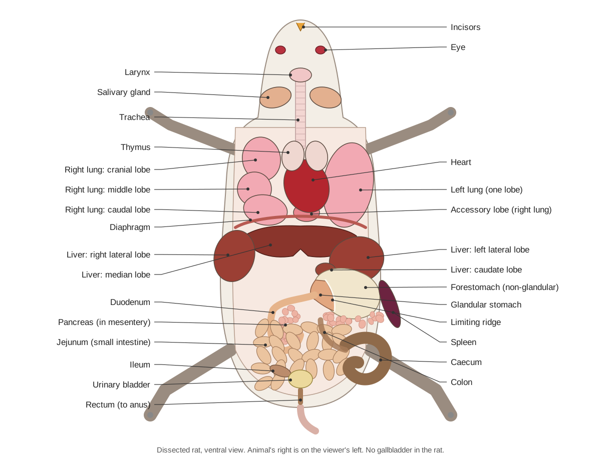
Compare the diagram with your specimen before you move anything. In a real rat the liver covers more of the stomach than shown, and the intestine fills the abdomen more tightly. Lift the liver lobes forward to see the stomach, and fan the intestine to one side to see the pancreas and mesentery.
External anatomy of the rat
Before cutting, identify the external features. The head carries the long vibrissae (whiskers), the nostrils, large eyes (pink-red in albino rats) and thin ear flaps (pinnae). Open the mouth to see the two upper and two lower incisors with orange enamel on their front faces, and the toothless gap behind them, the diastema. Rats have only 16 teeth: one incisor and three molars in each half of each jaw, with no canines or premolars.
Each forepaw has four clawed fingers and a tiny thumb with a flat nail; each hind foot has five clawed toes and the rat walks on the whole sole. The tail is covered in rings of scales with short hairs between them; in the Norway rat it is shorter than the head and body. Hunt's manual asks students to count the foot pads and to note that scales on a mammal's tail are unusual.
How to dissect a rat: step-by-step procedure
The order below follows Hunt's classic rat manual and modern school labs. Each numbered step matches a step in the 3D viewer above, so you can switch between the two.
1. External anatomy
- Lay the rat on its side. Identify the head, neck, trunk and tail.
- Find the vibrissae (whiskers), nostrils, eyes and pinnae (ear flaps).
- Open the mouth with forceps. Find the two upper and two lower incisors and the toothless gap (diastema) behind them.
- Count the digits: four clawed fingers plus a tiny thumb on each forepaw, five toes on each hind foot. Note the scaly rings on the tail.
Identify: Vibrissae, Nose and nostrils, Eye, Pinna, Upper incisors, Lower incisors, Forepaw, Hindpaw, Tail, Skin and fur.
2. Sexing the rat
- Turn the rat on its back and lift the tail. Find the anus under the base of the tail.
- Male: a large scrotum lies below the anus and a single urogenital opening (the prepuce) lies in front of it. The anogenital distance is long.
- Female: three openings in a row — anus, vaginal opening and urethral papilla — and a short anogenital distance. Look for six pairs of nipples in two rows along the belly (wet the fur to see them).
- Use the Female/Male switch on the model to compare.
Identify: Anus, Scrotum, Prepuce and urogenital opening, Vaginal opening, Urethral papilla, Nipples (teats).
3. Pin the rat and remove the ventral skin
- Pin or tie the rat on its back with the legs spread.
- Lift the skin at the midline just in front of the genital opening and make a small snip. Cut forward along the midline to the chin, keeping the scissors pointing up.
- Make side cuts towards each leg, then peel the skin back. Separate it from the body wall with your fingers or a probe; it comes away easily.
- In the neck find the salivary glands and the jugular veins. In a female, the thin mammary tissue stays on the skin. Look for small lymph nodes in the armpits and groin.
Caution: Cut only the skin. The abdominal muscles beneath it are thin and the organs lie just under them.
Identify: Salivary glands, Jugular veins, Mammary glands, Lymph nodes, Masseter.
4. Open the abdominal cavity
- Lift the abdominal muscles with forceps and cut along the midline (linea alba) from the pelvis to the bottom of the rib cage.
- Make cuts sideways just behind the ribs and in front of the hind legs, and pin the flaps back.
- Before moving anything, sketch what you see: the dark red liver at the front, the stomach on the left, coils of small intestine and the large caecum.
Caution: Do not cut the diaphragm yet; the thoracic cavity is opened later.
Identify: Liver: median lobe, Forestomach (non-glandular stomach), Glandular stomach, Jejunum, Caecum, Diaphragm, Urinary bladder.
5. The digestive tract
- Lift the liver forward. Find the oesophagus entering the stomach through the diaphragm.
- Identify the two regions of the stomach: the thin, translucent forestomach on the left and the thick, opaque glandular stomach on the right. Optionally open the stomach along its greater curvature to see the limiting ridge between them.
- Follow the gut from the pylorus: the U-shaped duodenum, the long jejunum and the darker ileum, then the caecum, the colon and the rectum.
- Spread the coils of intestine to see the thin, transparent mesentery and its blood vessels.
Identify: Oesophagus, Forestomach (non-glandular stomach), Glandular stomach, Limiting ridge, Duodenum, Jejunum, Ileum, Caecum, Colon, Rectum, Mesentery.
6. Liver, bile duct, pancreas and spleen
- Identify the four main liver lobes: median (with a deep notch), left lateral, right lateral and the small caudate lobe around the stomach.
- Look for a gallbladder: there is none. Find the thin bile duct running from the liver to the duodenum instead, with the larger hepatic portal vein beside it carrying blood from the gut to the liver.
- Find the pancreas: lift the duodenum and hold the mesentery up to the light. The pancreas is the pinkish, branching tissue spread in the duodenal loop and towards the spleen. White, smooth tissue is fat.
- Find the long, dark red spleen along the left side of the stomach.
Identify: Liver: median lobe, Liver: left lateral lobe, Liver: right lateral lobe, Liver: caudate lobe, Bile duct, Hepatic portal vein, Pancreas, Spleen, Duodenum.
7. Open the thoracic cavity
- Cut through the ribs on each side of the sternum, from the diaphragm to the front of the chest. Cut the diaphragm away from the body wall and lift the sternum forward.
- Find the pale thymus over the front of the heart, the heart in its pericardial sac, and the trachea in the neck. Just behind the larynx, look for the small, dark red lobes of the thyroid gland on each side of the trachea.
- Identify the lungs: one lobe on the left; cranial, middle, caudal and accessory lobes on the right. Lift the heart to find the accessory lobe behind it.
Caution: Rib ends are sharp. Cut with the tips of the scissors pointing up.
Identify: Thymus, Heart (ventricles), Trachea, Larynx, Thyroid gland, Left lung (one lobe), Right cranial lobe, Right middle lobe, Right caudal lobe, Accessory lobe, Diaphragm.
8. Heart and great vessels
- Remove the thymus and the pericardium. Identify the ventricles (apex pointing back and left) and the two atria at the base.
- Find the aorta arching to the left, the pulmonary trunk, the two cranial venae cavae (one each side) and the caudal vena cava passing through the diaphragm.
- Follow the common carotid arteries up the neck beside the trachea.
Identify: Heart (ventricles), Right atrium, Left atrium, Aorta, Pulmonary trunk, Cranial venae cavae (left and right), Caudal vena cava, Common carotid arteries.
9. Kidneys and urinary system
- Move the intestines to one side (or cut them free at the oesophagus and rectum and remove them).
- Find the kidneys against the dorsal body wall. The right kidney lies further forward than the left.
- Find the small adrenal gland at the front of each kidney, the renal artery and vein at each hilum, the aorta and caudal vena cava between the kidneys, and the ureters running back to the bladder.
- Optional: remove one kidney and slice it lengthwise to see the cortex, medulla and renal pelvis.
Identify: Kidneys, Adrenal glands, Renal arteries and veins, Ureters, Urinary bladder, Aorta, Caudal vena cava.
10. Male reproductive system
- Set the model to Male. Cut open the scrotum to expose the testes and the coiled epididymis on each.
- Trace each vas deferens up into the abdomen, over the ureter, to the urethra behind the bladder.
- Identify the large, curved seminal vesicles with the thin coagulating glands along their inner curve, and the prostate around the bladder neck.
- Find the preputial glands under the skin beside the penis. Cut the pubic symphysis to follow the urethra into the penis and find the bulbourethral glands.
Identify: Testes, Epididymis, Vas deferens, Seminal vesicles, Coagulating glands, Prostate, Bulbourethral glands, Preputial glands, Penis, Os penis, Urethra (male).
11. Female reproductive system (and the pregnant rat)
- Set the model to Female. Find the small ovaries behind the kidneys, each in a fatty capsule, with tiny coiled oviducts.
- Follow the two long uterine horns back to where they meet at the cervix above the bladder. The vagina runs from the cervix to its opening.
- Note that the urethra runs separately to the urethral papilla.
- If your rat is pregnant, each uterine horn looks like a string of beads. Count the embryos in each horn and measure one.
Identify: Ovaries, Oviducts, Uterine horns, Cervix, Vagina, Urethra (female), Clitoral glands, Mammary glands.
12. Brain (optional)
- Remove the skin of the head. With bone cutters, carefully chip away the top of the skull from behind the eyes to the back of the head.
- Lift the brain out by cutting the cranial nerves underneath. Identify the olfactory bulbs, the smooth cerebral hemispheres, the cerebellum and the brainstem.
- Cut one hemisphere lengthwise with a razor blade to look for the hippocampus inside.
Caution: Preserved brain tissue is soft. Remove bone in small pieces.
Identify: Olfactory bulbs, Cerebrum, Hippocampus, Cerebellum, Brainstem (medulla oblongata), Spinal cord, Skull (cranium).
13. The skeleton (review)
- Use the model to review the skeleton: skull and mandible, 7 cervical, 13 thoracic, 6 lumbar and 4 sacral vertebrae, the caudal vertebrae, 13 pairs of ribs, sternum, girdles and limbs.
- Compare it with your own skeleton: the rat has a clavicle like you, but a long tail, a plantigrade foot and a fused tibia and fibula.
Identify: Skull (cranium), Mandible, Cervical vertebrae (7), Thoracic vertebrae (13), Lumbar vertebrae (6), Sacrum (4 sacral vertebrae), Caudal vertebrae, Ribs (13 pairs), Sternum, Scapula, Clavicle, Humerus, Radius and ulna, Pelvis (os coxae), Femur, Tibia and fibula.
Rat digestive system labeled: the route of food
Food passes through the mouth, pharynx and oesophagus, into the forestomach (storage) and glandular stomach (acid and pepsin), then through the pylorus into the duodenum, jejunum and ileum. At the ileocaecal junction it enters the large intestine: the caecum (fermentation), the ascending, transverse and descending colon (water absorption and pellet formation) and the rectum. The accessory organs are the salivary glands, the liver (bile, with no gallbladder to store it) and the diffuse pancreas.
A good way to finish the digestive step is to cut the gut free at the oesophagus and rectum, lay it out straight on wet paper towel and measure each part. The small intestine is several times longer than the body; the caecum is the widest part of the tract.
Why dissect a rat? What it shows that frogs and worms cannot
A rat is a small, complete mammal. Every organ system in it has a direct human counterpart: a diaphragm separating thorax and abdomen, a four-chambered heart, lungs with lobes, a liver, kidneys with a cortex and medulla, a placenta-forming uterus, mammary glands and a brain with a cerebral cortex. That is why the rat is the classic mammal of school and first-year university labs, usually after the earthworm and frog. Hunt's 1924 laboratory manual made the same argument a century ago: the Norway rat and its albino variety are easy to obtain, of a convenient size and anatomically typical of mammals. (Hunt (1924), A Laboratory Manual of the Anatomy of the Rat, Internet Archive.)
The rat also differs from humans in instructive ways, and good lab questions use them. It has no gallbladder, a stomach with a non-glandular forestomach, a huge caecum, ever-growing incisors, a duplex uterus, a penile bone, and separate urethral and vaginal openings in the female. Spotting these differences, and explaining them, is the point of the dissection, not just naming organs.
Where do rats for dissection come from?
Most preserved rats used in schools are laboratory-type albino Norway rats (Rattus norvegicus) bred for the purpose and sold by biological supply companies. A Carolina Biological Supply paper on how the company procures animals says its specimens come from cultures (captive breeding), from managed habitats, and from individuals or organisations, that endangered species are never used, and that non-aquatic vertebrates such as rats are euthanised with pure carbon monoxide before being preserved. It also notes that movement of a body during perfusion with fixative is a post-mortem response, not a sign that the animal was alive. (Flagg (Carolina Biological Supply), Procuring and preserving animals for biological instruction, ABLE Tested Studies vol. 15, PDF.)
After euthanasia, supply-house rats are injected with a preservative (traditionally formalin-based, often replaced today by lower-odour fixatives) and many are double-injected with coloured latex: red in the arteries, blue in the veins. That is why the vessels in your rat may be bright red and blue and firm to the touch. Some teachers instead use frozen, unpreserved rats sold as food for pet reptiles. Fresh or thawed tissue is softer and its colours are natural, but it must be handled like raw meat and disposed of the same day.
If you object to dissecting an animal, ask your teacher about an alternative assignment. The 3D model on this page follows the same thirteen steps as the bench dissection and works with the same worksheet questions.
Sexing a rat: anogenital distance, nipples and the three openings
Lift the tail and look at the perineum (the area around the anus). In rats and other rodents the anogenital distance, the gap between the anus and the genital opening, is normally about twice as long in males as in females. The difference is produced by androgens (male hormones) acting on the fetus. Male rats also lack nipples: the nipple buds regress before birth under androgen control, so visible nipples are a quick sign of a female. (Schwartz et al. (2021), Areola/nipple retention as a biomarker for anti-androgenic effects in rat toxicity studies, Front. Toxicol..)
An adult male is unmistakable: two large testes fill a scrotum below the anus, and the single urogenital opening sits in a hairy prepuce in front of the scrotum. A female has three openings in a line: the anus under the tail, the vaginal opening just below it, and the urethral opening on top of a small cone, the urethral papilla. Hunt points out that this is unusual among mammals, most of which have a single urogenital opening in the female. (Hunt (1924), A Laboratory Manual of the Anatomy of the Rat, Internet Archive.)
Count the nipples on a female. Rats of both sexes have six pairs of mammary glands, three in the cervicothoracic region and three in the inguinoabdominal region, but only females have nipples. Mice have five pairs (three and two). Wetting the fur with a little water makes the nipples easier to see in a preserved specimen. (NTP Nonneoplastic Lesion Atlas: mammary gland.)
Opening the ventral body wall: what you see first
The skin of a rat is loose. After the first midline snip, most of it can be separated from the muscles with a finger or a blunt probe. The abdominal muscles underneath are thin, so open them separately and with the scissor tips pointing up. Once the flaps are pinned back, the view is dominated by four things: the dark red liver against the diaphragm at the front, the stomach on the left, the coiled small intestine filling the middle, and the large, dark caecum in the lower left.
The organs are held by thin membranes. The mesentery suspends the intestine and carries its blood vessels; the stomach's mesentery expands into a sac, the greater omentum, which often holds fat and part of the pancreas. In adult laboratory rats the small intestine is roughly 1 to 1.5 m long, most of it jejunum: a surgical-anatomy study gives the duodenum about 10 cm, the jejunum 90 to 135 cm and the ileum only 2.5 to 3.5 cm. (Hunt, measuring the smaller rats of 1924, gave about 70 cm.) Uncoiling it is a good way to show how much surface a small body packs inside. (Vdoviaková et al. (2016), Surgical anatomy of the gastrointestinal tract and its vasculature in the laboratory rat, Gastroenterol Res Pract (PMC).)
The rat stomach and why rats cannot vomit
The rat stomach has two regions that look different even from outside. The left part, the forestomach (non-glandular stomach), has a thin wall lined with keratinised squamous epithelium like the oesophagus. The right part, the glandular stomach, is thick and opaque, with folds (rugae) and glands that secrete acid and pepsin. Inside, the two are separated by a raised fold called the limiting ridge. (NTP Atlas of the rodent stomach (forestomach, limiting ridge, glandular stomach), PDF.)
Hunt described the forestomach as thin enough, in a full preserved stomach, to see the food inside, and the glandular region as yellowish, opaque and folded. Open the stomach along its greater curvature to see the limiting ridge for yourself. The oesophagus opens into the forestomach, just beside the ridge. (Hunt (1924), A Laboratory Manual of the Anatomy of the Rat, Internet Archive.)
Rats cannot vomit, and the limiting ridge is often given as the reason. The best evidence points elsewhere. Horn and colleagues gave standard emetic drugs to rats, mice and other rodents and none retched or vomited. They also found anatomical features that would make vomiting hard (relatively less diaphragm muscle with a larger central tendon, a stomach shape poorly suited to moving contents towards the oesophagus, a long abdominal oesophagus), but in recordings from the brainstem of rats and mice, the coordinated nerve activity that drives vomiting in shrews never appeared. They concluded that a missing brainstem neural component is the most likely cause. (Horn et al. (2013), Why can't rodents vomit? A comparative behavioral, anatomical, and physiological study, PLoS ONE (PMC).)
Rat liver lobes and the missing gallbladder
The rat liver is deeply divided. Most lab guides use four main lobes: a large median lobe partly split by a notch, a left lateral lobe behind it on the left, a right lateral lobe almost divided into front and back lobules, and a small caudate lobe whose two lobules fit around the lesser curvature of the stomach. The back lobule of the right lobe has a hollow, the renal impression, that holds the front end of the right kidney. (Hunt (1924), A Laboratory Manual of the Anatomy of the Rat, Internet Archive.)
Names and counts vary between books. Vdoviaková and colleagues, describing rat liver morphology for surgeons, distinguish six lobes (left and right medial, left and right lateral, caudate and quadrate) and review authors who use four. If your worksheet says 'four lobes', it is using the traditional grouping; neither is wrong. The same paper is clear on one point: laboratory rats have no gallbladder. (Vdoviaková et al. (2016), Importance of rat liver morphology and vasculature in surgical research, Med Sci Monit (PMC).)
Without a gallbladder to store it, bile flows continuously from the liver into the bile duct, which runs in the ligament between the liver and duodenum, crosses the back of the pancreas and enters the duodenum about 3 cm from the pylorus. Small pancreatic ducts join it on the way, so the bile duct carries pancreatic juice too. (Hunt (1924), A Laboratory Manual of the Anatomy of the Rat, Internet Archive.)
How to find the pancreas in a rat dissection
Students miss the rat pancreas because they expect a compact organ like the human pancreas. In the rat it is irregular and divided into many lobes, some joined to each other, spread thinly in the mesentery of the duodenum and in the descending part of the greater omentum. It sends a lobe towards the pylorus, and its left end lies dorsal to the spleen. (Hunt (1924), A Laboratory Manual of the Anatomy of the Rat, Internet Archive.)
To find it, lift the stomach forward and pull the duodenum gently to the right so its U-shaped loop spreads out. Hold the mesentery up against the light. The pancreas is the soft, pinkish-tan, granular tissue lying in the loop and running along the stomach towards the spleen. Fat looks whiter, smoother and more glossy, and lymph nodes are firm, rounded beads. If your rat is injected, the pancreas may be tinted by latex in its many small vessels.
The caecum and large intestine
The caecum is the large blind sac where the ileum meets the colon. In an adult laboratory rat it is about 5 to 7 cm long (Vdoviaková and colleagues), shaped like a comma or a short spiral, and full of dark contents. The ileum is darker than the jejunum because of what it carries. From the caecum the ascending colon runs forward, the transverse colon circles to the left near the stomach, and the descending colon runs back along the midline, beaded with faecal pellets 'shaped like cotton seeds', to the rectum and anus. (Hunt (1924), A Laboratory Manual of the Anatomy of the Rat, Internet Archive.)
The caecum lies on the left side of the abdomen. That is why research protocols for intraperitoneal injection in rats direct the needle into the right lower quadrant: to avoid penetrating the caecum on the left side. (Virginia Tech SOP ARCD-0320: intraperitoneal injections in the rat, PDF.)
Thoracic cavity: heart, thymus and lung lobes
Open the thorax by cutting through the ribs on both sides of the sternum and freeing the diaphragm. The heart sits in the middle in its pericardial sac, with the pale thymus over its base. The left lung has a single lobe and the right lung has four: cranial, middle, caudal and accessory. A 2016 study of the bronchial tree in Wistar rats confirmed exactly this pattern. (Olivares Pérez-Montt & Gil (2016), Lobulación pulmonar de la rata blanca, Int. J. Morphol. 34(3).)
The accessory lobe is a trap. It is a small, pyramid-shaped lobe that sits behind the heart, mostly to the left of the midline, in its own pocket of pleura. Hunt warns that it may be mistaken for part of the left lung unless you trace its bronchus back to the right side. Hunt also describes two cranial (superior) venae cavae entering the right atrium, one on each side: rats keep the left one, which in humans disappears before birth. (Hunt (1924), A Laboratory Manual of the Anatomy of the Rat, Internet Archive.)
Rat kidney dissection
The kidneys lie against the dorsal body wall, behind the peritoneum, with their hilum facing the midline. The right kidney is further forward than the left: its front end fits into the renal impression of the right liver lobe, while the left kidney is displaced so far back that its hilum lies behind the level of the right kidney. Hunt suggests the stomach on the left pushes it back. A small, rounded adrenal (suprarenal) gland caps the front of each kidney. (Hunt (1924), A Laboratory Manual of the Anatomy of the Rat, Internet Archive.)
To see the inside, remove one kidney and slice it lengthwise along its outer edge with a scalpel or razor. You will see a pale outer cortex, a darker inner medulla and a single cone-shaped papilla projecting into the renal pelvis, where the ureter begins. Human kidneys have many papillae; the rat kidney is unipapillate, which makes it simpler to read. (Hunt (1924), A Laboratory Manual of the Anatomy of the Rat, Internet Archive.)
Male vs female rat: urogenital system labeled
Only the reproductive steps differ between sexes. Sex the rat externally first (step 2), then use the diagram below while you dissect.
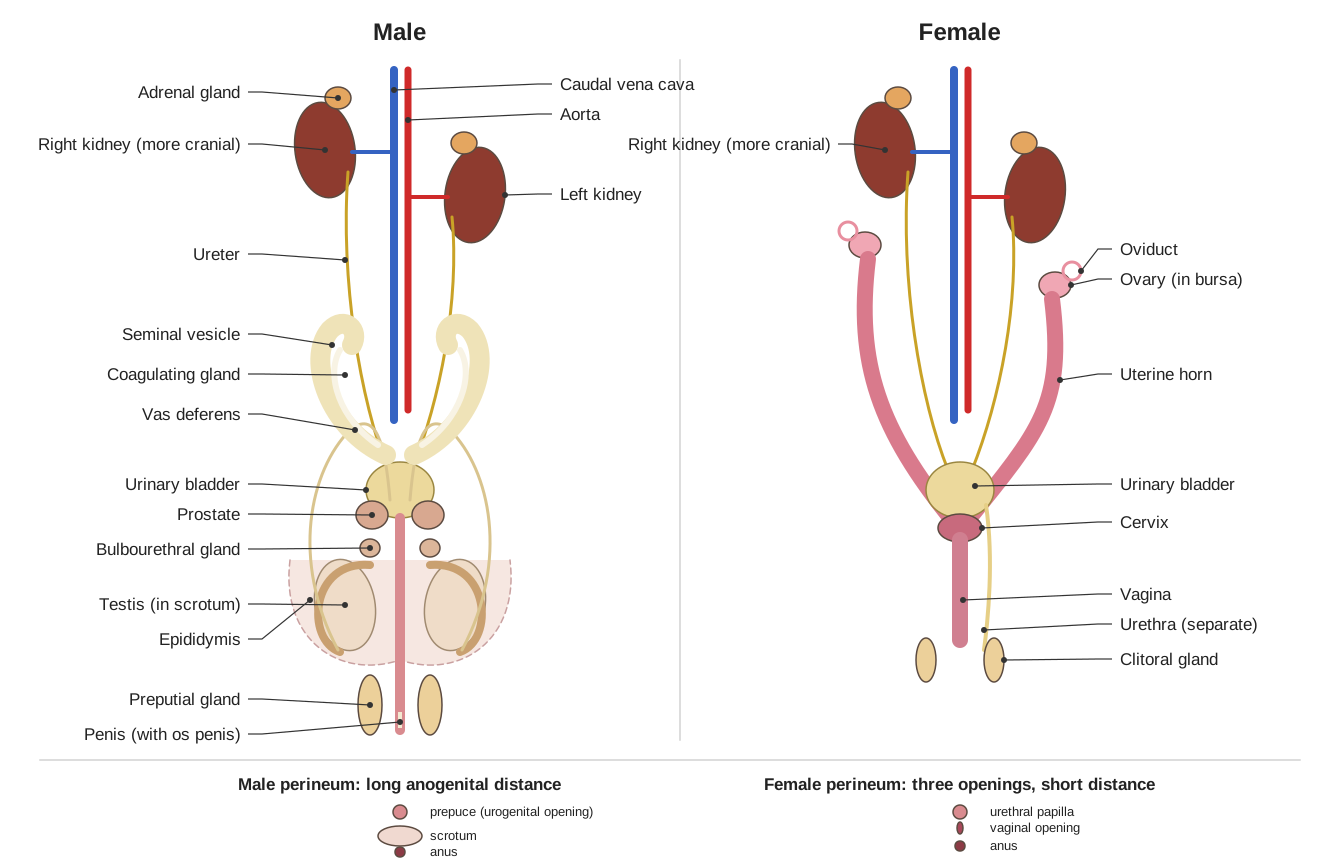
- Male: testes in the scrotum, epididymis, vas deferens looping over the ureter, large question-mark seminal vesicles with coagulating glands, prostate lobes around the bladder neck, bulbourethral glands, preputial glands and a penis with an os penis.
- Female: small ovaries in fatty capsules behind the kidneys, coiled oviducts, two long uterine horns joining at the cervix, a vagina below the rectum, a separate urethra opening on the urethral papilla, clitoral glands and six pairs of mammary glands.
Male rat reproductive system
The male accessory glands are the most surprising part of the rat dissection. Behind the bladder lie the seminal vesicles (vesicular glands): a pair of large, warty glands bent like question marks, hard and brittle in a preserved rat and full of coagulated secretion. Despite the old name they contain no sperm. Pressed into their concave side is a thin, flattened gland, the coagulating gland. The prostate has a dorsal part on the urethra and two prominent ventral lobes under the bladder. (Hunt (1924), A Laboratory Manual of the Anatomy of the Rat, Internet Archive.)
Each vas deferens (ductus deferens) comes up from the scrotum through the inguinal canal and loops around the ureter before joining the urethra, a loop produced when the testis descended during development. At the root of the penis the two bulbourethral (Cowper's) glands empty into the urethra. Under the skin beside the penis lie the preputial glands, whose duct contains a soapy secretion. The penis itself bends in a figure-7 and contains one or two small bones, the os penis, at its tip. (Hunt (1924), A Laboratory Manual of the Anatomy of the Rat, Internet Archive.)
Female rat reproductive system and the pregnant rat
The female tract is a Y. Each tiny ovary sits behind its kidney inside a thin capsule, the ovarian bursa, with a tightly coiled oviduct beside it. From each ovary a long uterine horn runs back to join its partner above the bladder. Hunt showed that the joining is only superficial: a probe passed down each horn emerges through its own opening into the vagina, so the rat uterus is a uterus duplex with two cervical canals. A 2019 study of laboratory rats, mice and guinea pigs confirmed two separate cervical canals in all three (uterus duplex bicollis). The vagina lies directly beneath the rectum. The urethra runs separately, ventral to the vagina, to the urethral papilla. (Hunt (1924), A Laboratory Manual of the Anatomy of the Rat, Internet Archive.)
Lab rats are sometimes pregnant. A pregnant uterus looks like two strings of beads: each swelling is one embryo with its placenta, spaced along both horns. The average gestation is 21 to 23 days and litters are usually 8 to 18 pups, so a late-pregnant rat can hold many fetuses. Count the embryos in each horn, measure the crown–rump length of one, and compare horns. (Merck Veterinary Manual: breeding and reproduction of rats.)
Rat brain dissection (brief)
The brain lies in a narrow braincase behind the eyes. Remove the skin of the head, then chip away the top of the skull in small pieces with bone cutters, working forward from the back of the head. Lift the brain out by cutting the cranial nerves underneath. From front to back you will find the two olfactory bulbs (very large compared with a human's, because rats rely on smell), the smooth cerebral hemispheres, the folded cerebellum and the brainstem continuing as the spinal cord. The cerebral surface has no folds: the rat brain is lissencephalic.
Hunt notes that the cribriform plate of the skull, the perforated bone between the nasal cavity and the braincase, holds the olfactory lobes; the olfactory nerves pass through its holes. Damaging this plate is the usual way the bulbs are lost during removal, so leave them for last. (Hunt (1924), A Laboratory Manual of the Anatomy of the Rat, Internet Archive.)
Rat dissection labeling worksheet and answer key
Use this blank diagram for "label the rat dissection" practice or as a pre-lab quiz. Write the name of each numbered structure, then check your answers.
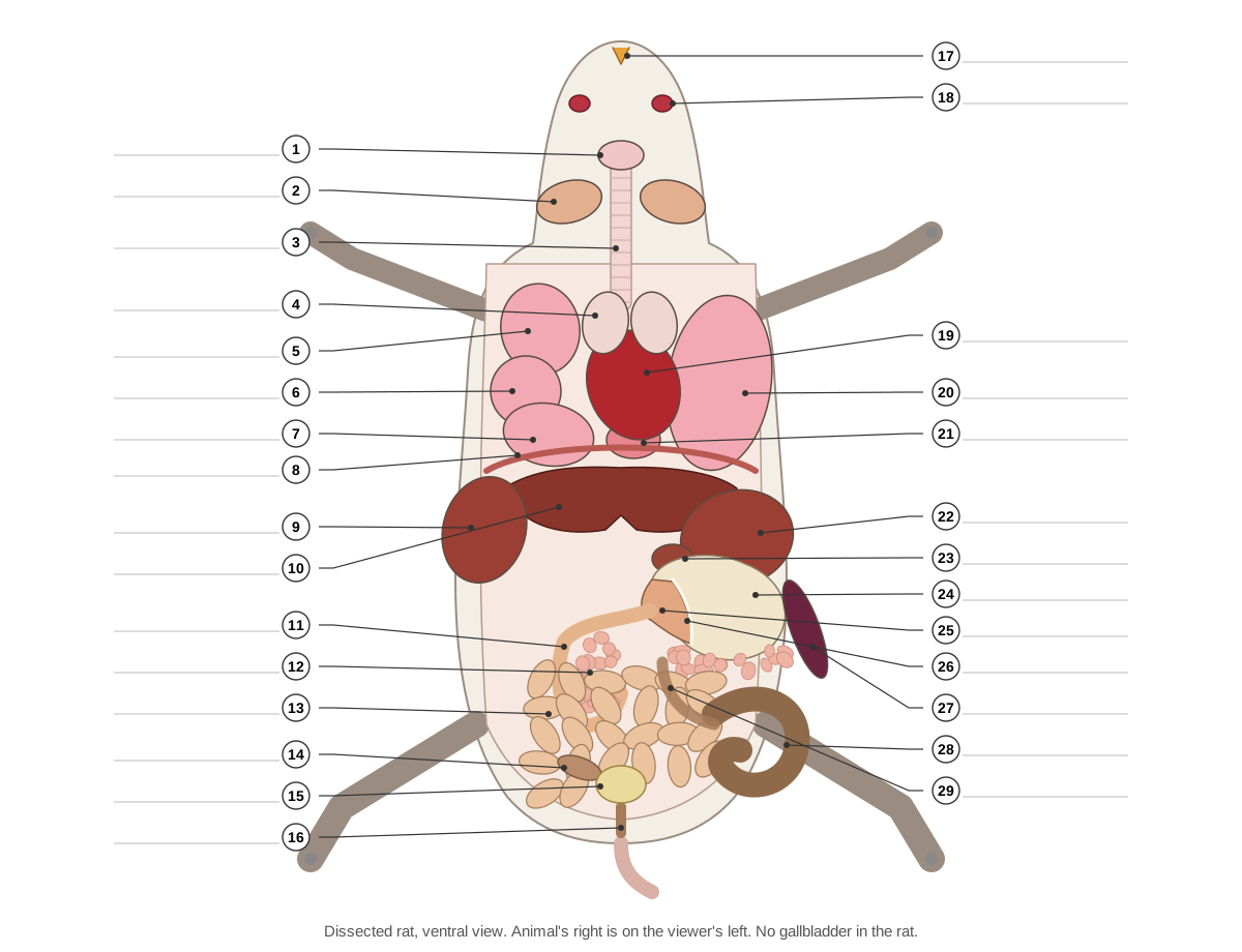
Show the answer key
- Larynx
- Salivary gland
- Trachea
- Thymus
- Right lung: cranial lobe
- Right lung: middle lobe
- Right lung: caudal lobe
- Diaphragm
- Liver: right lateral lobe
- Liver: median lobe
- Duodenum
- Pancreas (in mesentery)
- Jejunum (small intestine)
- Ileum
- Urinary bladder
- Rectum (to anus)
- Incisors
- Eye
- Heart
- Left lung (one lobe)
- Accessory lobe (right lung)
- Liver: left lateral lobe
- Liver: caudate lobe
- Forestomach (non-glandular)
- Glandular stomach
- Limiting ridge
- Spleen
- Caecum
- Colon
Rat dissection lab questions and answer key
Answer these during or after the dissection. Click a question to reveal the answer.
How can you tell a male rat from a female rat without opening it?
Look at the perineum. A male has a scrotum and a long anogenital distance (about twice the female's), with one urogenital opening. A female has three openings (anus, vaginal opening and urethral papilla), a short anogenital distance and six pairs of nipples.
Rats have the dental formula I 1/1, C 0/0, P 0/0, M 3/3. How many teeth does a rat have, and what is the diastema?
16 teeth: 4 incisors and 12 molars. The diastema is the toothless gap between the incisors and the molars; the cheeks fold into it to seal off the mouth while the rat gnaws.
Why do a rat's incisors stay sharp even though they never stop growing?
Enamel covers only their front surface. The softer dentine behind wears away faster, so the edge stays chisel-sharp. Constant growth replaces what is worn away.
Name the two regions of the rat stomach and the structure that separates them.
The non-glandular forestomach (thin, lined with keratinised squamous epithelium) and the glandular stomach (thick, secretes acid and pepsin). The limiting ridge separates them.
Why can't rats vomit?
Mainly because their brainstem lacks the neural circuit that coordinates vomiting (Horn et al. 2013). Their anatomy also works against it: a relatively weak diaphragm, a stomach shape poorly suited to pushing contents up and a long abdominal oesophagus. The limiting ridge is often blamed, but it is not the main reason.
Which organ do humans have that a rat lacks: gallbladder, spleen or caecum? Where does the rat's bile go?
The gallbladder. Bile flows continuously from the liver through the bile duct into the duodenum.
How do you find the pancreas in a rat?
Lift the stomach and duodenum and hold the mesentery to the light. The pancreas is the soft, pink, branching tissue in the loop of the duodenum, running towards the spleen. It is diffuse, not a compact organ; fat is whiter and smoother.
What is the function of the large caecum, and why is it important for herbivores and omnivores like rats?
It is a fermentation chamber: bacteria digest plant fibre and make vitamins. Rats re-eat some soft faeces (coprophagy) to absorb these products.
How many lobes do the left and right lungs of a rat have?
The left lung has one lobe. The right lung has four: cranial, middle, caudal and accessory.
Which kidney is further forward (cranial) in the rat, and why?
The right kidney. The left one is pushed back, probably by the stomach, which lies on the left (Hunt 1924).
Trace the path of sperm from where it is made to where it leaves the body.
Testis → epididymis → vas deferens (through the inguinal canal, over the ureter) → urethra → penis → out through the prepuce. The seminal vesicles, coagulating glands, prostate and bulbourethral glands add fluid along the way.
Describe the shape of the rat uterus. Where do embryos develop?
It is duplex: two long, separate uterine horns, each with its own opening into the vagina. Embryos implant in a row along both horns.
Name two structures found in a male rat that a male human does not have.
Any two of: os penis (penile bone), coagulating glands, preputial glands. (Humans do have seminal vesicles, but the rat's are relatively huge; rats also keep their inguinal canals open.)
Which part of the rat brain is relatively much larger than in a human, and what does that tell you?
The olfactory bulbs. Rats rely heavily on smell.
How rat anatomy differs from human anatomy
Most rat dissection reports end with a comparison to the human body. The table lists the differences you can see in the lab, with the laboratory mouse for comparison.
| Feature | Laboratory mouse | Laboratory rat | Human |
|---|---|---|---|
| Species | Mus musculus | Rattus norvegicus | Homo sapiens |
| Tail vs head-and-body length | About equal | Shorter | No tail |
| Teeth (dental formula) | 16 (I 1/1, C 0/0, P 0/0, M 3/3) | 16 (same) | 32 adult (I 2/2, C 1/1, P 2/2, M 3/3) |
| Incisors | Grow throughout life | Grow throughout life | Stop growing |
| Vertebrae | C7 T13 L6 S4 + ~28–30 caudal | C7 T13 L6 S4 + ~28 caudal | C7 T12 L5, sacrum (5 fused), coccyx |
| Stomach | Forestomach + glandular stomach | Forestomach + glandular stomach | Glandular only |
| Can vomit? | No | No | Yes |
| Gallbladder | Yes | No | Yes |
| Liver | Four main lobes, subdivided | Four main lobes, subdivided | Right and left lobes, plus small caudate and quadrate lobes |
| Lung lobes (left / right) | 1 / 4 | 1 / 4 | 2 / 3 |
| Cranial (superior) venae cavae | Left and right | Left and right | Right only |
| Caecum and appendix | Large caecum, no appendix | Large caecum, no appendix | Small caecum with appendix |
| Brain surface | Smooth (lissencephalic) | Smooth (lissencephalic) | Folded (gyri and sulci) |
| Olfactory bulbs | Very large | Very large | Small |
| Mammary glands (pairs, female) | 5 | 6 | 1 |
| Uterus | Two long horns (duplex/bicornuate) | Duplex: two horns, two cervical canals | Simplex (single body) |
| Female openings | Urethral, vaginal, anal: separate | Urethral, vaginal, anal: separate | Urethra and vagina open side by side within the vulval vestibule |
| Os penis (penile bone) | Present | Present | Absent |
| Gestation | About 19–21 days | 21–23 days | About 40 weeks |
For the mouse in more detail, including the mouse brain, heart, liver and hindlimb muscles, see our mouse anatomy page.
Paper rat dissection and virtual rat dissection
A 'paper rat dissection' is a worksheet in which students cut out paper organs and glue them onto an outline of a rat in the right order and position. The labeled diagrams on this page, and the blank version for labelling, do the same job. Work from deep to superficial: kidneys, ureters and bladder first; then the reproductive organs; then the intestines, caecum, stomach and spleen; then the liver, heart, lungs and thymus. If an organ cannot be placed, ask which organ lies on top of it in the real animal.
The 3D viewer is a virtual rat dissection. Each step removes the same layers you would remove at the bench and lists the structures to find; clicking any part shows its location and function. It is a practical alternative for students who opt out, for absent students and for revision before a practical exam.
Every structure in the 3D rat
The full list of 114 structures in the model, grouped by system. Click any of them in the viewer to zoom to it.
External
- Skin and fur (Integument, pelage)
- Loose skin covered by fur, white in albino laboratory rats and grey-brown above, paler below, in wild Norway rats. Protects the body, holds heat and carries touch receptors. The skin is loosely attached, so it lifts easily from the muscles in a dissection.
- Nose and nostrils (Rhinarium, external nares)
- The hairless tip of the snout, with two nostrils (external nares). Takes in air and odours. Rats rely heavily on smell; the nasal cavity leads back to the large olfactory bulbs of the brain.
- Vibrissae (Whiskers, mystacial vibrissae)
- Long, stiff sensory hairs arranged in rows on each side of the snout. Touch organs. Rats sweep (whisk) them back and forth to feel surfaces and gaps. Hunt (1924) noted that rats run faster along a wall, guided by touch through the vibrissae.
- Pinna (External ear, auricle)
- The thin, rounded, nearly hairless external ear flap. Collects sound and funnels it into the ear canal. Rats and mice hear well into ultrasound.
- Forepaw (Manus, front foot)
- The front foot: four clawed fingers and a tiny thumb with a flat, nail-like claw. Used for walking, climbing, digging and holding food while the rat sits up and gnaws.
- Hindpaw (Pes, hind foot)
- The long hind foot with five clawed toes; the rat walks on the whole sole (plantigrade). Pushes the body forward and supports the rat when it sits or stands up.
- Tail (Cauda)
- A long, nearly hairless tail covered in rings of overlapping scales. Balance when climbing, and heat loss: blood flow through the tail changes to keep the body temperature steady.
- Nipples (teats) (Papillae mammae) · females only
- Six pairs of nipples on the underside of a female rat: three pairs on the chest and three on the belly and groin. Each nipple drains one mammary gland; the pups suckle here.
- Scrotum (Scrotal sacs) · males only
- The large swelling below the base of the tail that holds the two testes. Keeps the testes slightly cooler than the body, which sperm production needs.
- Prepuce and urogenital opening (Foreskin, external urethral opening) · males only
- The furry fold that surrounds the tip (glans) of the penis; the single urogenital opening of the male lies here. Protects the glans. Urine and semen both leave the body through this opening.
- Urethral papilla (Urinary papilla, clitoral prepuce) · females only
- A small raised cone in front of the vaginal opening, with the urethral opening at its tip. Urine leaves through it. It is the female counterpart (homologue) of the male prepuce and contains the clitoris.
- Vaginal opening (Vaginal orifice) · females only
- The slit-like opening between the urethral papilla and the anus. Entrance to the reproductive tract: receives the penis at mating, and the pups are born through it.
- Anus
- The opening at the end of the digestive tract, just below the base of the tail. Faeces leave the body here as firm pellets formed in the colon.
Skeletal
- Skull (cranium) (Braincase, rostrum, zygomatic arches)
- The long, narrow skull: a braincase behind, a long snout (rostrum) in front, and a zygomatic arch on each side. Protects the brain and sense organs and anchors the jaw muscles.
- Mandible (Lower jaw (paired dentaries))
- The lower jaw, made of two halves joined loosely at the chin. Holds the lower incisors and molars. It slides forward to gnaw (incisors meet) and backward to chew (molars meet).
- Cervical vertebrae (7) (Neck vertebrae, atlas and axis)
- Seven neck vertebrae, as in almost all mammals. Support and move the head. The first (atlas) rocks on the skull; the second (axis) lets the head turn.
- Thoracic vertebrae (13)
- Thirteen vertebrae, each carrying a pair of ribs. Support the ribs and the trunk muscles.
- Lumbar vertebrae (6)
- Six large vertebrae of the lower back, with long transverse processes and no ribs. Carry the powerful back and hip muscles used in running and jumping.
- Sacrum (4 sacral vertebrae)
- Four sacral vertebrae, more or less fused into one bone. The first two sacral vertebrae join the ilium of each hip bone, so the weight of the hind body passes through them (Hunt 1924).
- Caudal vertebrae (Tail vertebrae)
- About 28 small tail vertebrae (rat) that become simpler and longer towards the tip. Support the tail and its tendons.
- Ribs (13 pairs)
- Thirteen pairs of ribs: 7 true ribs reach the sternum; 6 false ribs do not, and the last 3 of these float free. Protect the heart and lungs and move during breathing.
- Sternum (Breastbone, sternebrae, xiphoid process)
- The breastbone, made of six segments (sternebrae): the manubrium in front, four segments of the body and the xiphoid process behind. Anchors the true ribs and the chest muscles. In a dissection you cut beside it to open the thorax.
- Scapula (Shoulder blade)
- The flat, triangular shoulder blade with a ridge (spine) on its outer face. Anchors the shoulder muscles and forms the socket of the shoulder joint.
- Clavicle (Collarbone)
- A slender collarbone joining the shoulder to the sternum. Braces the shoulder, which helps rats and mice climb and handle food. (Many running mammals, such as dogs and horses, lack a functional clavicle.)
- Humerus
- The bone of the upper arm. Lever for the shoulder and elbow muscles.
- Radius and ulna
- The two forearm bones. The ulna forms the elbow hinge; the radius carries the wrist.
- Carpals, metacarpals and phalanges (Bones of the forepaw)
- The small wrist bones (carpals) in two rows, five metacarpals and the finger bones (phalanges). Support the hand. Each finger has three phalanges, except the thumb, which has two (Hunt 1924).
- Pelvis (os coxae) (Hip bones: ilium, ischium, pubis)
- Two hip bones, each made of the fused ilium, ischium and pubis, joined at the pubic symphysis. Carries the hindlimbs and protects the bladder, rectum and reproductive tract. The femur sits in its socket, the acetabulum.
- Femur (Thigh bone)
- The thigh bone. Lever for the large thigh muscles.
- Patella (Kneecap)
- A small bone in the tendon of the quadriceps, in front of the knee. Improves the leverage of the knee extensors.
- Tibia and fibula (Shin bones)
- The two bones of the lower leg; in rats and mice the slender fibula fuses to the tibia in its lower half. Support the body and anchor the calf and shin muscles.
- Tarsals, metatarsals and phalanges (Bones of the hind foot)
- Ankle bones (tarsals, including the heel bone, the calcaneus), five long metatarsals and the toe bones. Form the long, flat-footed lever used in running and jumping.
Muscular
- Masseter
- The big cheek muscle that closes the jaw and pulls it forward for gnawing. Rodents (the name means 'gnawers') have a very large, complex masseter that moves the jaw forward so the incisors meet.
- Diaphragm
- A thin dome of muscle and tendon that separates the thorax from the abdomen. The main breathing muscle: it flattens to draw air into the lungs.
- Quadriceps femoris (Rectus femoris, vastus muscles)
- Four muscles on the front of the thigh that straighten the knee. Knee extension when running and jumping.
- Gastrocnemius (Calf muscle)
- The two-headed calf muscle that inserts on the heel through the calcaneal (Achilles) tendon. Extends the ankle (plantarflexion) and helps bend the knee.
- Tibialis anterior
- The muscle on the front of the shin. Flexes the ankle (dorsiflexion), lifting the foot as the leg swings forward.
Nervous & senses
- Eye (Eyeball)
- A large, bulging eye on each side of the head; pink-red in albino laboratory rats, which lack pigment. Vision in dim light. The retina is dominated by rods; rats see poorly in bright daylight (Hunt 1924).
- Lens
- The clear, nearly spherical lens, which fills a large part of the small rodent eye. Focuses light on the retina.
- Olfactory bulbs
- Two large knobs at the front of the brain that receive the nerves of smell. First processing centre for odours. Their size reflects how much rats and mice depend on smell.
- Cerebrum (Cerebral hemispheres, cerebral cortex)
- The two smooth cerebral hemispheres, without the folds (gyri) of the human brain. Senses, movement, learning and memory.
- Hippocampus
- A curved, C-shaped structure inside each hemisphere, beneath the cortex. Memory and spatial navigation. Much of what is known about 'place cells' comes from rats and mice.
- Cerebellum
- The folded 'little brain' behind the cerebrum, with a central vermis and two side lobes. Balance and the timing and coordination of movement.
- Brainstem (medulla oblongata)
- The stalk of the brain that continues into the spinal cord. Controls breathing, heart rate and reflexes such as swallowing. Horn et al. (2013) found rat and mouse brainstems lack the coordinated motor pattern that produces vomiting.
- Spinal cord
- The cord of nerve tissue inside the vertebral canal. Carries signals between the brain and body and runs spinal reflexes.
- Sciatic nerve
- The largest nerve of the body, running down the back of the thigh. Supplies the muscles and skin of the hind leg.
Digestive & oral
- Upper incisors
- Two long, curved, chisel-like front teeth that grow throughout life. Gnawing. Hard enamel covers only the front face, so the softer dentine behind wears faster and the edge stays sharp (Hunt 1924).
- Lower incisors
- Two long lower front teeth, rooted far back in the mandible. Work against the upper incisors like scissors when the jaw is pulled forward.
- Molars (Cheek teeth)
- Three small grinding teeth in each half of each jaw (12 in total). Grind food. Unlike the incisors, the molars of rats and mice do not keep growing.
- Tongue
- A thick, muscular tongue with a raised hump at the back. Moves food to the molars and starts swallowing.
- Salivary glands (Submandibular (submaxillary) and parotid glands)
- Paired glands under the skin of the throat and below the ears: the large submandibular, the smaller sublingual beside it, and the flat, lobulated parotid. Produce saliva that moistens food and starts the digestion of starch.
- Oesophagus (Esophagus, gullet)
- The muscular tube that carries food from the throat to the stomach. Moves swallowed food by waves of contraction (peristalsis).
- Forestomach (non-glandular stomach) (Cardiac sac, nonglandular stomach)
- The thin-walled left part of the stomach, lined by tough, keratinised squamous epithelium like the oesophagus. Stores food before digestion. It makes no gastric juice.
- Glandular stomach (Corpus and pyloric antrum)
- The thick, opaque right part of the stomach, lined with gastric glands. Secretes acid and pepsin. Parietal cells make acid; chief cells make pepsinogen (NTP atlas).
- Limiting ridge (Margo plicatus, boundary fold)
- A raised fold on the inside of the stomach that marks the border between the forestomach and the glandular stomach. Separates the two linings. The oesophagus opens into the forestomach just beside it.
- Duodenum
- The first, U-shaped part of the small intestine, starting at the pylorus. Receives bile and pancreatic juice through the bile duct, which enters it about 3 cm from the pylorus (Hunt 1924).
- Jejunum
- The long, coiled middle part of the small intestine. Main site of digestion and absorption of nutrients.
- Ileum
- The last part of the small intestine, darker than the jejunum in a dissected rat. Absorbs remaining nutrients, vitamin B12 and bile salts.
- Caecum (Cecum)
- A large, comma-shaped blind sac where the small intestine meets the colon; often the biggest single loop in the abdomen. Fermentation chamber: bacteria break down plant fibre. Rats and mice also eat some of their own faeces (coprophagy) to recover vitamins made there.
- Colon (Ascending, transverse and descending colon)
- The large intestine: it runs forward from the caecum, crosses to the left, and runs back along the midline as a chain of faecal pellets. Absorbs water and forms the faecal pellets, which are shaped like cotton seeds (Hunt 1924).
- Rectum
- The final straight part of the large intestine, running through the pelvis to the anus. Stores pellets until they are passed.
- Liver: median lobe (Middle lobe)
- The largest liver lobe, at the front middle of the abdomen, partly split by a deep notch. Part of the liver: makes bile, stores glycogen, processes nutrients from the portal vein.
- Liver: left lateral lobe
- A large, thick lobe on the left, behind the median lobe. Part of the liver. With the median lobe it is removed in the classic two-thirds partial hepatectomy used to study liver regeneration.
- Liver: right lateral lobe (Right lobe (anterior and posterior lobules))
- The right lobe, nearly divided into a front and a back lobule. Part of the liver.
- Liver: caudate lobe (Spigelian lobe)
- Two small lobules that fit around the lesser curvature of the stomach. Part of the liver.
- Bile duct (Common hepatic duct, ductus choledochus)
- The thin duct that carries bile from the liver to the duodenum. Carries bile continuously, because the rat has no gallbladder to store it. Small pancreatic ducts join it as it crosses the pancreas (Hunt 1924).
- Pancreas
- A soft, pinkish, branching gland spread thinly through the mesentery, not a compact organ. Makes digestive enzymes (exocrine) and the hormones insulin and glucagon (islets, endocrine).
- Mesentery (Mesentery proper, greater omentum)
- The thin, transparent double sheet of peritoneum that holds the intestines and carries their blood vessels. Suspends the gut, carries vessels, nerves and lymphatics, and stores fat.
Respiratory
- Larynx (Voice box)
- The cartilage box at the top of the trachea. Guards the airway when swallowing and produces sound, including ultrasonic calls.
- Trachea (Windpipe)
- The windpipe, kept open by C-shaped rings of cartilage. Carries air to and from the lungs.
- Left lung (one lobe)
- A single, undivided lobe: the largest single lung lobe. Gas exchange.
- Right cranial lobe (Right anterior / apical lobe)
- The front lobe of the right lung. Gas exchange.
- Right middle lobe (Cardiac lobe)
- The middle lobe of the right lung, beside the heart. Gas exchange.
- Right caudal lobe (Right posterior lobe)
- The largest lobe of the right lung. Gas exchange.
- Accessory lobe (Postcaval / azygous / median lobe)
- A small, pyramid-shaped lobe of the right lung that sits behind the heart, mostly left of the midline. Gas exchange.
Heart & vessels
- Heart (ventricles)
- The four-chambered heart in its pericardial sac; this part shows the right and left ventricles, with the apex pointing back and to the left. The right ventricle pumps blood to the lungs; the thick-walled left ventricle pumps it to the body.
- Right atrium
- The thin-walled chamber that receives blood from the body. Receives oxygen-poor blood from the two cranial venae cavae and the caudal vena cava.
- Left atrium
- The chamber that receives oxygen-rich blood from the lungs. Passes blood from the pulmonary veins to the left ventricle.
- Aorta (Aortic arch, dorsal (abdominal) aorta, iliac arteries)
- The main artery: it arches over the heart, runs back under the spine and splits into the iliac arteries for the legs. Carries oxygen-rich blood to the body.
- Pulmonary trunk (Pulmonary arteries)
- The vessel that leaves the right ventricle and splits into the left and right pulmonary arteries. Carries oxygen-poor blood to the lungs.
- Cranial venae cavae (left and right) (Superior venae cavae, precaval veins)
- Two large veins, one on each side, that return blood from the head and forelimbs to the right atrium. Venous return from the front of the body.
- Caudal vena cava (Inferior vena cava, postcava)
- The large vein that returns blood from the hind body; it passes through the liver and diaphragm to the heart. Venous return from the legs, kidneys and gut (via the liver).
- Common carotid arteries
- Paired arteries running up each side of the trachea to the head. Supply the brain, face and head.
- Jugular veins (External jugular veins)
- Large, superficial veins on each side of the neck. Return blood from the head.
- Hepatic portal vein (Portal vein)
- The large vein that carries blood from the stomach, intestines, pancreas and spleen to the liver. Takes nutrients and other substances absorbed from the gut straight to the liver, which processes them before the blood reaches the rest of the body.
- Renal arteries and veins
- Short vessels linking each kidney with the aorta and caudal vena cava. About a fifth of the blood pumped by the heart passes through the kidneys to be filtered.
Lymphoid & brown fat
- Thymus
- A pale, two-lobed gland lying over the front of the heart; large in young rats, smaller in adults. Trains T lymphocytes (a type of white blood cell).
- Spleen
- A long, flat, dark red organ along the greater curvature of the stomach. Filters the blood, removes old red cells and makes immune cells.
- Lymph nodes (Cervical, axillary, inguinal, iliac and mesenteric nodes)
- Small, firm, bean-shaped nodes in the neck, armpits, groin, along the aorta and in the mesentery. Filter lymph and start immune responses.
- Brown fat (interscapular) (Brown adipose tissue, BAT)
- A butterfly-shaped pad of brownish fat between the shoulder blades. Produces heat (non-shivering thermogenesis) using uncoupling protein 1 (UCP1) in its mitochondria (Virtue & Vidal-Puig 2013).
Urinary & endocrine
- Thyroid gland (Thyroid lobes and isthmus; parathyroid glands)
- Two small, dark red lobes pressed against the sides of the trachea just behind the larynx, joined by a thin isthmus across the ventral trachea. Endocrine gland (it has no duct): makes the thyroid hormones (T4 and T3) that set the metabolic rate, and calcitonin from its C cells.
- Kidneys
- Two bean-shaped, dark red kidneys against the dorsal body wall; the right kidney is more cranial than the left. Filter blood and make urine.
- Adrenal glands (Suprarenal glands)
- Small, round, pinkish glands at the front end of each kidney. Endocrine glands with no duct: they release adrenaline and steroid hormones (corticosterone in rats) into the blood.
- Ureters
- Thin tubes running from each kidney back to the bladder. Carry urine to the bladder.
- Urinary bladder
- A small, thin-walled, oval sac in the floor of the pelvis. Stores urine.
- Urethra (female) · females only
- The tube from the bladder to the urethral papilla; separate from the vagina. Carries urine only.
- Urethra (male) · males only
- The tube from the bladder through the pelvis and penis; it carries both urine and semen. Receives the vasa deferentia and accessory gland ducts near the bladder neck.
Reproductive
- Testes · males only
- Two large, oval testes, about 2 cm long in an adult rat (Hunt 1924). Make sperm and testosterone.
- Epididymis (Caput, corpus and cauda epididymidis) · males only
- A coiled duct along the edge of each testis, with a head, body and tail. Stores and matures sperm.
- Vas deferens (Ductus deferens) · males only
- The sperm duct from the tail of the epididymis to the urethra. Carries sperm at ejaculation.
- Seminal vesicles (Vesicular glands) · males only
- A pair of large, knobbly glands shaped like question marks: the most conspicuous male organ in the lower abdomen. Produce a thick secretion that forms part of the semen and the copulatory plug. Despite the old name, they store no sperm (Hunt 1924).
- Coagulating glands (Anterior prostate) · males only
- Thin, pale glands lying along the inner (concave) curve of each seminal vesicle. Their secretion clots the seminal-vesicle fluid to form a copulatory plug in the female's vagina.
- Prostate (Ventral and dorsolateral prostate) · males only
- Lobed glands around the neck of the bladder: two ventral lobes under the bladder and a dorsal part on the urethra. Adds fluid to the semen.
- Bulbourethral glands (Cowper's glands) · males only
- Small, oval glands at the root of the penis. Secrete fluid into the urethra before ejaculation.
- Preputial glands · males only
- A pair of flat, yellowish glands under the skin on each side of the penis. Produce a waxy, scented secretion used in social and sexual communication (Hunt 1924).
- Penis · males only
- The penis, bent in a figure-7 shape, lying under the skin with its tip (glans) inside the prepuce. Carries urine and semen out of the body.
- Os penis (Baculum, penile bone) · males only
- A small bone inside the tip of the penis. Stiffens the glans. Rats and mice have one; humans do not.
- Ovaries · females only
- Two small, knobbly ovaries, each wrapped in a thin capsule (ovarian bursa) and set in fat. Make eggs and the hormones oestrogen and progesterone.
- Oviducts (Uterine (Fallopian) tubes) · females only
- Tiny, tightly coiled tubes between each ovary and its uterine horn. Carry eggs to the uterus; fertilisation happens here.
- Uterine horns (Cornua uteri (duplex uterus)) · females only
- Two long, separate horns of the uterus, forming a Y with the vagina; the embryos develop in a row in each horn. Hold the embryos during the roughly three-week pregnancy.
- Cervix · females only
- The firm neck where the uterine horns open into the vagina. Closes the uterus. It contains two separate canals, one for each horn, in rats (Hunt 1924) and in mice (Bertram et al. 2019).
- Vagina · females only
- The muscular tube from the cervix to the vaginal opening. Receives the penis and is the birth canal.
- Clitoral glands (Female preputial glands) · females only
- Paired glands under the skin beside the urethral papilla, the female counterpart of the preputial glands. Produce a waxy, scented secretion (Hunt 1924).
- Mammary glands · females only
- Thin sheets of glandular tissue under the skin of the chest and belly, one gland per nipple. Make milk after the pups are born.
Frequently asked questions
Where do rats for dissection come from?
Most are albino laboratory-type Norway rats bred for the purpose and sold preserved by biological supply companies. Carolina Biological Supply has stated that rats are euthanised with carbon monoxide before preservation and that no threatened species are used. Some schools use frozen feeder rats sold as reptile food instead.
Where is the pancreas in a rat dissection?
It is spread thinly through the mesentery in the loop of the duodenum and along the stomach towards the spleen. Lift the stomach and duodenum and hold the mesentery to the light: the pancreas is the pink, branching, granular tissue. Fat is whiter and smoother.
Do rats have a gallbladder?
No. Laboratory rats have no gallbladder. Bile flows continuously from the liver down the bile duct, which crosses the pancreas and enters the duodenum about 3 cm from the stomach. Mice and humans do have a gallbladder.
Why can't rats vomit?
Mainly because their brainstem lacks the neural circuit that coordinates vomiting. Rats also have relatively weak diaphragm muscle, a stomach shape poorly suited to pushing contents up and a long abdominal oesophagus. The limiting ridge in the stomach is often blamed, but research points to the brainstem.
How can you tell if a dissected rat is male or female?
Males have a scrotum below the anus and a long anogenital distance, about twice the female's. Females have three openings in a row (anus, vaginal opening and urethral papilla) and six pairs of nipples along the belly. Inside, look for testes and large seminal vesicles, or ovaries and two long uterine horns.
How many lobes do the rat liver and lungs have?
Lab guides give the rat liver four main lobes: median, left lateral, right lateral and caudate, some of them partly divided. The left lung has one lobe and the right lung has four: cranial, middle, caudal and accessory.
What is the big dark organ in the lower left of the abdomen?
The caecum: a large, comma-shaped blind sac at the junction of the small and large intestine, full of dark contents. Bacteria in it ferment plant fibre. Rats have no appendix.
What will I find inside a pregnant rat?
Both uterine horns look like strings of beads. Each swelling is an embryo with its placenta. Rat pregnancy lasts 21–23 days and litters are usually 8–18, so count the embryos in each horn and measure one.
Is there a virtual rat dissection?
Yes. The 3D rat on this page follows the same 13 steps as a bench dissection, removes the right layers at each step and lists the structures to find. Students who opt out can use it with the same worksheet and answer key.
References
- Hunt, H. R. (1924). A Laboratory Manual of the Anatomy of the Rat. New York: Macmillan. Internet Archive.
- Horn, C. C. et al. (2013). Why can't rodents vomit? A comparative behavioral, anatomical, and physiological study. PLoS ONE 8, e60537.
- Vdoviaková, K. et al. (2016). Importance of rat liver morphology and vasculature in surgical research. Medical Science Monitor 22, 4716–4728.
- National Toxicology Program. Nonneoplastic Lesion Atlas: mammary gland (rat and mouse).
- National Toxicology Program. Atlas of the rodent stomach: forestomach, limiting ridge and glandular stomach.
- Olivares Pérez-Montt, R. & Gil, J. (2016). Lobulación pulmonar de la rata blanca (Rattus norvegicus albinus). International Journal of Morphology 34(3), 901–903.
- Schwartz, C. L. et al. (2021). On the use and interpretation of areola/nipple retention as a biomarker for anti-androgenic effects in rat toxicity studies. Frontiers in Toxicology 3.
- Flagg, R. O. (Carolina Biological Supply Company). Procuring and preserving animals for biological instruction. Association for Biology Laboratory Education, Tested Studies for Laboratory Teaching, vol. 15.
- Virginia Tech Office of the University Veterinarian. SOP ARCD-0320: Intraperitoneal injections in the rat.
- Merck Veterinary Manual. Breeding and reproduction of rats.
- Vdoviaková, K. et al. (2016). Surgical anatomy of the gastrointestinal tract and its vasculature in the laboratory rat. Gastroenterology Research and Practice (2016).
- Bertram, C. A. et al. (2019). Uterus duplex bicollis, vagina simplex in laboratory guinea pigs, rats and mice. Anatomia, Histologia, Embryologia 48(4), 388–393.
- Virtue, S. & Vidal-Puig, A. (2013). Assessment of brown adipose tissue function. Frontiers in Physiology 4, 128.
More interactive anatomy models
- Mouse anatomyLaboratory mouse anatomy: brain, heart, organs and skeleton.
- Human anatomy1,000+ bones, muscles, organs, vessels and nerves from real anatomical data.
- Octopus anatomyThree hearts, a ring-shaped brain, eight arms and a beak, in 3D.
- Squid anatomyPen, eight arms, two tentacles, giant axons and three hearts of the longfin squid.
- Squid dissectionStep-by-step guided squid dissection with every structure labeled.
- Fish anatomyExternal and internal anatomy of a bony fish: fins, gills, swim bladder, heart and brain.