

Horse anatomy at a glance
A horse is a large, hoofed grazing mammal built for endurance running. Its anatomy is shaped by three facts: it stands on one toe per leg, it digests grass mainly by fermentation in a huge hindgut, and it is a prey animal with eyes and ears that watch in almost every direction. Most of what makes horse anatomy different from human anatomy follows from these three.
This page describes an adult light riding horse, such as a Thoroughbred or Warmblood, of about 500 kg. Ponies, draught horses and donkeys share the same organs and bones; the main differences are size, proportions and a few counts, such as the number of vertebrae, which vary between individuals and breeds.
Directions used here: cranial means towards the head and caudal towards the tail; dorsal is the back and ventral the belly. On the lower legs, veterinarians say dorsal for the front, and palmar (front leg) or plantar (hind leg) for the back. Medial is towards the midline and lateral away from it. The 3D model opens on the horse's left side, the side shown in most lab diagrams; the near side in horsemanship is also the left.
Points of the horse: external anatomy
The "points of the horse" are the named regions of its outside, from the poll behind the ears to the hoof. Learning them is the first step in equine anatomy because owners, vets, farriers and judges all use them to describe where an injury, a swelling or a conformation fault is.
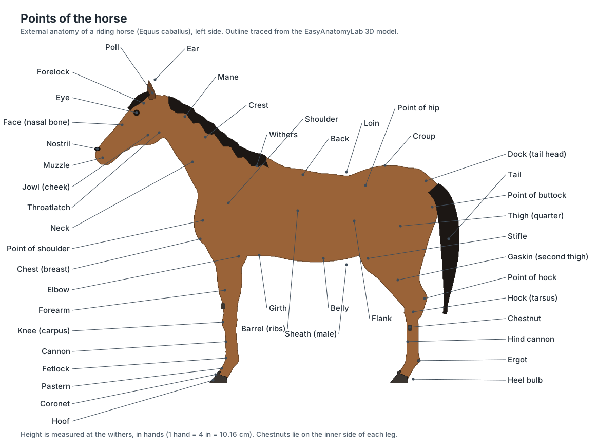
Several names are confusing at first. The knee of the front leg is the carpus, the equivalent of your wrist. The stifle is the true knee and sits high on the hind leg, at the level of the flank, and the hock is the equivalent of your ankle. The gaskin (second thigh) is the hind leg's shin and calf, and the cannon is the long lower leg over the cannon bone. The chestnuts are horny patches on the inside of each leg, and the ergots are small horny nubs at the back of each fetlock.
| Region | Points of the horse |
|---|---|
| Head | Poll, forelock, ears, forehead, eye, face, nostrils, muzzle (lips and chin), jowl, throatlatch |
| Neck and topline | Crest, mane, neck, withers, back, loin, croup, dock (tail head), tail |
| Trunk | Chest (breast), girth, barrel (ribs), belly, flank, point of hip, sheath or udder |
| Front leg | Point of shoulder, shoulder and arm, elbow, forearm, chestnut, knee, cannon, fetlock, ergot, pastern, coronet, hoof |
| Hind leg | Point of buttock, thigh (quarter), stifle, gaskin, point of hock, hock, chestnut, cannon, fetlock, ergot, pastern, coronet, hoof |
A horse's height is measured from the ground to the top of the withers, in hands of 4 inches (10.16 cm). The withers are used because they are the highest point of the back that does not move with the head and neck: they are formed by the long spinous processes of the first thoracic vertebrae.
Horse anatomy labeled: every structure in the model
This is the text version of the 3D model: every labeled structure, grouped by body system, with what it does. Click any name in the panel beside the model to find it in 3D.
Points of the horse
- Muzzle (Lips, chin and nose)
- The front of the face: nostrils, upper and lower lips and chin, covered by fine skin with long tactile whiskers. Selects food: the mobile upper lip sorts grass and feed before the incisors bite it off.
- Nostrils (Nares)
- Two large, flexible openings on the muzzle that can flare widely during hard breathing. The only airway into the body: horses are obligate nasal breathers and cannot breathe through the mouth.
- Forelock
- The tuft of mane hair that grows from the poll and falls forward between the ears over the forehead. Shelters the eyes and forehead from flies and sun.
- Poll
- The highest point of the head, just behind the ears, over the joint between skull and first neck vertebra. Where the head pivots on the neck; the funicular part of the nuchal ligament attaches to the skull here.
- Ears (Pinnae)
- Two tall, mobile external ears that the horse can turn independently. Funnel sound to the ear canal and signal attention and mood to other horses.
- Head (face, forehead and jowl) (Facial region, cheek, jowl)
- The long head of the horse: a flat forehead between the eyes, the long straight face, and the rounded jowl over the lower jaw. Holds the brain, eyes, ears, the long nasal passages and the large grinding teeth.
- Throatlatch (Throttle)
- The narrow area where the underside of the head joins the neck, behind the jaw. Space for the larynx, pharynx and guttural pouches; a clean throatlatch lets the horse flex at the poll.
- Crest
- The curved top line of the neck from the poll to the withers, from which the mane grows. Covers the nuchal ligament and a firm fatty pad, the nuchal fatty crest.
- Mane
- Long coarse hair growing from the crest, usually trained to fall to one side, traditionally the right (off) side. Insulates and sheds rain from the neck and protects it from flies.
- Neck
- The long, muscular neck carried by seven cervical vertebrae, flattened from side to side. Moves the head up, down and sideways for grazing and balance; the jugular groove along its lower side marks the jugular vein.
- Withers
- The ridge between the shoulder blades, formed by the long spinous processes of the cranial thoracic vertebrae. The highest fixed point of the back: a horse's height is measured from the ground to the top of the withers.
- Back
- The part of the top line behind the withers, over the caudal thoracic vertebrae, where the saddle sits. Carries the rider and transmits the thrust of the hindlimbs to the forehand.
- Loin (Coupling)
- The short region over the lumbar vertebrae, between the last rib and the croup. Couples the back to the hindquarters; a short, strong loin transfers propulsion efficiently.
- Croup (Rump)
- The top of the hindquarters from the loin to the root of the tail, over the sacrum and pelvis. Covers the large gluteal muscles that drive the horse forward.
- Tail (dock and tail hair) (Dock)
- The dock, the fleshy part containing the caudal vertebrae, and the long tail hair that grows from it. Swats flies, signals mood, and helps balance; the hair grows from the skin of the dock.
- Point of the hip (Tuber coxae)
- The bony prominence at the front corner of the hindquarters, formed by the tuber coxae of the ilium. Origin of the internal abdominal oblique and tensor fasciae latae muscles.
- Point of the buttock (Tuber ischii region)
- The rearmost point of the hindquarters, over the tuber ischii of the pelvis, with the hamstring muscles below it. Body length is measured from the point of the shoulder to the point of the buttock.
- Thigh (quarter)
- The muscular hindquarter from the hip joint down to the stifle, over the femur. Holds the biceps femoris, tensor fasciae latae and quadriceps, the main muscles for propulsion and jumping.
- Stifle
- The hindlimb joint equivalent to the human knee, at the lower front of the thigh near the flank. Femorotibial and femoropatellar joints; it can be locked by the patella to rest the hindlimb.
- Gaskin (Second thigh, crus)
- The muscular region between the stifle and the hock, over the tibia. Holds the gastrocnemius behind and the digital extensors in front.
- Hock (Tarsus)
- The large angled hindlimb joint, equivalent to the human ankle, with the point of the hock at the back. Bends and straightens with the stifle through the reciprocal apparatus; most hindlimb thrust passes through it.
- Cannon
- The lower leg between the knee (or hock) and the fetlock, over the cannon bone and the tendons behind it. A long, light lever: it has no muscles of its own, only tendons and ligaments.
- Fetlock (Metacarpo-/metatarsophalangeal joint)
- The joint between the cannon bone and the long pastern bone, with the proximal sesamoid bones at its back. Sinks under load and springs back, storing energy in the suspensory apparatus.
- Pastern
- The short sloping region between the fetlock and the hoof, over the long and short pastern bones. Absorbs concussion by flexing at the pastern joint and transmitting load to the hoof.
- Chest (breast)
- The broad front of the trunk between the forelegs, below the neck. Covers the pectoral muscles and the thoracic inlet.
- Point of the shoulder
- The bony prominence at the front of the shoulder joint, formed by the greater tubercle of the humerus. Landmark for measuring body length and shoulder angle.
- Shoulder and arm
- The sloping region over the scapula, and below it the arm over the humerus, between the withers and the elbow. A long, sloping shoulder gives a long stride because the forelimb is not fixed to the trunk by bone.
- Elbow
- The joint between the arm and the forearm, with the point of the elbow (olecranon) behind it, close to the chest wall. Extended by the triceps brachii, which inserts on the olecranon.
- Forearm (Antebrachium)
- The muscular upper foreleg between the elbow and the knee, over the radius and ulna. Holds the extensor and flexor muscles of the carpus and digit.
- Knee (Carpus)
- The horse's 'knee' on the foreleg is anatomically the carpus, equivalent to the human wrist. A compound joint of 7–8 carpal bones that locks straight when the horse stands.
- Barrel (ribs)
- The rounded side of the trunk over the ribs, between the shoulder and the flank. Encloses the lungs, heart and much of the gut; a well-sprung rib cage gives room for the large intestine.
- Girth (Heart girth)
- The underside of the chest just behind the elbows, where the saddle girth passes. Heart girth measured with a tape here is used, with body length, to estimate body weight.
- Belly (Abdomen (ventral))
- The underside of the abdomen between the girth and the sheath or udder. Supported by strong abdominal muscles and the yellow elastic tunic (tunica flava) that carry the heavy gut.
- Flank
- The side of the abdomen between the last rib and the thigh. Its movement is watched to count breathing rate; horses have no hollow paralumbar fossa like cattle.
- Chestnuts
- Small, flat, horny patches on the inside of each leg: above the knee in front, below the hock behind. No known function in the living horse.
- Ergots
- Small horny nubs at the back of each fetlock, often hidden in the feather. No clear function; they are thought to be remnants of foot pads.
- Sheath (Prepuce) · males only
- The double fold of skin on the belly of stallions and geldings that encloses the retracted penis. Protects the penis, which is drawn up inside it except when urinating or breeding.
- Scrotum · males only
- The skin pouch between the hind legs of a stallion that holds the two testes. Keeps the testes cooler than the body, which sperm production requires.
- Udder (Mammary glands) · females only
- Two small mammary glands between the hind legs of the mare, each with one teat. Secretes colostrum and then milk for the foal.
Hoof
- Hoof wall
- The hard outer horn of the hoof, visible when the horse stands; divided into toe, quarters and heels. Bears most of the horse's weight and protects the foot; it is anchored to the coffin bone by the laminae.
- Periople (Stratum externum)
- A thin, shiny, varnish-like layer of soft horn on the outside of the upper wall, just below the coronet. Reduces moisture loss from the wall.
- Coronet (coronary band) (Coronary corium, coronary dermis)
- The band at the top of the hoof where the hair ends; under it lies the coronary corium that produces the wall. Grows the hoof wall: horn tubules form over its papillae and are pushed down towards the ground.
- Laminae (Lamellae; sensitive (dermal) and insensitive (epidermal) laminae)
- Hundreds of interlocking leaf-like folds that join the inside of the hoof wall to the coffin bone. Suspend the coffin bone and the horse's weight inside the hoof.
- White line
- The pale line on the bottom of the foot where the wall meets the sole. A somewhat flexible junction between wall and sole; farriers use it as a guide for safe nail placement.
- Sole
- The slightly concave plate of horn that covers most of the bottom of the hoof. Protects the coffin bone and solar corium from below; on soft ground it shares some of the load with the frog.
- Frog
- The V-shaped, rubbery wedge of horn in the back half of the sole, pointing towards the toe. Absorbs shock, gives grip and, with the digital cushion, helps pump blood out of the foot at each step.
- Bars
- Ridges of wall horn that turn forward from each heel along the sides of the frog. Continuations of the wall that brace the heels and add bearing surface.
- Heel bulbs (Bulbs of the heel)
- The two soft, rounded swellings at the back of the foot above the frog. Cushion the back of the foot; they cover the digital cushion.
- Digital cushion (Plantar or palmar cushion)
- A wedge of fibrous and fatty tissue that fills the back of the foot above the frog. A major shock absorber; squeezed at each step, it also helps push blood out of the foot.
- Collateral cartilages (Ungual or lateral cartilages)
- Two curved plates of cartilage attached to the wings of the coffin bone, one on each side of the back of the foot. Let the heels expand under load and support the digital cushion and venous plexuses.
- Navicular bursa (Podotrochlear bursa)
- A small fluid-filled sac between the navicular bone and the deep digital flexor tendon. Lets the deep digital flexor tendon glide over the navicular bone with little friction.
Hoof section (enlarged)
- Hoof section (enlarged ×4) (Sagittal section of a front foot)
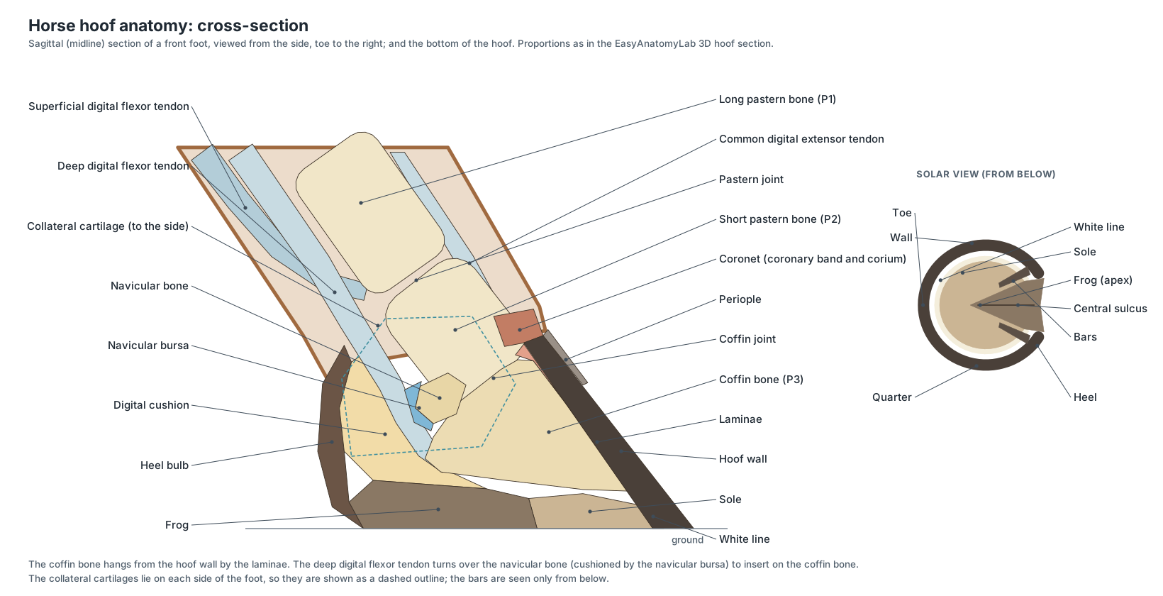
- A front hoof cut down the midline and enlarged four times, standing on a plinth in front of the horse. Shows the parts hidden inside the hoof: laminae, coffin and navicular bones, navicular bursa, deep digital flexor tendon, digital cushion and collateral cartilage.
Skeleton
- Incisive bone (Premaxilla)
- The paired front bones of the upper jaw; each carries three upper incisors, six in all. Holds the upper incisor teeth and frames the nasal opening (nasoincisive notch).
- Nasal bones
- Two long, narrow bones forming the roof of the nasal cavity along the bridge of the nose. Roof the long nasal passages that warm and filter inhaled air.
- Maxilla
- The large upper jaw bone on each side of the face, carrying the upper cheek teeth. Holds the six upper cheek teeth and encloses the maxillary sinuses; its facial crest anchors the masseter.
- Lacrimal bone
- A small bone forming the front rim of the orbit. Houses the start of the nasolacrimal duct that drains tears to the nostril.
- Zygomatic bone (Malar bone)
- The bone below the orbit that forms the front of the zygomatic arch. Part of the cheek arch that braces the skull against chewing forces.
- Frontal bones
- The paired bones of the forehead between and behind the eyes. Roof the front of the braincase; the supraorbital process forms a complete bony ring behind the eye.
- Parietal bones
- Paired bones forming the roof of the braincase behind the frontal bones. Protect the cerebral hemispheres; the temporalis muscle attaches along the temporal lines.
- Temporal bones
- The side walls of the braincase, which carry the jaw joint and the ear. Form the temporomandibular joint with the mandible and contain the middle and inner ear.
- Occipital bone
- The bone at the back of the skull, around the foramen magnum. Its condyles articulate with the atlas; the nuchal crest anchors the nuchal ligament and neck muscles.
- Mandible (Lower jaw)
- The lower jaw: two long halves joined at the chin, each with a tall ramus behind. Holds the lower incisors and cheek teeth; moved side to side by the masseter and pterygoid muscles to grind forage.
- Hyoid apparatus
- A chain of small bones that suspends the tongue and larynx from the skull. Anchors the tongue, pharynx and larynx.
- Atlas (C1)
- The first neck vertebra: a ring with broad wings and no body. Carries the head; the atlanto-occipital joint lets the horse nod.
- Axis (C2)
- The second neck vertebra, the longest vertebra in the body, with a blade-like spinous process. Its dens forms a pivot with the atlas so the head can rotate.
- Cervical vertebrae C3–C7
- The remaining five neck vertebrae; long bodies that run low in the neck in an S-shaped curve. Support and move the neck; their transverse foramina carry the vertebral artery.
- Thoracic vertebrae (T1–T18)
- Eighteen vertebrae that carry the ribs; the tall spinous processes of the cranial ones form the withers. Anchor the ribs and the back muscles and support the rider.
- Lumbar vertebrae (L1–L6)
- Usually six vertebrae with long, flat transverse processes, in the loin. Form a stiff bridge that carries hindlimb thrust forward.
- Sacrum
- Five sacral vertebrae fused into one triangular bone under the croup. Transmits hindlimb thrust from the pelvis to the spine through the sacroiliac joints.
- Caudal vertebrae (Coccygeal vertebrae, tail bones)
- The vertebrae of the dock, usually 15–21, getting smaller and simpler towards the tip. Support and move the tail.
- True ribs (sternal ribs 1–8)
- The first eight pairs of ribs, whose costal cartilages join the sternum directly. Form a stiff front to the chest that protects the heart and carries the forelimb muscles.
- False ribs (asternal ribs 9–18)
- The last ten pairs of ribs, whose cartilages join the one in front to form the costal arch. Enclose the caudal lungs, liver, stomach and spleen; their movement aids breathing.
- Costal cartilages
- The bars of cartilage that continue each rib to the sternum or to the costal arch. Give the rib cage flexibility for breathing.
- Sternum (Breastbone)
- A canoe-shaped breastbone of seven or eight segments (sternebrae) joined by cartilage, with a keel-like cartilage at the front. Anchors the true ribs and the pectoral muscles.
- Scapula (Shoulder blade)
- The long, triangular shoulder blade with a spine but no acromion, topped by a wide scapular cartilage. Attached to the trunk only by muscles: horses have no clavicle (collarbone).
- Humerus
- The short, stout arm bone between the shoulder joint and the elbow. Lever for the shoulder and elbow muscles.
- Radius
- The main weight-bearing bone of the forearm. Carries body weight from the elbow to the carpus.
- Ulna
- A reduced bone fused to the back of the radius; only its top, the olecranon, is large. The olecranon is the lever arm for the triceps brachii, which extends the elbow.
- Carpal bones (Carpus, 'knee')
- Seven or eight small bones in two rows: radial, intermediate, ulnar and accessory above; (first), second, third and fourth below. Form the knee joint, which flexes the foreleg and locks straight under load.
- Accessory carpal bone
- A disc-shaped bone projecting from the back of the knee. A lever for the flexor muscles of the carpus; it forms the wall of the carpal canal.
- Cannon bones (Third metacarpal (MC III) and third metatarsal (MT III))
- The single large bone between the knee or hock and the fetlock, the main bone of the horse's one remaining toe. Carries all the weight of the limb; its distal condyle forms the fetlock joint.
- Splint bones (MC/MT II (medial) and MC/MT IV (lateral))
- Two slender bones lying along the back edges of each cannon bone and ending about two-thirds of the way down. Help support the carpus and tarsus; they are the remains of digits II and IV of three-toed ancestors.
- Proximal sesamoid bones
- A pair of small pyramid-shaped bones behind each fetlock joint. Form a groove for the flexor tendons and receive the suspensory ligament.
- Long pastern bone (P1) (Proximal phalanx, first phalanx)
- The first bone of the digit, between the fetlock and the pastern joint. Transfers load from the fetlock to the short pastern bone.
- Short pastern bone (P2) (Middle phalanx, second phalanx)
- A short cube of bone between the pastern joint and the coffin joint, half inside the hoof. The superficial digital flexor tendon inserts on it.
- Coffin bone (P3) (Pedal bone, distal phalanx, third phalanx)
- The crescent-shaped last bone of the digit, entirely inside the hoof and shaped like a small hoof. Suspended from the wall by the laminae; the common digital extensor and deep digital flexor tendons insert on it.
- Navicular bone (Distal sesamoid bone)
- A small boat-shaped bone behind the coffin joint, over which the deep digital flexor tendon turns. Acts as a pulley that keeps the deep digital flexor tendon at a constant angle of insertion.
- Ilium
- The largest part of the pelvis: a wide wing with the point of the hip and the point of the croup. Joins the sacrum at the sacroiliac joint and anchors the gluteal and abdominal muscles.
- Ischium
- The caudal part of the pelvis, ending in the point of the buttock. Origin of the hamstring muscles; forms the back of the pelvic floor.
- Pubis
- The smallest part of the pelvis, forming the front of the pelvic floor. Anchors the prepubic tendon, which carries the weight of the abdominal muscles and viscera.
- Femur (Thigh bone)
- The heavy thigh bone between the hip and the stifle, with a large third trochanter on its side. Lever for the powerful gluteal and thigh muscles; its trochlea guides the patella.
- Patella (Kneecap)
- A sesamoid bone at the front of the stifle, held by three patellar ligaments. Increases the leverage of the quadriceps; with the medial patellar ligament it locks the stifle.
- Tibia
- The main bone of the gaskin, between the stifle and the hock. Carries body weight; its tuberosity receives the patellar ligaments.
- Fibula
- A thin, incomplete bone on the lateral side of the tibia. Gives attachment to muscles; it carries no significant weight.
- Talus (Tibial tarsal bone)
- The hock bone with a deep, spiral trochlea that meets the tibia. Forms the hinge of the tibiotarsal joint.
- Calcaneus (Fibular tarsal bone, point of the hock)
- The long hock bone that projects up and back to form the point of the hock. The lever arm on which the gastrocnemius and superficial digital flexor pull to straighten the hock.
- Distal tarsal bones (Central, third, fourth and fused first-second tarsal bones)
- The flat bones of the lower hock, in two rows below the talus and calcaneus. Form the low-motion intertarsal and tarsometatarsal joints that absorb shock.
Teeth
- Incisors
- Six upper and six lower chisel-shaped front teeth, arranged in an arc: central, intermediate and corner. Crop grass close to the ground.
- Canine teeth (Tushes) · males only
- Small pointed teeth in the gap between incisors and cheek teeth, usually present only in males. Used for fighting in stallions; no role in eating.
- Wolf teeth (First premolars)
- Small, often missing teeth just in front of the upper cheek teeth. No function; they are often removed because they can interfere with the bit.
- Cheek teeth (Premolars 2–4 and molars 1–3)
- Six large, square grinding teeth in each row, with long (hypsodont) crowns that keep erupting through life. Grind fibrous forage between their ridged enamel tables.
Muscles
- Masseter
- The large, flat chewing muscle of the cheek. Closes the jaw and, working with the opposite medial pterygoid, grinds the teeth side to side.
- Brachiocephalicus
- A long strap muscle from the head and neck to the arm. Pulls the forelimb forward (protraction) or, with the limb fixed, bends the neck to one side.
- Sternocephalicus (Sternomandibularis)
- A narrow muscle along the underside of the neck from the sternum to the jaw. Flexes the head and neck; its upper border forms the lower edge of the jugular groove.
- Splenius
- A broad, flat muscle in the upper neck under the crest. Raises and extends the head and neck.
- Serratus ventralis
- A fan-shaped sling muscle running from the neck and ribs to the inner face of the scapula. The main muscle that carries the trunk between the forelimbs.
- Trapezius
- A thin triangular muscle over the withers and scapular spine. Elevates and swings the scapula.
- Deltoideus
- The muscle on the back of the shoulder, below the scapular spine. Flexes the shoulder joint.
- Biceps brachii
- A spindle-shaped muscle on the front of the humerus, packed with tendinous tissue. Flexes the elbow and, as a near-passive cable, holds the shoulder extended under load as part of the forelimb stay apparatus.
- Triceps brachii
- The large triangular muscle mass between the shoulder blade and the elbow. Extends the elbow so the foreleg can bear weight.
- Pectoral muscles
- The superficial and deep chest muscles between the forelegs. Hold the forelegs against the chest (adduction); the deep pectorals pull the limb back in the power stroke.
- Latissimus dorsi
- A broad, fan-shaped muscle from the back to the inside of the arm. Pulls the forelimb back and flexes the shoulder.
- Longissimus dorsi
- The longest muscle in the body, lying along each side of the spine. Extends and stabilises the back and bends it to the side.
- External abdominal oblique
- The outermost abdominal muscle, from the ribs and thoracolumbar fascia to a wide tendinous sheet (aponeurosis) on the belly. Supports the heavy gut and helps forced expiration.
- Gluteal muscles (Middle and superficial gluteal)
- The massive muscles of the croup; the middle gluteal is one of the largest muscles in the horse. Extend the hip, driving the horse forward and powering rearing, kicking and jumping.
- Tensor fasciae latae
- A triangular muscle at the front of the thigh from the point of the hip to the stifle. Flexes the hip and extends the stifle by tensing the fascia lata.
- Biceps femoris
- The large muscle of the side of the thigh, one of the hamstrings. Extends the hip and helps extend the hock in propulsion.
- Semitendinosus
- A hamstring muscle along the back of the thigh. Extends the hip and hock and flexes the stifle.
- Gastrocnemius
- The calf muscle at the back of the gaskin. Extends the hock through the common calcanean (Achilles) tendon.
- Long digital extensor
- The main extensor muscle on the front of the gaskin. Extends the hind digit and flexes the hock.
- Extensor carpi radialis
- The largest muscle at the front of the forearm. Extends the knee; linked to the biceps by the lacertus fibrosus as part of the stay apparatus.
- Common digital extensor
- The forearm muscle whose long tendon runs down the front of the cannon and pastern to the coffin bone. Extends the digit and knee, lifting the toe as the leg swings forward.
Tendons & stay apparatus
- Nuchal ligament
- A strong elastic ligament from the poll to the withers that holds up the head and neck. Supports the weight of the head without muscular effort and helps raise it after grazing.
- Suspensory ligament (Interosseous medius (interosseous tendon))
- A strong, partly elastic band behind the cannon bone that slings the fetlock. Supports the fetlock so it does not sink to the ground under load; the core of the suspensory apparatus.
- Superficial digital flexor tendon (SDFT)
- The flexor tendon nearest the skin at the back of the cannon, which inserts on the pastern bones. Flexes the digit and, anchored by its check ligament, supports the fetlock passively.
- Deep digital flexor tendon (DDFT)
- The flexor tendon that runs under the SDFT, over the navicular bone, to the solar surface of the coffin bone. Flexes the coffin joint and supports the fetlock and pastern.
- Accessory ligament of the SDFT (Proximal (superior or radial) check ligament)
- A short band from the back of the radius to the SDFT above the knee. Turns the SDFT into a passive support that resists sinking of the fetlock.
- Accessory ligament of the DDFT (Distal (inferior or carpal) check ligament)
- A strong band from the back of the knee (or hock) that joins the DDFT in the upper cannon. Lets the DDFT support the digit without muscle effort.
- Distal sesamoidean ligaments (Straight, oblique and cruciate sesamoidean ligaments)
- Ligaments from the base of the sesamoid bones down to the pastern bones. Continue the suspensory apparatus from the sesamoids to the digit.
- Lacertus fibrosus
- A tendinous band that links the biceps brachii to the extensor carpi radialis. Forms a fibrous 'cable' from the scapula to the cannon bone that holds the shoulder and knee without effort.
- Patellar ligaments (Medial, middle and lateral)
- Three strong ligaments from the patella to the tibial tuberosity. The patella with its medial and middle ligaments forms a loop that hooks over the medial trochlear ridge and locks the stifle.
- Fibularis (peroneus) tertius
- An entirely tendinous cord down the front of the tibia from the femur to the hock and cannon. With the SDFT behind, forms the reciprocal apparatus: the hock must flex and extend whenever the stifle does.
Digestive
- Tongue
- The long muscular tongue that fills the mouth between the cheek-tooth rows. Gathers food and moves it between the cheek teeth and back for swallowing.
- Salivary glands (Parotid, mandibular and sublingual glands)
- Large glands around the jaw; the parotid lies behind the jaw below the ear. Produce saliva only while chewing, to moisten dry forage.
- Esophagus
- The muscular tube, about 1.2 m long, that carries food from the pharynx to the stomach. Moves swallowed food to the stomach.
- Stomach
- A small, J-shaped stomach that holds only about 8–15 L, about a tenth of the gut's volume. Begins protein digestion with acid and pepsin; food passes through quickly, so horses eat little and often.
- Liver
- A large lobed organ on the abdominal face of the diaphragm, mostly to the right. Makes bile and processes absorbed nutrients; horses have no gallbladder, so bile flows continuously.
- Pancreas
- A triangular gland in the dorsal abdomen near the duodenum. Secretes digestive enzymes and buffer into the duodenum and insulin into the blood.
- Duodenum
- The first part of the small intestine, held close to the body wall. Mixes food with bile and pancreatic juice.
- Jejunum
- The long, coiled main part of the small intestine, about 19–20 m long. Digests and absorbs sugars, starch, fat and protein.
- Ileum
- The short, thick-walled last part of the small intestine. Pushes ingesta into the cecum.
- Cecum (Caecum)
- A huge, comma-shaped, blind-ended fermentation vat, about 1.2–1.5 m long and holding about 27–30 L. Microbes ferment fibre into volatile fatty acids, a major energy source for the horse.
- Right ventral colon
- The first part of the large colon, wide and sacculated, running forward along the right abdominal floor. Continues microbial fermentation of fibre.
- Left ventral colon
- The sacculated colon running back along the left abdominal floor to the pelvic flexure. Fermentation and absorption of water and fatty acids.
- Pelvic flexure
- The tight hairpin bend near the pelvis where the large colon turns forward and narrows sharply. Slows the passage of coarse fibre back into the dorsal colon.
- Left dorsal colon
- The narrow, smooth colon running forward above the left ventral colon from the pelvic flexure. Carries digesta forward to the diaphragmatic flexure.
- Right dorsal colon
- The widest part of the colon, running back along the right side after the diaphragmatic flexure. Absorbs water and nutrients before the narrow transverse colon.
- Transverse colon
- A short colon segment, about 10 cm wide, that crosses from right to left in front of the cranial mesenteric artery. Links the large colon to the small colon.
- Small colon (Descending colon)
- A long, narrow, sacculated colon, about 3–3.6 m long, where the familiar fecal balls are formed. Absorbs water and forms the feces into balls.
- Rectum
- The last part of the gut, running through the pelvic cavity to the anus. Stores feces before defecation; veterinarians palpate abdominal organs through its wall.
- Anus
- The muscular opening of the rectum under the tail. Controls defecation with the internal and external anal sphincters.
Respiratory
- Nasal diverticulum (False nostril)
- A blind, hair-lined skin pouch in the upper part of each nostril. Allows the nostril to dilate widely during hard breathing.
- Nasal cavity and conchae (Turbinates; dorsal, ventral and ethmoidal conchae)
- The long nasal passages, divided into meatuses by scroll-like dorsal and ventral conchae. Warm, moisten and filter inhaled air; the ethmoidal conchae carry the smell receptors.
- Paranasal sinuses (Conchofrontal and maxillary sinuses)
- Air-filled spaces in the skull bones: the conchofrontal sinus above and the rostral and caudal maxillary sinuses below. Lighten the large skull and drain into the nasal cavity.
- Pharynx and soft palate
- The shared airway and food passage behind the mouth, divided by a long soft palate. The soft palate seals around the larynx so air passes from the nose straight into the larynx.
- Guttural pouches
- Two large air sacs, outpouchings of the auditory (Eustachian) tubes; among domestic animals only equids have them. Their role is uncertain; one hypothesis is that they help cool blood in the internal carotid artery on its way to the brain. The carotid arteries and cranial nerves IX–XII run in their walls.
- Larynx
- The voice box at the top of the trachea, made of cricoid, thyroid, arytenoid and epiglottic cartilages. Opens the airway during breathing, closes it during swallowing and produces the whinny.
- Epiglottis
- A leaf-shaped flap of cartilage at the front of the larynx. Helps direct food around the larynx during swallowing.
- Trachea (Windpipe)
- The windpipe, stiffened by C-shaped cartilage rings, running down the underside of the neck. Carries air between the larynx and the lungs.
- Bronchi
- The two main airways that branch from the trachea into the lungs. Distribute air through the bronchial tree to the alveoli.
- Lungs
- Two large lungs filling the chest, long and sloping to follow the diaphragm. Gas exchange. A galloping horse can move very large volumes of air, breathing once per stride.
- Diaphragm
- The dome-shaped muscle between chest and abdomen, which bulges far forward into the chest. The main muscle of inspiration.
Heart & vessels
- Heart
- A large four-chambered heart in the lower chest between the elbows. Pumps blood through the lungs (right side) and the body (left side).
- Aorta
- The main artery: it leaves the left ventricle, arches up and runs back under the spine. Carries oxygen-rich blood to the whole body.
- Pulmonary trunk
- The artery from the right ventricle that divides into the left and right pulmonary arteries. Carries oxygen-poor blood to the lungs.
- Cranial vena cava
- The large vein that returns blood from the head, neck and forelimbs to the right atrium. Formed by the joining of the jugular and subclavian (axillary) veins.
- Caudal vena cava
- The large vein that returns blood from the abdomen, pelvis and hindlimbs to the right atrium. Collects the hepatic veins and the veins of the kidneys and hindlimbs.
- Common carotid arteries
- Paired arteries running up the neck beside the trachea to the head. Supply the head; at the throat they divide into internal and external carotid arteries.
- External jugular veins
- The large superficial veins in the jugular groove on each side of the neck. Drain the head; the usual site for blood sampling and intravenous injection.
- Spleen
- A large, flat, sickle-shaped organ on the left side that stores red blood cells. Contracts during exercise and excitement, releasing stored red cells to carry more oxygen.
- Digital arteries and veins
- Paired vessels running down both sides of each fetlock and pastern into the hoof. Supply the hoof; the digital pulse felt here strengthens in laminitis and foot abscesses.
Urinary
- Kidneys
- Two kidneys under the lumbar spine: the right heart-shaped and further forward, the left bean-shaped. Filter the blood and make urine.
- Ureters
- Two thin tubes carrying urine from the kidneys to the bladder. Carry urine by peristalsis.
- Urinary bladder
- A muscular sac on the floor of the pelvis that stores urine. Stores urine and empties it through the urethra.
Reproductive
- Testes · males only
- Two oval testes held in the scrotum. Produce sperm and testosterone.
- Epididymis · males only
- The coiled duct along each testis where sperm mature and are stored. Matures and stores sperm.
- Ductus deferens (Vas deferens) · males only
- The duct that carries sperm from the epididymis through the inguinal canal to the urethra. Transports sperm at ejaculation.
- Accessory sex glands (Ampullae, vesicular glands, prostate, bulbourethral glands) · males only
- The stallion has all four sets: ampullae, vesicular glands (seminal vesicles), prostate and bulbourethral glands. Produce most of the fluid of semen.
- Penis · males only
- A musculocavernous ('vascular') penis that enlarges greatly with blood when erect. Carries urine and semen; the urethral process protrudes at the tip of the glans.
- Ovaries · females only
- Two large, kidney-shaped ovaries with an ovulation fossa, hanging below the kidneys. Produce eggs, estrogen and progesterone.
- Uterine tubes (Oviducts) · females only
- Small coiled tubes from each ovary to the tip of a uterine horn. Catch the egg and are the site of fertilisation.
- Uterus · females only
- A uterus with a large body and two horns, giving a T- or Y-shape. Holds the developing foal for about 11 months.
- Cervix · females only
- The thick muscular neck of the uterus that projects into the vagina. Separates the uterus from the vagina and protects it from infection.
- Vagina and vestibule · females only
- The muscular passage from the cervix to the vulva. Receives the penis at mating and forms the birth canal.
- Vulva · females only
- The external genital opening of the mare, a vertical slit below the anus. Closes the genital tract; its lips (labia) protect it from contamination.
Nervous & senses
- Cerebrum
- The two cerebral hemispheres, the largest part of the brain. Senses, learning, memory and voluntary movement.
- Cerebellum
- The folded 'little brain' at the back of the braincase. Coordinates movement and balance.
- Brainstem
- The stalk of the brain that connects the cerebrum to the spinal cord. Controls breathing, heart rate and swallowing; most cranial nerves arise from it.
- Spinal cord
- The cord of nerve tissue in the vertebral canal, ending in the sacral region. Carries signals between the brain and body and controls spinal reflexes.
- Brachial plexus
- The network of nerves from the last neck and first thoracic spinal nerves that supplies the forelimb. Forms the suprascapular, radial, median, ulnar and other forelimb nerves.
- Sciatic nerve
- The largest nerve in the body, supplying most of the hindlimb. Controls the hamstrings and, through its tibial and fibular branches, the muscles below the stifle.
- Vagus nerve and recurrent laryngeal nerve
- Cranial nerve X runs down the neck with the carotid artery; its recurrent laryngeal branch returns up the neck to the larynx. Parasympathetic supply to the heart, lungs and gut; the recurrent branch moves the laryngeal cartilages.
- Palmar and plantar digital nerves
- The nerves that run down each side of the pastern into the foot. Carry sensation from the hoof, including the heel and navicular region.
- Eye
- A very large eye set on the side of the head, with a horizontally oval pupil. Gives an almost all-round view: about 340° of peripheral vision, with 65–80° of binocular overlap in front.
Endocrine glands
- Thyroid gland
- Two oval, red-brown lobes, one on each side of the trachea just behind the larynx. Secretes the thyroid hormones (thyroxine, T4, and triiodothyronine, T3) that set the body's metabolic rate, and calcitonin.
- Adrenal glands
- Two small, flattened glands, one against the cranial pole and medial border of each kidney. The cortex secretes cortisol, aldosterone and small amounts of sex hormones; the medulla releases adrenaline and noradrenaline in the fight-or-flight response.
Horse hoof anatomy: parts of the hoof
The hoof is the hard keratin capsule at the end of each leg, together with everything inside it. Seen from outside, it has a coronet (coronary band) at the hairline, a shiny periople, and the hoof wall, divided into toe, quarters and heels. Underneath are the white line, the sole, the two bars, the V-shaped frog and the heel bulbs. Inside are the laminae, the coffin bone, the navicular bone and bursa, the end of the deep digital flexor tendon, the digital cushion and the two collateral cartilages.
To see the inside of the hoof in 3D, press Hoof section (enlarged) in the row of system buttons under the model. A front hoof cut down the middle, four times life size, appears on a stand in front of the horse; it also appears by itself when you click any part of the hoof, such as the frog or the coffin bone.
The outside of the hoof
- Coronet (coronary band): The band at the top of the hoof where the hair ends; under it lies the coronary corium that produces the wall. Grows the hoof wall: horn tubules form over its papillae and are pushed down towards the ground.
- Periople: A thin, shiny, varnish-like layer of soft horn on the outside of the upper wall, just below the coronet. Reduces moisture loss from the wall.
- Hoof wall: The hard outer horn of the hoof, visible when the horse stands; divided into toe, quarters and heels. Bears most of the horse's weight and protects the foot; it is anchored to the coffin bone by the laminae.
- White line: The pale line on the bottom of the foot where the wall meets the sole. A somewhat flexible junction between wall and sole; farriers use it as a guide for safe nail placement.
- Sole: The slightly concave plate of horn that covers most of the bottom of the hoof. Protects the coffin bone and solar corium from below; on soft ground it shares some of the load with the frog.
- Bars: Ridges of wall horn that turn forward from each heel along the sides of the frog. Continuations of the wall that brace the heels and add bearing surface.
- Frog: The V-shaped, rubbery wedge of horn in the back half of the sole, pointing towards the toe. Absorbs shock, gives grip and, with the digital cushion, helps pump blood out of the foot at each step.
- Heel bulbs: The two soft, rounded swellings at the back of the foot above the frog. Cushion the back of the foot; they cover the digital cushion.
Anatomy of a horse's foot: what is inside the hoof
Anatomists define the equine foot as the hoof and its contents. That includes the coffin bone, the lower half of the short pastern bone, the navicular bone, two joints (the coffin joint and part of the pastern joint), the tendons that end there, and the soft tissues that cushion the back of the foot.
- Laminae: Hundreds of interlocking leaf-like folds that join the inside of the hoof wall to the coffin bone. Suspend the coffin bone and the horse's weight inside the hoof.
- Coffin bone (P3): The crescent-shaped last bone of the digit, entirely inside the hoof and shaped like a small hoof. Suspended from the wall by the laminae; the common digital extensor and deep digital flexor tendons insert on it.
- Navicular bone: A small boat-shaped bone behind the coffin joint, over which the deep digital flexor tendon turns. Acts as a pulley that keeps the deep digital flexor tendon at a constant angle of insertion.
- Navicular bursa: A small fluid-filled sac between the navicular bone and the deep digital flexor tendon. Lets the deep digital flexor tendon glide over the navicular bone with little friction.
- Deep digital flexor tendon (DDFT): The flexor tendon that runs under the SDFT, over the navicular bone, to the solar surface of the coffin bone. Flexes the coffin joint and supports the fetlock and pastern.
- Digital cushion: A wedge of fibrous and fatty tissue that fills the back of the foot above the frog. A major shock absorber; squeezed at each step, it also helps push blood out of the foot.
- Collateral cartilages: Two curved plates of cartilage attached to the wings of the coffin bone, one on each side of the back of the foot. Let the heels expand under load and support the digital cushion and venous plexuses.
Which tissue makes which part of the hoof?
Each horny part grows from its own layer of living dermis (corium), the "quick" of the hoof. Damage to a dermis shows up later as a defect in the horn it makes.
| Horn (insensitive) | Made by (sensitive dermis) | Notes |
|---|---|---|
| Periople | Perioplic dermis | Thin, shiny outer layer; limits drying |
| Hoof wall | Coronary dermis | Grows down at roughly 6–10 mm a month; the toe is the oldest horn |
| Horny laminae | Laminar dermis | About 600 laminar units suspend the coffin bone |
| Sole | Solar dermis | Softer than the wall; flakes as it grows |
| Frog | Frog dermis | Softest part; shock absorption and grip |
How the hoof works
When the foot lands, the heels and frog meet the ground and the frog presses up on the digital cushion, which spreads outward against the collateral cartilages and lets the heels expand. This compresses the network of veins in the foot and helps push blood back up the leg. The wall then carries the load and passes it through the laminae to the coffin bone. Laminitis, inflammation of the laminae, breaks this link: in its chronic form, called founder, the coffin bone can rotate or sink inside the hoof. Farriers trim most horses every six to eight weeks because the wall grows continuously and an overgrown toe changes how load passes through the foot.
Horse leg anatomy: front leg and hind leg
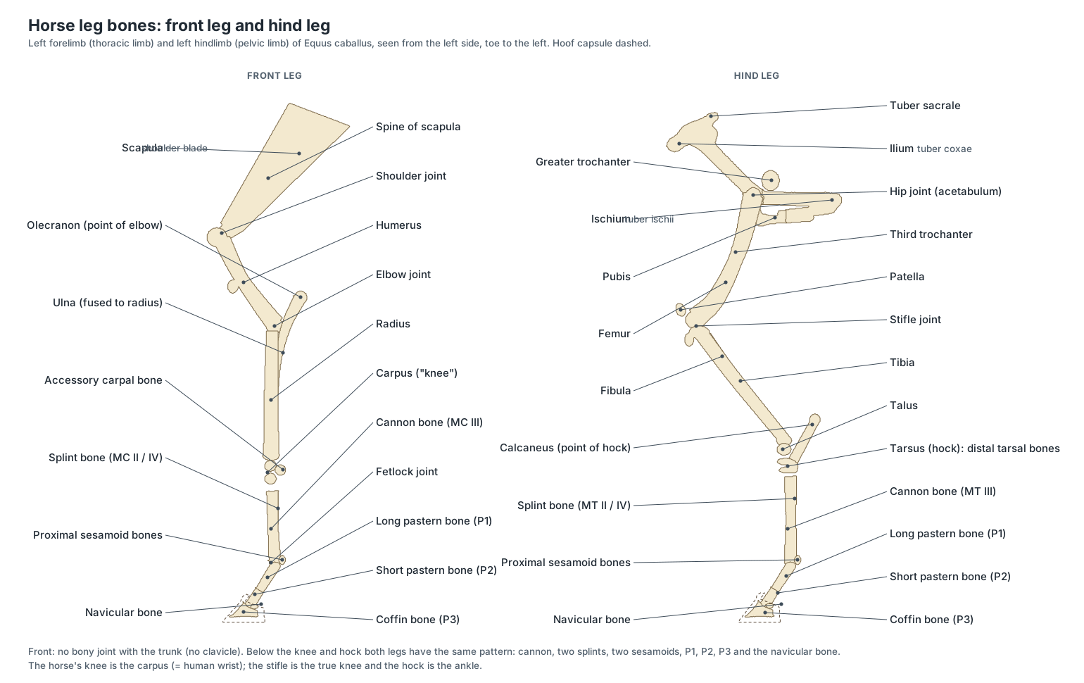
A horse leg has the same basic bones as a human arm or leg, but the horse stands on the tip of its single third digit. The front leg runs from the scapula through the humerus, radius and ulna, the carpus ("knee"), the cannon bone with its two splint bones, the fetlock, the long and short pastern bones and the coffin bone. The hind leg runs from the pelvis through the femur, the patella and stifle, the tibia and reduced fibula, the tarsus (hock), and then the same pattern of cannon, splints, sesamoids and phalanges.
| Horse name | Anatomical name | Human equivalent |
|---|---|---|
| Shoulder | Scapulohumeral joint | Shoulder |
| Elbow | Cubital joint | Elbow |
| Knee (front leg) | Carpus | Wrist |
| Fetlock | Metacarpo- or metatarsophalangeal joint | Knuckle at the base of the middle finger or toe |
| Pastern joint | Proximal interphalangeal joint | First finger joint |
| Coffin joint | Distal interphalangeal joint | Last finger joint |
| Stifle | Femorotibial and femoropatellar joints | Knee |
| Hock | Tarsus | Ankle |
The distal limb: cannon, fetlock and pastern
Below the knee and hock there are no muscle bellies, only bone, tendon, ligament, vessels and nerves under the skin. This keeps the lower leg light, which saves energy at speed, but it also means tendons carry very high loads. At the back of the cannon you can feel, from the skin inward, the superficial digital flexor tendon, the deep digital flexor tendon and the suspensory ligament; at the fetlock, the paired proximal sesamoid bones form a pulley for the flexor tendons.
Horse hock anatomy
The hock (tarsus) has six bones in three rows: the talus and calcaneus above, the central tarsal bone, and the third, fourth and fused first-and-second tarsal bones below. Almost all of its movement happens at the tibiotarsal joint, between the tibia and the deep spiral trochlea of the talus; the lower joints barely move and act as shock absorbers. The point of the hock is the top of the calcaneus, where the gastrocnemius tendon attaches and over which the superficial digital flexor tendon passes like a cap. Arthritis of the low hock joints is called bone spavin; swelling of the large tibiotarsal joint capsule is bog spavin.
Horse stifle anatomy
The stifle joins the femur, tibia and patella and contains the femorotibial and femoropatellar joints, with menisci and cruciate ligaments like a human knee. Unlike a human, a horse has three patellar ligaments (medial, middle and lateral), and the medial ridge of the femoral trochlea is enlarged into a hook. Together they form the locking mechanism described below.
The stay apparatus: how horses sleep standing up
The stay apparatus is a set of ligaments and tendons that lets a horse stand, and doze, with very little muscular effort. In the forelimb, the lacertus fibrosus links the biceps to the extensor carpi radialis and forms a fibrous cable from the scapula to the cannon bone; the suspensory ligament, distal sesamoidean ligaments and the check ligaments of the flexor tendons hold the fetlock and pastern. In the hindlimb the patella locks over the femur, and the reciprocal apparatus (fibularis tertius in front, superficial digital flexor behind) makes the hock follow the stifle, so locking one locks both.
- Lacertus fibrosus: A tendinous band that links the biceps brachii to the extensor carpi radialis. Forms a fibrous 'cable' from the scapula to the cannon bone that holds the shoulder and knee without effort.
- Suspensory ligament: A strong, partly elastic band behind the cannon bone that slings the fetlock. Supports the fetlock so it does not sink to the ground under load; the core of the suspensory apparatus.
- Accessory ligament of the SDFT: A short band from the back of the radius to the SDFT above the knee. Turns the SDFT into a passive support that resists sinking of the fetlock.
- Accessory ligament of the DDFT: A strong band from the back of the knee (or hock) that joins the DDFT in the upper cannon. Lets the DDFT support the digit without muscle effort.
- Distal sesamoidean ligaments: Ligaments from the base of the sesamoid bones down to the pastern bones. Continue the suspensory apparatus from the sesamoids to the digit.
- Patellar ligaments: Three strong ligaments from the patella to the tibial tuberosity. The patella with its medial and middle ligaments forms a loop that hooks over the medial trochlear ridge and locks the stifle.
- Fibularis (peroneus) tertius: An entirely tendinous cord down the front of the tibia from the femur to the hock and cannon. With the SDFT behind, forms the reciprocal apparatus: the hock must flex and extend whenever the stifle does.
- Nuchal ligament: A strong elastic ligament from the poll to the withers that holds up the head and neck. Supports the weight of the head without muscular effort and helps raise it after grazing.
A resting horse usually locks one hind leg and rests the other on the toe, swapping from time to time. Light sleep is possible standing; deep (REM) sleep requires the horse to lie down.
Horse muscle anatomy
Muscle makes up a large share of a fit horse's body mass, and the biggest muscles sit high on the body, close to the trunk: the gluteals and hamstrings of the hindquarters, which drive the horse forward, and the shoulder and chest muscles that hold the forelimbs on. The model shows the main superficial muscles as a layer just under the skin; set the "Skin & muscles" slider to about 70% and hide the skin regions to see them.
- Masseter: The large, flat chewing muscle of the cheek. Closes the jaw and, working with the opposite medial pterygoid, grinds the teeth side to side.
- Brachiocephalicus: A long strap muscle from the head and neck to the arm. Pulls the forelimb forward (protraction) or, with the limb fixed, bends the neck to one side.
- Splenius: A broad, flat muscle in the upper neck under the crest. Raises and extends the head and neck.
- Trapezius: A thin triangular muscle over the withers and scapular spine. Elevates and swings the scapula.
- Serratus ventralis: A fan-shaped sling muscle running from the neck and ribs to the inner face of the scapula. The main muscle that carries the trunk between the forelimbs.
- Triceps brachii: The large triangular muscle mass between the shoulder blade and the elbow. Extends the elbow so the foreleg can bear weight.
- Biceps brachii: A spindle-shaped muscle on the front of the humerus, packed with tendinous tissue. Flexes the elbow and, as a near-passive cable, holds the shoulder extended under load as part of the forelimb stay apparatus.
- Latissimus dorsi: A broad, fan-shaped muscle from the back to the inside of the arm. Pulls the forelimb back and flexes the shoulder.
- Pectoral muscles: The superficial and deep chest muscles between the forelegs. Hold the forelegs against the chest (adduction); the deep pectorals pull the limb back in the power stroke.
- Longissimus dorsi: The longest muscle in the body, lying along each side of the spine. Extends and stabilises the back and bends it to the side.
- External abdominal oblique: The outermost abdominal muscle, from the ribs and thoracolumbar fascia to a wide tendinous sheet (aponeurosis) on the belly. Supports the heavy gut and helps forced expiration.
- Gluteal muscles: The massive muscles of the croup; the middle gluteal is one of the largest muscles in the horse. Extend the hip, driving the horse forward and powering rearing, kicking and jumping.
- Biceps femoris: The large muscle of the side of the thigh, one of the hamstrings. Extends the hip and helps extend the hock in propulsion.
- Semitendinosus: A hamstring muscle along the back of the thigh. Extends the hip and hock and flexes the stifle.
- Gastrocnemius: The calf muscle at the back of the gaskin. Extends the hock through the common calcanean (Achilles) tendon.
Horse digestive system: stomach, cecum and large colon
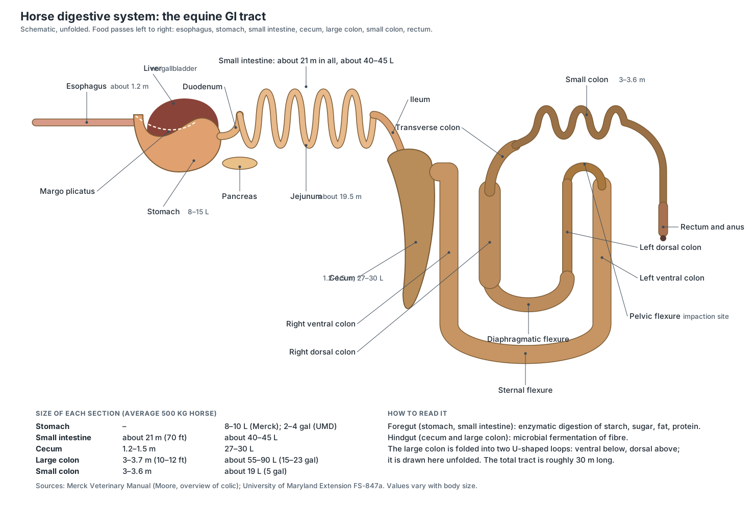
Food passes from the mouth through a 1.2 m esophagus to a small stomach, then through about 21 m of small intestine (duodenum, jejunum and ileum) into the cecum, the large colon, the small colon, the rectum and the anus. The whole tract is roughly 30 m long. The stomach and small intestine digest starch, sugar, fat and protein with enzymes; the cecum and large colon, together called the hindgut, ferment fibre with microbes.
| Section | Typical length | Typical capacity |
|---|---|---|
| Stomach | – | 8–10 L (Merck); 2–4 US gallons, 7.5–15 L (UMD) |
| Small intestine | About 21 m (70 ft); jejunum about 19.5 m | About 40–45 L (10–12 gal) |
| Cecum | About 1.2–1.5 m | About 27–30 L |
| Large colon | About 3–3.7 m (10–12 ft) | About 55–90 L (15–23 gal) |
| Small colon | About 3–3.6 m | About 19 L (5 gal) |
Horse large intestine length
The horse's large intestine is about 7–8 m long in an average horse: roughly 1.2–1.5 m of cecum, 3–3.7 m of large colon and 3–3.6 m of small colon. It is much shorter than the small intestine but holds far more: the large colon alone holds about 55–90 L. Figures differ between sources because they depend on body size and on how the gut is measured.
The large colon: a folded double horseshoe
The large colon starts at the cecum and runs forward along the right floor of the abdomen as the right ventral colon, turns left at the sternal flexure, runs back as the left ventral colon, and makes a hairpin turn at the pelvic flexure. It then runs forward again above itself as the narrow left dorsal colon, turns at the diaphragmatic flexure, and runs back along the right as the wide right dorsal colon, before the short transverse colon crosses to the left and joins the small colon.
- Stomach: A small, J-shaped stomach that holds only about 8–15 L, about a tenth of the gut's volume. Begins protein digestion with acid and pepsin; food passes through quickly, so horses eat little and often.
- Liver: A large lobed organ on the abdominal face of the diaphragm, mostly to the right. Makes bile and processes absorbed nutrients; horses have no gallbladder, so bile flows continuously.
- Cecum: A huge, comma-shaped, blind-ended fermentation vat, about 1.2–1.5 m long and holding about 27–30 L. Microbes ferment fibre into volatile fatty acids, a major energy source for the horse.
- Pelvic flexure: The tight hairpin bend near the pelvis where the large colon turns forward and narrows sharply. Slows the passage of coarse fibre back into the dorsal colon.
- Right dorsal colon: The widest part of the colon, running back along the right side after the diaphragmatic flexure. Absorbs water and nutrients before the narrow transverse colon.
- Small colon: A long, narrow, sacculated colon, about 3–3.6 m long, where the familiar fecal balls are formed. Absorbs water and forms the feces into balls.
Horse head anatomy: teeth, skull and sinuses
Most of a horse's long head is taken up by teeth, sinuses and nasal passages; the brain is small and lies far back, behind the eyes. An adult horse has 36–44 teeth: 12 incisors, 24 cheek teeth (premolars and molars), usually 4 canines in males, and 0–4 small wolf teeth.
- Incisors: Six upper and six lower chisel-shaped front teeth, arranged in an arc: central, intermediate and corner. Crop grass close to the ground.
- Canine teeth: Small pointed teeth in the gap between incisors and cheek teeth, usually present only in males. Used for fighting in stallions; no role in eating.
- Wolf teeth: Small, often missing teeth just in front of the upper cheek teeth. No function; they are often removed because they can interfere with the bit.
- Cheek teeth: Six large, square grinding teeth in each row, with long (hypsodont) crowns that keep erupting through life. Grind fibrous forage between their ridged enamel tables.
The skull contains large paranasal sinuses: a conchofrontal sinus above the eyes and rostral and caudal maxillary sinuses in the cheek, around the roots of the last upper cheek teeth. This is why an infected tooth root can cause a smelly nasal discharge from one nostril. For the individual skull bones see the horse skeleton page.
Equine respiratory anatomy
Air enters only through the nostrils: the horse is an obligate nasal breather, because the soft palate and palatopharyngeal folds seal tightly around the entrance to the larynx. From the nostrils air passes through the long nasal cavity, the pharynx and larynx, the trachea and bronchi, to two large lungs that follow the slope of the diaphragm far back under the ribs.
- Nasal diverticulum: A blind, hair-lined skin pouch in the upper part of each nostril. Allows the nostril to dilate widely during hard breathing.
- Paranasal sinuses: Air-filled spaces in the skull bones: the conchofrontal sinus above and the rostral and caudal maxillary sinuses below. Lighten the large skull and drain into the nasal cavity.
- Guttural pouches: Two large air sacs, outpouchings of the auditory (Eustachian) tubes; among domestic animals only equids have them. Their role is uncertain; one hypothesis is that they help cool blood in the internal carotid artery on its way to the brain. The carotid arteries and cranial nerves IX–XII run in their walls.
- Pharynx and soft palate: The shared airway and food passage behind the mouth, divided by a long soft palate. The soft palate seals around the larynx so air passes from the nose straight into the larynx.
- Larynx: The voice box at the top of the trachea, made of cricoid, thyroid, arytenoid and epiglottic cartilages. Opens the airway during breathing, closes it during swallowing and produces the whinny.
- Trachea: The windpipe, stiffened by C-shaped cartilage rings, running down the underside of the neck. Carries air between the larynx and the lungs.
- Lungs: Two large lungs filling the chest, long and sloping to follow the diaphragm. Gas exchange. A galloping horse can move very large volumes of air, breathing once per stride.
- Diaphragm: The dome-shaped muscle between chest and abdomen, which bulges far forward into the chest. The main muscle of inspiration.
The guttural pouches are found, among domestic animals, only in equids (tapirs, rhinoceroses, hyraxes and some bats have them too). Each is an air-filled outpouching of the auditory tube behind the skull, holding about 300–500 mL. The internal and external carotid arteries, cranial nerves IX, X, XI and XII and the sympathetic trunk run in their walls, so infections of the pouches can cause nosebleeds or difficulty swallowing.
Horse heart and circulation
The horse heart lies in the lower chest between the elbows, tilted so that its apex points down and back towards the sternum. It has the same four chambers and valves as a human heart. The aorta arches up from the left ventricle and runs back under the spine; the jugular veins run in the jugular groove on each side of the neck, where blood samples are taken.
- Heart: A large four-chambered heart in the lower chest between the elbows. Pumps blood through the lungs (right side) and the body (left side).
- Aorta: The main artery: it leaves the left ventricle, arches up and runs back under the spine. Carries oxygen-rich blood to the whole body.
- External jugular veins: The large superficial veins in the jugular groove on each side of the neck. Drain the head; the usual site for blood sampling and intravenous injection.
- Spleen: A large, flat, sickle-shaped organ on the left side that stores red blood cells. Contracts during exercise and excitement, releasing stored red cells to carry more oxygen.
- Digital arteries and veins: Paired vessels running down both sides of each fetlock and pastern into the hoof. Supply the hoof; the digital pulse felt here strengthens in laminitis and foot abscesses.
Horse eye anatomy and vision
Horses have very large eyes set on the sides of the head, with a horizontally oval pupil. They see movement through an arc of about 340° and have a binocular field of about 65–80° in front; their blind spots are directly behind the body and just in front of the forehead.
- Eye: A very large eye set on the side of the head, with a horizontally oval pupil. Gives an almost all-round view: about 340° of peripheral vision, with 65–80° of binocular overlap in front.
Horse reproductive anatomy: male and female
Male and female horses differ in their reproductive organs and in a few external features: stallions and geldings have a sheath on the belly, stallions have a scrotum, and mares have a small udder between the hind legs and a vulva below the anus. Switch the model between Male and Female to compare them.
| Male (stallion) | Female (mare) | |
|---|---|---|
| Gonads | Two testes in the scrotum | Two kidney-shaped ovaries below the kidneys, with an ovulation fossa |
| Ducts | Epididymis and ductus deferens, ending in a large ampulla | Uterine tubes leading to the tips of the uterine horns |
| Glands | Vesicular glands, prostate and bulbourethral glands | Mammary glands (udder) with two teats |
| Organ | Musculocavernous penis without a sigmoid flexure, retracted into the sheath | Uterus with a large body and two horns, cervix, vagina and vulva |
| Notes | Cryptorchidism (retained testis) is more common than in other domestic animals | Pregnancy lasts about 11 months; foals get antibodies only from colostrum |
- Testes: Two oval testes held in the scrotum. Produce sperm and testosterone.
- Accessory sex glands: The stallion has all four sets: ampullae, vesicular glands (seminal vesicles), prostate and bulbourethral glands. Produce most of the fluid of semen.
- Penis: A musculocavernous ('vascular') penis that enlarges greatly with blood when erect. Carries urine and semen; the urethral process protrudes at the tip of the glans.
- Ovaries: Two large, kidney-shaped ovaries with an ovulation fossa, hanging below the kidneys. Produce eggs, estrogen and progesterone.
- Uterus: A uterus with a large body and two horns, giving a T- or Y-shape. Holds the developing foal for about 11 months.
- Cervix: The thick muscular neck of the uterus that projects into the vagina. Separates the uterus from the vagina and protects it from infection.
Nervous system
The brain sits in the braincase behind the eyes and continues as the spinal cord through the vertebral canal to the sacrum. Spinal nerves C6–T2 form the brachial plexus that supplies the forelimb, and L6–S2 contribute to the sciatic nerve of the hindlimb.
- Cerebrum: The two cerebral hemispheres, the largest part of the brain. Senses, learning, memory and voluntary movement.
- Cerebellum: The folded 'little brain' at the back of the braincase. Coordinates movement and balance.
- Spinal cord: The cord of nerve tissue in the vertebral canal, ending in the sacral region. Carries signals between the brain and body and controls spinal reflexes.
- Vagus nerve and recurrent laryngeal nerve: Cranial nerve X runs down the neck with the carotid artery; its recurrent laryngeal branch returns up the neck to the larynx. Parasympathetic supply to the heart, lungs and gut; the recurrent branch moves the laryngeal cartilages.
- Palmar and plantar digital nerves: The nerves that run down each side of the pastern into the foot. Carry sensation from the hoof, including the heel and navicular region.
Endocrine glands
Two endocrine glands are shown in the model: the thyroid gland on the trachea just behind the larynx, and the adrenal glands against the kidneys. The pituitary gland, at the base of the brain, is too small to show.
- Thyroid gland: Two oval, red-brown lobes, one on each side of the trachea just behind the larynx. Secretes the thyroid hormones (thyroxine, T4, and triiodothyronine, T3) that set the body's metabolic rate, and calcitonin.
- Adrenal glands: Two small, flattened glands, one against the cranial pole and medial border of each kidney. The cortex secretes cortisol, aldosterone and small amounts of sex hormones; the medulla releases adrenaline and noradrenaline in the fight-or-flight response.
Horse skeleton
The horse skeleton has about 205 bones: a long skull, a vertebral column of usually C7 T18 L6 S5 and 15–21 caudal vertebrae, 18 pairs of ribs, and four limbs with no collarbone. For a labeled skeleton diagram, the vertebral formula and every bone of the front and hind leg, see the horse skeleton page, which uses the same 3D model.
Why the horse's gut is built backwards compared with a cow's
A horse is a hindgut fermenter. Cattle ferment fibre before the true stomach, in the rumen; the horse digests the easy nutrients first, in a small stomach and a long small intestine, and only then sends the fibrous remainder to the cecum and large colon, where microbes ferment it into volatile fatty acids. The stomach holds only about 8–10 L, the jejunum alone is about 19.5 m long, and the cecum holds about 27–30 L. These proportions explain much of practical horse care: small, frequent meals, plenty of forage, and gradual changes of diet so the hindgut microbes can adapt. (Merck Veterinary Manual: Overview of Colic in Horses (Moore).)
The design has costs. A strong valve at the entrance to the stomach lets food in but not out, so a horse with an obstructed small intestine can rupture its stomach instead of vomiting. The long mesentery of the jejunum lets loops twist or become trapped, and the large colon narrows sharply at the pelvic flexure, the most common site of impaction. Several of the large colon's segments are not attached to the body wall and can displace, for example over the ligament between the spleen and the left kidney. When you study the 3D model, trace the gut in order and mark each place where the tube narrows or is free to move: those are the places where colic usually begins. (Merck Veterinary Manual: Overview of Colic in Horses (Moore).)
The hoof is a living organ, not just a hard shoe
Every part of the hoof capsule is produced by a specialised layer of dermis (corium) under it: the coronary dermis grows the wall, the laminar dermis forms the horny laminae on its inner face, and the sole and frog have their own dermis. About 600 laminar units interlock to carry most or all of the body weight in a standing horse, which is why inflammation of these laminae, laminitis, is so painful. A shod horse on hard ground bears its weight almost entirely on the laminae; an unshod horse on soft ground shares some of the load with the sole and frog. (University of Minnesota: Lab 4, the equine foot.)
The wall itself has three layers: a thin outer stratum externum (the periople), a thick stratum medium of tubular and intertubular horn that bears the load, and an inner stratum internum carrying the laminae that interdigitate with the dermal laminae on the coffin bone. The capsule is not rigid: in galloping Thoroughbreds the heels have been measured to deform by up to almost 16 mm. Foals are born with a soft deciduous capsule over the sole and frog, which wears off after birth. (Hobbs et al. (2022), Hoof Matters: developing an athletic Thoroughbred hoof, Animals 12: 3119.)
Because horn grows down from the coronet at a little under a centimetre a month, the toe is the oldest, driest and least flexible part of the wall, and the heels are the most flexible. A defect at the coronet takes months to grow out. The digital cushion above the frog changes with age too: fibro-fatty in foals, it becomes firmer and fibrocartilaginous in adults, and when the frog is pressed it pushes the cushion out against the collateral cartilages. (Extension Horses: the digital cushion.)
See the coffin bone, navicular bone and pastern bones in the context of the whole limb on the page about the bones of the horse's leg and hoof.
One toe, a frog and two splints: how the horse's foot evolved
Modern horses stand on a single digit, the third. The proximal parts of digits II and IV survive as the splint bones beside the cannon bone; early Equus embryos even pass through a stage with five digit condensations. A recent hypothesis proposed that the far ends of the lost toes survive inside the hoof as part of the frog, but a study of fossil phalanges and footprints found no evidence for this and showed that a frog was present in many three-toed equids too. (Vincelette et al. (2023), Hipparion tracks and horses' toes, R Soc Open Sci 10: 230358.)
This matters when you label a horse leg diagram. The cannon bone (MC III or MT III), the long pastern bone, the short pastern bone and the coffin bone are the bones of one finger or toe. The chestnut and the ergot are horny skin structures, not hidden bones, and their origin is still debated.
Horse eyes and ears: built for a prey animal
A horse's large, laterally placed eyes give almost all-round vision, with blind spots only directly behind the body and just in front of the forehead, and a frontal binocular overlap of about 65–80°. In bright light the pupil constricts to a horizontal oval and the corpora nigra on its margins shade it. A fibrous tapetum lucidum improves vision in dim light, and horses are dichromats: they see blues and yellow-greens and confuse some blue-green shades with grey. (Roth & McGreevy (2025), Horse vision through two lenses, Front Vet Sci 12: 1647911.)
The lens can change shape only a little, so horses focus poorly on close objects, and they detect movement throughout a peripheral arc of about 340°. A third eyelid sweeps across the eye from the inner corner. These features help explain why horses react to movement at the edge of their vision before they have identified it, and why many horses raise or lower the head to bring an object into focus. (Merck Veterinary Manual: Eye Structure and Function in Horses (Gelatt).)
Horse anatomy for drawing: proportions that make a horse look right
Artists usually build a horse from a few proportions. In a typical riding horse, the length of the body from the point of the shoulder to the point of the buttock is about the same as the height at the withers, so the trunk fits in a square. The elbow sits at roughly half the height of the withers, and the knee well below the middle of the foreleg. The head is long, a little over a third of the height at the withers, and is carried sloping down and forward when the horse stands relaxed. The 3D model on this page follows these proportions, so you can rotate it to study foreshortening.
Draw the skeleton first: the sloping scapula, the short humerus angled back to the elbow, the vertical forearm and cannon, and the sloping pastern. In the hind leg, the femur slopes forward to the stifle, the tibia back to the hock, and the hind cannon is vertical. Then add the big muscle masses that shape the outline: the triceps behind the shoulder, the gluteals on the croup and the hamstrings behind the thigh. Below the knee and hock there are no muscle bellies, only tendons over bone, which is why the lower legs look so lean.
Horse anatomy quiz
Test yourself with the 3D model or the diagrams above. Click a question to see the answer.
Which joint is the horse's 'knee'?
The carpus, equivalent to the human wrist. The true knee is the stifle.
Which bone lies entirely inside the hoof?
The coffin bone (P3), together with the small navicular bone behind it.
What joins the hoof wall to the coffin bone?
The interlocking laminae (lamellae).
Name the V-shaped structure on the sole.
The frog.
Which organ of the gut is the main fermentation vat on the right side?
The cecum, about 1.2–1.5 m long and holding about 27–30 L.
Where does the large colon narrow sharply and often become impacted?
At the pelvic flexure, near the pelvic inlet on the left.
Which organ do horses lack that people have under the liver?
The gallbladder.
Why can a horse not breathe through its mouth?
The soft palate and palatopharyngeal folds seal around the larynx: horses are obligate nasal breathers.
What are the guttural pouches?
Air-filled outpouchings of the auditory tubes; among domestic animals only equids have them.
What locks the stifle so a horse can rest a hind leg?
The patella and its medial and middle patellar ligaments hooking over the medial trochlear ridge of the femur.
Frequently asked questions
How long is a horse's intestine?
About 30 m (100 ft) from mouth to anus in an average horse. The small intestine is about 21 m (70 ft) long, most of it jejunum (about 19.5 m). The large intestine adds roughly 7–8 m: the cecum about 1.2 m, the large colon about 3–3.7 m and the small colon about 3–3.6 m. The large intestine holds far more than the small intestine: the large colon alone holds roughly 55–90 L.
Do horses have a gallbladder?
No. Horses, like many other herbivores, have no gallbladder. Bile from the liver flows continuously into the duodenum, which suits an animal that grazes for many hours a day.
How big is a horse's stomach?
Small for the size of the animal: about 8–15 L, only about a tenth of the gut's total volume. Food passes through it quickly, which is why horses are healthiest when they eat small amounts often.
Why can't horses vomit?
The valve where the esophagus enters the stomach (the cardia) is strong and lets food in but not out. A horse with a blocked small intestine can therefore rupture its stomach instead of vomiting.
What are the parts of a horse's hoof?
From outside: the coronet (coronary band) where the hoof grows, the periople, the hoof wall (toe, quarters and heels), and on the bottom the white line, sole, bars, frog and heel bulbs. Inside are the laminae, the coffin bone (P3), the navicular bone and bursa, the end of the deep digital flexor tendon, the digital cushion and the two collateral cartilages.
Can horses breathe through their mouth?
No. Horses are obligate nasal breathers. The soft palate and palatopharyngeal folds form a tight seal around the larynx, so air travels from the nostrils straight into the larynx and the mouth is closed off from the airway.
How do horses sleep standing up?
Through the stay apparatus, a system of ligaments and tendons that holds the joints without muscle effort. In the forelimb, the lacertus fibrosus, check ligaments and suspensory apparatus fix the shoulder, elbow, knee and fetlock. In the hindlimb the patella locks over the femur and the reciprocal apparatus locks the hock, so the horse can doze while resting one hind leg. Deep (REM) sleep still requires lying down.
What are chestnuts and ergots on a horse?
Chestnuts are flat horny patches on the inside of the legs, above the knees and below the hocks; ergots are small horny nubs at the back of the fetlocks. They have no known function. Most authors interpret them as vestiges of footpads of ancestral equids rather than remnants of toes, but their origin is still debated.
How many teeth does a horse have?
Adult stallions and geldings usually have 40–44 teeth and mares 36–40, because canine teeth are usually present only in males and small wolf teeth are present in some horses only. Foals have 24 deciduous (milk) teeth.
References
- Cox, V., Clarkson, T., Brown, A. & Fletcher, T. (2013). Ungulate Anatomy Dissection Labs 1–20. University of Minnesota College of Veterinary Medicine.
- Moore, J. N. Overview of colic in horses. Merck Veterinary Manual (professional version).
- Easley, J. Introduction to digestive disorders of horses; Dental development of horses. Merck Veterinary Manual (owner version).
- Easley, J. Overview of general dentistry in horses. Merck Veterinary Manual.
- Gelatt, K. N. Eye structure and function in horses. Merck Veterinary Manual.
- Rush, B. R. Guttural pouch disease in horses. Merck Veterinary Manual.
- Momont, H. W. The gonads and genital tract of horses. Merck Veterinary Manual.
- Durgam, S. S. Intermittent upward fixation of the patella in horses. Merck Veterinary Manual.
- Burk, A. Teaching Basic Equine Nutrition, Part 1: the equine digestive tract model (FS-847a). University of Maryland Extension.
- Hepworth, K., Neary, M. & Kenyon, S. Hoof anatomy, care and management in livestock (ID-321). Purdue Extension.
- Hobbs, S. J. et al. (2022). Hoof Matters: developing an athletic Thoroughbred hoof. Animals 12, 3119.
- Barreto-Vianna, A. R. C. et al. (2013). Density of primary and secondary epidermal laminae of equine hoof. Pesquisa Veterinária Brasileira 33(4).
- Vincelette, A. R. et al. (2023). Hipparion tracks and horses' toes: the evolution of the equid single hoof. Royal Society Open Science 10, 230358.
- Roth, L. S. V. & McGreevy, P. (2025). Horse vision through two lenses. Frontiers in Veterinary Science 12, 1647911.
- Spoormakers, T. J. P. et al. (2021). A comparative study of breed differences in the anatomical configuration of the equine vertebral column. Journal of Anatomy 239, 829–838.
More interactive anatomy models
- Horse skeletonThe equine skeleton: skull, spine, forelimb, hindlimb and the bones of the hoof.
- Human anatomy1,000+ bones, muscles, organs, vessels and nerves from real anatomical data.
- Octopus anatomyThree hearts, a ring-shaped brain, eight arms and a beak, in 3D.
- Squid anatomyPen, eight arms, two tentacles, giant axons and three hearts of the longfin squid.
- Squid dissectionStep-by-step guided squid dissection with every structure labeled.
- Fish anatomyExternal and internal anatomy of a bony fish: fins, gills, swim bladder, heart and brain.