

Anatomy of a spider at a glance
A spider has two body parts: the prosoma (cephalothorax) in front and the opisthosoma (abdomen) behind, joined by a thin waist called the pedicel. The prosoma carries the eyes, the chelicerae with their fangs, two pedipalps and eight legs, and it holds the brain, venom glands and sucking stomach. The abdomen holds the heart, most of the gut, the book lungs, the silk glands with their spinnerets, and the reproductive organs.
Spiders (order Araneae) are arachnids, not insects. They have no antennae, no wings and no compound eyes, and every spider can make silk. About 90% of species belong to the Araneomorphae, the "modern" spiders such as orb weavers, wolf spiders and jumping spiders; most of the rest are Mygalomorphae, the tarantulas and trapdoor spiders (Mariano-Martins et al., 2020).
This page uses the European garden spider, Araneus diadematus, as its reference animal. It is a common orb weaver of gardens in Europe and North America, recognised by the white cross on its abdomen. Where tarantulas, jumping spiders, wolf spiders, widows, huntsmen or cellar spiders differ, the text says so.
Directions: anterior is towards the chelicerae, posterior towards the spinnerets, dorsal is the back (carapace side) and ventral the underside. Legs are numbered I to IV from front to back.
Spider anatomy diagram: external parts from above
The external anatomy of a spider, seen from above, shows the carapace with its eyes and fovea, the chelicerae and pedipalps at the front, four pairs of legs, and the rounded abdomen. The carapace is the hard shield over the prosoma; the abdomen is covered by softer, stretchable cuticle.
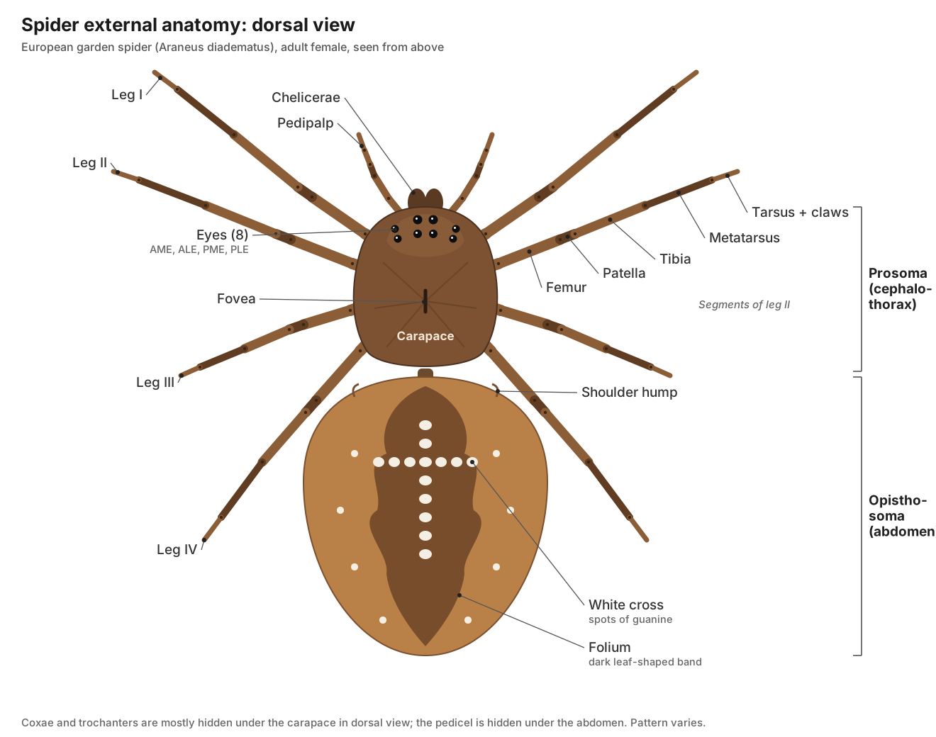
- Carapace (prosoma): Protects the brain, sucking stomach and venom glands, and anchors the muscles of the stomach and the muscles that pressurise the legs.
- Fovea: Marks the internal attachment point (apodeme) of the dorsal dilator muscles of the sucking stomach.
- Pedicel: Lets the abdomen swing in many directions, for spinning silk, while carrying the gut, aorta, nerves and tracheae between the two body parts.
- Opisthosoma (abdomen): Stores food reserves, makes silk and eggs, and breathes through the book lungs and tracheae.
- Dorsal cross and folium: Camouflage and species recognition; the white marks are a quick identification feature for Araneus diadematus.
Spider ventral anatomy: the underside
On the underside of a spider you can see the mouthparts, the sternum ringed by the leg coxae, and on the abdomen the book-lung covers, the epigastric furrow, the epigyne of the female, the tracheal spiracle and the spinnerets. This is the view lab manuals use for sexing and identifying spiders.
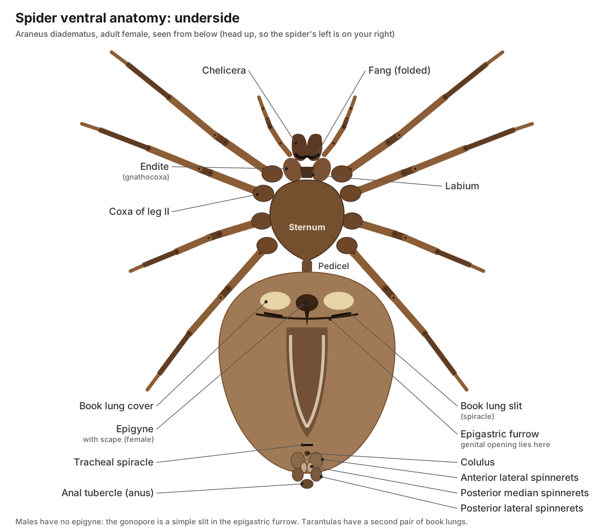
- Sternum: Protects the underside of the synganglion and gives the legs a firm base.
- Labium: Closes the mouth from below; together with the endites it forms the pre-oral cavity where liquefied food is filtered.
- Endites: Hold and squeeze the prey; their dense fringes of hairs filter solid particles out of the liquefied food before it is sucked in.
- Epigastric furrow: Contains the genital opening (gonopore) of both sexes and the openings of the book lungs.
- Book lung openings: Admit air to the atrium of each book lung; they can be narrowed to reduce water loss.
- Tracheal spiracle: Lets air into the tracheal tubes.
- Anal tubercle: Releases faeces and excretory waste from the stercoral pocket and rectum.
Labeled spider diagram: every part and its function
This is the text version of the 3D model: every labeled structure of the garden spider, grouped by body system, with what it does. Structures marked "females only" or "males only" appear when you switch the model's sex.
Body & exoskeleton
- Carapace (prosoma) (Cephalothorax shield; prosoma)
- The hard dorsal shield of the prosoma (cephalothorax), the front body part that carries the eyes, mouthparts, pedipalps and all eight legs. Protects the brain, sucking stomach and venom glands, and anchors the muscles of the stomach and the muscles that pressurise the legs.
- Fovea (Thoracic groove; median furrow)
- A short groove or pit on the midline of the carapace. Marks the internal attachment point (apodeme) of the dorsal dilator muscles of the sucking stomach.
- Sternum
- The heart-shaped ventral plate of the prosoma, surrounded by the coxae of the legs. Protects the underside of the synganglion and gives the legs a firm base.
- Labium (Lower lip)
- A small plate at the front of the sternum that forms the floor of the mouth. Closes the mouth from below; together with the endites it forms the pre-oral cavity where liquefied food is filtered.
- Endites (Gnathocoxae, maxillae)
- Flattened, hairy plates on the coxae of the pedipalps that flank the mouth. Hold and squeeze the prey; their dense fringes of hairs filter solid particles out of the liquefied food before it is sucked in.
- Pedicel (Waist; petiolus)
- The narrow waist that joins the prosoma to the opisthosoma (abdomen). Lets the abdomen swing in many directions, for spinning silk, while carrying the gut, aorta, nerves and tracheae between the two body parts.
- Opisthosoma (abdomen) (Abdomen)
- The soft, unsegmented rear body part, which holds the heart, most of the gut, the book lungs, silk glands and reproductive organs. Stores food reserves, makes silk and eggs, and breathes through the book lungs and tracheae.
- Dorsal cross and folium (Abdominal pattern)
- The white cross of spots and the darker leaf-shaped band on the back of the garden spider's abdomen. Camouflage and species recognition; the white marks are a quick identification feature for Araneus diadematus.
- Epigastric furrow (Genital furrow)
- A transverse groove across the underside of the front of the abdomen. Contains the genital opening (gonopore) of both sexes and the openings of the book lungs.
- Anal tubercle (Anal papilla)
- A small knob at the rear tip of the abdomen that carries the anus. Releases faeces and excretory waste from the stercoral pocket and rectum.
Eyes & sense organs
- Anterior median eyes (AME) (Principal eyes; main eyes)
- The front middle pair of the eight eyes, and the only 'principal' eyes. Form images. They have a movable retina and no light-reflecting tapetum. In jumping spiders they are huge and give sharp, telephoto vision.
- Anterior lateral eyes (ALE)
- The front pair of side eyes. Secondary eyes that mainly detect movement. In jumping spiders they face forward and help judge distance before a leap.
- Posterior median eyes (PME)
- The rear middle pair of eyes. Secondary eyes with a reflective tapetum that improves sensitivity in dim light.
- Posterior lateral eyes (PLE)
- The rear pair of side eyes. Secondary eyes that watch the sides and rear for movement.
- Setae and leg spines (Hairs, macrosetae)
- Hairs that cover the body and legs; many are touch or taste receptors, and the stout ones on the legs are spines. Sense touch and chemicals, grip silk, and protect the cuticle. Leg spines help hold struggling prey.
- Trichobothria (Hair sensilla)
- Very fine, long hairs set in cup-shaped sockets that swing in the faintest air movement. Detect air currents and low-frequency sound, such as the wing beats of a flying insect.
- Slit sensilla and lyriform organs (Slit sense organs)
- Tiny slits in the cuticle that sense strain; groups of parallel slits are called lyriform organs. Act as strain gauges: they measure how the exoskeleton bends, so the spider senses its own leg loads, gravity and vibrations in the web.
Chelicerae, fangs & venom
- Chelicerae (Jaws; paturon (basal segment))
- The first pair of appendages: two stout jaws hanging below the eyes, each ending in a fang. Hold and crush prey and inject venom through the fangs. They are also used for digging, carrying egg sacs and, in some species, making sounds.
- Fangs (Cheliceral fangs, unguis)
- The sharp, hollow, curved tips of the chelicerae. Pierce the prey's cuticle and inject venom, which flows out through a small opening near the tip.
- Venom glands (Poison glands)
- A pair of elongated glands that make venom and pump it through ducts to the fangs. Produce venom that paralyses or kills prey and starts digestion. Muscles wrapped around each gland squeeze the venom out.
Legs & pedipalps
- Pedipalps (Palps)
- The second pair of appendages: short, leg-like feelers with six segments, between the chelicerae and the first legs. Touch, taste and handle prey. In adult males the tip is transformed into a sperm-transfer organ. Females of many web spiders have a small claw at the tip.
- Coxa
- The first, basal segment of each leg, which joins it to the prosoma. Swings the leg forward and back.
- Trochanter
- The short second leg segment, between the coxa and femur. Lets the leg rise and fall.
- Femur
- The long third segment, usually the thickest part of the leg. Lifts the body; contains strong flexor muscles.
- Patella (Knee segment)
- The short 'knee' segment between femur and tibia. Forms the knee. The femur–patella joint has no extensor muscle and is straightened by hemolymph pressure.
- Tibia
- The long fifth segment, after the knee. Carries trichobothria and many spines; its distal joint is one of the hydraulically extended joints.
- Metatarsus
- The sixth leg segment, long and thin. Extends the reach of the leg. The tibia–metatarsus joint is straightened hydraulically.
- Tarsus (Foot)
- The last leg segment, which carries the claws. Grips silk and the ground. It carries a chemoreceptive tarsal organ.
- Tarsal claws
- Two or three small hooked claws at the tip of each tarsus. Web builders like Araneus have three claws: two toothed upper claws and a small middle claw that hooks silk threads. Hunting spiders often have two claws plus a claw tuft.
Muscles & endosternite
- Endosternite (Internal sternal plate)
- A plate of tough connective tissue floating inside the prosoma. An internal 'skeleton' to which many leg and dorsoventral muscles attach.
- Prosomal dorsoventral muscles (Hydraulic pump muscles)
- Muscles that run from the carapace down to the endosternite on each side of the prosoma. When they contract they pull the carapace down and raise hemolymph pressure, which extends the legs at the joints that have no extensor muscles.
Nervous
- Brain (supraesophageal ganglion) (Protocerebrum; synganglion (upper part))
- The upper part of the fused central nervous system, above the esophagus. Processes vision (it receives the optic nerves) and contains higher centres such as the arcuate body and, in hunting spiders, mushroom bodies.
- Subesophageal mass (Subesophageal ganglion; synganglion (lower part))
- The large lower part of the fused central nervous system, lying on the floor of the prosoma. Controls the pedipalps, legs and abdomen; it is made of fused ganglia that in other arthropods form a chain.
- Leg and palp nerves
- Thick nerves that radiate from the subesophageal mass into each pedipalp and leg. Carry motor commands to the leg muscles and bring back signals from hairs, trichobothria and slit sensilla.
- Opisthosomal nerve (cauda equina)
- A bundle of nerves that runs from the rear of the synganglion through the pedicel into the abdomen. Controls the heart, book lungs, spinnerets, silk glands and reproductive organs.
Digestive
- Pharynx
- A muscular tube that leads up from the mouth. Sucks liquid food in; a fine plate of hairs filters out particles.
- Esophagus
- A narrow tube from the pharynx to the sucking stomach. Carries liquefied food to the stomach.
- Sucking stomach (Pumping stomach)
- A muscular foregut chamber in the middle of the prosoma that works like a pump. Dilator muscles attached to the carapace (at the fovea) and endosternite pull its walls apart, sucking the liquefied prey in, then pump it into the midgut.
- Prosomal midgut diverticula (Anterior digestive caeca)
- Blind branches of the midgut that spread through the prosoma towards the leg bases. Absorb and store nutrients close to the leg muscles.
- Midgut (intestine)
- The central tube of the gut, running from the sucking stomach through the pedicel to the stercoral pocket. Carries food to the diverticula, where most digestion and absorption happen.
- Opisthosomal midgut diverticula (Digestive gland; posterior digestive caeca; 'liver')
- A large mass of branched, blind gut tubes that fills most of the abdomen. Finish digestion inside their cells, absorb nutrients and store food, which lets many spiders survive weeks or months without eating.
- Stercoral pocket (Cloacal chamber)
- A sac at the end of the midgut where faeces and excretory waste collect. Stores waste from the gut and the Malpighian tubules until it is released through the rectum and anus.
Excretory
- Malpighian tubules
- Very long, thin, branched tubes in the abdomen that remove nitrogenous waste. Collect waste, mainly guanine, from the hemolymph and pass it into the stercoral pocket.
- Coxal glands
- Small excretory glands in the prosoma that open at the bases of the legs. Filter hemolymph and help regulate water and salts.
Circulatory
- Heart (Dorsal vessel)
- A muscular tube along the back of the abdomen that pumps hemolymph (blood). Pumps hemolymph forward to the prosoma and backward through the abdomen in an open circulatory system.
- Ostia
- Slit-like valved openings in the heart wall through which hemolymph enters. Let hemolymph from the surrounding sinus flow into the heart when it relaxes, and close when it contracts.
- Anterior aorta and prosomal arteries
- The main artery that carries hemolymph from the heart through the pedicel into the prosoma, where it branches to the legs and brain. Supplies the brain, eyes, venom glands and leg muscles, and delivers the pressure that extends the legs.
- Posterior aorta
- The artery that continues from the back of the heart to the rear of the abdomen. Supplies the silk glands, gut and spinnerets.
Respiratory
- Book lungs
- Air-breathing organs made of stacks of thin, blood-filled plates, like the pages of a book. Gas exchange: air in the spaces between the lamellae exchanges oxygen with hemolymph inside them.
- Book lung openings (Lung slits, pulmonary spiracles)
- Small slits on the underside of the abdomen through which air enters the book lungs. Admit air to the atrium of each book lung; they can be narrowed to reduce water loss.
- Tracheal spiracle
- A single small opening on the underside of the abdomen that leads into the tracheae. Lets air into the tracheal tubes.
- Tracheae
- Air-filled cuticular tubes that carry air directly to tissues. Supplement the book lungs; they evolved from the second pair of book lungs.
Silk glands & spinnerets
- Anterior lateral spinnerets (ALS)
- The front pair of spinnerets: short, stout and conical. Carry the spigots of the major ampullate (dragline) and piriform (attachment cement) glands.
- Posterior median spinnerets (PMS)
- The small middle pair of spinnerets, partly hidden between the others. Carry spigots of the minor ampullate and aciniform glands, and of the tubuliform glands in females.
- Posterior lateral spinnerets (PLS)
- The rear pair of spinnerets. Carry the flagelliform and aggregate spigots that make the sticky capture spiral, plus aciniform spigots.
- Colulus
- A small, non-spinning bump in front of the spinnerets. A vestige of the fourth pair of spinnerets; in cribellate spiders the same pair forms the silk-spinning cribellum plate.
- Major ampullate glands
- The largest silk glands: long tails, a wide storage sac (ampulla) and a long S-shaped duct. Make dragline silk, used for safety lines and for the frame and radii of the orb web.
- Minor ampullate glands
- Smaller ampulla-shaped glands next to the major ampullate glands. Make the non-sticky auxiliary (temporary) spiral that guides orb construction.
- Flagelliform glands
- Glands that make the very stretchy core thread of the capture spiral. Produce the elastic capture-spiral fibre that absorbs the impact of flying insects.
- Aggregate glands
- Large, lobed glands that make the watery glue of the capture spiral. Coat the capture spiral with glue that beads up into regularly spaced sticky droplets.
- Aciniform glands
- Many small, grape-like glands. Make wrapping silk for swathing prey and the inner layer of the egg sac.
- Piriform glands (Pyriform glands)
- Pear-shaped glands that make attachment cement. Glue the dragline and web threads to surfaces and to each other (attachment discs).
- Tubuliform glands (Cylindrical glands) · females only
- Long tubular silk glands present only in adult females. Make the tough outer layer of the egg sac.
Reproductive
- Cymbium · males only
- The spoon-shaped last segment of the male pedipalp, which cradles the palpal bulb. Holds and protects the copulatory bulb.
- Palpal bulb (Palpal organ, copulatory bulb, bulbus) · males only
- The complex sperm-transfer organ on the tip of each male pedipalp. Stores sperm in a coiled duct (spermophor) and delivers it to the female. Membranous sacs (haematodochae) inflate with hemolymph to unfold the bulb during mating.
- Embolus · males only
- The slender tip of the palpal bulb, through which sperm leaves. The actual intromittent part: it is inserted into a copulatory opening of the female's epigyne and releases the sperm.
- Epigyne (Epigynum) · females only
- A hardened plate on the underside of the female's abdomen that contains the copulatory openings. Receives the male's embolus; ducts lead from it to the spermathecae. Its shape fits only the palp of the right species.
- Spermathecae (Seminal receptacles) · females only
- Small, hard-walled sacs that store sperm after mating. Keep sperm alive until the eggs are laid, sometimes for months.
- Ovaries · females only
- Paired elongated organs studded with developing eggs. Produce eggs, which can fill much of the abdomen before laying.
- Oviducts and uterus externus · females only
- Ducts that carry eggs from the ovaries to the genital opening in the epigastric furrow. Eggs pass sperm from the spermathecae on their way out and are fertilised as they are laid.
- Testes · males only
- Paired tubular organs that produce sperm. Make sperm, which leaves the body through the gonopore in the epigastric furrow.
- Vasa deferentia (Sperm ducts) · males only
- Paired ducts that carry sperm from the testes to the male gonopore. Transport sperm out of the body.
Spider internal anatomy
Inside a spider, the prosoma holds the synganglion (the fused brain and nerve cord), the venom glands, the sucking stomach and the muscles that move the legs. The abdomen holds the tubular heart along the back, the branched midgut diverticula that fill most of the space, the Malpighian tubules and stercoral pocket, the book lungs and tracheae, the silk glands, and the ovaries or testes.
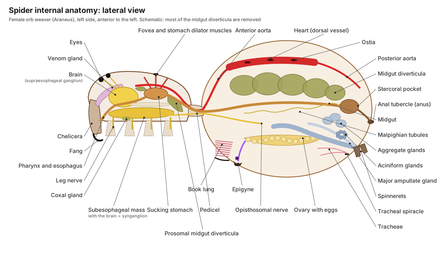
Fox's orb-weaver lab guide notes that the central nervous system forms a single white mass in the middle of the prosoma, with no ventral nerve cord in the abdomen, and that the posterior digestive caeca fill much of the abdomen. In the 3D model, set the exoskeleton slider to zero and hide the digestive system to see the silk glands and gonads below.
Spider eyes: how many eyes do spiders have?
Most spiders have eight eyes, arranged in two or three rows on the front of the carapace. They come in four pairs: the anterior median eyes (AME), anterior lateral eyes (ALE), posterior median eyes (PME) and posterior lateral eyes (PLE). Some spiders, such as recluses, spitting spiders and woodlouse hunters, have six, and some cave spiders have none.
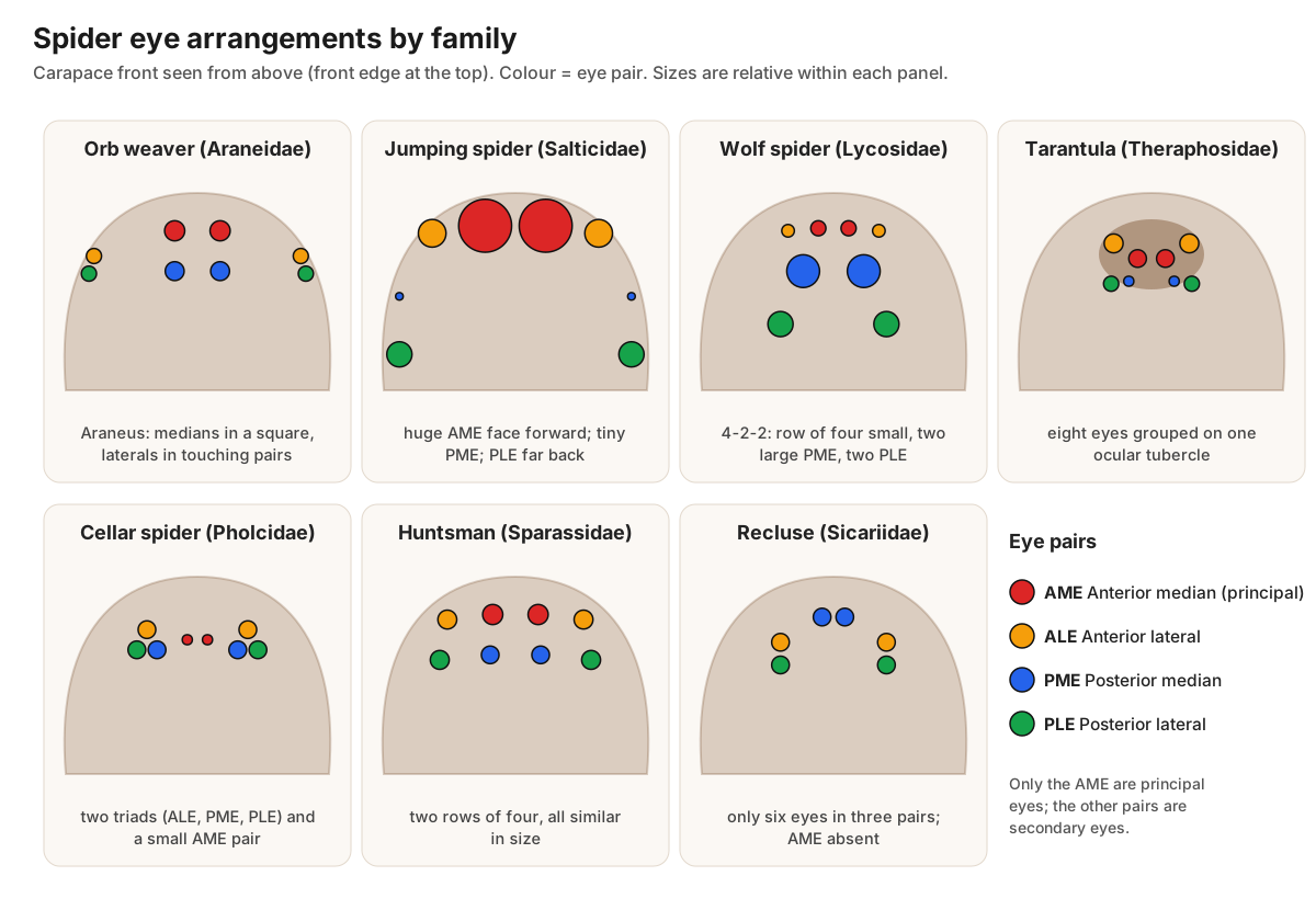
Only the AME are principal eyes, with a movable retina. The other three pairs are secondary eyes, which evolved from the compound eyes of the spiders' ancestors and are mainly motion detectors (Fox). Many secondary eyes have a reflective layer (tapetum), which is why wolf spider eyes shine in torchlight. A 2026 comparative study from the Museum für Naturkunde Berlin found that visually hunting spiders have repeatedly evolved forward-facing eye arrangements, while orb weavers show a more even, halo-like layout.
- Anterior median eyes (AME): Form images. They have a movable retina and no light-reflecting tapetum. In jumping spiders they are huge and give sharp, telephoto vision.
- Anterior lateral eyes (ALE): Secondary eyes that mainly detect movement. In jumping spiders they face forward and help judge distance before a leap.
- Posterior median eyes (PME): Secondary eyes with a reflective tapetum that improves sensitivity in dim light.
- Posterior lateral eyes (PLE): Secondary eyes that watch the sides and rear for movement.
Jumping spider anatomy: eyes, mouth and legs
Jumping spiders (family Salticidae) have the sharpest vision of any spider. Their two huge anterior median eyes face forward like headlights and work like small telescopes, with a long eye tube moved by six muscles and a retina in four layers. The anterior lateral eyes beside them also face forward and help judge distance; the posterior median eyes are tiny; and the posterior lateral eyes, set far back, watch the sides and rear, so together the eyes cover almost 360° (Jackson & Pollard, 1996).
A jumping spider's mouthparts are the same as an orb weaver's: short chelicerae with sideways-closing fangs, endites and a labium. Its legs end in two claws with dense tufts of hairs that let it walk on glass. Jumping spiders do not build capture webs; they stalk prey, fix a silk safety line and leap, powering the jump mainly with the hind legs and using the same hydraulic leg extension as other spiders.
Spider mouth anatomy: chelicerae, fangs and venom
A spider's "mouth" is a set of parts. The chelicerae are the jaws, each ending in a hollow fang connected to a venom gland. Behind them, the endites (plates on the pedipalp bases) and the labium (lower lip) frame the small mouth opening. Spiders cannot chew; they inject venom and digestive fluid and suck up the liquefied prey.
| Feature | Araneomorphs (e.g. garden spider) | Mygalomorphs (tarantulas, trapdoor spiders) |
|---|---|---|
| Fang movement | Fangs swing sideways and close towards each other like pincers (labidognath) | Fangs point backwards side by side and strike downwards (orthognath, paraxial) |
| How they bite | Can bite from almost any position | Must raise the front of the body to strike down |
| Venom glands | Extend back from the chelicerae into the prosoma | Stay inside the chelicerae |
- Chelicerae: Hold and crush prey and inject venom through the fangs. They are also used for digging, carrying egg sacs and, in some species, making sounds.
- Fangs: Pierce the prey's cuticle and inject venom, which flows out through a small opening near the tip.
- Venom glands: Produce venom that paralyses or kills prey and starts digestion. Muscles wrapped around each gland squeeze the venom out.
Spider leg anatomy: seven segments and hydraulic joints
Every spider leg has seven segments: coxa, trochanter, femur, patella, tibia, metatarsus and tarsus, ending in two or three claws. Insect legs have five segments and no patella or metatarsus. A spider has eight legs; the shorter pedipalps in front of them are not legs.
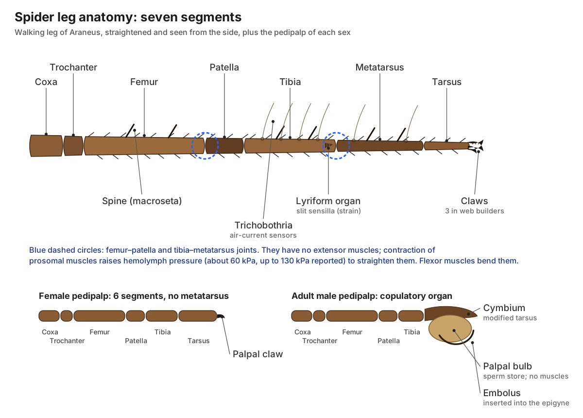
Spiders bend their legs with muscles, but two joints, the femur–patella and tibia–metatarsus joints, have no extensor muscles. They are straightened by hemolymph pressure created by muscles in the prosoma (Göttler et al., 2021). The legs are also the spider's main sense organs: they carry touch hairs, air-sensing trichobothria and strain-sensing slit sensilla.
- Coxa: Swings the leg forward and back.
- Trochanter: Lets the leg rise and fall.
- Femur: Lifts the body; contains strong flexor muscles.
- Patella: Forms the knee. The femur–patella joint has no extensor muscle and is straightened by hemolymph pressure.
- Tibia: Carries trichobothria and many spines; its distal joint is one of the hydraulically extended joints.
- Metatarsus: Extends the reach of the leg. The tibia–metatarsus joint is straightened hydraulically.
- Tarsus: Grips silk and the ground. It carries a chemoreceptive tarsal organ.
- Tarsal claws: Web builders like Araneus have three claws: two toothed upper claws and a small middle claw that hooks silk threads. Hunting spiders often have two claws plus a claw tuft.
- Prosomal dorsoventral muscles: When they contract they pull the carapace down and raise hemolymph pressure, which extends the legs at the joints that have no extensor muscles.
Spider pedipalps and male vs female spider anatomy
You can tell an adult male spider from a female by its pedipalps: in males the tips are swollen into shiny palpal bulbs that look like boxing gloves. Adult females of most araneomorphs have an epigyne, a hard plate on the underside of the abdomen just in front of the epigastric furrow. Juveniles of both sexes look alike until the final moult.
| Female spider anatomy | Male spider anatomy | |
|---|---|---|
| Pedipalps | Slender, leg-like; often a small claw at the tip | Tip transformed into cymbium, palpal bulb and embolus |
| Abdomen | Large, swells with eggs | Smaller; males are often slimmer with relatively longer legs |
| Genital opening | Epigyne with copulatory openings; spermathecae inside | Simple gonopore in the epigastric furrow |
| Internal organs | Ovaries, oviducts, spermathecae; tubuliform silk glands for egg sacs | Testes and sperm ducts; epiandrous silk spigots for the sperm web |
| Size in widows | Larger | One-half to two-thirds the female's length (UC IPM) |
Male spiders transfer sperm indirectly. Because the testes open on the abdomen but the copulatory organs are on the pedipalps, a mature male first spins a small sperm web, deposits a drop of sperm on it and draws the sperm into his palpal bulbs. During mating he inserts the slender embolus into the female's epigyne. In the garden spider, as in most entelegyne spiders, the bulb contains no muscles; it is unfolded by hemolymph pressure that inflates membranous sacs called haematodochae (Sentenská et al., 2017).
- Pedipalps: Touch, taste and handle prey. In adult males the tip is transformed into a sperm-transfer organ. Females of many web spiders have a small claw at the tip.
- Cymbium: Holds and protects the copulatory bulb.
- Palpal bulb: Stores sperm in a coiled duct (spermophor) and delivers it to the female. Membranous sacs (haematodochae) inflate with hemolymph to unfold the bulb during mating.
- Embolus: The actual intromittent part: it is inserted into a copulatory opening of the female's epigyne and releases the sperm.
- Epigyne: Receives the male's embolus; ducts lead from it to the spermathecae. Its shape fits only the palp of the right species.
- Spermathecae: Keep sperm alive until the eggs are laid, sometimes for months.
- Ovaries: Produce eggs, which can fill much of the abdomen before laying.
- Testes: Make sperm, which leaves the body through the gonopore in the epigastric furrow.
Spider book lungs and tracheae: how spiders breathe
Spiders breathe through book lungs and tracheae. A book lung is a pocket in the front of the abdomen filled with a stack of thin, blood-filled plates (lamellae), like the pages of a book. Air enters through a slit (spiracle) into an atrium and passes between the plates; small struts called trabeculae keep the air spaces from collapsing (Küntzel et al., 2019).
Most araneomorphs, including the garden spider, have one pair of book lungs, and their second pair has been replaced by tracheae, fine air tubes that open through a single spiracle near the spinnerets (Australian Museum). Tarantulas and other mygalomorphs keep two pairs of book lungs (Garrison et al., 2016), which you can see as four pale patches under the abdomen.
- Book lungs: Gas exchange: air in the spaces between the lamellae exchanges oxygen with hemolymph inside them.
- Tracheae: Supplement the book lungs; they evolved from the second pair of book lungs.
Spider heart and circulatory system
Yes, spiders have a heart. It is a muscular tube lying along the top of the abdomen, inside a sac of hemolymph, with paired slit-like valves called ostia. When the heart relaxes, hemolymph enters through the ostia; when it contracts, the ostia close and blood is pumped forward through the anterior aorta into the prosoma and backward through the posterior aorta.
The circulation is open: arteries empty into spaces (sinuses) around the organs, and blood returns to the heart after passing through the book lungs. Spider blood carries oxygen on hemocyanin, a copper-containing protein, so it looks bluish rather than red. In the garden spider the heart has three pairs of ostia and gives off three pairs of cardiac arteries (Runge & Wirkner, 2016); across spiders the count of ostia varies from five pairs to two (Gerdinand, 2019).
- Heart: Pumps hemolymph forward to the prosoma and backward through the abdomen in an open circulatory system.
- Ostia: Let hemolymph from the surrounding sinus flow into the heart when it relaxes, and close when it contracts.
- Anterior aorta and prosomal arteries: Supplies the brain, eyes, venom glands and leg muscles, and delivers the pressure that extends the legs.
- Posterior aorta: Supplies the silk glands, gut and spinnerets.
Spider brain and nervous system
A spider's brain and nerve cord are fused into one mass, the synganglion, which sits in the prosoma. Its upper part, the supraesophageal ganglion (brain), receives the optic nerves from the eyes. Its lower part, the subesophageal mass, is made of fused ganglia that control the pedipalps, legs and abdomen. The esophagus runs through the gap between them, which is one reason spiders can swallow only liquid food.
Unlike insects, spiders have no chain of ganglia in the abdomen; a bundle of nerves (the cauda equina) runs back through the pedicel instead. Hunting spiders that depend on vision, like jumping and wolf spiders, have larger visual brain centres than web builders.
- Brain (supraesophageal ganglion): Processes vision (it receives the optic nerves) and contains higher centres such as the arcuate body and, in hunting spiders, mushroom bodies.
- Subesophageal mass: Controls the pedipalps, legs and abdomen; it is made of fused ganglia that in other arthropods form a chain.
- Leg and palp nerves: Carry motor commands to the leg muscles and bring back signals from hairs, trichobothria and slit sensilla.
- Opisthosomal nerve (cauda equina): Controls the heart, book lungs, spinnerets, silk glands and reproductive organs.
Spider digestive and excretory systems
Spiders digest their food mostly outside the body. They pump digestive fluid into the prey, then suck up the liquid through the pharynx and esophagus into the sucking stomach. The midgut carries it to the midgut diverticula, which finish digestion, absorb nutrients and store reserves. Waste collects in the stercoral pocket and leaves through the anus on the anal tubercle.
- Pharynx: Sucks liquid food in; a fine plate of hairs filters out particles.
- Sucking stomach: Dilator muscles attached to the carapace (at the fovea) and endosternite pull its walls apart, sucking the liquefied prey in, then pump it into the midgut.
- Prosomal midgut diverticula: Absorb and store nutrients close to the leg muscles.
- Midgut (intestine): Carries food to the diverticula, where most digestion and absorption happen.
- Opisthosomal midgut diverticula: Finish digestion inside their cells, absorb nutrients and store food, which lets many spiders survive weeks or months without eating.
- Stercoral pocket: Stores waste from the gut and the Malpighian tubules until it is released through the rectum and anus.
- Malpighian tubules: Collect waste, mainly guanine, from the hemolymph and pass it into the stercoral pocket.
- Coxal glands: Filter hemolymph and help regulate water and salts.
Spinnerets and silk glands
Spinnerets are small, finger-like appendages under the rear of the abdomen. Most araneomorphs, including the garden spider, have six: anterior lateral, posterior median and posterior lateral pairs. Each is studded with spigots, the openings of the silk glands. Many mygalomorphs have four spinnerets (Mariano-Martins et al., 2020). In front of the garden spider's spinnerets is the colulus, a small bump that is a remnant of a fourth pair; in cribellate spiders the same pair forms a silk-spinning plate called the cribellum.
| Silk gland | Silk it makes (orb weavers) |
|---|---|
| Major ampullate | Dragline, web frame and radii |
| Minor ampullate | Temporary auxiliary spiral |
| Flagelliform | Stretchy core fibre of the capture spiral |
| Aggregate | Glue droplets on the capture spiral |
| Piriform | Attachment cement for anchoring threads |
| Aciniform | Prey wrapping; inner layer of the egg sac |
| Tubuliform (cylindrical) | Tough outer layer of the egg sac (females only) |
Gland functions follow Zhao et al. (2021).
Spider web anatomy: parts of an orb web
An orb web has a frame of strong threads fixed to plants or walls by anchor lines, radii running from the frame to the central hub, and a sticky capture spiral laid from the outside in. While building, the spider first lays a widely spaced, non-sticky auxiliary spiral to stabilise the radii, then removes it as it adds the capture spiral. The frame, radii and auxiliary spiral are dry; only the capture spiral carries glue droplets (Zhao et al., 2021).
The garden spider usually sits head-down on the hub or waits in a retreat at the edge, connected to the hub by a signal thread. Through its legs it feels the vibrations of anything that hits the web.
Tarantula anatomy: how mygalomorphs differ
Tarantulas (family Theraphosidae) have the same body plan as other spiders, but they keep several older features. Their fangs are paraxial: they point backwards and strike downwards instead of closing like pincers. They have two pairs of book lungs and no tracheae, their eight small eyes sit together on a raised ocular tubercle, and they usually have four spinnerets, the rear pair long and finger-like.
Many New World tarantulas (subfamilies Theraphosinae and Aviculariinae) also carry urticating setae, barbed hairs on the abdomen or pedipalps that they release against predators and that irritate skin and eyes (Kaderka et al., 2019). Old World tarantulas generally lack them. Females of many species live for decades, and mature males of many species have tibial hooks on their first legs to hold the female's fangs during mating.
Wolf spider, black widow and other spiders compared
All spiders share the two-part body, eight legs, chelicerae with fangs, pedipalps and spinnerets. This table shows how common spiders differ from the garden spider used in the model.
| Spider | Distinctive anatomy |
|---|---|
| Garden spider (Araneus diadematus) | Orb weaver; three tarsal claws; medians in a square, laterals in touching pairs; white cross on the abdomen |
| Wolf spiders (Lycosidae) | Eyes in three rows (4-2-2) with large forward-facing PME; no capture web; female carries the egg sac on her spinnerets and the young ride on her abdomen (Montana State Extension) |
| Black widow (Latrodectus) | Shiny black female with a red hourglass under the abdomen; comb-footed family (Theridiidae) with a comb of bristles on tarsus IV; tangled cobweb (UC IPM) |
| Jumping spiders (Salticidae) | Huge AME, tiny PME; two claws with hair tufts; stalk and leap instead of building webs |
| Tarantulas (Theraphosidae) | Paraxial fangs, two pairs of book lungs, eye tubercle, urticating setae in New World species |
| Huntsman spiders (Sparassidae) | Flattened body; legs twisted so they spread sideways (laterigrade); eight eyes in two rows of four |
| Cellar spiders (Pholcidae) | Very long, thin legs; eyes in two triads plus a small AME pair (some species six eyes); no hardened epigyne |
| Recluses (Loxosceles) | Six eyes in three pairs; violin mark on the carapace |
Wolf spiders are hunters with good eyesight. Their eight eyes are arranged as one row of four small eyes, then two rows of two larger eyes (University of Minnesota Extension), which separates them at a glance from the similar-looking nursery-web and grass spiders.
Spider vs insect: what is the difference?
Spiders are not insects. Both are arthropods with a jointed exoskeleton, but spiders are arachnids, with a different body plan.
| Feature | Spider | Insect |
|---|---|---|
| Body parts | Two: prosoma and opisthosoma | Three: head, thorax and abdomen |
| Legs | Eight, each with seven segments | Six, each with five segments |
| Antennae | None (pedipalps do a similar sensing job) | One pair |
| Wings | None | Usually two pairs (some wingless) |
| Eyes | Simple eyes only, usually eight | Two compound eyes, often plus simple ocelli |
| Mouthparts | Chelicerae with fangs; liquid diet | Mandibles and other mouthparts; many chew |
| Breathing | Book lungs and tracheae | Tracheae with spiracles along the body |
| Silk | All spiders, from spinnerets on the abdomen | Only some insects, mostly larvae such as silkworms, from glands near the mouth |
| Development | Hatchlings look like small adults (no metamorphosis) | Many go through larva and pupa (metamorphosis) |
Camel spiders, sea spiders and spider mites: not true spiders
Several animals called "spiders" are not spiders (order Araneae) at all.
- Camel spiders (sun spiders, order Solifugae) are separate arachnids. They have huge pincer-like chelicerae, no venom glands and no spinnerets, and their long pedipalps end in an evertable adhesive organ used to grab prey and climb (Klann et al., 2008).
- Sea spiders (class Pycnogonida) are marine arthropods with a tiny body, a sucking proboscis and usually eight long legs; branches of the gut run into the legs. They are not arachnids.
- Spider mites are tiny plant-feeding mites (Acari), not spiders. Adults have eight legs but newly hatched larvae have six, and they spin silk webbing on leaves (UC IPM).
- Harvestmen ("daddy longlegs", order Opiliones) look like they have one oval body because the two body parts are broadly joined, with no narrow waist. They have two eyes and no silk or venom. The name "daddy longlegs" is also used for cellar spiders, which are true spiders.
Spider anatomy for kids
A spider's body is simple to remember:
- Two body parts. The front part (cephalothorax) has the eyes, mouth and legs. The back part (abdomen) makes silk.
- Eight legs. Insects have six. Each spider leg bends in seven pieces and ends in tiny hooks.
- Lots of eyes. Most spiders have eight eyes. Jumping spiders have two very big ones in front.
- Fangs. Two fangs inject venom to stop the spider's food from moving. Very few spiders can harm people.
- Feelers. Two short "arms" by the mouth, called pedipalps, touch and taste.
- Silk. Spinnerets at the back of the abdomen squeeze out silk for webs, safety lines and egg sacs.
- Breathing. Spiders breathe through book lungs, which have thin layers like the pages of a book.
Hydraulic legs: why a spider needs blood pressure to walk
A spider bends its legs with muscles but straightens two of its main leg joints with fluid pressure. The femur–patella and tibia–metatarsus joints have no extensor muscles. Instead, muscles in the prosoma contract, squeeze the body and push hemolymph (spider blood) into the legs. Göttler and colleagues summarise measured pressures of about 60 kPa, with values up to 130 kPa reported, which is far higher than the resting pressure in a human artery. The flexor muscles inside the leg then work against this pressure to bend the joint again. (Göttler et al. (2021): folding of jumping spider leg joints, Advanced Science.)
This design explains several things students notice. A spider that has lost a lot of fluid, or has died, curls its legs under its body, because nothing opposes the flexors any more. Spiders also seal a leg quickly if it is lost, so that the pressure system does not leak. When you study the leg diagram, mark the two hydraulic joints and remember that the 'missing' muscle is replaced by the circulatory system: the heart, the anterior aorta and the hemolymph sinuses of the prosoma all take part in movement.
How spiders sense the world without ears or antennae
Most spiders see poorly and rely on touch, vibration and air movement. Their sense organs are built into the exoskeleton. Ordinary setae (hairs) respond to touch and many carry taste receptors. Trichobothria are extremely fine hairs set in sockets on the tibiae and metatarsi that swing in the slightest air current, such as the wing beat of a fly. Slit sensilla are narrow slits in the cuticle that act as strain gauges. Groups of parallel slits, called lyriform organs, sit close to the leg joints, oriented roughly at right angles to the stress lines expected during walking. (Blickhan, Weihmann & Barth (2021): strain in spider exoskeletons.)
An orb weaver sitting at the hub of its web uses these organs to 'listen' to the threads: the metatarsal lyriform organs register tiny vibrations, and the spider can tell a trapped insect from wind or rain. Jumping spiders are the exception to the poor-vision rule. Their huge anterior median eyes have a long, movable eye tube and a tiered retina, and let them identify mates, rivals and prey from 30 body lengths or more away, while the secondary eyes detect motion all around them. (Jackson & Pollard (1996): predatory behavior of jumping spiders, Annual Review of Entomology.)
The silk system: from gland to web
Silk is a liquid protein (fibroin, mostly spidroins) made and stored in glands inside the abdomen. Each gland type opens through microscopic nozzles called spigots on a particular spinneret, and each makes a different silk. An orb weaver may have up to seven types: major and minor ampullate, aciniform, tubuliform, aggregate, piriform and flagelliform glands. The spinnerets themselves are modified abdominal appendages that develop from limb buds like legs; males of some species also have epiandrous glands near the genital furrow that spin the sperm web. (Mariano-Martins, Lo-Man-Hung & Torres (2020): evolution of spinnerets.)
In an orb web, the major ampullate glands make the dragline, frame and radial threads; the minor ampullate glands make the temporary auxiliary spiral; the flagelliform glands make the elastic core of the capture spiral, which the aggregate glands coat with glue that beads into droplets; piriform cement fixes threads to surfaces; aciniform silk wraps prey and lines the egg sac; and tubuliform (cylindrical) silk forms the tough outer egg-sac wall. The frame, radii and auxiliary spiral are not sticky; only the capture spiral is. (Zhao, Morita & Sakamoto (2021): silk glands and orb web glue.)
See the parts of an orb web, from frame threads to hub, in spider web anatomy.
How to study a real spider specimen
Spiders are small, so school labs normally examine preserved specimens in alcohol under a hand lens or dissecting microscope rather than cut them open. Fox's orb-weaver lab guide works through the animal in a logical order: first the carapace, eyes, chelicerae, pedipalps and legs; then the underside, with the sternum, labium, endites, book lung slits, epigynum and spinnerets; and only then the internal organs. Following the same order with the 3D model works well: start with the exoskeleton opaque, then fade it to see the synganglion in the prosoma and the heart, gut, silk glands and gonads in the abdomen. (R. Fox, Lander University: Argiope (orb weaver) anatomy lab.)
Three checks make identification easier. Count the leg segments from the body outwards and confirm there are seven, ending in claws. Look at the tips of the pedipalps: swollen, shiny bulbs mean an adult male. Turn the specimen over and find the epigastric furrow; in adult females of most araneomorphs, the hardened epigyne lies just in front of it, between the pale book-lung covers. If the fangs point straight back side by side, rather than closing towards each other, you are looking at a mygalomorph such as a tarantula or trapdoor spider. (Australian Museum: spider structure.)
Digestion outside the body
Spiders cannot chew or swallow solid food. After venom subdues the prey, the spider releases digestive fluid onto or into it and sucks up the resulting soup; this is called extra-oral digestion. Proteomic work on a tarantula (Acanthoscurria geniculata) and a social araneomorph (Stegodyphus mimosarum) found similar abundant enzymes in both, especially astacin-like metalloproteases, which suggests the system is shared across the two main spider lineages. (Walter et al. (2017): spider digestive fluids and extra-oral digestion.)
The liquid is filtered by hairs on the endites and in the pharynx, then pumped by the sucking stomach, whose dilator muscles pull on the carapace at the fovea. From there the midgut carries it to the branched diverticula that fill most of the abdomen and also reach into the prosoma. These diverticula finish digestion inside their cells and store reserves, which is why many spiders can survive long periods without a meal. (R. Fox, Lander University: Argiope (orb weaver) anatomy lab.)
Spider anatomy worksheet: test yourself
Use the 3D model and the diagrams to answer these. Answers are below each question.
- Name the two main body parts of a spider and the structure that joins them. Answer: prosoma (cephalothorax) and opisthosoma (abdomen), joined by the pedicel.
- List the seven segments of a spider leg in order. Answer: coxa, trochanter, femur, patella, tibia, metatarsus, tarsus.
- Which pair of eyes are the principal eyes? Answer: the anterior median eyes (AME).
- Which external feature shows that a spider is an adult male? Answer: the swollen palpal bulbs at the tips of the pedipalps.
- How many pairs of book lungs does a garden spider have, and how many does a tarantula have? Answer: one pair in the garden spider (plus tracheae); two pairs in a tarantula.
- Which two leg joints are straightened by hemolymph pressure? Answer: the femur–patella and tibia–metatarsus joints.
- Where is the spider's brain, and what passes through it? Answer: in the prosoma, as part of the synganglion; the esophagus passes through it.
- Which silk gland makes the dragline? Answer: the major ampullate gland.
Frequently asked questions
What are the main body parts of a spider?
A spider has two main body parts: the prosoma (cephalothorax), which carries the eyes, chelicerae with fangs, two pedipalps and eight legs, and the opisthosoma (abdomen), which holds the heart, most of the gut, the book lungs, silk glands and reproductive organs. A narrow waist called the pedicel joins them.
How many legs and eyes does a spider have?
All spiders have eight legs, each with seven segments ending in claws. Most have eight eyes in two or three rows, named anterior median, anterior lateral, posterior median and posterior lateral. Some have six, and a few cave spiders have none.
Do spiders have a heart and blood?
Yes. A spider's heart is a muscular tube along the back of the abdomen with slit-like openings called ostia. It pumps hemolymph, a bluish blood that carries oxygen on the copper protein hemocyanin, through an open circulatory system.
How do spiders breathe?
Spiders breathe with book lungs, stacks of thin blood-filled plates in the front of the abdomen, and with tracheae, air tubes that open through a spiracle. Most araneomorph spiders have one pair of book lungs plus tracheae; tarantulas have two pairs of book lungs.
Do spiders have a brain?
Yes. A spider's central nervous system is fused into a single mass, the synganglion, in the prosoma. Its upper part (the brain or supraesophageal ganglion) handles vision, and its lower subesophageal mass controls the legs and abdomen. The esophagus passes between the two parts.
What are pedipalps used for?
Pedipalps are short, six-segmented appendages beside the mouth. Both sexes use them to touch, taste and hold prey. In adult males the tip is swollen into a palpal bulb that stores sperm and inserts it into the female's epigyne.
How can you tell a male spider from a female?
Adult males have swollen, club-like tips on their pedipalps (the palpal bulbs) and usually a smaller abdomen and relatively longer legs. Adult females of most species have an epigyne, a hardened plate on the underside of the abdomen in front of the epigastric furrow.
Why do spiders curl up when they die?
Spiders extend two of their leg joints, femur–patella and tibia–metatarsus, with hemolymph pressure instead of extensor muscles. When a spider dies or dries out, the pressure drops and the flexor muscles pull the legs in, so the legs curl under the body.
Are spiders insects?
No. Spiders are arachnids (class Arachnida, order Araneae). Insects have three body parts, six legs, antennae, compound eyes and often wings. Spiders have two body parts, eight legs, no antennae, only simple eyes, no wings, and chelicerae with fangs.
Where are a spider's spinnerets and silk glands?
The spinnerets are finger-like appendages under the tip of the abdomen; most araneomorphs have three pairs. Inside the abdomen, up to seven types of silk gland in orb weavers feed them through tiny spigots, each gland making a different silk for draglines, capture spirals, glue, wrapping or egg sacs.
References
- Fox, R. Invertebrate Anatomy OnLine: Argiope, orb weaving spider. Lander University laboratory guide.
- Australian Museum. Spider structure.
- Garrison, N. L. et al. (2016). Spider phylogenomics: untangling the Spider Tree of Life. PeerJ 4, e1719.
- Küntzel, N., Dunlop, J. A. & Scholtz, G. (2019). Morphology and evolution of spider book lungs (Araneae). Arthropod Systematics & Phylogeny 77, 267–284.
- Göttler, C., Elflein, K., Siegwart, R. & Sitti, M. (2021). Spider origami: folding principle of jumping spider leg joints for bioinspired fluidic actuators. Advanced Science 8, 2003890.
- Blickhan, R., Weihmann, T. & Barth, F. G. (2021). Measuring strain in the exoskeleton of spiders: virtues and caveats. Journal of Comparative Physiology A 207, 191–204.
- Jackson, R. R. & Pollard, S. D. (1996). Predatory behavior of jumping spiders. Annual Review of Entomology 41, 287–308.
- Sentenská, L. et al. (2017). Neurons and a sensory organ in the pedipalps of male spiders reveal that it is not a numb structure. Scientific Reports 7, 12209.
- Mariano-Martins, P., Lo-Man-Hung, N. & Torres, T. T. (2020). Evolution of spiders and silk spinning: mini review of the morphology, evolution, and development of spiders' spinnerets. Frontiers in Ecology and Evolution 8, 109.
- Zhao, Y., Morita, M. & Sakamoto, T. (2021). Inorganic component imaging of aggregate glue droplets on spider orb webs by TOF-SIMS. Journal of Surface Analysis 27, 78–94.
- Runge, J. & Wirkner, C. S. (2016). The hemolymph vascular system in Araneus diadematus with special focus on intraspecific variability in artery systems. Journal of Arachnology 44, 153–164.
- Gerdinand, K. (2019). Evolutionary morphology of the hemolymph vascular system of Araneae (Arachnida). Dissertation summary, University of Rostock.
- Walter, A. et al. (2017). Characterisation of protein families in spider digestive fluids and their role in extra-oral digestion. BMC Genomics 18, 600.
- Kaderka, R., Bulantová, J., Heneberg, P. & Řezáč, M. (2019). Urticating setae of tarantulas (Araneae: Theraphosidae). PLOS ONE 14, e0224384.
- Pande, A. et al. (2026). Hunting behaviour drives the evolution of spider eye arrangements (press release, Museum für Naturkunde Berlin; Current Biology).
- University of California IPM. Pest Notes: Spiders (widow spiders).
- Montana State University Extension. Wolf spiders fact sheet; University of Minnesota Extension. Wolf spiders.
- University of California IPM. Pest Notes: Spider mites.
- Klann, A. E. et al. (2008). The anatomy and ultrastructure of the suctorial organ of Solifugae (Arachnida). Arthropod Structure & Development 37, 3–12.
More interactive anatomy models
- Human anatomy1,000+ bones, muscles, organs, vessels and nerves from real anatomical data.
- Octopus anatomyThree hearts, a ring-shaped brain, eight arms and a beak, in 3D.
- Squid anatomyPen, eight arms, two tentacles, giant axons and three hearts of the longfin squid.
- Squid dissectionStep-by-step guided squid dissection with every structure labeled.
- Fish anatomyExternal and internal anatomy of a bony fish: fins, gills, swim bladder, heart and brain.
- Perch dissectionGuided yellow perch (fish) dissection, from scales to the swim bladder.