

Sheep brain dissection at a glance
A sheep brain dissection has three stages. First you study the outside: the meninges, the dorsal surface with its hemispheres, gyri, sulci and cerebellum, and the ventral surface with the olfactory bulbs, optic chiasm, pituitary, brainstem and cranial nerves. Then you make a midsagittal cut through the longitudinal fissure to see the corpus callosum, thalamus, ventricles and the arbor vitae of the cerebellum. Finally you cut coronal (frontal) slices to find the hippocampus, thalamus and internal capsule, and to compare grey and white matter. Allow one to two lab periods.
The sheep brain is about a tenth of the mass of a human brain, but nearly every structure on a human brain diagram is there. The main differences are proportions: bigger olfactory bulbs, a smaller frontal lobe and a brainstem that runs horizontally. Those differences are explained in the sheep vs human brain section.
Materials and safety
You need one preserved sheep brain per pair of students, a dissecting tray, a long sharp blade for the big cuts, small tools and protective equipment. Check whether your brain came with the dura mater intact or removed: it changes step 1.
Materials
- One preserved sheep brain (dura intact or removed; note which you have)
- Dissecting tray or cutting board with a wax or rubber mat
- A long, sharp blade for the midsagittal and coronal cuts (brain knife, long scalpel blade or a single-edged razor blade, as your teacher allows)
- Scalpel, fine scissors, forceps and a blunt probe
- Ruler (for 5 mm coronal slices) and a hand lens
- Nitrile gloves, safety glasses and a lab coat or apron
- Paper towels, a wash bottle of water and a labeled bag or container for storing the halves
- Printed worksheet or this page's labeled diagrams
Safety
- Wear gloves and eye protection. Preserved brains are stored in fixative or a preserving solution: rinse the brain in water and work in a well-ventilated room.
- Keep the brain in the tray when you cut. Never hold it in your hand, and always cut away from your fingers and your partner.
- Avoid cuts and punctures. Biosafety guidance for brain tissue names accidental cuts and ingestion as the main routes of exposure, so cover any broken skin before the lab and report any cut at once.
- Do not eat, drink or touch your face in the lab. Wash your hands with soap when you finish, even if you wore gloves.
- Put tissue, gloves and paper towels in the waste container your teacher provides, and blades in the sharps box. Wipe the tray and tools as instructed.
Scrapie, the prion disease of sheep, is covered in where sheep brains come from. In short, there is no evidence that it infects people, and the safety rules above are the precautions that matter.
How to dissect a sheep brain: step-by-step procedure
The order below follows university sheep brain lab manuals: the outside first (meninges, dorsal surface, ventral surface and cranial nerves), then the midsagittal cut, then coronal sections. Each step matches a step in the 3D guide above.
1. The meninges and the dura mater
- Rinse the brain and place it dorsal side up in the tray, with the olfactory bulbs pointing away from you.
- Look for a tough, shiny, whitish sheet over the hemispheres: the dura mater. Supply brains are sold with the dura intact or removed, and often only torn pieces remain.
- If the dura is present, lift it with forceps and cut it with fine scissors along the longitudinal fissure. Leave the ventral dura for now: the pituitary gland and several cranial nerves are attached to it.
- Under the dura, look for the thin, web-like arachnoid. Then peel a small piece of the transparent pia mater from a gyrus with forceps to feel how thin it is. Its blood vessels show where it lies.
Caution: Cut the dura with scissors, not a scalpel, so you do not slice into the cortex.
Identify: Dura mater, Arachnoid mater, Pia mater.
2. Dorsal surface: hemispheres, gyri and sulci
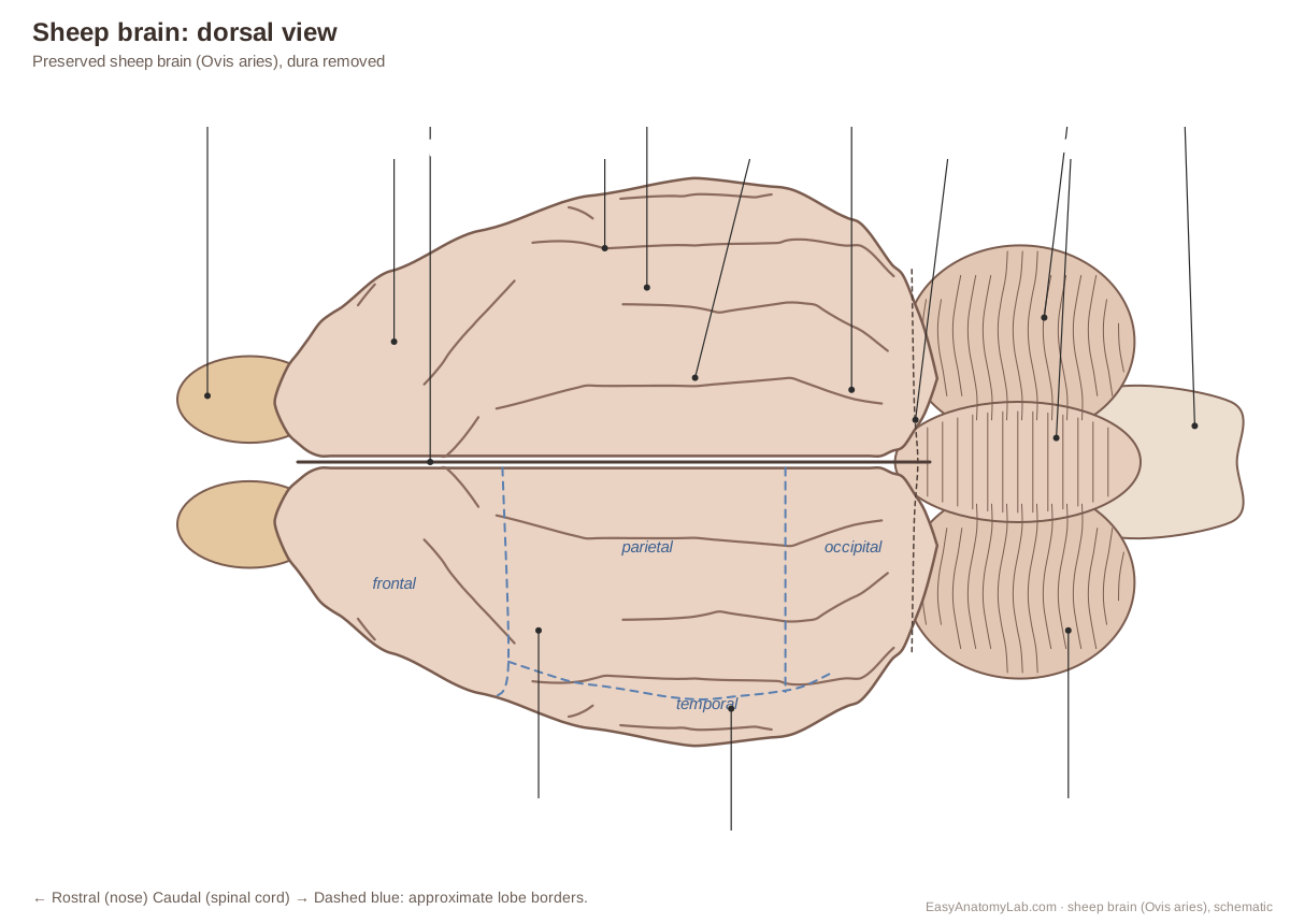
- Identify the two cerebral hemispheres and the longitudinal fissure between them.
- Find gyri (ridges) and sulci (grooves). Most sheep sulci run rostrocaudally.
- Locate the approximate frontal, parietal, temporal and occipital lobes. In the sheep these borders are not marked by clear sulci, so they are estimates based on the human brain.
- Find the cerebellum caudal to the cerebrum, the transverse fissure between them, the midline vermis and the two cerebellar hemispheres.
- Note the medulla oblongata projecting caudally beyond the cerebellum: in the sheep the brainstem runs horizontally.
Identify: Cerebral hemispheres, Longitudinal fissure, Gyri and sulci, Frontal lobe, Parietal lobe, Temporal lobe, Occipital lobe, Transverse fissure, Vermis, Cerebellar hemispheres, Medulla oblongata.
3. Under the cerebellum: pineal gland and colliculi
- Hold the brain dorsal side up. Gently bend the cerebellum caudally (towards the medulla) to open the transverse fissure. The model hides the cerebellum to show this view.
- On the midline, find the small, round pineal gland.
- Caudal to it, find the four bumps of the corpora quadrigemina: the larger superior colliculi in front and the smaller inferior colliculi behind.
- Just caudal to the inferior colliculi, look for the thread-like trochlear nerve (CN IV), the only cranial nerve that leaves the dorsal brainstem.
Caution: Bend the cerebellum gently. If you pull hard it tears off the brainstem.
Identify: Pineal gland, Superior colliculi, Inferior colliculi, Trochlear nerve (CN IV), Midbrain.
4. Ventral surface
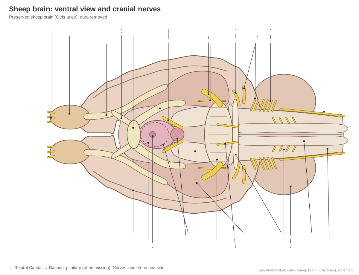
- Turn the brain over. At the rostral end find the olfactory bulbs and the olfactory tracts running back from them to the piriform lobes.
- Find the X-shaped optic chiasm, the optic nerve stumps in front of it and the optic tracts behind it.
- Caudal to the chiasm, find the pituitary gland (if present) on its stalk, the infundibulum. It is often missing; then look for a small hole in the hypothalamus.
- Find the single, round mammillary body (fused in the sheep) and, behind it, the two cerebral peduncles.
- Find the pons, the narrow trapezoid body behind it, and the medulla oblongata with its two pyramids.
Identify: Olfactory bulbs, Olfactory tracts, Piriform lobe, Optic chiasm, Optic nerve (CN II), Optic tracts, Pituitary gland, Infundibulum, Hypothalamus, Mammillary body, Cerebral peduncles, Pons, Trapezoid body, Pyramids.
5. Cranial nerves
- Use a probe and a hand lens. Work from rostral to caudal.
- Usually visible: the optic nerves (II), the flat oculomotor nerves (III) beside the mammillary body, and the thick trigeminal nerves (V) on the sides of the pons.
- Often visible: the abducens nerves (VI) at the trapezoid body and, on careful specimens, the thin trochlear nerves (IV) curving around the brainstem.
- Sometimes visible: the facial (VII) and vestibulocochlear (VIII) nerves at the lateral end of the trapezoid body, and the accessory nerve (XI) along the medulla.
- Usually torn: the olfactory nerves (I) are sheared off at the skull, and IX, X and XII remain as short rootlets or are missing.
- If the ventral dura and pituitary are still attached, free them carefully: cut through the dura close to it (not at the brain) around each nerve so the nerve stumps stay on the brain, cut the infundibulum, then lift the dura and gland away from the caudal end and keep the pituitary.
Caution: Nerves break easily. Move them with a blunt probe, not forceps.
Identify: Olfactory nerve (CN I), Optic nerve (CN II), Oculomotor nerve (CN III), Trochlear nerve (CN IV), Trigeminal nerve (CN V), Abducens nerve (CN VI), Facial nerve (CN VII), Vestibulocochlear nerve (CN VIII), Glossopharyngeal nerve (CN IX), Vagus nerve (CN X), Accessory nerve (CN XI), Hypoglossal nerve (CN XII).
6. Separating the hemispheres: the corpus callosum
- Turn the brain dorsal side up again.
- Place your thumbs in the longitudinal fissure and gently spread the hemispheres apart.
- Look down into the fissure: the bright white band joining the hemispheres is the corpus callosum.
Identify: Longitudinal fissure, Corpus callosum, Cerebral hemispheres.
7. The midsagittal cut
- Place the brain ventral side down. Line the long blade up exactly in the longitudinal fissure.
- Cut straight down through the cerebrum, cerebellum and brainstem with long, smooth strokes, rather than sawing or pressing hard. You now have a left and a right half. The model hides the left half.
- On the cut face, find the corpus callosum (rostrum, genu, body and splenium), the fornix beneath it and the thin septum pellucidum between them.
- Find the thalamus and the round interthalamic adhesion in its centre, surrounded by the third ventricle. Below it is the hypothalamus with the mammillary body; in front, the cut optic chiasm and the small, round anterior commissure.
- Caudally, find the pineal gland, the superior and inferior colliculi, the cerebral aqueduct under them, the midbrain, the pons and the medulla.
- In the cerebellum, find the tree-like arbor vitae and the fourth ventricle beneath it.
Caution: Keep your other hand flat on top of the brain, away from the blade's path.
Identify: Corpus callosum, Fornix, Septum pellucidum, Thalamus, Interthalamic adhesion, Third ventricle, Hypothalamus, Mammillary body, Optic chiasm, Anterior commissure, Pineal gland, Superior colliculi, Inferior colliculi, Cerebral aqueduct, Midbrain, Pons, Medulla oblongata, Arbor vitae, Fourth ventricle.
8. The ventricles and choroid plexus
- On the right half, break through the thin septum pellucidum with a probe to open the lateral ventricle.
- Follow the lateral ventricle forward over the caudate nucleus and back and down towards the temporal region. The model hides the cortex so you can see the whole C-shaped cavity.
- Look for the dark, crumpled choroid plexus in the ventricle floor and in the roofs of the third and fourth ventricles.
- Trace the CSF route: lateral ventricles → third ventricle → cerebral aqueduct → fourth ventricle.
Identify: Lateral ventricles, Septum pellucidum, Caudate nucleus, Choroid plexus, Third ventricle, Cerebral aqueduct, Fourth ventricle.
9. Coronal sections through the thalamus and hippocampus
- Take the other (left) half, or a whole second brain, and lay it flat. Starting at the rostral end, cut coronal (frontal) slices about 5 mm thick, perpendicular to the long axis.
- Rostral slices show the genu of the corpus callosum, the grey caudate nucleus beside the lateral ventricle, the white internal capsule and, lateral to it, the grey putamen.
- At the level of the optic chiasm and thalamus, find the thalamus on each side of the slit-like third ventricle, the internal capsule lateral to it and the hypothalamus below.
- Just caudal to that, the curved, pale hippocampus appears in the floor of each lateral ventricle, above and lateral to the thalamus.
- In every slice compare grey matter (cortex, nuclei) with white matter (corpus callosum, internal capsule). The model hides the cortex to show these deep structures in place.
Identify: Hippocampus, Thalamus, Internal capsule, Caudate nucleus, Putamen, Corpus callosum, Lateral ventricles, Third ventricle, Fornix.
10. The cerebellum and arbor vitae
- Look again at the cut vermis on the midsagittal face.
- Identify the grey cortex on the outside of each folium and the branching white matter, the arbor vitae ('tree of life'), inside.
- Note the fourth ventricle between the cerebellum and the pons and medulla.
- Finish by storing or discarding the halves as your teacher directs, cleaning your tools and washing your hands.
Identify: Arbor vitae, Vermis, Fourth ventricle, Medulla oblongata.
Sheep brain dissection labeled diagrams
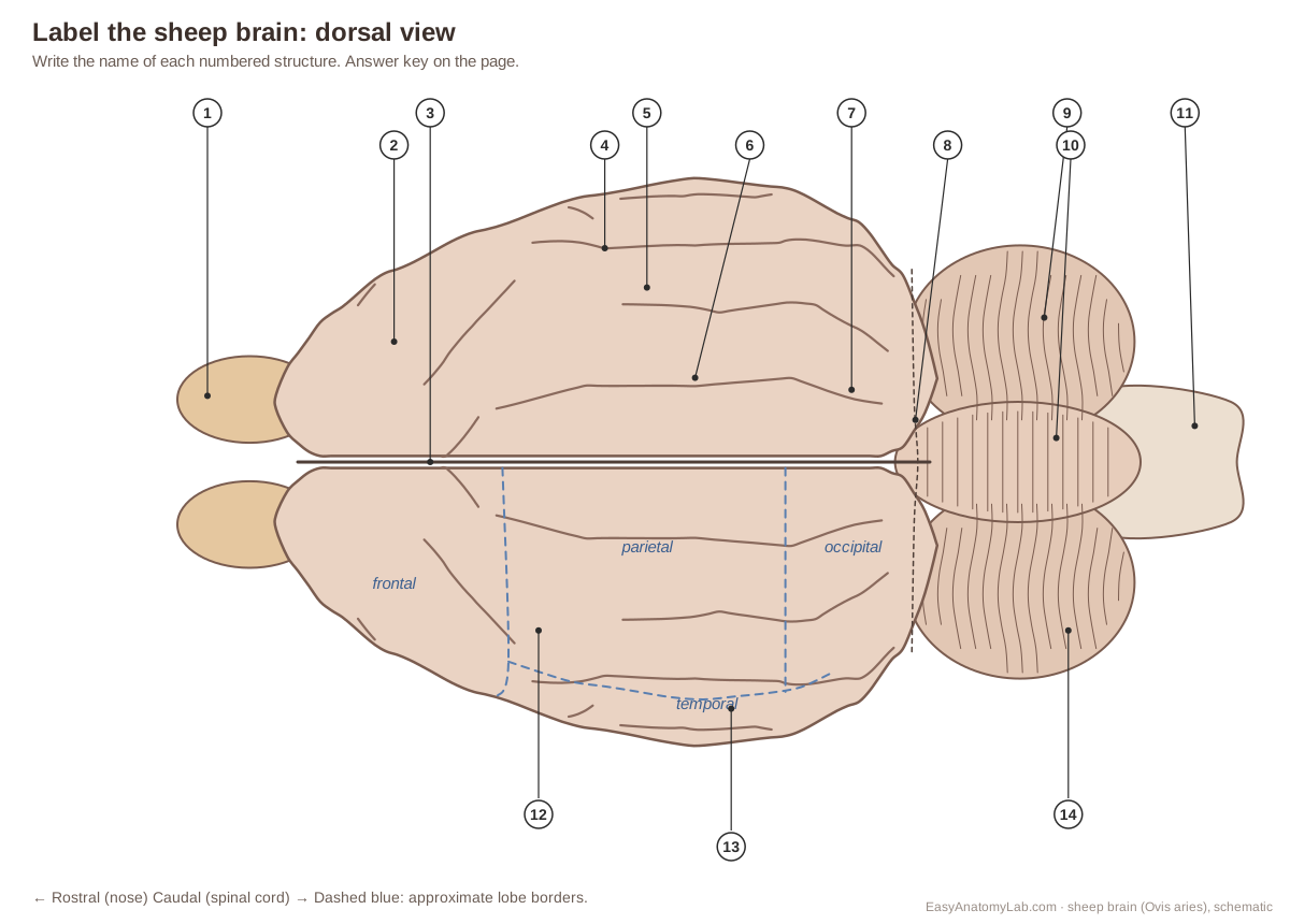
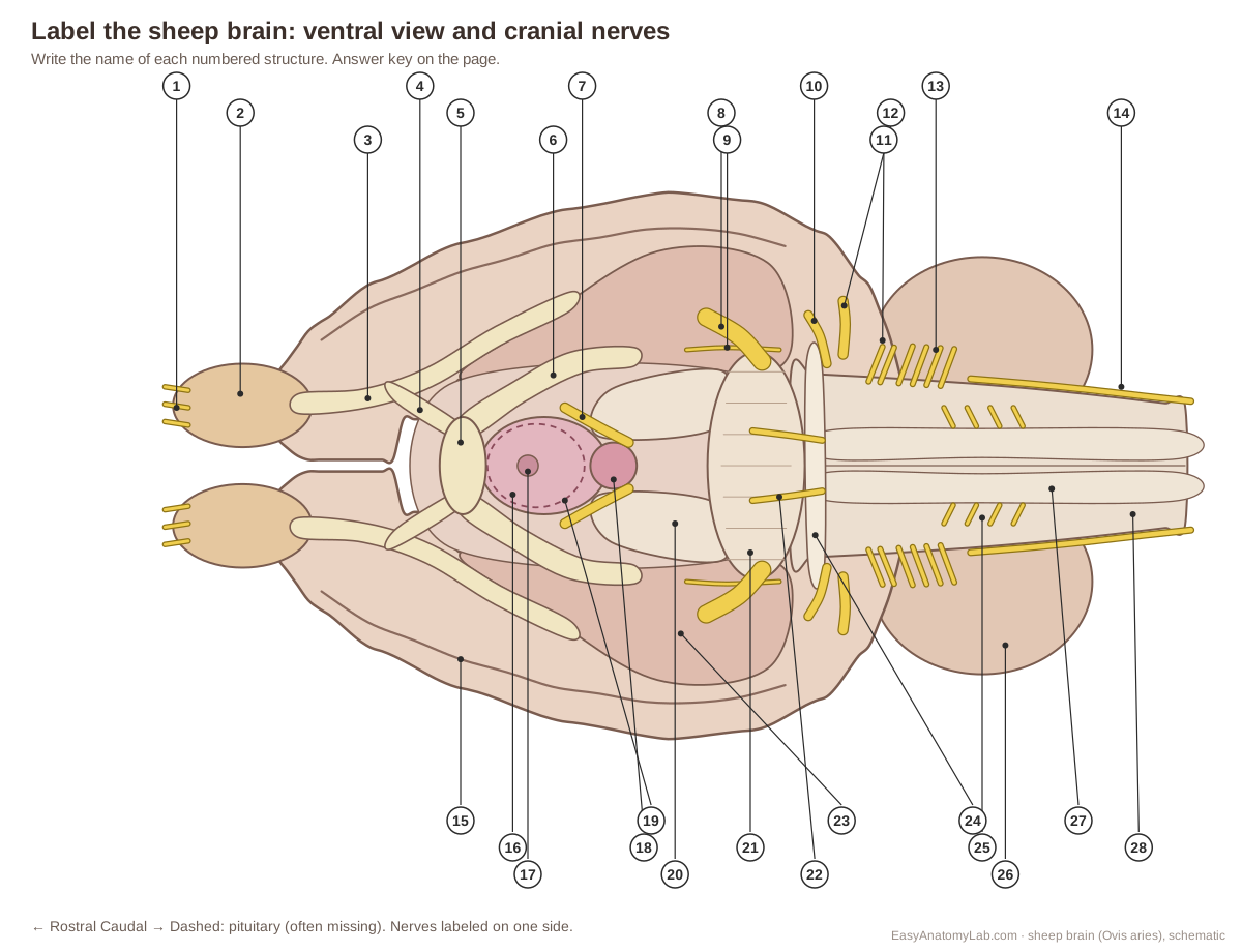
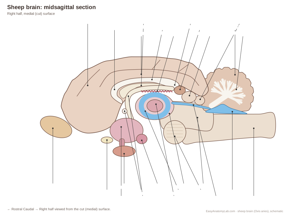
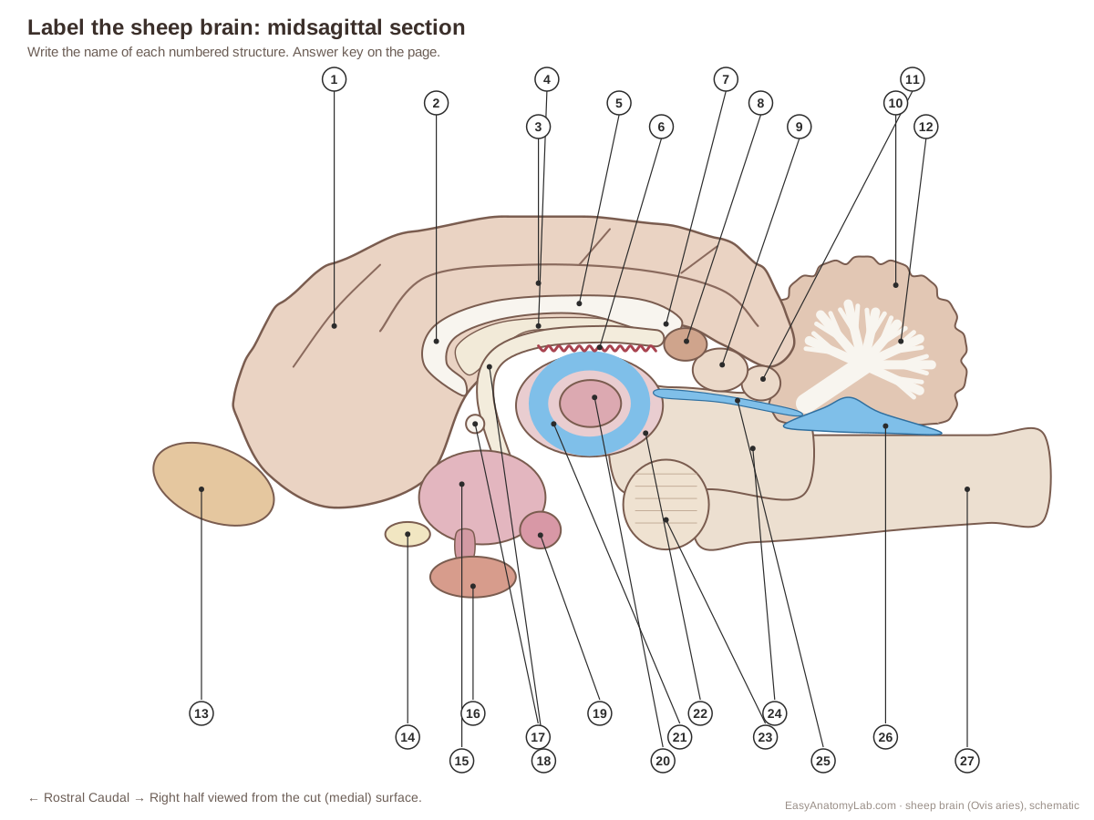
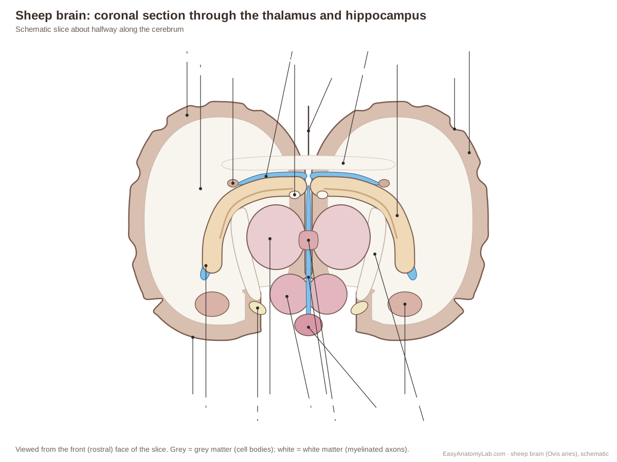
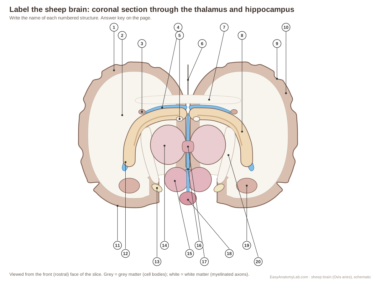
These four labeled diagrams match the four views of a sheep brain lab: dorsal, ventral with the cranial nerves, midsagittal and coronal. Each has a numbered "label the brain" version for worksheets; the answer keys are in the worksheet section.
Sheep brain dorsal view, labeled
Sheep brain ventral view and cranial nerves, labeled
Sheep brain midsagittal section, labeled
Sheep brain coronal section through the thalamus and hippocampus, labeled
Sheep brain structures and functions
Every structure in the 3D model, where to find it on a sheep brain and what it does. Structures are grouped by region, from the coverings inward.
Meninges
| Structure | Where to find it | Function |
|---|---|---|
| Dura materDura; includes the falx cerebri and tentorium cerebelli | Outermost of the three meninges. On supply specimens it is often present only as torn sheets over the dorsal surface, or has been removed entirely. | Protects the brain and anchors it inside the skull. Its folds, the falx cerebri between the hemispheres and the tentorium cerebelli between cerebrum and cerebellum, limit movement of the brain. |
| Arachnoid materArachnoid membrane | Between the dura mater and the pia mater. It bridges over the sulci rather than dipping into them. | Encloses the subarachnoid space, which in life is filled with cerebrospinal fluid (CSF) that cushions the brain. Large blood vessels run in this space. |
| Pia materPia; shown here by its surface blood vessels | Directly on the brain, following every gyrus and dipping into every sulcus. | Carries the small blood vessels that supply the cortex. The model shows it as the fine red vessels on the surface, because that is how you recognise it on a specimen. |
Cerebrum (cortex)
| Structure | Where to find it | Function |
|---|---|---|
| Cerebral hemispheresCerebrum; medial surface and cingulate gyrus | Dorsal and rostral part of the brain, separated by the longitudinal fissure. Highlighted here: the flat medial surface that faces the other hemisphere, with the cingulate gyrus just above the corpus callosum. | The cerebral cortex (outer grey matter) handles perception, voluntary movement, learning and memory. The white matter beneath connects cortical areas to each other and to the rest of the nervous system. |
| Longitudinal fissureCerebral fissure, median fissure | Dorsal midline, from the frontal poles back to the cerebellum. The corpus callosum forms its floor. | Holds the falx cerebri (a fold of dura) in life. In the lab it is your guide for the midsagittal cut. |
| Gyri and sulciConvolutions: ridges (gyri) and grooves (sulci) | Whole surface of both hemispheres. In sheep most main sulci run front-to-back (rostrocaudally). | Folding packs a large sheet of cortex into a small skull. More than two-thirds of the human cortex is hidden inside sulci. |
| Frontal lobeApproximate region in sheep | Front of each hemisphere, above the olfactory bulbs. Its caudal border in sheep is not marked by a clear sulcus like the human central sulcus. | Contains the motor cortex and, in sheep, also the primary somatosensory (touch) area. In humans it is also the seat of planning, decision-making and personality. |
| Parietal lobeApproximate region in sheep | Top of each hemisphere, about halfway along. Boundaries in sheep are approximate. | In humans: touch, body position, spatial attention and some language. Sheep lab manuals use the human names as a guide. |
| Temporal lobeApproximate region in sheep | Side of each hemisphere, below the suprasylvian region and above the rhinal sulcus. | Hearing (auditory cortex) and, with the hippocampus beneath it, memory. |
| Occipital lobeApproximate region in sheep | Back of each hemisphere, rostral to the transverse fissure and cerebellum. | Vision: the primary visual cortex receives signals relayed from the eyes through the thalamus. |
| Piriform lobeOlfactory (pear-shaped) cortex; rhinencephalon | Ventral surface, lateral to the hypothalamus and cerebral peduncles, below the rhinal sulcus. | Primary olfactory cortex: it receives smell information from the lateral olfactory tract. |
White matter & deep nuclei
| Structure | Where to find it | Function |
|---|---|---|
| Corpus callosumRostrum, genu, body and splenium | Floor of the longitudinal fissure. In the midsagittal view it is the white arch above the lateral ventricle and fornix. | Lets the left and right cerebral hemispheres exchange information. |
| Fornix | Midsagittal view: under the body of the corpus callosum, curving down in front of the thalamus to the mammillary body. | The main output of the hippocampus, carrying signals to the mammillary body and other limbic areas. Part of the memory circuit. |
| Septum pellucidum | Midline, ventral to the body of the corpus callosum and dorsal to the fornix. | Separates the left and right lateral ventricles. |
| Anterior commissureRostral commissure (veterinary term) | Midsagittal view: a small white circle below the fornix columns, rostral to the thalamus. | Connects parts of the left and right hemispheres, including olfactory and temporal regions. |
| Caudate nucleus | Floor and lateral wall of the front part of each lateral ventricle; its tail thins out caudally. | Helps plan and control movement and supports learning of habits, with the putamen (together, the striatum). |
| PutamenLentiform nucleus (with the globus pallidus) | Deep in each hemisphere, lateral to the internal capsule and caudate nucleus, at the level of the anterior commissure. | With the caudate nucleus (together, the striatum) it helps select and smooth movements and learn motor habits. |
| Internal capsuleWhite matter between the basal nuclei and thalamus | Deep in each hemisphere, between the caudate nucleus and thalamus (medially) and the putamen (laterally). | Carries almost all fibres running between the cortex and the thalamus, brainstem and spinal cord. |
| Hippocampus | In the sheep it starts dorsally under the corpus callosum, caudal to the thalamus, and curves laterally and ventrally into the temporal region. Reach it by opening the lateral ventricle. | Forms new long-term memories and spatial memory (maps of places). |
Diencephalon
| Structure | Where to find it | Function |
|---|---|---|
| Thalamus | Midsagittal view: the large round area below the fornix and corpus callosum, around the interthalamic adhesion. | Relay station: almost all sensory signals except smell pass through it on their way to the cortex. |
| Interthalamic adhesionMassa intermedia | Centre of the midsagittal section, surrounded by the third ventricle. | Connects the two halves of the thalamus. Its function is not well understood. |
| Hypothalamus | Ventral surface between the optic chiasm and mammillary body; in the midsagittal view, ventral to the thalamus. | Controls body temperature, hunger, thirst, the sleep cycle and the pituitary gland: the link between the nervous and endocrine systems. |
| Mammillary bodyCorpus mamillare; single and fused in sheep | Ventral surface, caudal to the infundibulum and rostral to the cerebral peduncles. | A relay in the memory circuit: it receives the fornix from the hippocampus and projects to the thalamus. |
| InfundibulumPituitary stalk | Ventral midline, just caudal to the optic chiasm. | Carries nerve fibres and blood vessels from the hypothalamus to the pituitary. |
| Pituitary glandHypophysis | Ventral midline, caudal to the optic chiasm, sitting in a pocket of dura in life. | The 'master gland': it releases hormones that control growth, reproduction, the thyroid, the adrenal glands and milk production. |
| Pineal glandPineal body, epiphysis | Between the caudal ends of the hemispheres and the cerebellum, just rostral to the superior colliculi. Pull the cerebellum back to see it. | Makes melatonin at night. In sheep, day-length signals from melatonin time the breeding season. |
Brainstem
| Structure | Where to find it | Function |
|---|---|---|
| MidbrainMesencephalon (tegmentum) | Under the caudal end of the cerebrum. Its roof carries the colliculi, its floor the cerebral peduncles. | Contains the cerebral aqueduct, eye-movement nuclei (CN III and IV) and dopamine-producing neurons (substantia nigra). |
| Superior colliculiRostral colliculi (veterinary term); with the inferior pair: corpora quadrigemina | Dorsal midbrain, caudal to the pineal gland, hidden between the cerebrum and cerebellum. | Visual reflexes: turning the eyes and head towards sudden movement. |
| Inferior colliculiCaudal colliculi (veterinary term) | Dorsal midbrain, just caudal to the superior colliculi and rostral to the cerebellum. | Hearing reflexes: locating where a sound comes from. |
| Cerebral pedunclesCrus cerebri | Ventral surface, caudal to the optic tracts and lateral to the mammillary body. | Carry motor fibres from the cortex to the brainstem and spinal cord. |
| Pons | Ventral surface, caudal to the cerebral peduncles. The trigeminal nerves attach to its sides. | A relay from the cortex to the cerebellum, and a centre for breathing rhythm and sleep. |
| Trapezoid body | Ventral surface of the rostral medulla, behind the pons. The abducens nerve emerges here. | Fibres of the hearing pathway crossing the midline. |
| Medulla oblongataMedulla, myelencephalon | Caudal to the pons, under the cerebellum, ending where the specimen was cut from the spinal cord. | Controls breathing, heart rate, blood pressure, swallowing and vomiting. All signals between brain and spinal cord pass through it. |
| PyramidsPyramidal tracts (medullary pyramids) | Ventral medulla, caudal to the trapezoid body. | Carry the corticospinal (pyramidal) tract for voluntary movement; most fibres cross at the pyramidal decussation. |
Cerebellum
| Structure | Where to find it | Function |
|---|---|---|
| VermisCerebellar vermis | Dorsal midline of the cerebellum, between the two cerebellar hemispheres. | Controls posture, balance and the muscles of the trunk. |
| Cerebellar hemispheres | Either side of the vermis, caudal to the cerebrum. | Coordinate and fine-tune movements of the limbs by comparing intended and actual movement. |
| Arbor vitaeCerebellar white matter ('tree of life') | Inside the cerebellum; visible on the cut face of the vermis in the midsagittal section. | Carries signals into and out of the cerebellar cortex (grey matter). |
| Transverse fissureTransverse cerebral fissure | Between the caudal poles of the hemispheres and the rostral edge of the cerebellum. | Holds the tentorium cerebelli (a fold of dura) in life. |
Ventricles & CSF
| Structure | Where to find it | Function |
|---|---|---|
| Lateral ventriclesFirst and second ventricles | Under the corpus callosum, separated by the septum pellucidum. The caudate nucleus forms the front of the floor and the hippocampus the back. | Filled with cerebrospinal fluid (CSF) made by the choroid plexus. |
| Third ventricle | Midsagittal view: the space surrounding the interthalamic adhesion. | Carries CSF from the lateral ventricles to the cerebral aqueduct. |
| Cerebral aqueductMesencephalic aqueduct, aqueduct of Sylvius | Midsagittal view: a thin channel ventral to the corpora quadrigemina, joining the third and fourth ventricles. | Carries CSF from the third to the fourth ventricle. |
| Fourth ventricle | Midsagittal view: the triangular space under the cerebellum, above the pons and medulla. | Passes CSF into the central canal of the spinal cord and out into the subarachnoid space. |
| Choroid plexus | Floor of the lateral ventricles (along the hippocampus), roof of the third ventricle and roof of the fourth ventricle. | Produces cerebrospinal fluid from blood plasma. |
Olfactory & optic pathways
| Structure | Where to find it | Function |
|---|---|---|
| Olfactory bulbs | Ventral surface, at the rostral end of the brain. They are often damaged when the brain is removed. | First relay for smell. Olfactory nerve fibres from the nose end here and form synapses with mitral cells. |
| Olfactory tractsLateral and medial olfactory tracts | Ventral surface, from the olfactory bulbs back towards the piriform lobe. | Carry smell information from the bulb to olfactory cortex, mostly the piriform lobe, without passing through the thalamus. |
| Optic chiasmOptic chiasma | Ventral surface, caudal to the olfactory tracts and just rostral to the pituitary stalk. | Here fibres from the inner (nasal) half of each retina cross to the opposite side. |
| Optic tracts | From the chiasm caudolaterally around the cerebral peduncles to the lateral side of the thalamus. | Carry visual signals to the lateral geniculate nucleus of the thalamus and to the superior (rostral) colliculus. |
Cranial nerves
| Structure | Where to find it | Function |
|---|---|---|
| Olfactory nerve (CN I) | Rostroventral surface of each olfactory bulb (shown as short stubs). | Sensory: smell. |
| Optic nerve (CN II) | Ventral surface, rostral to the optic chiasm, angled towards the eyes. | Sensory: vision. |
| Oculomotor nerve (CN III) | Ventral surface, on the medial side of each cerebral peduncle, about halfway between the optic chiasm and the pons. | Motor: most eye muscles and the upper eyelid; parasympathetic fibres constrict the pupil. |
| Trochlear nerve (CN IV) | Emerges just caudal to the inferior (caudal) colliculi and curves ventrally around the side of the brainstem, often hidden by the trigeminal nerve. | Motor: the superior oblique muscle of the eye. |
| Trigeminal nerve (CN V) | Lateral side of the pons, near its junction with the medulla. | Mixed: sensation from the face, cornea, teeth and dura; motor to the chewing muscles. |
| Abducens nerve (CN VI) | Ventral surface at the trapezoid body, close to the midline beside the pyramids. | Motor: the lateral rectus muscle, which turns the eye outward. |
| Facial nerve (CN VII) | Lateral to the trapezoid body, caudal to the trigeminal nerve. | Mixed: muscles of facial expression, salivary and tear glands, taste from the front two-thirds of the tongue. |
| Vestibulocochlear nerve (CN VIII)Auditory or acoustic nerve | Lateral end of the trapezoid body, just caudal and lateral to CN VII. | Sensory: hearing (cochlear part) and balance (vestibular part). |
| Glossopharyngeal nerve (CN IX) | Lateral medulla, caudal to CN VIII, rostral to the vagus rootlets. | Mixed: taste and sensation from the back of the tongue and throat; swallowing; salivation. |
| Vagus nerve (CN X) | Lateral medulla, between the glossopharyngeal and accessory rootlets. | Mixed: the heart, lungs, stomach and gut, plus the larynx and pharynx. |
| Accessory nerve (CN XI)Spinal accessory nerve | Lateral medulla, its rootlets continuing caudally along the cut end of the brainstem. | Motor: neck and shoulder muscles (in sheep, muscles that move the head and neck). |
| Hypoglossal nerve (CN XII) | Ventral surface of the caudal medulla, lateral to the pyramids. | Motor: the muscles of the tongue. |
Sheep brain vs human brain
The sheep brain has the same parts as the human brain in different proportions. The sheep cerebrum is smaller relative to the brainstem and cerebellum, the olfactory bulbs are much larger, the frontal lobe is smaller and the whole brain lies horizontally. Sources: Banstola & Reynolds (2022), Murray & Mitchell (2022) and the University of Guelph lab manual.
| Feature | Sheep | Human | Why it differs |
|---|---|---|---|
| Brain mass | About 130–140 g | About 1,300–1,400 g | Humans have a much larger cerebrum relative to body size. |
| Overall shape | Smaller, elongated | Larger, rounded | The human cerebrum has expanded up and forward. |
| Orientation of brainstem | Horizontal; runs straight back to the spinal cord | Vertical; bends at the cephalic flexure | Four-legged versus upright posture. |
| Olfactory bulbs | Large and well developed | Small | Sheep rely on smell for lamb recognition, feeding and mating. |
| Piriform (olfactory) lobe | Large bulge on the ventral surface | Small, hidden on the medial temporal lobe | Matches the size of the olfactory bulbs. |
| Frontal lobe | Small; contains motor and somatosensory areas | Very large; somatosensory cortex is in the parietal lobe | Planning, language and reasoning take up much of the human frontal lobe. |
| Lobe boundaries | Approximate; no reliable sulcal landmarks | Marked by the central and lateral sulci | Different folding patterns. |
| Cerebellum | Smaller; lies behind the cerebrum | Larger; tucked under the occipital lobes | Straight versus bent neuraxis and a much bigger human cerebrum. |
| Mammillary bodies | Fused into one midline body | Two, side by side | A species difference in shape, not in function. |
| Pineal gland | Large and round | Small and pine-cone shaped | Melatonin times the seasonal breeding of sheep. |
| Optic chiasm | More pronounced | Less pronounced | Sheep eyes face sideways; more fibres cross. |
| Trapezoid body | Visible on the ventral surface behind the pons | Hidden inside the brainstem | The large human pons covers it. |
| Hippocampus | Starts dorsally under the corpus callosum and curves into the temporal region | In the medial temporal lobe | Same arc; in humans it lies wholly inside the large temporal lobe. |
Why biology classes dissect a sheep brain
The sheep brain is the standard teaching brain because it is large enough to handle, cheap, and built on the same plan as the human brain. An adult sheep brain weighs about 130–140 g, roughly a tenth of an adult human brain (1,300–1,400 g), against only 1–2 g for a rat. Its cortex is folded (gyrencephalic), and it has well-defined basal nuclei: a caudate nucleus and putamen separated by an internal capsule, as in humans. Those features are the reason neuroscientists also use sheep as a large-animal model for human brain disease. (Murray & Mitchell (2022): the translational benefits of sheep as large animal models of human neurological disorders, Front. Vet. Sci. 9:831838.)
Nearly every structure on a human brain diagram has a counterpart you can see with the naked eye on a sheep brain: the three meninges, the hemispheres and corpus callosum, the thalamus and hypothalamus, the colliculi, pons and medulla, the cerebellum and its arbor vitae, and the ventricles. Most structures come in pairs, one per hemisphere; the pineal gland is the classic unpaired exception. The differences, covered below, are mostly differences of proportion, and each one tells you something about how a sheep lives. (MIT OpenCourseWare 9.10 Cognitive Neuroscience (Spring 2006): Sheep Brain Dissection Guide.)
Orientation: rostral, caudal, dorsal and ventral on a sheep brain
Because the sheep walks on four legs, its brain and spinal cord lie in one nearly straight, horizontal line. The olfactory bulbs point forward (rostral), the medulla and spinal cord point back (caudal), the top surface is dorsal and the underside, where most cranial nerves leave, is ventral. In an upright human the brainstem points down instead, the foramen magnum sits under the skull rather than at the back, and the neuraxis bends at the cephalic flexure. That is why human atlases use superior and inferior where sheep lab manuals use dorsal and ventral. (Human Anatomy Laboratory Manual (LibreTexts), 11.7 Sheep Brain Dissection.)
The same straight neuraxis explains why the sheep cerebellum sits behind the cerebrum rather than tucked under the occipital lobes, and why the medulla projects caudally beyond the cerebellum on a dorsal view. Murray and Mitchell point out that the almost linear sheep neuraxis is one of the anatomical differences researchers must account for when they compare head injuries in sheep and humans. (Murray & Mitchell (2022): the translational benefits of sheep as large animal models of human neurological disorders, Front. Vet. Sci. 9:831838.)
Veterinary atlases also use their own names for a few structures. The superior and inferior colliculi of human anatomy are the rostral and caudal colliculi in a veterinary text, and the anterior commissure is the rostral commissure. Learn both: lab quizzes for biology and psychology courses usually use the human terms. (University of Minnesota veterinary neuroanatomy: sheep diencephalon, dorsal view.)
External anatomy: meninges, dorsal and ventral surfaces
The brain is wrapped in three meninges. The dura mater is a tough, opaque shell; the arachnoid beneath it often comes away with the dura; and the pia mater is a transparent film that follows every convolution of the surface. Many supply brains arrive with the dura already removed. When the dura is present, the tough, transparent tissue over the pituitary is also dura, and several cranial nerves pass through it, so it should be removed carefully and only where your instructor asks. (California State University, Northridge: Sheep brain Lab 1, external structures and cranial nerves.)
On the dorsal surface, the ridges are gyri and the grooves are sulci. Students are usually asked to locate the frontal, parietal, temporal and occipital lobes, but in the sheep those borders are not marked by reliable sulci as they are in humans, so treat sheep lobe borders as approximate. More than two-thirds of the human cortex is hidden inside sulci; the sheep cortex is folded less deeply. (MIT OpenCourseWare 9.10 Cognitive Neuroscience (Spring 2006): Sheep Brain Dissection Guide.)
The ventral surface is where most of the identification happens. From rostral to caudal: the olfactory bulbs and tracts, the optic nerves, optic chiasm and optic tracts, the pituitary gland and its stalk, the mammillary body, the cerebral peduncles, the pons, the trapezoid body and the pyramids of the medulla. Two sheep-specific details help: the mammillary bodies are fused into one round midline body, and the trapezoid body forms a clear transverse ridge behind the pons, where the abducens nerve emerges. (University of Guelph, Department of Psychology: sheep brain lab manual, 'The Ventral Surface'.)
Lateral to the hypothalamus, the large, smooth piriform lobes are the olfactory cortex. On the University of Minnesota sheep specimen you can follow the olfactory bulb into lateral and medial olfactory tracts, past the rostral perforated area, to the piriform lobe. The lateral rhinal sulcus separates this old, smell-related cortex from the neocortex above it. (University of Minnesota veterinary neuroanatomy: sheep telencephalon, ventral view (rhinencephalon).)
Sheep brain cranial nerves: which ones you can actually find
All twelve cranial nerves exist in the sheep, but a removed brain keeps only some of them. The University of Guelph manual says that often you will only be able to see nerves II to VII and XI. The olfactory nerve (I) is sheared off by the cribriform plate when the brain is taken out, so you identify the olfactory bulb in its place. The optic nerve (II) is large; the oculomotor (III) leaves the ventral midbrain about halfway between the pons and the chiasm; the trigeminal (V) is very large and leaves the side of the pons; and the abducens (VI) emerges behind the pons at the trapezoid body. (University of Guelph, Department of Psychology: sheep brain lab manual, 'The Ventral Surface'.)
The trochlear nerve (IV) is a very thin, thread-like nerve that leaves the dorsal brainstem just behind the inferior colliculi and curves ventrally, often hidden by the bulk of the trigeminal nerve. It is the only cranial nerve that leaves the dorsal side of the brainstem. CSUN's lab manual warns that nerves VII to XII were quite likely stripped from your sheep brain, so many courses only ask students to know their functions and whether they are sensory, motor or mixed. (California State University, Northridge: Sheep brain Lab 1, external structures and cranial nerves.)
A quick function summary: I smell; II vision; III, IV and VI eye movements (III also constricts the pupil and lifts the eyelid); V face sensation and chewing; VII facial expression, tears, saliva and taste on the front of the tongue; VIII hearing and balance; IX taste and swallowing; X heart, lungs and gut; XI neck and shoulder muscles; XII tongue. A common mnemonic is 'On Old Olympus' Towering Top, A Finn And German Viewed Some Hops'. (California State University, Northridge: Table 1, the cranial nerves.)
Reading the midsagittal section
The midsagittal cut divides the brain into equal left and right halves along the longitudinal fissure. Guelph's manual starts the tour with the massa intermedia (interthalamic adhesion), where the two halves of the thalamus join across the midline. Dorsal to it lies the fornix, and above that the corpus callosum with its genu (front), body and splenium (back). The cingulate gyrus lies just above the corpus callosum, and the thin septum pellucidum, which closes off the lateral ventricle, is often partly torn. (University of Guelph sheep brain lab manual, 'The Mid-Sagittal Cut'.)
The ventricles show as thin spaces. The third ventricle is a narrow slit between the two thalami, so on the cut face you see the smooth ventricle wall around the adhesion rather than a hollow chamber. The cerebral aqueduct runs under the colliculi and opens into the third ventricle at one end and the fourth ventricle at the other. The fourth ventricle widens under the cerebellum. The roof of the midbrain above the aqueduct, which carries the four colliculi, is the tectum or lamina quadrigemina (quadrigeminal plate). (California State University, Northridge: Sheep brain Lab 3, mid-sagittal section.)
The University of Minnesota median view of a sheep brain marks the same landmarks: the interthalamic adhesion, the pineal body, the third ventricle, the mesencephalic aqueduct and the fourth ventricle. In the cerebellum, the cut through the vermis shows the arbor vitae: white matter branching like a tree into folia covered by grey cortex. (University of Minnesota veterinary neuroanatomy: sheep brain, median view.)
Coronal sections: thalamus, hippocampus and grey versus white matter
Coronal (frontal) slices about half a centimetre thick show how the deep structures fit together. Near the front, the genu of the corpus callosum and the corona radiata appear with the caudate nucleus and putamen. Further back the lateral ventricles widen, the internal capsule appears as a mass of white fibres lateral to the caudate, and the anterior commissure crosses the midline near the optic chiasm. At the level of the thalamus, the fimbria of the hippocampus starts to fill the lateral ventricle, the third ventricle is a thin slit with the hypothalamus on either side, and the optic tracts run along the bottom. (University of Guelph sheep brain lab manual, 'Coronal Cuts'.)
The hippocampus of the sheep is easy to find. Cutting down from the dorsal surface, you meet the lateral ventricle first, then the caudate nucleus rostrally and the hippocampus caudally. Once you reach the dorsal part of the hippocampus, you can follow it as it curves laterally and ventrally through the temporal region; dissected out whole, it shows why it was named after a seahorse. The shiny white fornix follows its medial face. (MIT OpenCourseWare 9.10 Cognitive Neuroscience (Spring 2006): Sheep Brain Dissection Guide.)
Every slice is also a lesson in grey and white matter. Grey matter is coloured by neuron cell bodies (the cortex, caudate, thalamus, hippocampus); white matter is coloured by the fatty myelin that wraps axons (the corpus callosum, internal capsule, optic tracts and arbor vitae). Myelin insulates axons and speeds up conduction, which matters most for long fibres. (MIT OpenCourseWare 9.10 Cognitive Neuroscience (Spring 2006): Sheep Brain Dissection Guide.)
Why is the sheep brain different? Olfactory bulbs, frontal lobe and pineal gland
In a comparison table of the sheep and human central nervous system, Banstola and Reynolds list the sheep brain as smaller and elongated with a small frontal lobe, large and well-developed olfactory bulbs, a more pronounced optic chiasm, a smaller cerebellum placed behind the cerebrum, and a large, round pineal gland; the human brain is larger and rounded, with a very large frontal lobe, small olfactory bulbs and a small, pine-cone-shaped pineal. They also note that the sheep somatosensory area lies in the frontal lobe, while the human one is in the parietal lobe. (Banstola & Reynolds (2022): mapping sheep to human brain, the need for a sheep brain atlas, Front. Vet. Sci. 9:961413.)
The big olfactory bulbs reflect how much sheep rely on smell. Ungulate social behaviour depends heavily on olfaction, and the main olfactory system is needed for maternal behaviour and for the ewe to recognise her own lamb at birth. Recordings from the olfactory bulb show that after birth many mitral cells start responding to lamb odours, and some respond preferentially to the ewe's own lamb, so coding of the familiar lamb's smell already begins in the bulb. (Keller & Lévy (2012): the main but not the accessory olfactory system processes socially relevant chemosignals in ungulates, Front. Neuroanat. 6:39.)
Field studies agree: a ewe identifies her lamb at the udder by smell alone, while at a distance she uses sight and the lamb's call. Humans, by contrast, depend mainly on vision and hearing for social recognition, and their olfactory bulbs are small. The large human frontal lobe, in turn, supports planning, language and abstract reasoning, abilities that take up comparatively little of the sheep cortex. (Searby & Jouventin (2003): mother–lamb acoustic recognition in sheep, Proc. R. Soc. B 270.)
Where do sheep brains for dissection come from?
Sheep brains for dissection are a by-product of the meat industry. Ward's Science, a major supplier, states that its preserved sheep materials are a byproduct of the food industry and that sheep are not raised specifically for dissection. Suppliers sell brains with the dura mater intact or removed, preserved in formalin or in formalin-free preserving solutions; some listings note that the hypophysis (pituitary) and cranial nerves are intact, while cheaper grades may lack them. (Ward's Science: Pure Preserved fully extracted sheep brains (product description).)
Scrapie is a fatal prion disease of the central nervous system of sheep and goats, one of the transmissible spongiform encephalopathies. The California Department of Food and Agriculture states that there is no evidence that scrapie poses a risk to human health. All US states take part in the National Scrapie Eradication Program, which began in 2001. (California Department of Food and Agriculture: scrapie fact sheet.)
USDA notes that prions bind tightly to surfaces and are difficult to decontaminate, and that scrapie tests need brain or lymphoid tissue. (USDA APHIS: scrapie.)
Prions are therefore treated with respect in the lab. A university biosafety guide classes animal prions such as scrapie as Biosafety Level 2 agents, notes that formalin-fixed brain tissue can remain infectious for long periods, and names accidental cuts, punctures and ingestion as the main hazards; there are no documented laboratory-acquired prion infections. For a classroom, that translates into simple rules: gloves, no eating or drinking, careful blade work, covered cuts and hand-washing. (Michigan State University EHS: recommended biosafety practices for handling prions and prion-infected tissues.)
Rules differ by country. Under EU Regulation 999/2001, which also applies in UK law, the skull including the brain and eyes of sheep and goats over 12 months old, or with a permanent incisor erupted, is specified risk material that must be removed from the food chain. Schools there should buy specimens only from suppliers that follow these rules, and follow their local science-safety advice. (Regulation (EC) No 999/2001, Annex V: specified risk material (legislation.gov.uk).)
If you cannot or prefer not to dissect a real brain, the 3D model on this page follows the same ten steps and lets you hide the left half for a true midsagittal view, so you can complete the same worksheet.
Sheep brain dissection worksheet and answer key
Match the structure to the description
Write the letter of the matching structure next to each description.
- Branching white matter inside the cerebellum
- Tough outer covering of the brain
- X-shaped crossing of the optic nerves
- Front pair of midbrain bumps for visual reflexes
- Rear pair of midbrain bumps for hearing reflexes
- Ventral bulge that relays signals from the cortex to the cerebellum
- Unpaired gland that releases melatonin
- Relay station for sensory signals on their way to the cortex
- C-shaped CSF cavity inside each hemisphere
- Large pad under the frontal lobe that receives smell signals
- White band that connects the two cerebral hemispheres
- Controls breathing and heart rate; joins the spinal cord
- Controls the pituitary, body temperature, hunger and thirst
- Coordinates movement and balance
- A. Pons
- B. Medulla oblongata
- C. Arbor vitae
- D. Lateral ventricle
- E. Superior colliculi
- F. Cerebellum
- G. Olfactory bulb
- H. Dura mater
- I. Hypothalamus
- J. Pineal gland
- K. Optic chiasm
- L. Corpus callosum
- M. Thalamus
- N. Inferior colliculi
Show the answer key
1–C (Arbor vitae); 2–H (Dura mater); 3–K (Optic chiasm); 4–E (Superior colliculi); 5–N (Inferior colliculi); 6–A (Pons); 7–J (Pineal gland); 8–M (Thalamus); 9–D (Lateral ventricle); 10–G (Olfactory bulb); 11–L (Corpus callosum); 12–B (Medulla oblongata); 13–I (Hypothalamus); 14–F (Cerebellum).
Label the brain: answer keys
Print the blank numbered diagrams, label them during the dissection, then check your answers here.
Dorsal view (blank diagram)
- Olfactory bulb
- Frontal lobe (approx.)
- Longitudinal fissure
- Sulcus
- Gyrus
- Parietal lobe (approx.)
- Occipital lobe (approx.)
- Transverse fissure
- Cerebellar hemisphere
- Vermis
- Medulla oblongata
- Cerebral hemisphere
- Temporal lobe (approx.)
- Cerebellum
Ventral view and cranial nerves (blank diagram)
- I: olfactory nerve fibres
- Olfactory bulb
- Olfactory tract
- II: optic nerve
- Optic chiasm
- Optic tract
- III: oculomotor
- V: trigeminal
- IV: trochlear
- VII: facial
- IX: glossopharyngeal
- VIII: vestibulocochlear
- X: vagus
- XI: accessory
- Rhinal sulcus
- Hypothalamus
- Infundibulum
- Mammillary body
- Pituitary gland (if present)
- Cerebral peduncle
- Pons
- VI: abducens
- Piriform lobe
- Trapezoid body
- XII: hypoglossal
- Cerebellum
- Pyramid
- Medulla oblongata
Midsagittal section (blank diagram)
- Cerebral hemisphere (medial surface)
- Genu of corpus callosum
- Cingulate gyrus
- Septum pellucidum
- Corpus callosum (body)
- Choroid plexus
- Splenium of corpus callosum
- Pineal gland
- Superior colliculus
- Cerebellum (vermis)
- Inferior colliculus
- Arbor vitae
- Olfactory bulb
- Optic chiasm
- Hypothalamus
- Pituitary gland
- Anterior commissure
- Fornix
- Mammillary body
- Interthalamic adhesion
- Third ventricle
- Thalamus
- Pons
- Midbrain
- Cerebral aqueduct
- Fourth ventricle
- Medulla oblongata
Coronal section through the thalamus and hippocampus (blank diagram)
- Cerebral cortex (grey matter)
- White matter
- Tail of caudate nucleus
- Lateral ventricle
- Fornix (fimbria)
- Longitudinal fissure
- Corpus callosum
- Hippocampus
- Sulcus
- Gyrus
- Piriform lobe
- Temporal horn of lateral ventricle
- Optic tract
- Thalamus
- Hypothalamus
- Third ventricle
- Interthalamic adhesion
- Mammillary body
- Amygdala
- Internal capsule
Sheep brain dissection pre-lab questions
Answer these before the lab. Click a question to reveal the answer.
Name the three meninges from outside to inside.
Dura mater, arachnoid mater, pia mater (remember PAD from the inside out).
What is the difference between grey matter and white matter?
Grey matter is made mostly of neuron cell bodies (the cortex and nuclei such as the thalamus and caudate). White matter is made of myelinated axons, which look white because myelin is fatty (corpus callosum, internal capsule, arbor vitae).
Define the directional terms rostral, caudal, dorsal and ventral for a sheep brain.
Rostral = towards the nose (olfactory bulbs); caudal = towards the tail (medulla); dorsal = the top surface; ventral = the underside, where most cranial nerves are.
What is a midsagittal cut, and what is a coronal (frontal) cut?
A midsagittal cut runs along the midline, through the longitudinal fissure, and divides the brain into equal left and right halves. A coronal cut is perpendicular to the long axis and slices the brain from front to back into cross-sections.
Which sense does not relay through the thalamus?
Smell (olfaction). Olfactory signals go from the olfactory bulb to olfactory cortex such as the piriform lobe directly.
Predict: which brain parts will look larger in the sheep than in the human, relative to the rest of the brain?
The olfactory bulbs and piriform (olfactory) cortex, and the brainstem and cerebellum relative to the cerebrum. The frontal lobe will look smaller.
Sheep brain dissection post-lab and analysis questions
Answer these after the dissection, using your specimen and the diagrams.
Why are the olfactory bulbs of the sheep so much larger than those of a human?
Sheep depend heavily on smell for social behaviour: a ewe learns her own lamb's odour within hours of birth and accepts only that lamb at the udder, and smell helps them find food and detect rams and predators. Humans rely mostly on vision, so their olfactory bulbs are small.
Why is the frontal lobe smaller in the sheep?
In humans the frontal lobe is greatly enlarged for planning, decision-making, language and social reasoning. The sheep cerebrum as a whole is smaller relative to the brainstem, and its frontal region is small and contains mainly motor and body-sense areas.
Why does the sheep brainstem run horizontally, while the human brainstem points down?
Sheep walk on four legs, so the spinal cord runs horizontally and leaves the skull through a foramen magnum at the back of the skull. Humans stand upright, the foramen magnum is under the skull, and the neuraxis bends at the cephalic flexure.
Which cranial nerves did you find, and why are some usually missing?
Usually II, III and V; often IV and VI; sometimes VII, VIII and XI. Nerve I is sheared off by the cribriform plate when the brain is removed, and IX, X and XII are thin rootlets on the medulla that tear easily.
Trace the flow of cerebrospinal fluid through the ventricles you saw.
CSF is made by the choroid plexus. It flows from each lateral ventricle through the interventricular foramen into the third ventricle, through the cerebral aqueduct into the fourth ventricle, and then into the central canal and the subarachnoid space.
Where is the hippocampus in the sheep, and how is that different in humans?
In the sheep a large part lies dorsally, in the floor of the lateral ventricle under the corpus callosum and caudal to the thalamus, and it curves ventrally into the temporal region. In humans it lies only in the medial temporal lobe.
What does the arbor vitae tell you about grey and white matter in the cerebellum?
The grey matter (cortex) is on the outside of each folium and the white matter (arbor vitae) is inside, branching like a tree, which is the same pattern as in the cerebrum: cortex outside, fibres inside.
Name two structures that are unpaired (single, on the midline) in the sheep brain.
The pineal gland, the mammillary body (fused into one in the sheep), the pituitary gland, the optic chiasm, the interthalamic adhesion and the vermis are all midline structures.
Virtual sheep brain dissection
The 3D viewer at the top of the page is a free virtual sheep brain dissection. The ten steps match the real procedure: the dura and arachnoid fade away, the cerebellum lifts off to show the colliculi, the left half disappears for the midsagittal cut, and the cortex hides to show the ventricles, hippocampus, thalamus and internal capsule in place. Click any structure for its location and function, switch on "All labels" to quiz yourself, or rotate to the ventral surface to count the cranial nerves. Students who opt out of a physical dissection can complete the same worksheet with it.
For other dissection labs, see the fetal pig dissection and dogfish shark dissection guides.
Frequently asked questions
What are the main parts of a sheep brain?
The cerebrum (two cerebral hemispheres joined by the corpus callosum), the diencephalon (thalamus, hypothalamus, pineal and pituitary glands), the brainstem (midbrain, pons and medulla oblongata) and the cerebellum. It is covered by three meninges and has four ventricles filled with cerebrospinal fluid in life.
How is a sheep brain different from a human brain?
It is about one-tenth the mass (130–140 g versus 1,300–1,400 g), more elongated, and lies horizontally, with the brainstem running straight back to the spinal cord. The sheep has much larger olfactory bulbs and piriform lobes, a smaller frontal lobe, a cerebellum behind rather than under the cerebrum, a single fused mammillary body and a large, round pineal gland.
Why are the olfactory bulbs of a sheep so large?
Sheep rely heavily on smell. A ewe learns the odour of her own lamb within hours of birth and accepts only that lamb at the udder, and smell also guides feeding and reproduction. Humans rely mainly on vision, so their olfactory bulbs are small.
Which cranial nerves can you see on a sheep brain?
Usually the optic (II), oculomotor (III) and trigeminal (V) nerves; often the abducens (VI) and, with care, the thread-like trochlear (IV); sometimes the facial (VII), vestibulocochlear (VIII) and accessory (XI). The olfactory nerves (I) are torn off at the skull, and IX, X and XII usually remain only as short rootlets.
Where do sheep brains for dissection come from?
They are a by-product of the meat industry: supply companies collect them from sheep slaughtered for food and preserve them. Suppliers state that sheep are not raised specifically for dissection.
Is it safe to dissect a sheep brain? What about scrapie?
Yes, with normal lab precautions. Scrapie is a prion disease of sheep, and there is no evidence that it poses a risk to human health. Because prions resist ordinary disinfection and the main lab hazard is a cut or ingestion, wear gloves and eye protection, keep the brain in the tray when cutting, cover any broken skin, never eat in the lab and wash your hands afterwards.
Why is the pituitary gland missing from my sheep brain?
The pituitary sits in a pocket of dura below the brain, so it usually tears off with the dura when the brain is removed from the skull. Look for the infundibulum (stalk) or a small hole in the hypothalamus just behind the optic chiasm.
Where is the hippocampus in a sheep brain?
In the floor of the lateral ventricle. In the sheep it starts dorsally, under the corpus callosum and behind the thalamus, then curves laterally and ventrally into the temporal region. Open the lateral ventricle from above, or cut coronal slices behind the optic chiasm, to see it.
What is the arbor vitae?
The branching white matter inside the cerebellum, seen when the cerebellum is cut in half. Its name means 'tree of life'. The white branches are nerve fibres; the grey covering of each fold (folium) is cerebellar cortex.
Is there a virtual sheep brain dissection?
Yes. The 3D model on this page follows the same ten steps as a real dissection: the meninges, dorsal and ventral surfaces, cranial nerves, the midsagittal cut (hide the left half), the ventricles and coronal-level deep structures. Students who opt out of a physical dissection can complete the same worksheet with it.
References
- University of Guelph, Department of Psychology. Sheep brain lab manual: The Ventral Surface; The Mid-Sagittal Cut; Coronal Cuts.
- University of Guelph sheep brain lab manual: The Mid-Sagittal Cut.
- University of Guelph sheep brain lab manual: Coronal Cuts.
- Schneider, A. E. Sheep Brain Dissection Guide. MIT OpenCourseWare 9.10 Cognitive Neuroscience, Spring 2006.
- California State University, Northridge. Sheep brain dissection, Lab 1: external structures and cranial nerves on the ventral surface.
- California State University, Northridge. Sheep brain dissection, Lab 3: mid-sagittal section.
- University of Minnesota College of Veterinary Medicine. Neuroanatomy lab: sheep diencephalon, dorsal and ventral views; sheep brain median view; sheep rhinencephalon.
- Human Anatomy Laboratory Manual (LibreTexts / Lumen Learning), 11.7 Sheep Brain Dissection.
- Murray, S. J. & Mitchell, N. L. (2022). The translational benefits of sheep as large animal models of human neurological disorders. Frontiers in Veterinary Science 9, 831838.
- Banstola, A. & Reynolds, J. N. J. (2022). Mapping sheep to human brain: the need for a sheep brain atlas. Frontiers in Veterinary Science 9, 961413.
- Keller, M. & Lévy, F. (2012). The main but not the accessory olfactory system is involved in the processing of socially relevant chemosignals in ungulates. Frontiers in Neuroanatomy 6, 39.
- Searby, A. & Jouventin, P. (2003). Mother–lamb acoustic recognition in sheep: a frequency coding. Proceedings of the Royal Society B 270, 1765–1771.
- California Department of Food and Agriculture. Scrapie fact sheet.
- Michigan State University Environmental Health & Safety. Recommended biosafety practices for handling prions and prion-infected tissues.
- Regulation (EC) No 999/2001, Annex V: specified risk material (legislation.gov.uk).
More interactive anatomy models
- Cow eye dissectionGuided cow eye dissection: cornea, lens, retina and tapetum lucidum.
- Sheep heart dissectionGuided sheep (and pig) heart dissection: chambers, valves and great vessels.
- Human anatomy1,000+ bones, muscles, organs, vessels and nerves from real anatomical data.
- Octopus anatomyThree hearts, a ring-shaped brain, eight arms and a beak, in 3D.
- Squid anatomyPen, eight arms, two tentacles, giant axons and three hearts of the longfin squid.
- Squid dissectionStep-by-step guided squid dissection with every structure labeled.