

Honeybee anatomy at a glance
A honeybee has three body parts: a head with two compound eyes, three ocelli, two elbowed antennae, mandibles and a long proboscis; a thorax with two pairs of wings and three pairs of legs; and an abdomen that holds the honey stomach, gut, heart, wax glands and, in females, the sting. A very short, narrow waist (the petiole) joins the thorax to the abdomen.
This page describes the worker of the western honeybee, Apis mellifera, the bee kept in hives around the world, and flags where the queen and drone differ. Workers are sterile females and the smallest caste; the queen has the same-sized head and thorax but a longer, plumper abdomen; drones have larger heads and thoraxes and a bullet-shaped abdomen (UF/IFAS EDIS EENY-568). The classic reference for everything here is R. E. Snodgrass's The Anatomy of the Honey Bee (1910), still cited in modern studies.
| Body part | Main structures |
|---|---|
| Head | Compound eyes, 3 ocelli, antennae (scape + flagellum), mandibles, proboscis (glossa, galeae, labial palps), brain, hypopharyngeal and mandibular glands |
| Thorax | Forewings and hindwings with hamuli, 6 legs (antenna cleaner, pollen basket), indirect flight muscles, thoracic salivary glands |
| Petiole | Narrow waist (abdominal segment 2); aorta, oesophagus and nerve cord pass through |
| Abdomen | Honey stomach, proventriculus, ventriculus, Malpighian tubules, rectum, heart, air sacs, wax glands, Nasonov gland, ovaries, sting apparatus |
Honeybee external anatomy: labeled diagram
The external anatomy of a worker honeybee includes the compound eyes, ocelli, antennae and mouthparts on the head; the scutum, scutellum, tegulae and propodeum on the thorax; two pairs of wings; six legs; and the striped abdomen with tergites above, sternites below, wax mirrors, spiracles and the sting.
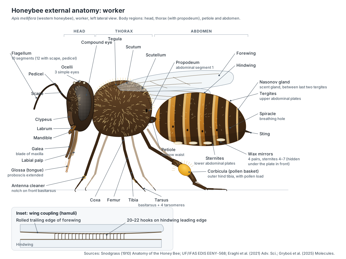
In bees, ants and wasps the "thorax" includes the first abdominal segment, the propodeum. So the narrow waist is abdominal segment 2 (UF/IFAS EDIS EENY-568), and a worker's visible abdomen shows only six segments (Snodgrass, 1910). The dense, branched hairs that cover the bee trap pollen and insulate the flight muscles.
- Scutum: Forms the roof of the flight box. The flight muscles flex it slightly to drive the wings.
- Scutellum: Part of the flexible roof of the thorax that moves the wings.
- Propodeum: Carries a large spiracle and forms the joint with the waist.
- Petiole: Lets the abdomen bend, for example to aim the sting. The oesophagus, aorta and nerve cord all pass through it.
- Sternites: Protect the belly. In workers, sternites 4–7 carry the wax mirrors.
- Wax mirrors: The wax glands secrete liquid wax through these plates; it hardens into thin scales in the pockets below.
Honeybee head: eyes, antennae and proboscis
A honeybee has five eyes: two large compound eyes on the sides of the head and three small ocelli in a triangle on top (Snodgrass, 1910). Each compound eye is made of thousands of facets (ommatidia), each with its own lens and 8 or 9 photoreceptors (Kelber & Somanathan, 2019). Bees see ultraviolet, blue and green light. Drone eyes are so large that they meet on top of the head, pushing the ocelli forward onto the face.
The antennae are elbowed, with a long scape and a flagellum (including the small pedicel). Workers and queens have 12 antennal segments; drones have 13 (Snodgrass, 1910). The antennae carry the bee's sense of smell, taste and touch.
Honeybee mouthparts combine biting and lapping. The mandibles mould wax and handle propolis. The proboscis is a temporary tube formed by the hairy glossa (tongue) in the middle, two blade-like galeae from the maxillae, and two labial palps; nectar rises up it by capillary action and the sucking pump of the pharynx. The proboscis is much longer in workers than in queens or drones (Snodgrass, 1910).
- Compound eyes: See colour, shapes and movement. Each facet (ommatidium) has its own lens and 8 or 9 photoreceptors and acts like one pixel (Kelber & Somanathan, 2019).
- Ocelli: Measure light level and help keep the bee level in flight.
- Antennal flagellum: Smells flowers, pheromones and the hive, tastes, and senses touch, humidity and sound (air vibrations).
- Mandibles: Mould wax and propolis, carry debris, feed larvae and fight. Bees do not chew solid food.
- Glossa: Laps up nectar, honey and water, which rise between the proboscis parts by capillary action and suction.
- Galeae: With the labial palps they form a temporary tube around the glossa through which liquid is drawn up.
- Labial palps: Help close the feeding tube and taste food.
Wings, flight muscles and hamuli
A honeybee has two pairs of clear, veined wings. In flight each hindwing is locked to the forewing by a row of tiny hooks, the hamuli, which catch a rolled fold on the trailing edge of the forewing; a worker has 20–22 hooks on each hindwing (Eraghi et al., 2021). The wings are driven indirectly. Large dorsal longitudinal musclesarch the roof of the thorax and push the wings down; upright dorsoventral muscles pull the roof down and flip the wings up. Neither muscle attaches to the wings themselves.
- Hamuli: Hook into a rolled fold on the trailing edge of the forewing, coupling the wings in flight.
- Dorsal longitudinal muscles: Contract to arch the thorax roof, which pushes the wings down. They are 'indirect' because they move the thorax, not the wings.
- Dorsoventral muscles: Contract to pull the roof down, which flips the wings up. They alternate with the longitudinal muscles to beat the wings.
- Tegulae: Protect the delicate wing hinge.
Honeybee legs: antenna cleaner and pollen basket
Every leg has a coxa, trochanter, femur, tibia and five-part tarsus with claws and a sticky pad. Each pair also has a special job. The front legs carry the antenna cleaner, a notch with a comb on the basitarsus closed by a flat spur on the tibia. The middle legs carry a long tibial spur. The hind legs of workers carry the corbicula (pollen basket), the pollen comb and the pollen press.
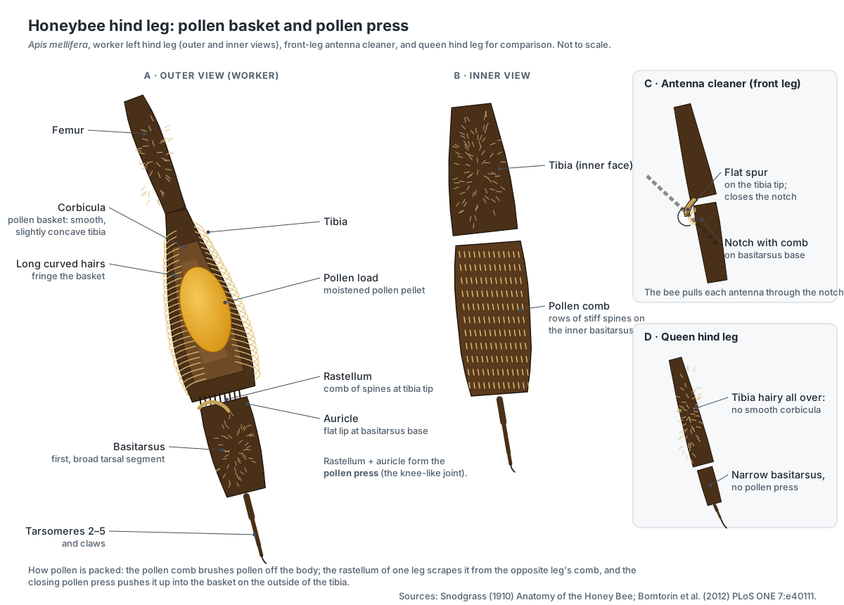
- Antenna cleaner: The bee pulls an antenna through the notch to comb it clean (Snodgrass, 1910).
- Corbicula: Holds a pellet of moistened pollen, or propolis, for the flight home (Snodgrass, 1910).
- Pollen comb: Brushes pollen off the body hairs; the opposite leg's pollen press then pushes it into the basket (Snodgrass, 1910).
- Pollen press: Scrapes pollen from the opposite leg's pollen comb and presses it up into the pollen basket as the joint opens and closes.
- Middle tibial spur: Used to clean the wings and to pry pollen pellets out of the pollen baskets in the hive.
Honeybee internal anatomy diagram
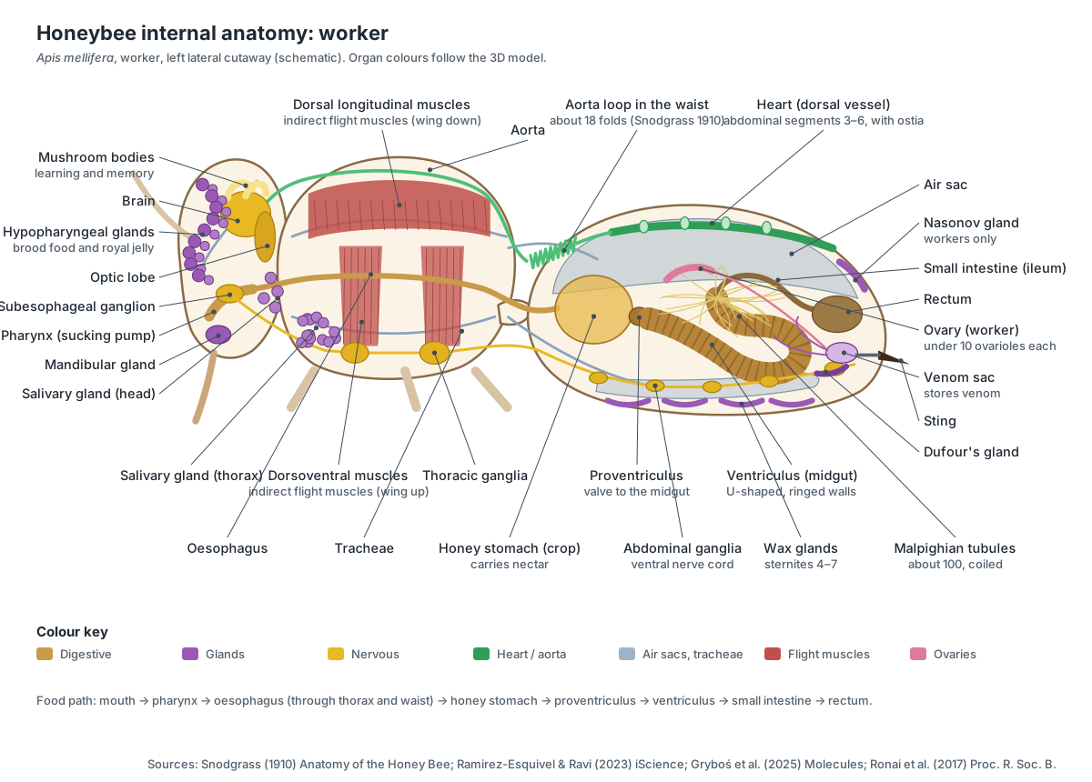
Inside a honeybee are the digestive tract (pharynx, oesophagus, honey stomach, proventriculus, ventriculus, Malpighian tubules, small intestine and rectum), a tubular heart and aorta, a breathing system of spiracles, tracheae and air sacs, a brain and ventral nerve cord, the flight muscles, many glands, the reproductive organs and the sting apparatus.
Honey stomach and digestion
Nectar passes up the proboscis, through the pharynx and the long oesophagus, and into the honey stomach (the crop), a stretchy bag at the front of the abdomen. Foragers carry nectar home in it and regurgitate it to house bees. The four-lipped proventriculus keeps the load in, but strains out pollen grains and passes them into the ventriculus, where food is digested. About one hundred Malpighian tubules remove waste, and the rectum stores faeces (Snodgrass, 1910).
- Honey stomach: Carries nectar and water home. Foragers regurgitate the load to house bees; nectar is not digested here.
- Proventriculus: Keeps the nectar load in the honey stomach, but strains pollen grains out of it and passes them on to be digested.
- Ventriculus: Digests pollen and sugars and absorbs nutrients (Snodgrass, 1910).
- Malpighian tubules: Excretory organs (insect kidneys) that remove nitrogen wastes from the hemolymph (Snodgrass, 1910).
- Rectum: Stores faeces through the winter so bees can wait for a 'cleansing flight' instead of soiling the hive.
Heart, aorta and air sacs
The heart is a muscular tube under the top of the abdomen. Snodgrass described four chambers in abdominal segments 3 to 6, continued forward as the aorta, which forms a convoluted loop of about 18 folds where it passes through the waist and then runs through the thorax to the head. Bees breathe through ten pairs of spiracles (two on the thorax, eight on the abdomen) that open into tracheae and large, balloon-like air sacs, which the abdomen pumps like bellows.
Honeybee brain
The honeybee brain sits between the eyes, above the oesophagus. Its large optic lobes process vision, the antennal lobes process smell, and two pairs of cup-shaped mushroom bodies on top handle learning and memory. A subesophageal ganglion below controls the mouthparts. The mushroom bodies grow as a worker ages and starts foraging (Kraft et al., 2025).
- Brain: Processes vision, smell and touch and controls complex behaviour such as navigation and the waggle dance.
- Mushroom bodies: Centres for learning, memory and combining senses.
- Optic lobes: Process images and colour from the compound eyes.
- Air sacs: Act as bellows: the abdomen pumps air through them, ventilating the flight muscles.
- Aorta: Carries hemolymph forward through the thorax and empties it into the head, where it bathes the brain before flowing back.
Honeybee glands: royal jelly, wax and scent
Worker honeybees are packed with glands. In the head, the hypopharyngeal glands of young nurse bees make brood food and royal jelly, and the mandibular glands add fatty acids; in the queen, the mandibular glands make queen mandibular pheromone. Under the abdomen, four pairs of wax glands secrete beeswax. Near the tip, the Nasonov gland releases an orientation scent, found only in workers (Gryboś et al., 2025).
- Hypopharyngeal glands: In young nurse bees they make the protein-rich part of royal jelly and brood food.
- Mandibular glands: In workers they add fatty acids to royal jelly and make an alarm scent. In the queen they make queen mandibular pheromone (QMP), which holds the colony together.
- Wax glands: Secrete beeswax. Wax-making workers hang in chains, and the wax scales are passed forward to the mandibles to build comb (Snodgrass, 1910).
- Nasonov gland: Releases a lemon-like scent (geraniol, citral and related terpenoids) that guides bees to the hive entrance, a swarm cluster or water.
Honeybee sting apparatus
The honeybee sting is a modified egg-laying tube, so only females (workers and the queen) have one. It is made of a single stylet and two barbed lancets that slide alternately to drive the sting into skin, plus plates and muscles that power them, the venom gland and venom sac, Dufour's gland and Koschevnikov's gland (Ramirez-Esquivel & Ravi, 2023).
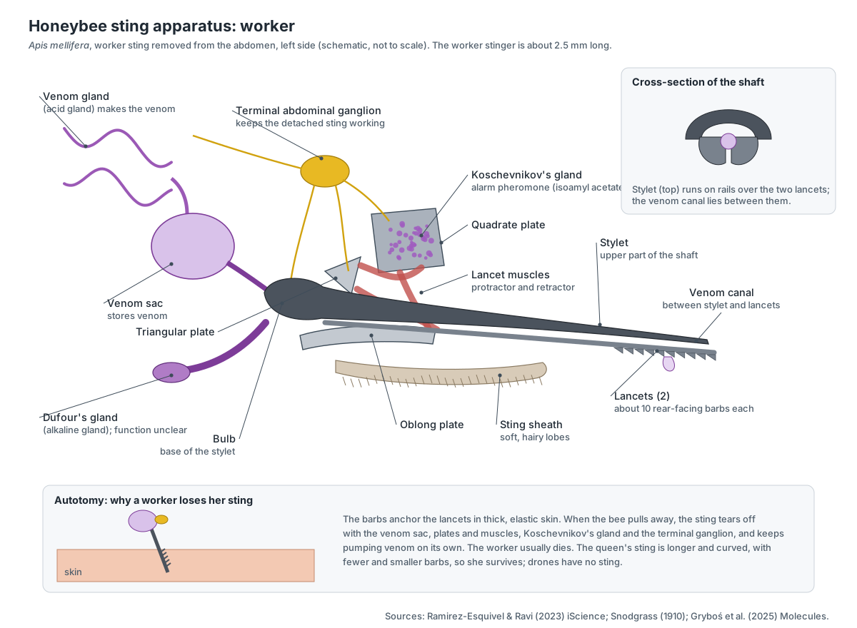
- Stylet: Forms the top of the venom canal and guides the two lancets on rails.
- Lancets: They move alternately, digging deeper with each stroke; rear-facing barbs anchor them in skin.
- Sting plates & muscles: Lever the lancets in and out. They tear away with the sting together with the terminal ganglion, which keeps the sting working on its own (Ramirez-Esquivel & Ravi, 2023).
- Venom gland: Produces venom, which collects in the venom sac (Ramirez-Esquivel & Ravi, 2023).
- Venom sac: Feeds venom into the sting bulb. It tears away with the sting and keeps emptying into the wound.
- Koschevnikov's gland: Makes the sting alarm pheromone. Its main component, isoamyl acetate, smells of bananas and makes other bees attack (Gryboś et al., 2025).
Worker vs queen vs drone honeybee
A colony normally has a single queen, many thousands of workers and, in season, drones. Workers and queens develop from fertilised eggs and are both female; the difference comes from larval diet: larvae fed copious royal jelly become queens. Drones develop from unfertilised eggs (UF/IFAS EDIS EENY-568). Use the caste switch above the 3D model to compare them.
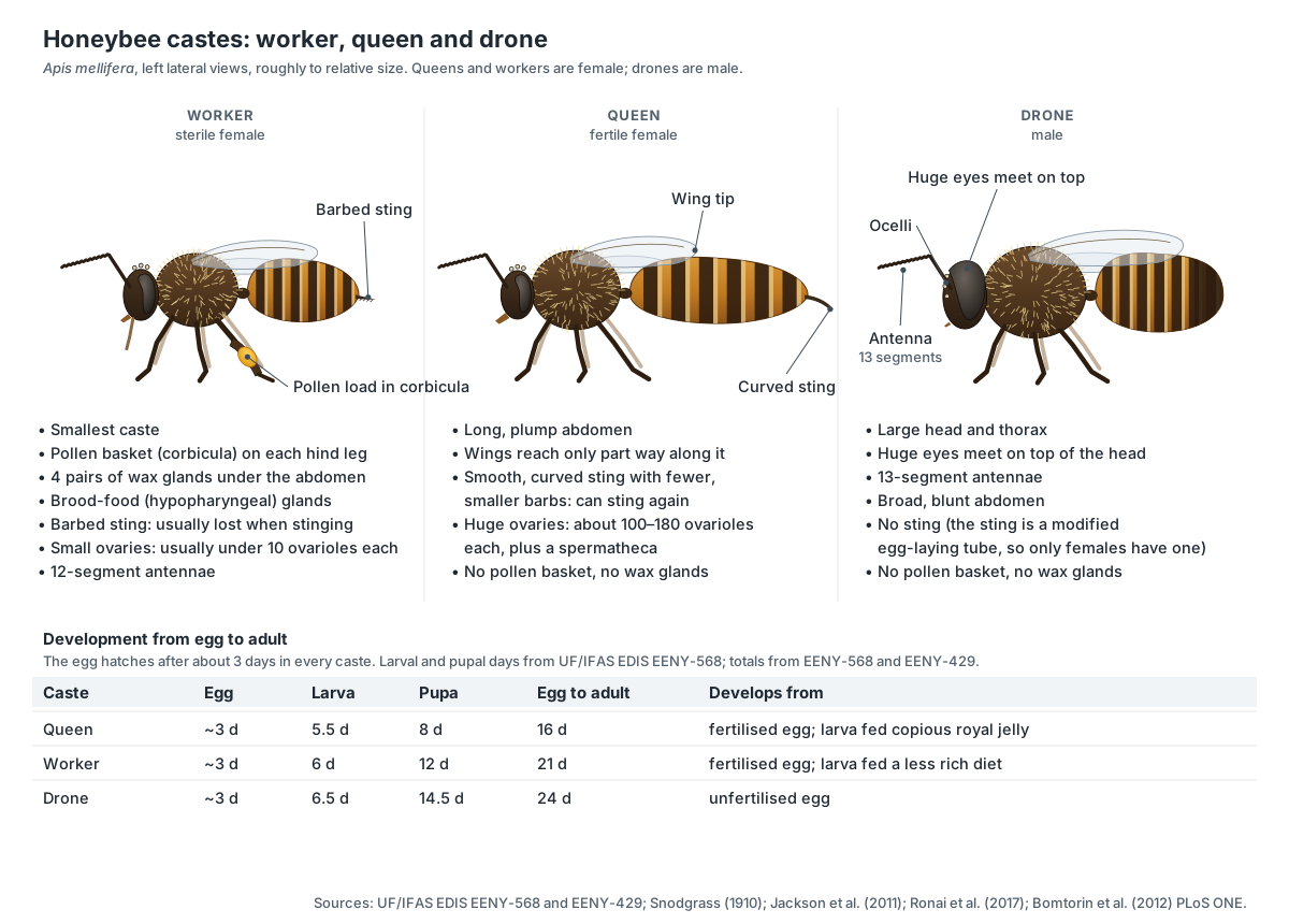
| Feature | Worker | Queen | Drone |
|---|---|---|---|
| Sex | Female, sterile | Female, fertile | Male |
| Egg to adult | 21 days | 16 days | 24 days |
| Body | Smallest | Long, plump abdomen | Large head and thorax, blunt abdomen |
| Eyes | Separate on the sides | Separate on the sides | Huge, meeting on top of the head |
| Antennal segments | 12 | 12 | 13 |
| Proboscis | Long | Shorter | Shorter |
| Pollen basket | Yes | No | No |
| Wax glands, Nasonov gland | Yes | No | No |
| Hypopharyngeal glands | Large | Rudimentary | Absent |
| Sting | Barbed; usually lost on stinging | Longer, curved, few small barbs | None |
| Ovarioles per ovary | Usually fewer than 10 | About 100–180 | — (testes) |
| Spermatheca | No | Yes | — |
Development times are from UF/IFAS EDIS EENY-568 and EENY-429; gland and antennal details from Snodgrass (1910); the ovariole counts from Jackson et al. (2011) and Ronai et al. (2017). A virgin queen mates with about 15 drones in her first two weeks and stores their sperm in her spermatheca for life (UF/IFAS EDIS EENY-568). During mating in flight, the drone's endophallus is everted into the queen and the drone dies (Snodgrass, 1910).
- Ovaries: Produce eggs. A queen can lay up to 1,500 eggs a day (UF/IFAS EDIS EENY-568).
- Spermatheca: Stores sperm from the 15 or so drones she mates with in her first two weeks of life, for the rest of her life (UF/IFAS EDIS EENY-568).
- Testes: Produce sperm during development; in adults sperm is stored in the seminal vesicles.
- Endophallus: Turns inside out into the queen's sting chamber during mating in flight; the drone dies as it breaks away (Snodgrass, 1910).
Internal anatomy of a honeybee vs a dragonfly
Honeybees and dragonflies share the basic insect plan, but their internal anatomy reflects very different lives: the bee is a social nectar and pollen feeder, the dragonfly a solitary aerial predator. The table summarises the main differences students are asked to compare.
| System | Honeybee (Hymenoptera) | Dragonfly (Odonata) |
|---|---|---|
| Flight muscles | Indirect: deform the thorax; wings coupled by hamuli | Mainly direct, attached to the wing bases; fore- and hindwings move independently |
| Foregut | Honey stomach for carrying nectar | Crop and gizzard for prey |
| Mouthparts | Mandibles plus a long sucking proboscis | Strong biting mandibles |
| Body plan | Narrow waist (petiole); sting in females | No waist; long slender abdomen; no sting |
| Development | Complete metamorphosis; larvae in wax cells | Incomplete metamorphosis; aquatic nymphs |
| Social glands | Wax, Nasonov, hypopharyngeal, pheromone glands | None of these |
The honeybee column follows the sources on this page. The dragonfly column is a general comparison from standard insect anatomy rather than a single cited study.
How the honeybee sting works, and why it tears out
The worker's stinger is only about 2.5 mm long but it is a complete machine. Its shaft is a single stylet above two lancets. The lancets slide along rails on the stylet and move alternately, so the sting walks into the skin a little further with each stroke. Rear-facing barbs, on average about 10 per lancet, catch in the tissue as each lancet is pulled back and anchor it while the other advances. Venom flows down a canal between the stylet and lancets. (Ramirez-Esquivel & Ravi (2023): functional anatomy of the worker honeybee stinger, iScience.)
When a worker stings a vertebrate with thick, elastic skin, the barbs can hold so firmly that the whole apparatus tears away from the bee: the piercing parts, the plates and muscles that drive them, the venom gland and venom sac, Koschevnikov's gland that releases the alarm pheromone, and the terminal abdominal ganglion that keeps the sting pumping on its own. This is stinger autotomy. It can kill the worker, but it keeps delivering venom after the bee has been brushed off. (Ramirez-Esquivel & Ravi (2023): functional anatomy of the worker honeybee stinger, iScience.)
The queen's sting is longer and curved, carries fewer and smaller barbs and is more solidly attached to her body (Snodgrass, 1910), so she can sting repeatedly without losing it. Queens use it mainly against rival queens. Drones have no sting at all, because the sting is a modified egg-laying organ (ovipositor) and only females have one. (Snodgrass (1910): The Anatomy of the Honey Bee, USDA Bureau of Entomology.)
Loading the pollen basket
A foraging worker collects pollen with her whole body. The branched hairs pick up grains as she moves over flowers. She then brushes them off with the stiff spines on the inner face of the hind basitarsus (the pollen comb), moistens them with nectar, and transfers them to the outer face of the opposite hind tibia. There, a smooth, slightly hollow area fringed with long curved hairs, the corbicula or pollen basket, holds a growing pellet (Snodgrass, 1910). The pollen press at the tibia–basitarsus joint pushes each new load up into the basket. (Snodgrass (1910): The Anatomy of the Honey Bee, USDA Bureau of Entomology.)
Only workers have this equipment. Bomtorin and colleagues showed that the hind tibia of the worker develops as an expanded, bristle-free corbicula, while in queens the structure is absent; the difference is set during larval development by caste-biased gene expression, including the Hox gene Ultrabithorax, in response to the different larval diets. Bumblebees, stingless bees and orchid bees carry pollen the same way, which is why this group is called the corbiculate bees. (Bomtorin et al. (2012): Hox genes and caste differences in honeybee hind legs, PLoS ONE.)
The honey stomach and the true stomach
In the front of the abdomen the oesophagus expands into a thin-walled sac, the crop, which in bees is called the honey stomach. Behind it is a short, rigid neck, the proventriculus, and then the large U-shaped ventriculus with thick, spongy, ringed walls, where digestion actually happens. A ring of about one hundred long, coiled Malpighian tubules opens at the start of the short small intestine; they are excretory organs, not part of digestion. The large intestine (rectum) follows (Snodgrass, 1910). (Snodgrass (1910): The Anatomy of the Honey Bee, USDA Bureau of Entomology.)
The proventriculus pushes into the rear of the honey stomach and has four triangular lips, each with a special band of muscle. These lips can close the honey stomach so a forager keeps her nectar load separate from her own food, and they can strain pollen grains out of the nectar and pass them back to be digested. That is why diagrams often call the proventriculus the 'honey stopper'. The nectar itself is regurgitated to house bees in the hive and turned into honey there. (Snodgrass (1910): The Anatomy of the Honey Bee, USDA Bureau of Entomology.)
Glands that run the hive
A honeybee colony is held together by glands. In young nurse workers the hypopharyngeal glands, long chains of small lobes coiled in the front of the head, make the protein-rich part of brood food and royal jelly. Snodgrass noted that they are entirely absent in drones and at best rudimentary in queens, which shows how closely they are tied to the worker's job. The mandibular glands add fatty acids. A larva fed copious royal jelly develops into a queen with about 100–180 ovarioles in each ovary (Jackson et al., 2011); a worker-fed larva becomes a worker with usually fewer than ten (Ronai et al., 2017). (Jackson, Tarpy & Fahrbach (2011): ovariole number in honey bee queens, J. Insect Science 11:82.)
The queen's own mandibular glands, paired sacs above the base of the mandibles, make queen mandibular pheromone (QMP), the best-studied honeybee pheromone and one that affects the whole colony. Workers have their own signals: the Nasonov gland, found only in workers, between the last two visible tergites, releases a terpene scent (geraniol, citral and related compounds) used to guide nestmates to the hive entrance, a swarm cluster or water; Koschevnikov's gland on the sting apparatus releases the sting alarm pheromone, whose main component isoamyl acetate smells of bananas. (Gryboś et al. (2025): the pheromone landscape of Apis mellifera, Molecules.)
How honeybee eyes see
A honeybee has five eyes: two compound eyes and three ocelli. Each compound eye is an apposition eye made of thousands of ommatidia. Each ommatidium has its own facet lens and eight or nine photoreceptors and acts like one pixel in the image, so a larger eye can have more pixels, larger facets, or both. Drones have the most extreme eyes, with enlarged regions of high resolution used to spot a queen against the sky. (Kelber & Somanathan (2019): spatial vision in Apidae, Insects.)
Durand and colleagues mapped the light-sensitive proteins (opsins) in all three castes. Bees see with UV-, blue- and green-sensitive photoreceptors. Workers and queens have three kinds of ommatidia in their compound eyes; in drones the upper two-thirds of the eye contains only two kinds, dominated by blue receptors, which suits a male looking upward for a flying queen. The ocelli contain UV- and green-sensitive receptors. (Durand et al. (2025): opsin distribution in the three honeybee castes, iScience.)
Vision feeds a brain that keeps changing. The mushroom bodies, the brain's learning centres, grow in their input regions as a worker ages and starts to forage. Kraft and colleagues found that long-lived winter bees keep this plasticity: their mushroom-body calyces expand again when spring foraging begins. (Kraft, Rössler & Groh (2025): mushroom body plasticity in winter bees, Developmental Neurobiology.)
Honeybee anatomy worksheet with answers
- How many eyes does a honeybee have? Answer: five, two compound eyes and three ocelli.
- Which three parts form the proboscis? Answer: the glossa, the galeae and the labial palps.
- What holds the forewing and hindwing together? Answer: the hamuli, 20–22 hooks on the hindwing.
- Where is the pollen basket? Answer: on the outside of each hind tibia of a worker.
- What is the difference between the honey stomach and the ventriculus? Answer: the honey stomach carries nectar; the ventriculus digests food.
- Which gland makes royal jelly? Answer: the hypopharyngeal glands of young workers, with the mandibular glands.
- Why does a worker usually die after stinging a mammal? Answer: the barbed lancets anchor the sting, which tears out with the venom sac, muscles and ganglion.
- How can you recognise a drone? Answer: huge eyes meeting on top of the head, 13-segment antennae, blunt abdomen, no sting.
Compare with ant anatomy: ants and bees share the narrow waist, the antenna cleaner and hooked wings, and both store food in a crop.
Every structure in the 3D honeybee model
The labeled list below matches the model (worker, queen and drone combined).
Exoskeleton & body regions
- Head (Head capsule)
- Body part 1: a flattened, triangular head seen from the front. Carries the eyes, antennae and mouthparts and holds the brain and the large brood-food glands.
- Thorax (Mesosoma)
- Body part 2: the round, hairy middle section that carries the wings and legs. A box of muscle that powers flight and walking. It is the three thoracic segments plus the first abdominal segment (propodeum) fused to them.
- Scutum (Mesoscutum)
- The large shield-shaped plate on top of the thorax. Forms the roof of the flight box. The flight muscles flex it slightly to drive the wings.
- Scutellum
- A smaller, half-moon plate behind the scutum. Part of the flexible roof of the thorax that moves the wings.
- Tegulae
- Small scale-like plates covering the base of each forewing. Protect the delicate wing hinge.
- Propodeum
- Abdominal segment 1, fused to the back of the thorax. Carries a large spiracle and forms the joint with the waist.
- Petiole (Waist; narrow front part of abdominal segment 2)
- The very short, narrow waist between the thorax and the abdomen. Lets the abdomen bend, for example to aim the sting. The oesophagus, aorta and nerve cord all pass through it.
- Abdomen (Gaster, metasoma)
- Body part 3: the striped rear section with overlapping plates. Holds the honey stomach, gut, heart, air sacs, reproductive organs and sting. Its plates slide in and out as the bee breathes.
- Sternites
- The plates on the underside of the abdomen. Protect the belly. In workers, sternites 4–7 carry the wax mirrors.
- Wax mirrors (Wax plates)
- Four pairs of smooth, shiny plates on the underside of a worker's abdomen. The wax glands secrete liquid wax through these plates; it hardens into thin scales in the pockets below.
- Branched body hairs (Plumose setae)
- A dense coat of feathery hairs over the head and thorax. Trap pollen grains, insulate the flight muscles and sense touch and air flow.
Head, eyes & mouthparts
- Compound eyes
- Two large, hairy eyes made of thousands of hexagonal facets. See colour, shapes and movement. Each facet (ommatidium) has its own lens and 8 or 9 photoreceptors and acts like one pixel (Kelber & Somanathan, 2019).
- Ocelli (Simple eyes)
- Three small simple eyes in a triangle on top of the head. Measure light level and help keep the bee level in flight.
- Antennal scape
- The long first segment of each antenna. Holds and aims the rest of the antenna.
- Antennal flagellum (Pedicel and flagellomeres)
- The chain of segments beyond the elbow, packed with sense organs. Smells flowers, pheromones and the hive, tastes, and senses touch, humidity and sound (air vibrations).
- Clypeus
- The plate on the lower face above the labrum. Anchors the muscles of the sucking pump inside the head.
- Labrum (Upper lip)
- A small flap below the clypeus. Closes the front of the mouth over the proboscis base.
- Mandibles (Jaws)
- A pair of spoon-shaped jaws on each side of the mouth. Mould wax and propolis, carry debris, feed larvae and fight. Bees do not chew solid food.
- Glossa (Tongue)
- The long, hairy tongue in the middle of the proboscis. Laps up nectar, honey and water, which rise between the proboscis parts by capillary action and suction.
- Galeae (Maxillary blades)
- Two long blades from the maxillae that wrap around the tongue. With the labial palps they form a temporary tube around the glossa through which liquid is drawn up.
- Labial palps
- A pair of slender jointed feelers lying alongside the glossa. Help close the feeding tube and taste food.
Legs & wings
- Coxae & trochanters
- The short basal segments that join each leg to the thorax. Swing the legs forward and back.
- Femora
- The thick upper leg segments. Hold the muscles that move the tibia.
- Tibiae
- The long leg segments below the femora. On the hind leg of a worker the tibia is broad and flat and forms the pollen basket.
- Tarsi (Basitarsus + 4 small tarsomeres)
- The foot: a large first segment (basitarsus) and four small ones. Walk, grip and groom. The hind basitarsus of a worker is broad and carries the pollen comb.
- Claws & arolium
- Two forked claws with a soft sticky pad (arolium) between them. Claws grip rough surfaces and flowers; the pad unfolds to stick to smooth surfaces.
- Antenna cleaner (Strigil)
- A semicircular notch with a comb of bristles on the front basitarsus, closed by a flat spur on the tibia. The bee pulls an antenna through the notch to comb it clean (Snodgrass, 1910).
- Middle tibial spur
- A single spine at the end of each middle tibia. Used to clean the wings and to pry pollen pellets out of the pollen baskets in the hive.
- Corbicula (Pollen basket)
- A smooth, slightly hollow area on the outside of the hind tibia fringed by long curved hairs. Holds a pellet of moistened pollen, or propolis, for the flight home (Snodgrass, 1910).
- Pollen press (Rastellum + auricle)
- A hinged joint between the hind tibia and basitarsus: a comb of spines (rastellum) above a flat lip (auricle). Scrapes pollen from the opposite leg's pollen comb and presses it up into the pollen basket as the joint opens and closes.
- Pollen comb (Pollen brush, scopa of basitarsus)
- Rows of short, stiff spines on the inner face of the hind basitarsus. Brushes pollen off the body hairs; the opposite leg's pollen press then pushes it into the basket (Snodgrass, 1910).
- Forewings
- The larger front pair of clear, veined wings. Generate lift and thrust; the wing beat is driven indirectly by the thorax muscles.
- Hindwings
- The smaller back pair of wings. Lock onto the forewing so each side works as a single surface.
- Hamuli (Wing hooks)
- A row of tiny hooks on the front edge of the hindwing. Hook into a rolled fold on the trailing edge of the forewing, coupling the wings in flight.
Flight muscles
- Dorsal longitudinal muscles (Indirect flight muscles (wing depressors))
- Large muscles running front to back along the top of the thorax. Contract to arch the thorax roof, which pushes the wings down. They are 'indirect' because they move the thorax, not the wings.
- Dorsoventral muscles (Indirect flight muscles (wing elevators))
- Upright muscles running from the roof to the floor of the thorax. Contract to pull the roof down, which flips the wings up. They alternate with the longitudinal muscles to beat the wings.
Digestive
- Pharynx (Sucking pump)
- A muscular pump in the head behind the mouth. Sucks liquid up the proboscis. The brood-food glands open onto a plate at its front (Snodgrass, 1910).
- Oesophagus
- A thin tube through the neck, thorax and waist. Carries nectar and food to the honey stomach.
- Honey stomach (Crop, honey sac)
- A thin-walled, stretchy bag at the front of the abdomen. Carries nectar and water home. Foragers regurgitate the load to house bees; nectar is not digested here.
- Proventriculus (Honey-stopper)
- A short valve with four lips between the honey stomach and the ventriculus. Keeps the nectar load in the honey stomach, but strains pollen grains out of it and passes them on to be digested.
- Ventriculus (Midgut, true stomach)
- A large U-shaped tube with ringed, spongy walls. Digests pollen and sugars and absorbs nutrients (Snodgrass, 1910).
- Malpighian tubules
- About one hundred long, fine, coiled tubes. Excretory organs (insect kidneys) that remove nitrogen wastes from the hemolymph (Snodgrass, 1910).
- Small intestine (Ileum)
- A short, narrow, coiled tube behind the ventriculus. Carries digested waste to the rectum and absorbs water.
- Rectum
- A large, stretchy sac at the end of the gut with six rectal pads. Stores faeces through the winter so bees can wait for a 'cleansing flight' instead of soiling the hive.
Glands
- Hypopharyngeal glands (Brood-food glands, pharyngeal glands)
- A pair of long chains of grape-like lobes coiled in the front of the head. In young nurse bees they make the protein-rich part of royal jelly and brood food.
- Mandibular glands
- A pair of sac-like glands above the base of each mandible. In workers they add fatty acids to royal jelly and make an alarm scent. In the queen they make queen mandibular pheromone (QMP), which holds the colony together.
- Labial (salivary) glands
- Two sets of glands, one in the back of the head and one in the thorax. Produce saliva that moistens food and dissolves sugar.
- Wax glands
- Four pairs of glandular cell layers above the wax mirrors. Secrete beeswax. Wax-making workers hang in chains, and the wax scales are passed forward to the mandibles to build comb (Snodgrass, 1910).
- Nasonov gland (Scent gland)
- A patch of gland cells near the tip of a worker's abdomen. Releases a lemon-like scent (geraniol, citral and related terpenoids) that guides bees to the hive entrance, a swarm cluster or water.
- Venom gland (Acid gland)
- A long, forked thread that makes bee venom. Produces venom, which collects in the venom sac (Ramirez-Esquivel & Ravi, 2023).
- Venom sac (Poison sac, venom reservoir)
- A clear, oval sac that stores venom. Feeds venom into the sting bulb. It tears away with the sting and keeps emptying into the wound.
- Dufour's gland (Alkaline gland)
- A small, unpaired tubular gland beside the venom sac. Its function is still unclear; it was once thought to lubricate the sting or mark eggs (Ramirez-Esquivel & Ravi, 2023).
- Koschevnikov's gland
- A granular mass of gland cells on the quadrate plate of the sting. Makes the sting alarm pheromone. Its main component, isoamyl acetate, smells of bananas and makes other bees attack (Gryboś et al., 2025).
Sting apparatus
- Stylet
- The upper, single piece of the sting shaft, swollen at its base into the bulb. Forms the top of the venom canal and guides the two lancets on rails.
- Lancets
- Two barbed blades that slide back and forth under the stylet. They move alternately, digging deeper with each stroke; rear-facing barbs anchor them in skin.
- Sting plates & muscles (Quadrate, oblong and triangular plates)
- Three pairs of small plates and their muscles that drive the lancets. Lever the lancets in and out. They tear away with the sting together with the terminal ganglion, which keeps the sting working on its own (Ramirez-Esquivel & Ravi, 2023).
Heart & breathing
- Heart (Dorsal vessel)
- A muscular tube under the abdominal tergites with paired valves (ostia). Pumps hemolymph forward into the aorta.
- Aorta
- A narrow tube from the heart to the head, with a tight coil in the waist. Carries hemolymph forward through the thorax and empties it into the head, where it bathes the brain before flowing back.
- Air sacs
- Large, balloon-like expansions of the tracheae, especially in the abdomen. Act as bellows: the abdomen pumps air through them, ventilating the flight muscles.
- Tracheae
- Branching air tubes that deliver oxygen to every tissue. Carry air from the spiracles directly to the cells.
- Spiracles
- Breathing holes on the sides of the thorax and abdomen. Open and close to let air into the tracheae.
Nervous
- Brain
- The main nerve centre in the head, above the oesophagus. Processes vision, smell and touch and controls complex behaviour such as navigation and the waggle dance.
- Mushroom bodies (Corpora pedunculata)
- Two pairs of cup-shaped centres (calyces) on top of the brain. Centres for learning, memory and combining senses.
- Optic lobes
- Large lobes on each side of the brain behind the compound eyes. Process images and colour from the compound eyes.
- Antennal lobes
- Round lobes at the front of the brain. First processing centre for smells and pheromones.
- Subesophageal ganglion
- A nerve centre below the oesophagus. Controls the mandibles and proboscis.
- Thoracic ganglia
- Two large nerve centres on the floor of the thorax. Control the legs and wings.
- Ventral nerve cord (With abdominal ganglia)
- A nerve cord along the belly with a chain of small abdominal ganglia. Links the brain to the abdomen; the last (terminal) ganglion controls the sting.
Reproductive
- Ovaries
- Pear-shaped egg organs made of ovarioles: tiny in workers, enormous in the queen. Produce eggs. A queen can lay up to 1,500 eggs a day (UF/IFAS EDIS EENY-568).
- Spermatheca
- A small round sperm-storage sac in the queen. Stores sperm from the 15 or so drones she mates with in her first two weeks of life, for the rest of her life (UF/IFAS EDIS EENY-568).
- Oviducts & vagina
- Tubes that carry eggs from the ovaries to the sting chamber. Each egg passes the spermatheca opening, where it may be fertilised, before it is laid.
- Testes
- Paired sperm-making organs of the drone. Produce sperm during development; in adults sperm is stored in the seminal vesicles.
- Seminal vesicles & mucus glands
- Paired sperm-storage tubes and two large mucus glands. Store sperm and add mucus to the ejaculate.
- Endophallus
- The drone's internal, eversible copulatory organ. Turns inside out into the queen's sting chamber during mating in flight; the drone dies as it breaks away (Snodgrass, 1910).
Frequently asked questions
What are the main body parts of a honeybee?
A honeybee has a head, a thorax and an abdomen. The head carries two compound eyes, three ocelli, two elbowed antennae, mandibles and a long proboscis. The thorax carries two pairs of wings and three pairs of legs. The abdomen holds the honey stomach, gut, heart, wax glands and, in females, the sting.
Do honeybees have two stomachs?
A worker has a honey stomach (crop) for carrying nectar and a true stomach (ventriculus) for digesting food. A valve called the proventriculus between them keeps nectar in the honey stomach but lets pollen through to be digested.
Where is the pollen basket on a bee?
The pollen basket, or corbicula, is a smooth, slightly hollow area on the outside of each hind tibia, fringed with long curved hairs. Only workers have one; queens and drones do not.
Why does a honeybee die after stinging?
A worker's two lancets carry about ten rear-facing barbs each. In thick skin the barbs hold the sting so firmly that it tears out of the bee, together with the venom sac, muscles and the last nerve ganglion. The torn-off sting keeps pumping venom, but the worker dies from the wound. Queens have a smoother sting and survive; drones have no sting.
How many eyes does a honeybee have?
Five: two large compound eyes on the sides of the head and three small ocelli in a triangle on top. Drones have much larger compound eyes that meet on top of the head.
Where does beeswax come from?
Young workers secrete wax from four pairs of wax glands on the underside of the abdomen. The wax passes through smooth plates (wax mirrors) and hardens into thin scales, which the bee pulls off and chews into comb.
What is the difference between a worker, a queen and a drone?
Workers are sterile females with pollen baskets, wax glands, brood-food glands and a barbed sting. The queen is a fertile female with a long abdomen, huge ovaries and a spermatheca, and her sting is smoother. Drones are males from unfertilised eggs, with huge eyes, 13-segment antennae, no sting and no pollen basket. Development takes 21 days for a worker, 16 for a queen and 24 for a drone (UF/IFAS EDIS).
Do bees have a heart and lungs?
Bees have a tube-shaped heart along the top of the abdomen that pumps hemolymph forward through an aorta, but no lungs. They breathe through spiracles that lead into tracheae and large air sacs.
What makes royal jelly?
Young nurse workers make royal jelly in their hypopharyngeal glands, in the front of the head, mixed with secretions of the mandibular glands. Larvae fed copious royal jelly develop into queens.
References
- Snodgrass, R. E. (1910). The Anatomy of the Honey Bee. USDA Bureau of Entomology, Technical Series 18. Washington: Government Printing Office.
- Mortensen, A. N., Schmehl, D. R. & Ellis, J. European Honey Bee, Apis mellifera. UF/IFAS EDIS EENY-568/IN1005.
- Ellis, J. D. & Ellis, A. African Honey Bee, Apis mellifera scutellata. UF/IFAS EDIS EENY-429/IN790.
- Ramirez-Esquivel, F. & Ravi, S. (2023). Functional anatomy of the worker honeybee stinger (Apis mellifera). iScience 26, 107103.
- Eraghi, S. H. et al. (2021). Wing coupling in bees and wasps: from the underlying science to bioinspired engineering. Advanced Science 8, 2004383.
- Gryboś, A., Staniszewska, P., Bryś, M. S. & Strachecka, A. (2025). The pheromone landscape of Apis mellifera. Molecules 30, 2369.
- Bomtorin, A. D. et al. (2012). Hox gene expression leads to differential hind leg development between honeybee castes. PLoS ONE 7, e40111.
- Jackson, J. T., Tarpy, D. R. & Fahrbach, S. E. (2011). Histological estimates of ovariole number in honey bee queens, Apis mellifera, reveal lack of correlation with other queen quality measures. Journal of Insect Science 11, 82.
- Ronai, I. et al. (2017). The dynamic association between ovariole loss and sterility in adult honeybee workers. Proc. R. Soc. B 284, 20162693.
- Cardoso-Júnior, C. A. M. et al. (2026). A sterility-associated long noncoding RNA involved in honey bee caste determination and adult queen and worker fertility. PNAS.
- Durand, A. et al. (2025). A comprehensive characterization of opsin distribution across the visual system in the three castes of the honeybee. iScience.
- Kelber, A. & Somanathan, H. (2019). Spatial vision and visually guided behavior in Apidae. Insects 10, 418.
- Kraft, N., Rössler, W. & Groh, C. (2025). Neuronal plasticity in the mushroom bodies of winter bees is retained despite substantially advanced age. Developmental Neurobiology.
More interactive anatomy models
- Insect body partsHead, thorax and abdomen: the three body parts every insect shares.
- Grasshopper anatomyExternal and internal anatomy of the grasshopper, with mouthparts.
- Grasshopper dissectionGuided grasshopper dissection: crop, gastric caeca, heart and tracheae.
- Butterfly anatomyScaled wings, proboscis, compound eyes and internal organs of a butterfly.
- Ant anatomyAnt body parts: elbowed antennae, mandibles, petiole and stinger.
- Human anatomy1,000+ bones, muscles, organs, vessels and nerves from real anatomical data.