

Anatomy of a chicken at a glance
A chicken is a bird built for walking, scratching and short bursts of flight. On the outside it has a fleshy comb and wattles, a horny beak with no teeth, feathers in distinct regions, and bare, scaly legs with four toes. Inside it has a crop to store food, a two-part stomach (the glandular proventriculus and the grinding gizzard), two long caeca, small fixed lungs ventilated by nine air sacs (some texts count eight), a four-chambered heart, kidneys but no bladder, and a single cloaca that ends at the vent.
This page describes the domestic chicken, Gallus gallus domesticus, using a laying hen as the reference animal, with notes on the rooster. Breeds differ in comb type, plumage, size, earlobe colour and even toe number, so where those differences matter they are pointed out.
Directions: cranial means towards the beak, caudal towards the tail, dorsal the back and ventral the belly and keel. Left and right are always the bird's own. Most diagrams, including the ones here, show the left side, because that is where the hen's ovary and oviduct lie.
Chicken anatomy diagram: external parts of a chicken
The external anatomy of a chicken includes the comb, beak, nostrils, eyes, ear openings, earlobes and wattles on the head; the hackles and cape on the neck; the back, saddle, breast and abdomen on the body; the wing coverts, secondaries and primaries on the wing; the tail feathers and preen gland; the vent; and the thigh, drumstick, hock, shank, spur, toes and claws on the leg.
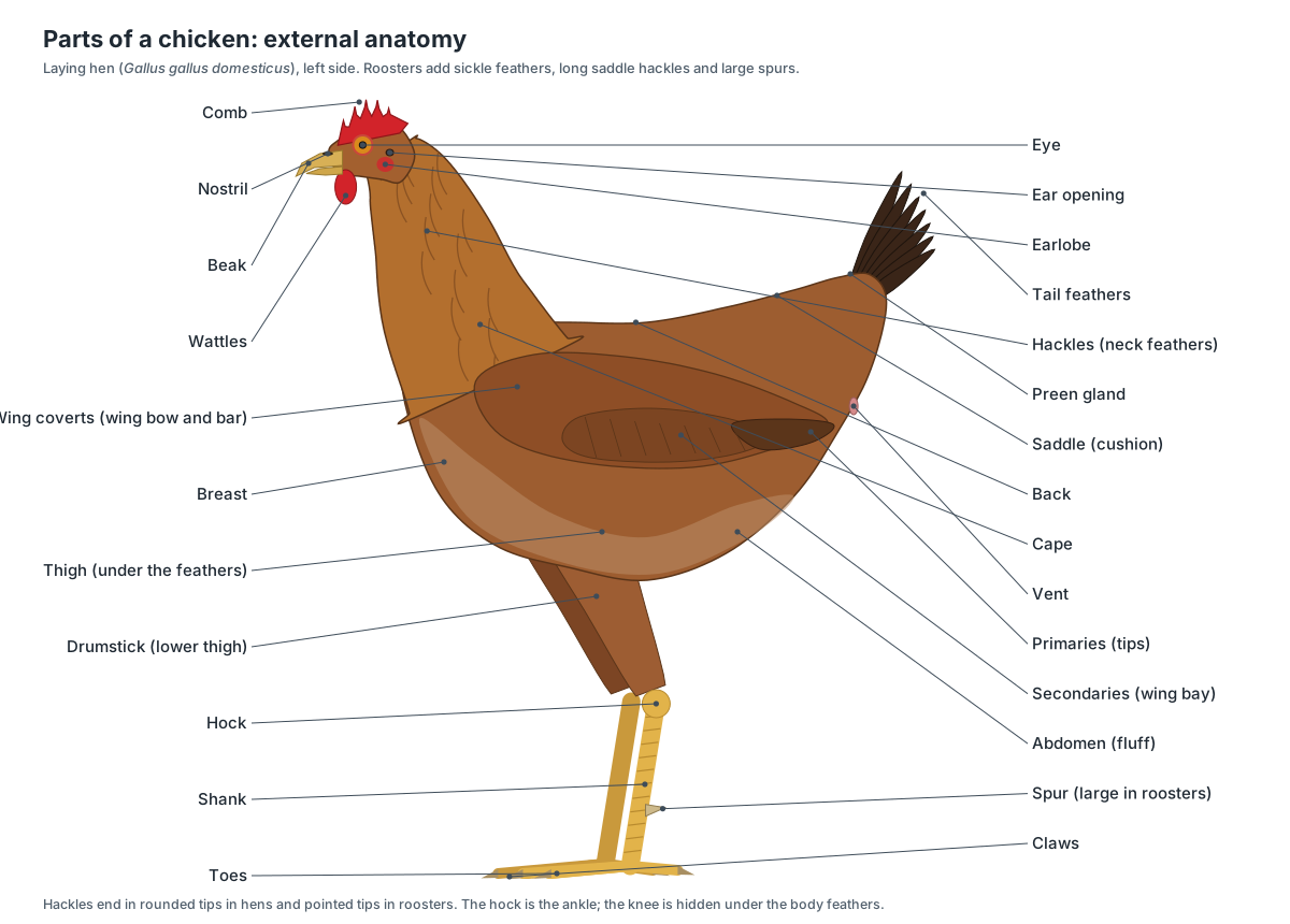
| Region | Parts you can see | Hen vs rooster |
|---|---|---|
| Head | Comb, wattles, earlobes, eye, ear opening, beak, nostrils | Rooster comb and wattles are larger |
| Neck | Hackles; cape at the base | Hackle tips rounded in hens, pointed in roosters |
| Body | Back, saddle (cushion in hens), breast, abdomen (fluff) | Roosters grow long, pointed saddle feathers |
| Wing | Coverts (wing bow and bar), secondaries (wing bay), primaries | Similar |
| Tail | Main tail feathers, coverts, preen gland | Only roosters have sickle feathers |
| Leg | Thigh, drumstick, hock, shank, spur, four toes, claws | Spurs much larger in roosters |
Two features cause the most confusion. First, the backward-bending joint at the bottom of the feathered leg is the hock, the bird's ankle; the knee is hidden under the body feathers. Second, the "claw" that some farm guides mention is the hind toe (the hallux), while the nails at the ends of all the toes are the claws proper.
The skin itself is thin and has almost no glands. Poultry Hub lists only two kinds: the preen (uropygial) gland at the base of the tail and the glands of the outer ear canal. Chickens have no sweat glands; they shed heat by panting, by holding the wings away from the body and through the blood-rich comb and wattles.
External vs internal anatomy of a chicken
External anatomy is what you can see and feel on a live bird: feathers, comb, beak, legs and vent. Internal anatomy is everything under the skin: the skeleton, muscles and organs. A few internal structures can be felt from outside, which is how poultry keepers check a bird's health.
| Feel or see from outside | What it tells you | Internal structure |
|---|---|---|
| Bulge at the base of the neck | Whether the bird has eaten | Crop |
| Ridge down the breast | Body condition (muscle on each side) | Keel of the sternum |
| Two thin bones beside the vent | A wide gap means the hen is laying | Pubic bones of the pelvis |
| Size and colour of the comb | Laying status and health | Ovary hormones and blood supply |
| Moist, oval vent | An active layer | Cloaca and oviduct |
Chicken head anatomy: comb, wattles, eyes and beak
The head carries the comb on top, the wattles below the beak, an earlobe and an ear opening on each side, two large eyes and the beak with its two nostrils. Inside, a light skull with huge eye sockets protects a small brain.
- Comb: A sexual ornament that signals maturity and health, and a heat radiator: its rich blood supply lets the bird lose heat.
- Wattles: Like the comb, they are an ornament for display and help shed heat.
- Earlobes: A breed and display feature; it has no role in hearing.
- Ear opening: Lets sound into the ear canal and onto the eardrum.
- Eye: Vision. The side-facing eyes give a very wide field of view for spotting predators.
- Nostrils: Air enters here for breathing and smelling. A flap (operculum) partly covers each nostril and keeps out dust.
- Beak: Picks up food, pecks, tears and preens. Chickens have no teeth, so the beak does not chew.
| Comb type | Shape | Examples and genetics |
|---|---|---|
| Single | One upright, serrated blade | Wild type; Leghorn, Rhode Island Red |
| Rose | Flat, fleshy, bumpy, with a backward spike | Dominant Rose-comb allele (Wyandotte) |
| Pea | Three low ridges | Dominant Pea-comb allele; also smaller wattles (Brahma) |
| Walnut | Rounded, like half a walnut | Rose + Pea together (Silkie) |
| Others | Cushion, buttercup, strawberry, V-shaped | Breed-specific forms |
Earlobes are red in many breeds and white in Mediterranean breeds such as the Leghorn. White-lobed breeds often lay white eggs and red-lobed breeds brown eggs, but that is only a rule of thumb, with exceptions.
Tear duct anatomy of a chicken: does a chicken have tear ducts?
Yes. A chicken's eye is kept moist by a small lacrimal gland and a large Harderian gland, and tears drain away through a duct. Poultry Hub describes the tears draining into a collecting sac and then, through a special duct, into the nasal cavity, in the same general way as the human nasolacrimal duct.
Each eye has an upper and a lower eyelid plus a third eyelid, the nictitating membrane, which sweeps across the eye to clean it. The Harderian gland, behind the eyeball, lubricates this third eyelid and is also an immune organ: Poultry Hub lists it among the places that receive antibody-making B-cells. Because tears drain into the nose, eye and nasal problems in chickens often appear together.
- Tear glands & duct: The Harderian gland lubricates the third eyelid and is also an immune organ; the duct drains tears into the nose.
- Eyeball & pecten: Forms images. Like most birds, chickens see colour well.
- Scleral ring: Stiffens the large eye and helps hold its shape.
Chicken mouth and throat anatomy
A chicken's mouth has no teeth and no soft palate, so the mouth and throat form one cavity, the oropharynx. The roof has a long slit, the choana, that opens into the nasal cavity. The narrow, arrow-shaped tongue has backward-pointing barbs, and behind it sits the raised slit of the glottis, the entrance to the windpipe.
- Tongue: Moves food and water to the back of the mouth to be swallowed.
- Mouth & pharynx: Takes in food, moistens it with saliva, and passes it to the oesophagus.
- Larynx & glottis: Closes the airway during swallowing.
Poultry Hub notes that the palate has five rows of backward-pointing papillae and that taste buds sit mainly at the base of the tongue and on the floor of the mouth. The larynx has no vocal cords and no epiglottis. The chicken's voice comes from the syrinx, deep in the chest.
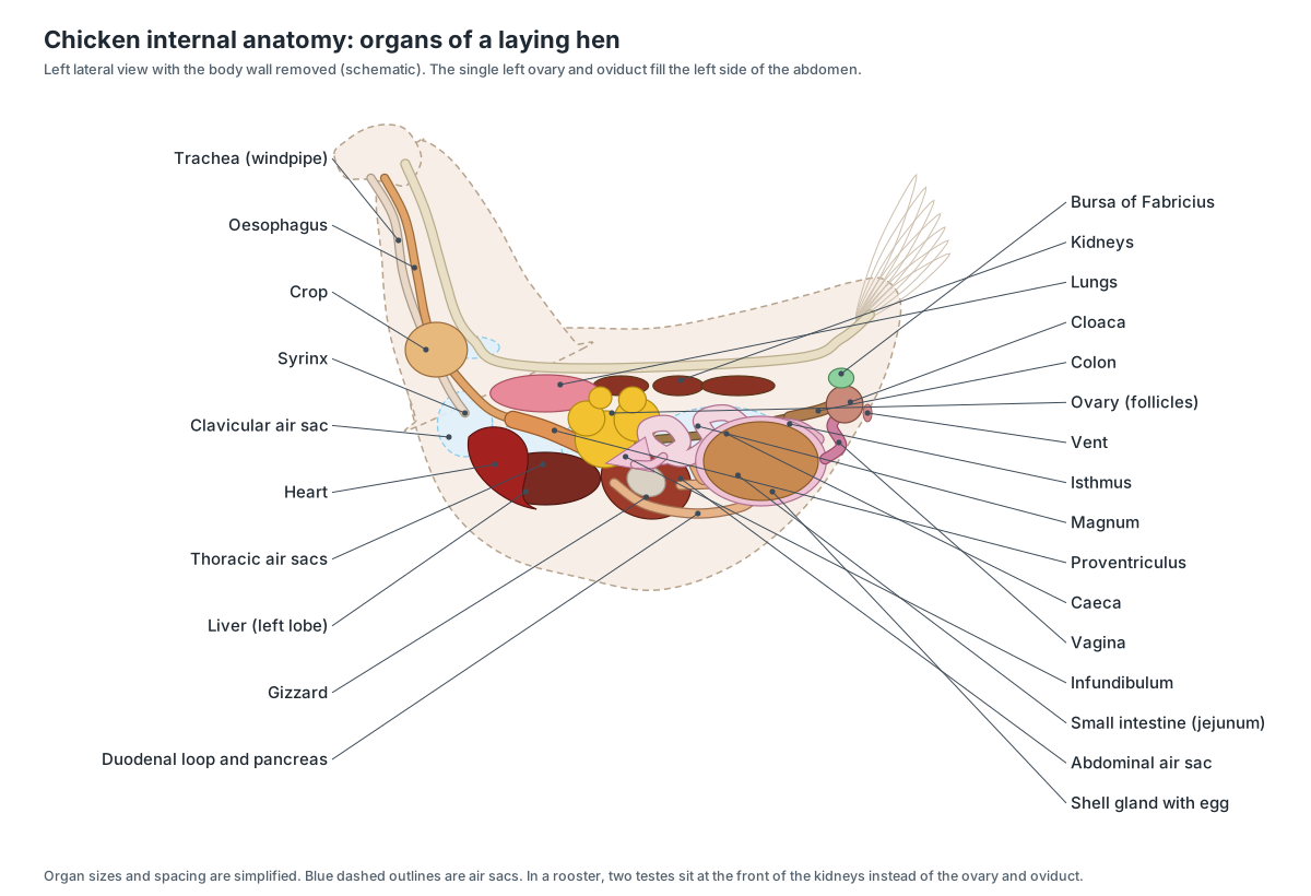
Internal anatomy of a chicken: organs labeled
Inside a laying hen, the crop sits at the base of the neck; the heart lies just behind it, cradled by the two lobes of the liver; the proventriculus and gizzard lie behind the liver; the intestines fill the right and lower abdomen; the left ovary and the long oviduct fill the left abdomen; and the lungs and kidneys are pressed against the backbone. Thin air sacs (nine in most poultry references) fill the spaces between the organs.
Chicken anatomy labeled: every part and its function
This is the text version of the 3D model: every labeled structure of the chicken, grouped by body system, with what it does.
External & plumage
- Comb (Single comb in this model)
- The red, fleshy crest on top of the head. This hen has a single comb: one upright blade with five to six points. A sexual ornament that signals maturity and health, and a heat radiator: its rich blood supply lets the bird lose heat.
- Wattles
- Two red, fleshy lobes that hang below the beak. Like the comb, they are an ornament for display and help shed heat.
- Earlobes
- A patch of bare skin below and behind the ear opening, red or white depending on the breed. A breed and display feature; it has no role in hearing.
- Ear opening (External acoustic meatus)
- A small hole in the side of the head, hidden under a tuft of fine feathers. Lets sound into the ear canal and onto the eardrum.
- Eye (With eyelids and nictitating membrane)
- A large eye on each side of the head, with upper and lower eyelids and a third eyelid. Vision. The side-facing eyes give a very wide field of view for spotting predators.
- Nostrils (Nares)
- Two slit-like openings at the base of the upper beak. Air enters here for breathing and smelling. A flap (operculum) partly covers each nostril and keeps out dust.
- Beak (Bill; upper and lower mandible)
- The hard, pointed mouthparts: a horny sheath over the bones of the upper and lower jaw. Picks up food, pecks, tears and preens. Chickens have no teeth, so the beak does not chew.
- Head feathers & face
- The small feathers of the crown and nape, and the bare red skin of the face around the eye. Covers and insulates the skull. The bare face, like the comb, is part of the bird's display.
- Hackles (Neck feathers)
- The long, narrow feathers that cover the neck. Protect and insulate the neck; roosters raise them as a ruff when threatening a rival.
- Cape
- Short feathers at the base of the neck, between the hackles and the back. Covers the shoulders and the joint between neck and back.
- Back
- The feathered area on top of the body between the shoulders and the saddle. Sheds rain and insulates the body; it covers the lungs and kidneys, which lie against the backbone.
- Saddle (Cushion (in hens))
- The lower back, just in front of the tail. In roosters it carries long, pointed saddle feathers. Covers the lower back and the base of the tail; rooster saddle feathers are part of his display plumage.
- Breast
- The rounded front and underside of the body, over the keel bone and breast muscles. The feathers cover the breast muscles (white meat) that power the wings.
- Abdomen & fluff
- The soft underside behind the keel, covered with downy fluff feathers. Covers the abdominal organs. In a laying hen it is deep and soft because the oviduct and developing eggs fill it.
- Wing coverts (Wing bow and wing bar)
- Rows of smaller feathers that cover the bases of the flight feathers. Smooth the airflow over the wing and protect the bases of the flight feathers.
- Secondary feathers (Secondaries; wing bay)
- The inner flight feathers, attached to the forearm (ulna). On the folded wing they form the triangular 'wing bay'. Form the lifting inner part of the wing in flight.
- Primary feathers (Primaries)
- The 10 long outer flight feathers of each wing, attached to the 'hand' bones. Provide thrust in flight. Chickens fly only short distances, in bursts.
- Tail feathers (Main tail, tail coverts)
- The stiff tail feathers (rectrices), held in a folded, upright fan, plus the smaller coverts at their base. Balance, braking and display.
- Sickle feathers · males only
- The long, curved display feathers that arch over the tail of a rooster. Display. They are one of the clearest outward signs that a bird is male.
- Preen gland (Uropygial gland, oil gland)
- A two-lobed oil gland on top of the tail base. Secretes an oily fluid that the bird collects with its beak and spreads over its feathers when preening.
- Vent (Cloacal opening)
- The single opening under the tail through which droppings, urates, eggs and sperm all leave the body. The exit of the cloaca. Mating is a brief 'cloacal kiss' in which the vents of rooster and hen touch.
- Thigh
- The upper part of the leg, mostly hidden under the body feathers. Contains the thigh muscles (dark meat) that swing the leg.
- Drumstick (lower thigh) (Crus)
- The feathered lower part of the leg between the knee and the hock. Contains the large calf muscles that extend the foot.
- Hock (Intertarsal (ankle) joint)
- The backward-pointing joint between the drumstick and the shank. It is the bird's ankle, not its knee. It bends the opposite way to a human knee because it is a different joint.
- Shank (Tarsometatarsus region)
- The bare, scaly part of the leg between the hock and the toes. Carries the bird's weight. The scales protect it.
- Spur
- A sharp, horn-covered spike on the back of the shank, large in roosters. A weapon used in fights between roosters.
- Toes (Digits I–IV)
- Four toes: three pointing forward and one (the hallux) pointing back. Spread the bird's weight, grip perches and scratch the ground for food.
- Claws (Toenails)
- Curved horny nails on the tips of the toes. Scratching and gripping.
Skeletal
- Skull (Cranium, premaxilla and upper beak bones)
- A light skull of thin, fused bones with huge eye sockets and a long upper beak bone (premaxilla). Protects the brain and eyes and supports the beak.
- Mandible (Lower jaw)
- The lower jaw bone, covered by the horny lower beak. Opens and closes the beak.
- Scleral ring (Scleral ossicles)
- A ring of tiny, overlapping bony plates in the wall of each eyeball. Stiffens the large eye and helps hold its shape.
- Cervical vertebrae (Neck vertebrae)
- About 13–14 neck vertebrae arranged in an S-curve (sources count 13 or 14). Make the neck very flexible, so the beak can reach the whole body for preening and pecking.
- Thoracic vertebrae & notarium
- Seven rib-bearing vertebrae; several of the front ones are fused into a rigid block called the notarium. Form a stiff backbone that the wing muscles can pull against.
- Synsacrum
- A long, rigid bone formed by the fusion of the last thoracic, the lumbar, the sacral and the first tail vertebrae. Makes a strong, light frame that carries the weight of the body on the legs.
- Free caudal vertebrae (Tail vertebrae)
- About five free tail vertebrae between the synsacrum and the pygostyle. Let the tail move up, down and sideways.
- Pygostyle ('Parson's nose' bone)
- A small, blade-shaped bone at the end of the spine, made of fused tail vertebrae. Anchors the tail feathers and the muscles that fan and raise them.
- Ribs & uncinate processes
- Seven pairs of ribs, most with an upper (vertebral) and lower (sternal) segment and a backward hook, the uncinate process. Form the rib cage. Because birds have no diaphragm, rib movements pump air through the air sacs.
- Sternum (Breastbone)
- A large, dish-shaped breastbone that covers much of the underside of the body. Protects the heart and liver and anchors the breast muscles.
- Keel (Carina of the sternum)
- The deep, blade-like ridge running down the middle of the sternum. Gives a large surface for the attachment of the main flight muscles (breast meat).
- Furcula (Wishbone (fused clavicles))
- The V-shaped wishbone, formed by the two collarbones fused at their lower ends. Acts like a spring that braces the shoulders apart during wing beats.
- Coracoid
- A strong, pillar-like bone from each shoulder to the front of the sternum. Braces the shoulder against the pull of the breast muscles.
- Scapula (Shoulder blade)
- A long, thin, sabre-shaped shoulder blade lying along the ribs. Forms part of the shoulder joint and anchors wing muscles.
- Humerus (Upper arm bone (drumette))
- The upper wing bone, from shoulder to elbow. It is the bone inside a wing 'drumette'. Transmits the force of the breast muscles to the wing.
- Radius
- The thinner of the two forearm bones of the wing. Works with the ulna to extend and fold the wing.
- Ulna
- The thicker forearm bone, which carries the secondary feathers. Anchors the secondary flight feathers; small bumps on the bone (quill knobs) mark their attachment.
- Carpometacarpus (Hand bones with carpals)
- The fused 'hand' bone, joined to the forearm by two small free wrist bones (carpals). Carries the primary feathers.
- Wing digits (Finger bones, alula)
- Three small digits on the hand; the first carries the alula (bastard wing). Support the outer primaries and the alula, which smooths airflow at low speeds.
- Pelvic girdle (Ilium, ischium and pubis)
- The hip bones, fused to each other and to the synsacrum into one broad, light plate. Supports the legs and carries the body's weight; the bones are open below so a large egg can pass.
- Femur (Thigh bone)
- The short, stout thigh bone, from the hip to the knee. Carries the thigh muscles; the knee sits close to the body.
- Patella (Kneecap)
- A small kneecap in the tendon at the front of the knee. Improves the leverage of the muscles that straighten the knee.
- Tibiotarsus (Shin bone (drumstick bone))
- The long shin bone of the drumstick: the tibia fused with the upper row of ankle bones. The longest leg bone; it carries the drumstick muscles.
- Fibula
- A thin, splinter-like bone on the outer side of the tibiotarsus. Anchors some leg muscles.
- Tarsometatarsus (Shank bone)
- The shank bone: the lower ankle bones fused with metatarsals II, III and IV. Lengthens the leg; birds stand on their toes with the 'heel' high off the ground.
- Toe bones (Phalanges and metatarsal I)
- The phalanges of the four toes, plus a small separate metatarsal for the hind toe. Form the jointed toes that grip and scratch.
- Medullary bone · females only
- A spongy calcium-store bone that grows inside the marrow cavities of a laying hen's long bones. Releases calcium quickly for the eggshell, much of which is made at night when the hen is not eating.
Muscular (meat)
- Pectoralis (Breast meat, pectoralis major)
- The large superficial breast muscle on each side of the keel: the chicken breast. Pulls the wing down: the power stroke of flight.
- Supracoracoideus (Tenderloin, 'pectoralis minor' (trade name), deep pectoral)
- The deep breast muscle that lies alongside the keel under the pectoralis: the chicken tenderloin (tender). Raises the wing. Its tendon runs up through the triosseal canal at the shoulder and over the top of the humerus like a rope over a pulley.
- Wing muscles (Biceps and triceps (drumette and flat))
- The muscles that fold and stretch the wing, around the humerus and forearm bones. The biceps bends the elbow and folds the wing; the triceps straightens it.
- Thigh muscles (Iliotibialis, iliofibularis, femorotibialis)
- The group of muscles around the femur that make up the thigh (dark meat). Swing the leg forward and back and straighten the knee for walking, scratching and perching.
- Drumstick muscles (Gastrocnemius, fibularis longus, toe flexors)
- The muscles around the tibiotarsus that form the meat of the drumstick. The gastrocnemius extends the foot at the hock; the long flexor tendons curl the toes.
Digestive
- Tongue
- A narrow, arrow-shaped tongue with backward-pointing barbs at its base. Moves food and water to the back of the mouth to be swallowed.
- Mouth & pharynx (Oropharynx with choana)
- The shared mouth-throat cavity. Its roof has a long slit, the choana, that opens into the nasal cavity. Takes in food, moistens it with saliva, and passes it to the oesophagus.
- Oesophagus (Esophagus, gullet)
- A thin-walled, stretchy tube from the throat to the stomach. Carries swallowed food; it widens into the crop at the base of the neck.
- Crop (Ingluvies)
- A stretchy pouch of the oesophagus at the base of the neck that stores food. Holds feed so the bird can eat quickly and digest later; food softens here before passing on.
- Proventriculus (Glandular stomach, true stomach)
- A spindle-shaped glandular stomach between the oesophagus and the gizzard. Secretes hydrochloric acid and pepsinogen, which start protein digestion.
- Gizzard (Ventriculus, muscular stomach)
- A thick, disc-shaped muscular stomach that grinds food, often with swallowed grit. Grinds food mechanically, doing the job teeth do in mammals.
- Duodenum (Duodenal loop)
- The first part of the small intestine: a long U-shaped loop with the pancreas held between its two arms. Receives bile and pancreatic juice; most digestion and absorption begins here.
- Pancreas
- A pale, elongated gland lying inside the duodenal loop. Makes digestive enzymes (for protein, fat and starch) and the hormones insulin and glucagon.
- Liver
- A large, dark red-brown organ with a right and a left lobe that cradle the heart. Makes bile, stores energy and makes the yolk proteins and fats that the hen sends to her ovary.
- Gallbladder
- A small green sac of bile on the right liver lobe. Stores bile, which helps digest fats and absorb vitamins A, D, E and K.
- Jejunum
- The long middle part of the small intestine, folded into loops held by the mesentery. Digests and absorbs nutrients.
- Meckel's diverticulum (Yolk-stalk remnant)
- A small blind nub on the small intestine: the remains of the stalk that joined the chick's gut to its yolk sac. In the embryo and newly hatched chick it carried yolk into the gut; in adults it contains lymphoid tissue.
- Ileum
- The last part of the small intestine, running between the two caeca. Absorbs the remaining nutrients and water.
- Caeca (Ceca (paired))
- Two long blind pouches branching from the junction of the small and large intestine. Bacteria ferment fibre, reabsorb water and make some B vitamins.
- Colon (Large intestine, rectum)
- A very short, straight large intestine from the caeca to the cloaca. Absorbs water from the gut contents and from urine pushed back into it from the cloaca.
- Cloaca (Coprodeum, urodeum, proctodeum)
- A shared chamber at the end of the digestive, urinary and reproductive tracts, with three compartments. The coprodeum receives faeces, the urodeum receives urine and the oviduct or sperm ducts, and the proctodeum opens at the vent.
Respiratory & air sacs
- Larynx & glottis
- A raised slit (the glottis) on the floor of the throat, just behind the tongue, that opens into the trachea. Closes the airway during swallowing.
- Trachea (Windpipe)
- The windpipe, held open by complete rings of cartilage. Carries air between the throat and the lungs.
- Syrinx (Voice box)
- The bird's voice box, at the lower end of the trachea where it divides into the two bronchi. Makes the crow, clucks and alarm calls: vibrating membranes in its wall produce the sound.
- Lungs
- Two small, bright pink, spongy lungs pressed into the ribs on each side of the backbone. Gas exchange. Air flows through tiny tubes (parabronchi) in one direction, during both breathing in and out.
- Cervical air sacs
- A pair of small air sacs at the base of the neck, just in front of the lungs. Part of the bellows system that moves air through the lungs; they extend into the vertebrae.
- Clavicular air sac (Interclavicular air sac (single))
- A single air sac at the entrance to the chest, wrapped around the syrinx and the base of the heart. Bellows for the lungs; its extension into the humerus makes that bone pneumatic.
- Thoracic air sacs (Cranial and caudal thoracic)
- Two pairs of air sacs along the sides of the body cavity, below the lungs. The cranial pair fills with air that has passed through the lungs; the caudal pair with fresh air on its way to the lungs.
- Abdominal air sacs
- The largest air sacs: a pair that extends among the intestines at the back of the body cavity. Hold fresh air that is pushed through the lungs when the bird breathes out.
Heart & vessels
- Heart
- A relatively large, cone-shaped, four-chambered heart. Pumps blood in two circuits: the right side to the lungs, the left side to the body.
- Aorta (Right aortic arch)
- The main artery, which leaves the left ventricle and arches to the right before running back along the spine. Carries oxygen-rich blood to the body.
- Brachiocephalic arteries
- Two large arteries that branch from the aorta just above the heart. Each divides into a carotid artery to the head and a large artery to the wing and breast muscles.
- Jugular veins
- Two veins running down the neck just under the skin. Drain blood from the head into the cranial venae cavae.
- Caudal vena cava
- The large vein that brings blood from the rear of the body back through the liver to the heart. Returns oxygen-poor blood from the legs, kidneys and organs.
Urinary
- Kidneys
- Two long, dark red-brown kidneys, each with three divisions (cranial, middle and caudal), set into hollows of the synsacrum. Filter the blood and remove nitrogen waste as uric acid.
- Ureters
- Two thin tubes that carry urine from the kidneys straight to the cloaca. There is no urinary bladder. Carry thick, pasty urine full of uric acid.
Reproductive
- Ovary (Left ovary with follicles) · females only
- A single ovary on the left side, like a bunch of grapes: yolks (follicles) of all sizes. Grows yolks and releases one at a time (ovulation); makes estrogen.
- Infundibulum (Funnel) · females only
- The thin-walled funnel at the start of the oviduct that catches the released yolk. Captures the yolk; fertilisation happens here if sperm are present. The yolk stays about 15–17 minutes.
- Magnum · females only
- The longest, thickest part of the oviduct, with a deeply folded glandular lining. Adds about half of the egg white (albumen), mainly the thick albumen, in about 3 hours.
- Isthmus · females only
- A narrower section of the oviduct where the shell membranes are added. Adds the inner and outer shell membranes and a little albumen, and sets the egg's shape (about 75 minutes).
- Shell gland (Uterus) · females only
- A muscular pouch where the egg spends 20 hours or more while its shell is made. First adds water and salts to the albumen ('plumping'), then lays down the calcium carbonate shell, the pigment and the cuticle.
- Vagina (With sperm storage tubules) · females only
- The short, muscular last part of the oviduct, which opens into the cloaca. Pushes the finished egg out during laying, and stores sperm in small glands near its junction with the shell gland.
- Egg in the shell gland · females only
- A forming egg receiving its shell. Inside: yolk, albumen, chalazae, shell membranes and air cell. Contains everything an embryo needs for the 21 days of incubation.
- Testes · males only
- Two bean-shaped, pale yellow testes inside the body, against the backbone. Make sperm and testosterone.
- Ductus deferens (Vas deferens, deferent duct) · males only
- A zig-zag duct from each testis that runs back beside the ureter to the cloaca. Carries and stores sperm; it is the main sperm store in the rooster.
- Papillae & phallus (Rudimentary copulatory organ) · males only
- Two small papillae where the sperm ducts open, and a tiny non-protruding phallus in the cloaca. Roosters have no penis: sperm passes from the papillae to the hen's cloaca when the vents touch.
Brain, nerves & eye
- Cerebrum (Cerebral hemispheres)
- The two smooth cerebral hemispheres, the largest part of the brain. Controls learning, behaviour, feeding and social recognition (pecking order).
- Olfactory bulbs
- Two small bulbs at the very front of the brain. Process smell.
- Optic lobes (Optic tectum)
- Two large, rounded lobes on the sides of the midbrain. Process vision.
- Cerebellum
- A ridged, finger-shaped lobe at the back of the brain. Coordinates balance, posture and movement.
- Brainstem (Medulla oblongata)
- The base of the brain, which joins it to the spinal cord. Controls breathing and heart rate.
- Spinal cord
- The nerve cord running inside the vertebrae from the brain almost to the tail. Carries messages between brain and body and controls reflexes.
- Eyeball & pecten (Retina, lens, pecten oculi)
- The large, flattened eyeball, with a comb-like structure (the pecten) projecting into it from the back. Forms images. Like most birds, chickens see colour well.
- Tear glands & duct (Harderian gland, lacrimal gland, nasolacrimal duct)
- Chickens do have tear glands and a tear duct. Tears drain into a small collecting sac and then through a duct into the nasal cavity. The Harderian gland lubricates the third eyelid and is also an immune organ; the duct drains tears into the nose.
- Sciatic nerve (Ischiadic nerve, lumbosacral plexus)
- The largest nerve of the leg, formed from the lumbosacral plexus beside the synsacrum. Carries motor and sensory signals for most of the leg below the hip.
Endocrine glands
- Thyroid glands
- Two separate, oval, dark red glands at the entrance to the chest, one on each side of the trachea. Make thyroid hormones (thyroxine, T4, and triiodothyronine, T3) that set the metabolic rate and control growth, moult and feather development.
- Adrenal glands
- Two small, yellow-orange glands at the front end of the kidneys. Make corticosterone (the main stress and metabolic hormone in birds), aldosterone, and adrenaline and noradrenaline.
Immune (lymphoid)
- Bursa of Fabricius
- A small, round lymphoid pouch on the dorsal wall of the cloaca, present only in young birds. Trains B-cells, the white blood cells that make antibodies. B-cells were discovered through this organ.
- Thymus
- A chain of pale lobes along each side of the neck. Trains T-cells.
- Spleen
- A small, round, dark red organ. Filters blood and is a meeting place for B- and T-cells; chickens have no true lymph nodes.
Chicken digestive system: crop, gizzard and caeca
Food passes from the beak through the oesophagus to the crop, on to the proventriculus (glandular stomach) and the gizzard (muscular stomach), then through the duodenum, jejunum and ileum. Undigested material goes through the short colon, and some enters the two caeca. Everything ends in the cloaca and leaves through the vent.
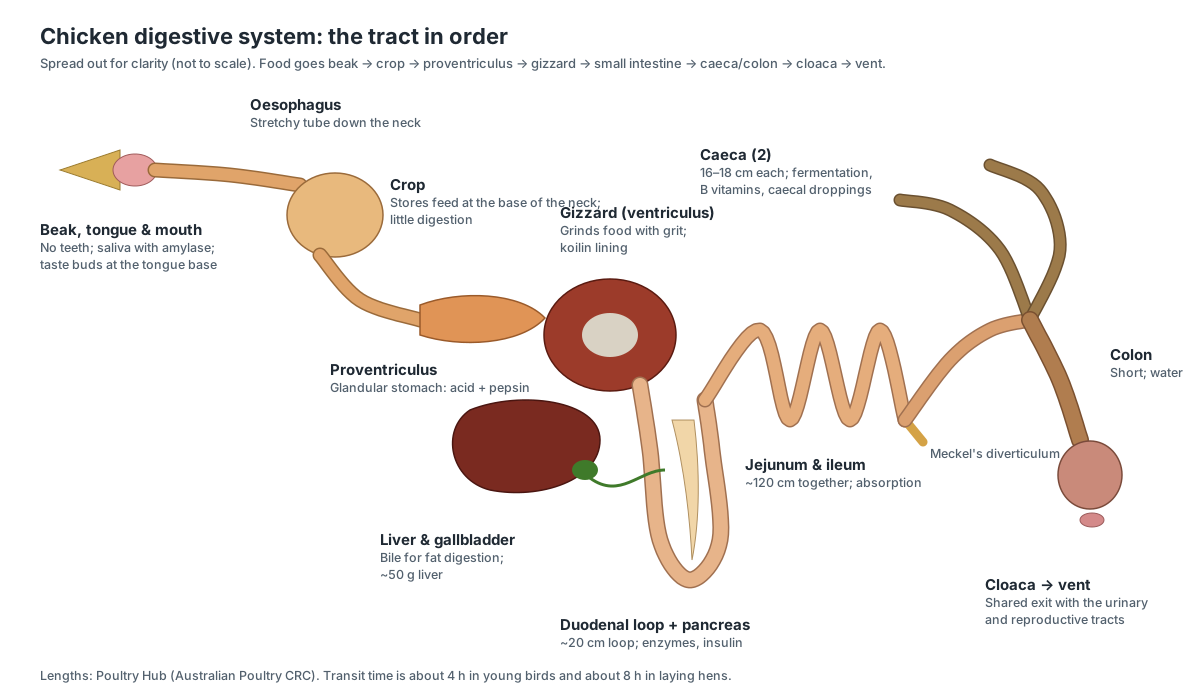
Poultry Hub gives these sizes for an adult chicken: a duodenal loop of about 20 cm, about 120 cm of jejunum and ileum together, and two caeca of 16–18 cm each. A meal takes about 4 hours to pass through a young bird, about 8 hours in a laying hen and about 12 hours in a broody hen.
Chicken crop anatomy
The crop (ingluvies) is a stretchy pouch of the oesophagus just before it enters the chest, at the base of the neck in front of the wishbone. It stores food so that a chicken can eat quickly and digest later. Very little digestion happens there, although some grain ferments.
A full crop is easy to feel as a soft lump at the front of the breast in the evening. It should be empty by the next morning. A crop that stays hard or full points to a blockage (impacted crop), and a sour-smelling, squashy crop to a yeast infection (sour crop).
Chicken gizzard anatomy
The gizzard (ventriculus) is the chicken's grinding stomach: a thick, disc-shaped organ made of two pairs of strong muscles, lined by a tough, yellowish layer called koilin. With swallowed grit, it crushes whole grains, doing the job that teeth do in mammals.
It works as a team with the proventriculus, the spindle-shaped glandular stomach in front of it. The proventriculus secretes hydrochloric acid and pepsinogen, but food passes through it quickly; most acid digestion happens in the gizzard, and food can move back and forth between the two. A cooked gizzard, sold as a giblet, still shows the shiny tendon sheet (tendinous centre) on each side and the peeled-away koilin lining.
- Proventriculus: Secretes hydrochloric acid and pepsinogen, which start protein digestion.
- Gizzard: Grinds food mechanically, doing the job teeth do in mammals.
- Duodenum: Receives bile and pancreatic juice; most digestion and absorption begins here.
- Pancreas: Makes digestive enzymes (for protein, fat and starch) and the hormones insulin and glucagon.
- Caeca: Bacteria ferment fibre, reabsorb water and make some B vitamins.
- Meckel's diverticulum: In the embryo and newly hatched chick it carried yolk into the gut; in adults it contains lymphoid tissue.
Chicken liver anatomy
The liver is the largest gland in the chicken: a dark red-brown organ with a right and a left lobe that wrap around the heart, lying just above the sternum. Poultry Hub gives its weight as about 50 g in an adult bird. Two bile ducts leave the right lobe, and the green gallbladder sits on that lobe.
In a laying hen the liver has an extra job: under the influence of estrogen it makes the proteins and fats of the egg yolk and sends them to the ovary in the blood. A chick's liver looks pale yellow for its first 10–14 days because it is full of absorbed yolk fat.
Chicken heart anatomy
A chicken has one cone-shaped heart with four chambers: two atria and two ventricles. It lies low in the chest, in the midline just behind the crop, with its tip tucked between the liver lobes. The right side pumps blood to the lungs and the left side to the body, as in mammals.
Two details are bird-specific. The valve between the right atrium and right ventricle is a strong muscular flap rather than a thin, three-cusped valve, and the aorta curves to the right, not the left, as it leaves the heart. Poultry Hub describes the bird heart as relatively large for the body size.
- Heart: Pumps blood in two circuits: the right side to the lungs, the left side to the body.
- Aorta: Carries oxygen-rich blood to the body.
- Brachiocephalic arteries: Each divides into a carotid artery to the head and a large artery to the wing and breast muscles.
- Jugular veins: Drain blood from the head into the cranial venae cavae.
- Caudal vena cava: Returns oxygen-poor blood from the legs, kidneys and organs.
Chicken lungs and air sacs
Chickens breathe with two small lungs that do not inflate, plus air sacs that act as bellows. Poultry Hub counts nine: one clavicular air sac (which holds the syrinx) and pairs of cervical, cranial thoracic, caudal thoracic and abdominal air sacs; the University of Kentucky also counts nine. Some anatomy texts count eight, because they treat sacs that are joined across the midline as a single sac. Air flows through the lungs in one direction, during both breathing in and breathing out.
The trachea is 15–18 cm long in a medium-sized adult, with about 108–125 complete cartilage rings. It ends at the syrinx, the voice box that makes the rooster's crow. Some air sacs extend into bones: the clavicular air sac enters the humerus, making it pneumatic (air-filled).
- Trachea: Carries air between the throat and the lungs.
- Syrinx: Makes the crow, clucks and alarm calls: vibrating membranes in its wall produce the sound.
- Lungs: Gas exchange. Air flows through tiny tubes (parabronchi) in one direction, during both breathing in and out.
- Cervical air sacs: Part of the bellows system that moves air through the lungs; they extend into the vertebrae.
- Clavicular air sac: Bellows for the lungs; its extension into the humerus makes that bone pneumatic.
- Thoracic air sacs: The cranial pair fills with air that has passed through the lungs; the caudal pair with fresh air on its way to the lungs.
- Abdominal air sacs: Hold fresh air that is pushed through the lungs when the bird breathes out.
Chicken skeleton anatomy: chicken bones labeled
The chicken skeleton is light but rigid. Many bones are fused: the hand bones, the shin and ankle bones, the back vertebrae into the notarium and synsacrum, and the tail tip into the pygostyle. The breastbone has a deep keel for the flight muscles, and the collarbones are fused into the wishbone (furcula).
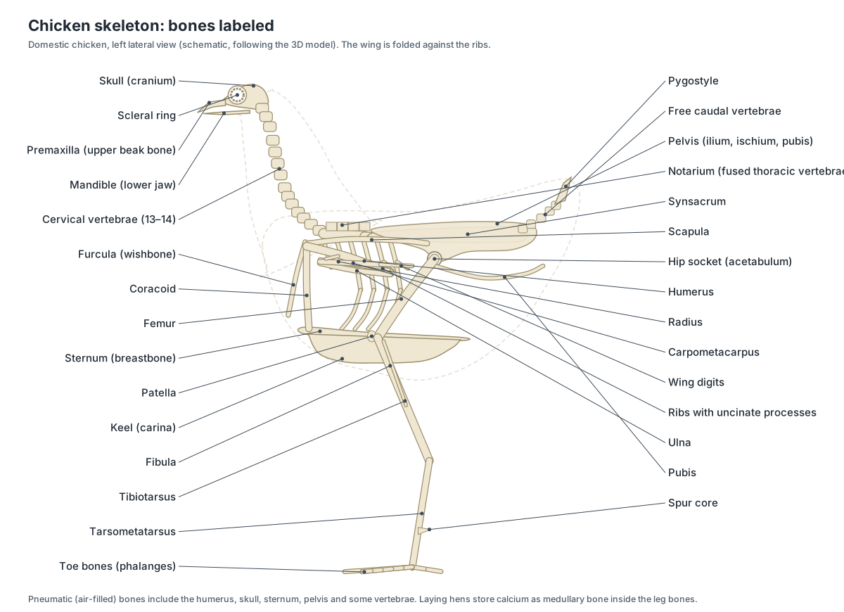
| Region | Bones | Notes |
|---|---|---|
| Skull | Cranium, premaxilla, mandible, quadrate, scleral ring | Thin, fused, pneumatic |
| Neck | About 13–14 cervical vertebrae | Poultry Hub lists 13; some texts count 14 |
| Trunk | 7 thoracic (front ones fused as the notarium), synsacrum | Last thoracic is fused into the synsacrum |
| Tail | About 5 free caudal vertebrae, pygostyle | Pygostyle anchors the tail feathers |
| Chest | 7 pairs of ribs with uncinate processes, sternum with keel | No diaphragm; ribs pump the air sacs |
| Shoulder | Scapula, coracoid, furcula | Together they form the triosseal canal |
| Wing | Humerus, radius, ulna, 2 carpals, carpometacarpus, 3 digits | Humerus is pneumatic |
| Hip | Ilium, ischium and pubis fused to the synsacrum | Pubic bones do not meet below |
| Leg | Femur, patella, tibiotarsus, fibula, tarsometatarsus | No separate ankle bones in the adult |
| Foot | Metatarsal I and toe phalanges (2, 3, 4, 5) | Some breeds have five toes |
Pneumatic bones, according to the University of Kentucky's extension article, include the skull, humerus, clavicle, keel, pelvic girdle and the lumbar and sacral vertebrae. The leg bones are not air-filled in the same way; in laying hens their marrow cavities fill with medullary bone instead.
Chicken wing anatomy
A chicken wing has the same bones as your arm, reduced and partly fused: the humerus in the upper wing, the radius and ulna in the forearm, and a fused hand (the carpometacarpus) with three small digits. Ten primary flight feathers grow from the hand and the secondaries grow from the ulna. Coverts overlap their bases.
| Wing segment (kitchen name) | Bones inside | Feathers on it |
|---|---|---|
| Drumette | Humerus | Small coverts |
| Flat (wingette) | Radius and ulna | Secondaries and their coverts |
| Tip | Carpometacarpus and digits | Primaries and the alula |
US poultry rules define the wing drummette as the humerus of the wing with its skin and meat, and the wing portion as the rest of the wing once the drummette is removed. When the wing is folded, the forearm and hand lie back along the body and the primaries tuck under the secondaries, so only the coverts, the triangular wing bay and the primary tips are visible.
- Humerus: Transmits the force of the breast muscles to the wing.
- Radius: Works with the ulna to extend and fold the wing.
- Ulna: Anchors the secondary flight feathers; small bumps on the bone (quill knobs) mark their attachment.
- Carpometacarpus: Carries the primary feathers.
- Wing digits: Support the outer primaries and the alula, which smooths airflow at low speeds.
- Primary feathers: Provide thrust in flight. Chickens fly only short distances, in bursts.
- Secondary feathers: Form the lifting inner part of the wing in flight.
Chicken leg and foot anatomy
A chicken leg has three long segments: the thigh (femur), the drumstick (tibiotarsus with the thin fibula) and the bare, scaly shank (tarsometatarsus), ending in four toes. The bird stands on its toes with the ankle (hock) high off the ground, and the knee is tucked up under the body feathers.
- Thigh: Contains the thigh muscles (dark meat) that swing the leg.
- Drumstick (lower thigh): Contains the large calf muscles that extend the foot.
- Hock: It is the bird's ankle, not its knee. It bends the opposite way to a human knee because it is a different joint.
- Shank: Carries the bird's weight. The scales protect it.
- Spur: A weapon used in fights between roosters.
- Toes: Spread the bird's weight, grip perches and scratch the ground for food.
- Claws: Scratching and gripping.
| Toe | Direction | Number of bones |
|---|---|---|
| I (hallux, hind toe) | Points backwards | 2 |
| II (inner toe) | Forwards, inner side | 3 |
| III (middle toe) | Forwards, longest | 4 |
| IV (outer toe) | Forwards, outer side | 5 |
The shank and the tops of the toes are covered with keratin scales attached to the skin, which Poultry Hub describes as a carry-over from the chicken's reptile-like ancestors. Spurs grow on the back of the shank from a bony core; they are much larger in roosters, especially older ones. Breeds such as the Dorking, Silkie, Houdan and Faverolles have a fifth toe.
Chicken muscle anatomy and meat cuts
Chicken meat is muscle, so every supermarket cut maps onto real anatomy. White meat comes from the breast and wing drumette, whose fibres are mostly fast white fibres with little myoglobin. Dark meat comes from the thigh and drumstick, which have more red fibres for sustained work such as walking.
| Cut | Muscles | Bone | Meat |
|---|---|---|---|
| Breast | Pectoralis (pectoralis major) | Keel and sternum | White |
| Tenderloin (tender) | Supracoracoideus | Alongside the keel | White |
| Wing drumette | Biceps, triceps and shoulder muscles | Humerus | White |
| Wing flat | Forearm muscles | Radius and ulna | White |
| Thigh | Iliotibialis, iliofibularis, femorotibialis and others | Femur | Dark |
| Drumstick | Gastrocnemius, fibularis longus, toe flexors | Tibiotarsus and fibula | Dark |
| Oysters | Small muscles on the back of the pelvis | Pelvis | Dark |
US rules separate drumsticks from thighs by a cut through the knee joint, take thighs off at the hip joint (they may include pelvic meat), and define a leg as the thigh and drumstick together. The tough white strands in a drumstick are tendons running down to the foot.
- Pectoralis: Pulls the wing down: the power stroke of flight.
- Thigh muscles: Swing the leg forward and back and straighten the knee for walking, scratching and perching.
- Drumstick muscles: The gastrocnemius extends the foot at the hock; the long flexor tendons curl the toes.
- Wing muscles: The biceps bends the elbow and folds the wing; the triceps straightens it.
Chicken tenderloin anatomy: which muscle is the tender?
The chicken tenderloin is the supracoracoideus, the deep breast muscle that lies alongside the keel underneath the main breast muscle. The meat trade often calls it the "pectoralis minor", but it is not the same muscle as the human pectoralis minor, and veterinary sources name it the supracoracoideus or deep pectoral muscle.
Its job is surprising: although it sits below the wing, it raises the wing. Its tendon runs up through the triosseal canal, a gap between the coracoid, scapula and furcula, and hooks over the top of the humerus like a rope over a pulley. The pectoralis on top of it pulls the wing down. Because the tenderloin sits in a tight compartment, its blood supply can fail in heavy broilers, causing deep pectoral myopathy (green muscle disease).
- Supracoracoideus: Raises the wing. Its tendon runs up through the triosseal canal at the shoulder and over the top of the humerus like a rope over a pulley.
Chicken reproductive anatomy: hen vs rooster
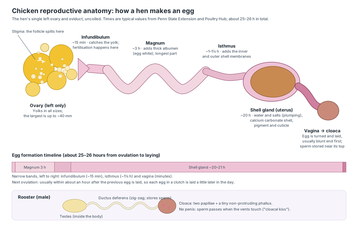
A hen has a single working ovary and oviduct, both on the left; the right ones regress during embryonic development and do not work in the adult. A rooster has two bean-shaped testes inside the body, against the backbone near the kidneys, and two sperm ducts that open into the cloaca. Neither sex has external genitals.
| Hen (female) | Rooster (male) | |
|---|---|---|
| Gonads | One left ovary with yolks of all sizes | Two internal testes; the left is often larger |
| Ducts | Oviduct: infundibulum, magnum, isthmus, shell gland, vagina | Two zig-zag ductus deferens, the main sperm store |
| In the cloaca | Oviduct opens into the urodeum | Two papillae and a tiny phallus |
| Outside | Smaller comb and wattles, rounded hackles | Big comb, sickle feathers, pointed hackles, spurs |
| Mating | Stores sperm for about 10 days to 2 weeks | Passes sperm in a brief 'cloacal kiss' |
Poultry Hub gives the active oviduct a length of about 70–80 cm, and US extension sources about 25–27 inches. Day-old chicks can be sexed by looking for the rudimentary male organ in the everted vent, a method developed in Japan, or by wing feathers and down colour in certain crosses.
- Ovary: Grows yolks and releases one at a time (ovulation); makes estrogen.
- Testes: Make sperm and testosterone.
- Ductus deferens: Carries and stores sperm; it is the main sperm store in the rooster.
- Papillae & phallus: Roosters have no penis: sperm passes from the papillae to the hen's cloaca when the vents touch.
How a hen lays an egg: egg formation step by step
A hen takes about 25–26 hours to turn a yolk into a laid egg (often rounded to "about a day"). The yolk is released from the ovary, caught by the infundibulum, and then coated in turn with egg white, shell membranes and shell as it moves down the oviduct.
- Ovulation. A mature yolk, up to about 40 mm across, splits out of its follicle along the stigma. The next yolk is usually released within about an hour after the previous egg is laid.
- Infundibulum (about 15–17 minutes). The muscular funnel moves to engulf the yolk. If sperm are present, fertilisation happens here.
- Magnum (about 3 hours). Glands add the thick albumen.
- Isthmus (about 75 minutes). The inner and outer shell membranes are added and the egg takes its shape.
- Shell gland (20 hours or more). Water and salts plump up the albumen, then the calcium carbonate shell is laid down. Brown pigment is added in the last hours.
- Vagina and vent. The egg enters the vagina small end first, turns, and is pushed out through the cloaca, usually blunt (large) end first.
Because the cycle is a little longer than a day, each egg in a clutch is laid a little later in the day. The University of Kentucky's extension article notes that hens almost never ovulate after about 3 p.m., so a hen that laid late in the day may skip a day before starting a new clutch. An egg needs no rooster: an unfertilised egg is laid in exactly the same way.
- Infundibulum: Captures the yolk; fertilisation happens here if sperm are present. The yolk stays about 15–17 minutes.
- Magnum: Adds about half of the egg white (albumen), mainly the thick albumen, in about 3 hours.
- Isthmus: Adds the inner and outer shell membranes and a little albumen, and sets the egg's shape (about 75 minutes).
- Shell gland: First adds water and salts to the albumen ('plumping'), then lays down the calcium carbonate shell, the pigment and the cuticle.
- Vagina: Pushes the finished egg out during laying, and stores sperm in small glands near its junction with the shell gland.
Chicken egg anatomy: the parts of an egg
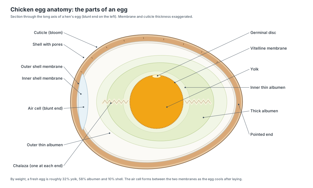
A chicken egg has, from outside in: the cuticle (bloom), the porous calcium-carbonate shell, the outer and inner shell membranes, the air cell at the blunt end, layers of thin and thick albumen (egg white), two twisted chalazae, and the yolk wrapped in the vitelline membrane, with the pale germinal disc on top.
| Part | What it is | Made in |
|---|---|---|
| Cuticle (bloom) | Thin coating that helps keep bacteria and dust out of the pores | Shell gland and vagina |
| Shell | Calcium carbonate with an organic matrix; thousands of pores (about 8,000) | Shell gland |
| Shell membranes | Inner and outer fibrous membranes | Isthmus |
| Air cell | Forms between the membranes at the blunt end as the egg cools | After laying |
| Albumen | Outer thin, thick, inner thin and chalaziferous layers | Magnum (and shell gland) |
| Chalazae | Twisted cords that hold the yolk in the centre | Oviduct (sources differ on where they first form) |
| Yolk and vitelline membrane | Food store for the embryo; ~32% of the egg | Ovary |
| Germinal disc | Pale spot holding the female cell; called the blastoderm once fertilised | Ovary |
By weight, a fresh egg is roughly 32% yolk, 58% albumen and 10% shell, according to the egg-quality handbook published by the Queensland Department of Primary Industries; Poultry Hub gives ranges of 32–35% yolk, 52–58% albumen and 9–14% shell. Mississippi State University Extension notes that the air cell grows as an egg ages, so a small air cell is a sign of a fresh egg, and that a chick pierces the air cell to take its first breaths before hatching.
Chicken cloaca and vent anatomy
The cloaca is the shared chamber at the end of the digestive, urinary and reproductive tracts, and the vent is its opening under the tail. It has three compartments: the coprodeum receives faeces from the colon, the urodeum receives urine from the ureters and the oviduct or sperm ducts, and the proctodeum opens at the vent.
Chickens have no urinary bladder. Their kidneys excrete nitrogen as uric acid, which reaches the cloaca as a thick white paste: the white cap on a chicken dropping. Poultry Hub notes that urine can be moved back into the colon by reverse peristalsis so that water is reabsorbed. The bursa of Fabricius, an immune organ of young birds, opens into the dorsal wall of the proctodeum.
- Cloaca: The coprodeum receives faeces, the urodeum receives urine and the oviduct or sperm ducts, and the proctodeum opens at the vent.
- Vent: The exit of the cloaca. Mating is a brief 'cloacal kiss' in which the vents of rooster and hen touch.
- Kidneys: Filter the blood and remove nitrogen waste as uric acid.
- Ureters: Carry thick, pasty urine full of uric acid.
- Bursa of Fabricius: Trains B-cells, the white blood cells that make antibodies. B-cells were discovered through this organ.
Chicken skin and feathers
Chicken skin has three layers, the epidermis, dermis and hypodermis, and is thin and loose. Feathers grow only from feather tracts called pterylae, separated by bare strips called apteria, which is why a plucked chicken shows rows of follicles. Poultry Hub lists five feather types in chickens: contour feathers, plumules, filoplumes, chick down and bristles.
The preen gland on top of the tail base secretes oil that the bird spreads over its feathers. Moult, the yearly replacement of feathers, usually happens in autumn, and males moult before females.
Chicken brain anatomy and senses
The chicken brain has smooth cerebral hemispheres at the front, large optic lobes on the sides of the midbrain, a ridged cerebellum at the back, small olfactory bulbs and a brainstem that joins the spinal cord. Poultry Hub describes the optic lobes as very large compared with those of other species, which fits an animal that relies mainly on sight.
- Cerebrum: Controls learning, behaviour, feeding and social recognition (pecking order).
- Optic lobes: Process vision.
- Cerebellum: Coordinates balance, posture and movement.
- Olfactory bulbs: Process smell.
- Brainstem: Controls breathing and heart rate.
- Spinal cord: Carries messages between brain and body and controls reflexes.
- Sciatic nerve: Carries motor and sensory signals for most of the leg below the hip.
Chickens have eyes on the sides of the head, giving a wide field of view. Only about 26° directly in front is seen with both eyes. Chicks reach their peak sharpness of vision within 48 hours of hatching. The ear has no outer flap, and a single bone, the columella, replaces the three middle-ear bones of mammals.
Chicken endocrine glands: thyroid and adrenals
A chicken's main hormone glands in the body cavity are two separate thyroid glands at the entrance to the chest and two small adrenal glands at the front end of the kidneys, next to the gonads. The thyroid hormones control metabolic rate, growth and moult; the adrenals make corticosterone, the main stress hormone of birds. The tiny parathyroid glands just behind the thyroids control blood calcium, which a laying hen needs for every eggshell.
- Thyroid glands: Make thyroid hormones (thyroxine, T4, and triiodothyronine, T3) that set the metabolic rate and control growth, moult and feather development.
- Adrenal glands: Make corticosterone (the main stress and metabolic hormone in birds), aldosterone, and adrenaline and noradrenaline.
Chicken immune organs: bursa, thymus and spleen
Chickens have two primary lymphoid organs: the bursa of Fabricius, where B-cells mature, and the thymus, where T-cells are trained. Poultry Hub notes that B-cells move into the bursa from about day 15 of incubation until about 10 weeks of age, and that early damage from Gumboro or Marek's disease stops this programming for life. The bursa is present in birds under about a year old.
- Bursa of Fabricius: Trains B-cells, the white blood cells that make antibodies. B-cells were discovered through this organ.
- Thymus: Trains T-cells.
- Spleen: Filters blood and is a meeting place for B- and T-cells; chickens have no true lymph nodes.
Medullary bone: how a hen pays for her eggshells
A laying hen makes a new shell almost every day, and much of it forms at night, when she is not eating. She bridges the gap with medullary bone: a spongy, temporary bone that grows inside the marrow cavities of her long bones. Canoville and colleagues describe it as a sex-specific, estrogen-dependent tissue that, among living reptiles, birds and mammals, is found only in female birds. In laying pullets it has been seen in most of the skeleton, although only in small amounts in the skull, humerus and feet. (Canoville, Schweitzer & Zanno (2019): systemic distribution of medullary bone in the avian skeleton, BMC Evolutionary Biology.)
The numbers show why it matters. Poultry Hub notes that medullary bone starts forming about 10–14 days before the first egg, and that a shortage of calcium can lead to cage layer fatigue. The University of Kentucky's extension article puts the share of shell calcium that comes from bone at 47%, with the rest from the diet. When you read a chicken skeleton diagram, remember that the hollow-looking leg bones of a laying hen are, in life, partly filled with this calcium store. (Poultry Hub (Australia): skeletal system of the fowl.)
Comb types and the first gene interaction ever described
The wild type is the single comb, one upright blade of tissue. Two dominant mutations change it. Pea comb, caused by an amplified stretch of DNA inside the SOX5 gene, shrinks the comb and wattles into three low rows; Wright and colleagues linked this to cold climates, where a small comb loses less heat. (Wright et al. (2009): copy number variation in SOX5 causes the Pea-comb phenotype, PLoS Genetics.)
Rose comb is caused by a large chromosomal inversion that switches on the MNR2 gene where it should be silent, giving a flat, bumpy comb with a spike at the back. In homozygous males the same rearrangement also reduces sperm motility. A bird with both rose and pea alleles has a walnut comb, the classic example of epistasis that Bateson and Punnett described over a century ago. (Imsland et al. (2012): the Rose-comb mutation in chickens, PLoS Genetics.)
Why the tenderloin can turn green: breast muscles in broilers
The two breast muscles sit in different compartments. The pectoralis lies on the surface; the supracoracoideus (the tenderloin) lies underneath it, pressed against the keel. In fast-growing meat chickens the deep muscle is tightly enclosed, and its blood supply can fail during vigorous wing flapping. The result is deep pectoral myopathy, seen at necropsy as a necrotic supracoracoideus under an apparently normal superficial breast muscle. (MSD Veterinary Manual: deep pectoral (supracoracoideus) myopathy in chickens (image).)
Researchers are still studying how the two muscles interact. A 2025 study of broilers examined injury and fibrosis where the pectoralis major and supracoracoideus join their tendinous sheets (the myoaponeurotic junction), and found coagulative necrosis in the supracoracoideus of one bird. For anatomy students, the takeaway is simple: the breast is two muscles with opposite jobs, one pulling the wing down and one lifting it, and both attach to the keel. (Kawasaki et al. (2025): pectoralis major and supracoracoideus myoaponeurotic junction in broilers, Journal of Poultry Science.)
How to read a chicken anatomy diagram without getting lost
Most chicken diagrams are drawn from the left side, because that is where the hen's single ovary and oviduct lie. Start with landmarks you can feel on a live bird: the crop at the base of the neck, the keel down the middle of the breast, the pubic bones on each side of the vent. Then place the organs between them. The heart sits just behind the crop, the liver lobes wrap around it, and the gizzard sits behind the liver. (Poultry Hub: digestive system of the fowl.)
Feathers make a chicken look much larger than its skeleton. In the 3D model, fade the feathers and notice how far the knee and the keel sit inside the outline. The visible bend at the bottom of the feathered leg is the hock (ankle), not the knee, and the thigh bone is hidden under the body plumage. Many worksheet mistakes come from labeling the hock as the knee. (Jacob, University of Kentucky / Extension Foundation: external anatomy of chickens.)
Chicken anatomy worksheet: test yourself
Use the 3D model or the diagrams above to answer these.
- Which joint is the visible backward bend of a chicken's leg? (The hock, or ankle.)
- Name the two parts of a chicken's stomach. (Proventriculus and gizzard.)
- On which side is the hen's working ovary? (The left.)
- Which muscle is the tenderloin? (The supracoracoideus.)
- Where does an egg spend most of its 25–26 hours? (The shell gland, about 20 hours.)
- Which bone is inside a wing drumette? (The humerus.)
- What is the white cap on a chicken dropping? (Uric acid from the kidneys.)
- How many air sacs does a chicken have? (Nine in most poultry references; some texts count eight.)
Frequently asked questions
Do chickens have tear ducts?
Yes. Chickens make tears in the lacrimal gland and the larger Harderian gland behind the eye. The nictitating membrane (third eyelid) spreads the fluid over the eye, and tears drain into a small sac and then through a duct into the nasal cavity, much like the human nasolacrimal duct. That is why eye and nose infections often occur together in poultry.
What part of the chicken is the tenderloin?
The tenderloin (or tender) is the supracoracoideus muscle, the deep breast muscle that lies alongside the keel under the main breast muscle (pectoralis). The meat trade calls it the 'pectoralis minor', but it is not the same as the human pectoralis minor. In the living bird it raises the wing, pulling on a tendon that runs over the shoulder like a rope over a pulley.
How long does it take a hen to make an egg?
About 25–26 hours from ovulation to laying. The yolk spends about 15 minutes in the infundibulum, about 3 hours in the magnum (egg white), about 75 minutes in the isthmus (shell membranes) and 20 hours or more in the shell gland (shell). The next yolk is usually released within about an hour after an egg is laid.
Why do hens have only one ovary?
Only the left ovary and oviduct develop in hens; the right ones regress during embryonic development and remain only as a tiny, non-functional remnant. One reason often given is that a single oviduct is enough for a bird that forms one large hard-shelled egg at a time, and it saves weight.
Do chickens pee?
Chickens have kidneys and ureters but no urinary bladder. Their urine is a thick paste of uric acid that goes straight into the cloaca and leaves with the droppings: it is the white cap on a chicken dropping. This saves water.
Do roosters have a penis?
No. A rooster has two internal testes and two sperm ducts that open on small papillae in the cloaca, and only a tiny, non-protruding phallus. Mating is a brief 'cloacal kiss': the rooster presses his vent against the hen's and passes sperm across.
What does a chicken's crop do?
The crop is a stretchy pouch of the oesophagus at the base of the neck. It stores food so a chicken can eat quickly and digest later. Little digestion happens there; food is passed in small amounts to the proventriculus (glandular stomach) and gizzard (grinding stomach).
Why do chickens eat grit?
Chickens have no teeth. Small stones (grit) collect in the muscular gizzard, where its strong walls rub them against hard food such as whole grains, grinding it up.
Which way does a chicken's knee bend?
Forward, like ours, but you cannot see it: the knee is hidden under the body feathers. The backward-bending joint at the bottom of the feathered leg is the hock, which is the bird's ankle.
Are chicken bones hollow?
Some are. The humerus, skull, sternum, pelvis and some vertebrae are pneumatic: air sacs extend into them. Leg bones such as the femur and tibiotarsus have marrow, and in laying hens they fill with medullary bone, a calcium store for eggshells.
References
- Martin & Clauer, Penn State Extension: hen reproduction
- Jacob, University of Kentucky / Extension Foundation: avian reproductive system, female
- Jacob, University of Kentucky / Extension Foundation: avian reproductive system, male
- Jacob, University of Kentucky / Extension Foundation: avian digestive system
- Jacob, University of Kentucky / Extension Foundation: external anatomy of chickens
- Jacob, University of Kentucky / Extension Foundation: avian skeletal system
- Extension Foundation: sexing day-old chicks
- Poultry Hub (Australia): skeletal system of the fowl
- Poultry Hub: digestive system of the fowl
- Poultry Hub: respiratory system and thermoregulation
- Poultry Hub: reproductive system of the fowl
- Poultry Hub: nervous system and sense organs
- Poultry Hub: the avian egg
- Wells, Mississippi State University Extension (2023): The Avian Embryo, Publication 1150 (PDF)
- Optimum Egg Quality handbook: internal and external egg quality
- Canoville, Schweitzer & Zanno (2019): systemic distribution of medullary bone in the avian skeleton, BMC Evolutionary Biology
- Wright et al. (2009): copy number variation in SOX5 causes the Pea-comb phenotype, PLoS Genetics
- Imsland et al. (2012): the Rose-comb mutation in chickens, PLoS Genetics
- US 9 CFR 381.170: standards for kinds and classes, and for cuts of raw poultry
- MSD Veterinary Manual: deep pectoral (supracoracoideus) myopathy in chickens (image)
More interactive anatomy models
- Bird skeletonHollow bones, keeled sternum, wishbone and fused wing bones of a bird.
- Bird anatomyBird digestive system, air sacs, heart and feather anatomy in 3D.
- Pigeon dissectionGuided pigeon dissection: flight muscles, crop, gizzard, air sacs and heart.
- Human anatomy1,000+ bones, muscles, organs, vessels and nerves from real anatomical data.
- Octopus anatomyThree hearts, a ring-shaped brain, eight arms and a beak, in 3D.
- Squid anatomyPen, eight arms, two tentacles, giant axons and three hearts of the longfin squid.