

Anatomy of a pig at a glance
A pig is an even-toed hoofed mammal (order Artiodactyla) with a simple, single-chambered stomach, a long intestine with a spiral colon, a snout stiffened by a unique rostral bone, four toes on each foot and a bicornuate uterus that carries large litters. Its heart, kidneys, skin and digestive tract are close enough to ours that pigs are a major model in medicine.
This page describes the domestic pig and uses a fetal pig for the 3D model, because that is what most students dissect. The fetus has all the adult organs plus the umbilical cord and fetal blood shunts. Where the adult differs, the text says so. Directions follow veterinary usage: cranial (towards the head), caudal (towards the tail), dorsal (back) and ventral (belly); left and right are the pig's own.
Pig external anatomy: labeled diagram
The external anatomy of a pig includes the snout with its flat rostral disk and nostrils, the eyes and eyelids, the ears, the jowl, neck, shoulder, back, loin, rump and tail, the ham, flank and belly, two rows of teats, and four legs ending in cloven hooves with dewclaws.
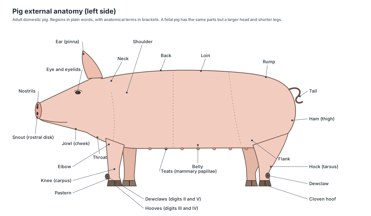
| Part | Where | What to notice |
|---|---|---|
| Snout (rostral disk) | Tip of the head | Flat, hairless digging disk with the two nostrils; supported by the rostral bone |
| Ears | Top of the head | Upright or floppy depending on breed; folded back in a fetus |
| Teats (mammary papillae) | Two rows on the belly | Present in both sexes; sows typically have 12–14 |
| Hooves | Each foot | Two main toes (digits III and IV) and two dewclaws (II and V) |
| Tail | Above the anus | Thin, often curled; 20–23 caudal vertebrae |
| Genitals | Ventral body | Female: genital papilla below the anus. Male: preputial opening behind the navel, scrotum below the anus |
| Umbilical cord | Belly (fetus only) | Two arteries, one vein and the allantoic duct |
Internal anatomy of a pig: organs diagram
Inside a pig, the chest holds the lungs, the heart in its pericardium and, in young pigs, a large thymus. The dome-shaped diaphragm separates the chest from the abdomen. Behind it lie the lobed liver with the gallbladder, the stomach and spleen on the left, the pancreas along the duodenum, the long coiled small intestine, the caecum and the cone-shaped spiral colon, the kidneys against the roof of the abdomen, and the bladder and reproductive organs at the back.
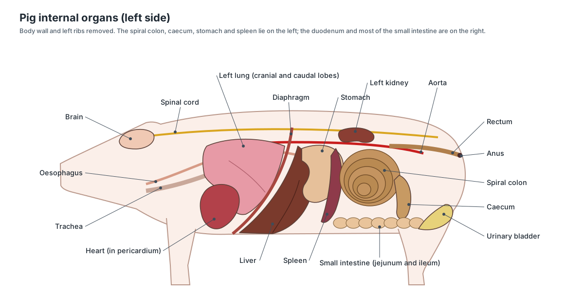
- Lungs: Spongy organs on both sides of the heart. The right lung has cranial, middle, caudal and accessory lobes; the left has cranial and caudal lobes. Exchange oxygen and carbon dioxide after birth. Before birth they are not inflated.
- Pericardium: The thin membrane sac around the heart. Holds the heart in place and lets it beat with little friction.
- Left ventricle: The lower left chamber, forming the apex of the heart. Pumps oxygenated blood into the aorta for the whole body.
- Thymus: A large, soft, pale gland in the neck and over the heart. It is much bigger in a fetus than in an adult. Trains T lymphocytes (immune cells) early in life.
- Liver: The largest organ in the abdomen: dark brown, deeply divided into lobes, and very large in the fetus. Makes bile, stores nutrients and processes blood from the gut. In the fetus it also makes blood cells.
- Stomach: A large, J-shaped, single-chambered (monogastric) stomach lying mostly on the left. Stores and churns food and begins protein digestion with acid and pepsin.
- Spleen: A long, flat, tongue-shaped red-brown organ along the stomach. Filters and stores blood and fights infection. It is not part of the digestive tract.
- Pancreas: Pale, bumpy, granular tissue lying under the stomach and in the loop of the duodenum. Makes digestive enzymes for the duodenum and the hormones insulin and glucagon.
- Jejunum and ileum: The long, coiled rest of the small intestine, held together by mesentery. Finishes digestion and absorbs nutrients into blood vessels of the mesentery.
- Spiral colon: The ascending colon, wound into a cone of tight coils: inward (centripetal) coils, then outward (centrifugal) coils. Absorbs water and salts and ferments fibre with its gut bacteria.
- Kidneys: Two bean-shaped organs on the roof of the abdomen, outside the peritoneum. Filter blood and make urine.
To find these organs in a real specimen, step by step, use our fetal pig dissection guide.
Pig anatomy labeled: every structure and its function
This is the text version of the 3D model: every labeled structure of the fetal pig, grouped by body system. Male-only and female-only structures are marked.
External
- Skin and body wall (Integument)
- Thin, almost hairless skin over the body. In a late fetus it is soft and wrinkled, with only fine, sparse hairs. Protects the body. In the adult it carries sparse bristles and a fat layer (the source of pork rind and bacon fat).
- Snout and rostral disk (Rostrum, planum rostrale)
- The flat, round, hairless disk at the tip of the snout, with the two nostrils in it. A tough, touch-sensitive digging tool. Pigs root in soil with it to find food.
- Nostrils (External nares)
- The two openings in the rostral disk. Let air into the nasal cavity for breathing and smell.
- Eyelids (Palpebrae)
- Upper and lower eyelids, still closed or barely open in most lab fetal pigs. Protect the eye. Piglets are born with their eyes open.
- Ears (Pinnae (auricles))
- The large, flat outer ear flaps. Collect sound and direct it into the ear canal.
- Mammary papillae (Teats, nipples)
- Two rows of small teats along the belly, present in both males and females. In sows they deliver milk to the piglets. In males they are not functional.
- Umbilical cord (Umbilicus)
- The cut cord on the belly. In cross-section you can see two umbilical arteries, one umbilical vein and the allantoic duct. Before birth it linked the fetus to the placenta, carrying blood and nutrients in and wastes out.
- Anus
- The opening of the digestive tract under the base of the tail. Passes faeces. In the fetus the gut is filled with meconium, the first faeces.
- Hooves (main digits) (Claws, digits III and IV)
- Two large hoofed toes on each foot: digits III and IV, which carry the weight. Support the body. Pigs walk on the tips of their toes (unguligrade).
- Dewclaws (Accessory digits II and V)
- Two small hoofed toes higher up at the back of each foot: digits II and V. Usually clear the ground on firm surfaces; they help on soft ground.
- Tail (Cauda)
- The short, thin tail above the anus. In the adult it is used in social signalling; it often curls.
- Genital papilla and urogenital opening (Urogenital papilla) · females only
- A small fleshy projection just below the anus, with the urogenital opening at its base: the sign of a female. Marks the external opening of the urogenital sinus (shared exit for urine and the reproductive tract).
- Urogenital opening (male) (Preputial opening) · males only
- A small opening on the belly just behind the umbilical cord: the tip of the prepuce, where the penis opens. Outlet for urine (and, in adults, semen).
- Scrotal sacs (Scrotum) · males only
- Paired swellings just below the anus that will hold the testes. Keep the testes cooler than the body after they descend.
Mouth & oral cavity
- Hard palate (Palatum durum)
- The bony front part of the roof of the mouth, covered by ridged mucous membrane. Separates the mouth from the nasal cavity and gives the tongue a firm surface to press food against.
- Soft palate (Velum palatinum)
- The boneless back part of the roof of the mouth. Closes off the nasopharynx during swallowing so food does not enter the nasal cavity.
- Tongue (Lingua)
- The long, muscular tongue filling the floor of the mouth. Moves food, helps with suckling and swallowing, and carries the taste buds.
- Tongue papillae (Lingual papillae, sensory papillae)
- Small bumps on the tongue. Some carry taste buds. Grip food and hold taste buds.
- Epiglottis
- A cone-shaped flap of cartilage at the back of the throat, in front of the glottis. Folds over the glottis during swallowing so that food goes into the oesophagus, not the trachea.
- Teeth
- In a late fetus, a few sharp teeth may already have erupted: the third incisors and canines (needle teeth). After birth, the needle teeth are used when piglets compete for teats.
- Salivary glands (Parotid and mandibular glands)
- Soft, lobed glands below and behind the ear and along the jaw. Produce saliva, which moistens food and starts the digestion of starch.
Digestive
- Oesophagus (Esophagus, gullet)
- The muscular tube that carries food from the pharynx to the stomach. Moves swallowed food to the stomach by waves of muscle contraction (peristalsis).
- Stomach (Ventriculus, gaster)
- A large, J-shaped, single-chambered (monogastric) stomach lying mostly on the left. Stores and churns food and begins protein digestion with acid and pepsin.
- Pyloric sphincter (Pylorus)
- The ring of muscle at the exit of the stomach. Controls the passage of food from the stomach into the small intestine.
- Duodenum
- The first, fairly straight part of the small intestine. Receives bile from the liver and enzymes from the pancreas.
- Jejunum and ileum (Small intestine)
- The long, coiled rest of the small intestine, held together by mesentery. Finishes digestion and absorbs nutrients into blood vessels of the mesentery.
- Mesentery
- The thin, transparent sheet that holds the coils of intestine and carries their vessels. Suspends the intestines and carries the mesenteric arteries, veins, lymph vessels and nerves.
- Caecum (Cecum)
- A blind-ended pouch where the small intestine joins the large intestine. Holds microbes that ferment fibre, which pigs digest better than humans.
- Spiral colon (Ascending colon, ansa spiralis coli)
- The ascending colon, wound into a cone of tight coils: inward (centripetal) coils, then outward (centrifugal) coils. Absorbs water and salts and ferments fibre with its gut bacteria.
- Rectum
- The last straight part of the large intestine, running to the anus. Stores faeces before they leave through the anus.
- Liver (Hepar)
- The largest organ in the abdomen: dark brown, deeply divided into lobes, and very large in the fetus. Makes bile, stores nutrients and processes blood from the gut. In the fetus it also makes blood cells.
- Gallbladder (Gall bladder)
- A small, greenish sac tucked under the liver. Stores bile and releases it into the duodenum.
- Bile duct (Common bile duct, ductus choledochus)
- The thin duct that carries bile from the gallbladder and liver to the duodenum. Delivers bile into the duodenum.
- Pancreas
- Pale, bumpy, granular tissue lying under the stomach and in the loop of the duodenum. Makes digestive enzymes for the duodenum and the hormones insulin and glucagon.
Respiratory
- Larynx (Voice box)
- The voice box: a short tube of cartilage at the top of the trachea. Guards the airway and produces the pig's squeals and grunts.
- Trachea (Windpipe)
- The windpipe, stiffened by C-shaped rings of cartilage. Carries air between the larynx and the bronchi.
- Bronchi (with tracheal bronchus) (Primary bronchi, bronchus trachealis)
- The branches of the trachea into the lungs, including a separate bronchus to the right cranial lobe. Carry air into each lung lobe.
- Lungs (Pulmones)
- Spongy organs on both sides of the heart. The right lung has cranial, middle, caudal and accessory lobes; the left has cranial and caudal lobes. Exchange oxygen and carbon dioxide after birth. Before birth they are not inflated.
- Diaphragm
- The dome-shaped sheet of muscle between the thorax and the abdomen. Contracts to enlarge the thorax and draw air in; it is the main breathing muscle.
Circulatory (fetal)
- Pericardium
- The thin membrane sac around the heart. Holds the heart in place and lets it beat with little friction.
- Right atrium
- The thin-walled chamber that receives blood from the venae cavae. Receives blood from the body. In the fetus much of it flows straight on to the left atrium through the foramen ovale.
- Left atrium
- The chamber that receives blood from the lungs. Passes blood to the left ventricle.
- Right ventricle
- The lower right chamber, which pumps blood into the pulmonary trunk. Pumps blood towards the lungs; in the fetus most of it is diverted to the aorta through the ductus arteriosus.
- Left ventricle
- The lower left chamber, forming the apex of the heart. Pumps oxygenated blood into the aorta for the whole body.
- Coronary vessels
- Arteries and veins running in the grooves on the surface of the heart. Supply the heart muscle itself with blood.
- Foramen ovale
- A flap-covered hole in the wall between the right and left atria. Fetal shunt: lets blood pass from the right atrium straight to the left atrium, bypassing the lungs.
- Aorta (Aortic arch, dorsal aorta)
- The largest artery: it leaves the left ventricle, arches to the left and runs back along the roof of the body cavity. Carries blood from the left ventricle to the whole body.
- Pulmonary trunk
- The large artery leaving the right ventricle, which divides into the pulmonary arteries. Carries blood towards the lungs.
- Ductus arteriosus
- A short, wide vessel joining the pulmonary trunk to the aorta. Fetal shunt: carries most of the right ventricle's output into the aorta, bypassing the uninflated lungs.
- Brachiocephalic trunk
- The first large branch of the aortic arch. Supplies the head (through the carotids) and the right forelimb (through the right subclavian artery).
- Common carotid arteries
- Paired arteries running up the neck beside the trachea. Supply the head and brain.
- Subclavian arteries
- The arteries to the forelimbs. Supply the forelimbs, chest wall and neck.
- Cranial vena cava (Anterior vena cava, precava)
- The large vein returning blood from the head, neck and forelimbs. Returns blood from the front half of the body to the heart.
- Caudal vena cava (Posterior vena cava, postcava)
- The large vein returning blood from the abdomen and hind limbs. Returns blood from the back half of the body. In the fetus it also carries oxygen-rich umbilical blood from the liver.
- Jugular veins
- Paired veins in the neck that drain the head. Return blood from the head and brain to the heart.
- Umbilical vein
- The single large vein running from the umbilical cord forward to the liver. Carries oxygen- and nutrient-rich blood from the placenta into the fetus.
- Ductus venosus
- A fetal channel through the liver from the umbilical vein to the caudal vena cava. Fetal shunt: lets part of the umbilical blood bypass the liver sinusoids on its way to the heart.
- Umbilical arteries
- Two arteries running from the iliac arteries along both sides of the bladder into the umbilical cord. Carry oxygen-poor blood and wastes from the fetus to the placenta.
- Renal arteries and veins
- Short vessels between the kidneys and the aorta and caudal vena cava. Carry blood into and out of the kidneys for filtering.
- Iliac arteries
- The terminal branches of the aorta to the pelvis and hind limbs. Supply the hind limbs and pelvis; the umbilical arteries branch from the internal iliacs.
- Coeliac and cranial mesenteric arteries (Celiac trunk, superior mesenteric artery)
- The two large unpaired branches of the abdominal aorta that supply the gut. The coeliac artery supplies the stomach, spleen, liver and pancreas; the cranial mesenteric artery fans out in the mesentery to the small intestine, caecum and most of the colon.
- Hepatic portal vein (Portal vein)
- A large vein that carries blood from the stomach, intestines, spleen and pancreas to the liver. Brings absorbed nutrients and gut blood to the liver to be processed before it reaches the rest of the body.
Glands & lymphoid organs
- Thymus
- A large, soft, pale gland in the neck and over the heart. It is much bigger in a fetus than in an adult. Trains T lymphocytes (immune cells) early in life.
- Thyroid gland
- A small, dark reddish-brown gland on the trachea. Secretes thyroid hormones that control growth and metabolism, and calcitonin.
- Adrenal glands
- Small, pale, elongated glands next to the cranial ends of the kidneys. Secrete stress hormones (adrenaline, cortisol) and hormones controlling salt balance.
- Spleen
- A long, flat, tongue-shaped red-brown organ along the stomach. Filters and stores blood and fights infection. It is not part of the digestive tract.
Urinary
- Kidneys
- Two bean-shaped organs on the roof of the abdomen, outside the peritoneum. Filter blood and make urine.
- Ureters
- Thin tubes from each kidney to the urinary bladder. Carry urine from the kidneys to the bladder.
- Urinary (allantoic) bladder (Allantoic bladder)
- A long, narrow, flattened sac between the two umbilical arteries. Stores urine. In the fetus it drains through the allantoic duct in the umbilical cord.
- Allantoic duct (Urachus, allantoic stalk)
- The thin tube in the umbilical cord that continues from the bladder. Drains fetal urine to the allantois, a fluid sac of the fetal membranes.
- Urethra
- The tube that carries urine from the bladder out of the body. Drains urine (and in males also carries semen).
Reproductive
- Testes · males only
- Paired oval male gonads. Make sperm and testosterone (after puberty).
- Epididymis · males only
- A tightly coiled tube cupped against each testis. Stores and matures sperm.
- Ductus (vas) deferens (Ductus deferens) · males only
- The sperm duct running from the epididymis through the inguinal canal to the urethra. Carries sperm to the urethra.
- Bulbourethral glands (Cowper's glands) · males only
- A pair of long glands beside the pelvic urethra, very large in boars. Add gel-like fluid to semen; in boars this forms a plug in the sow's cervix.
- Penis · males only
- A firm tube under the skin of the belly, running from the pelvis forward to the urogenital opening behind the umbilical cord. Carries urine and semen out of the body.
- Ovaries · females only
- Small, bean-shaped female gonads. Hold all the eggs the sow will ever have and produce oestrogen and progesterone.
- Oviducts (Fallopian tubes, uterine tubes) · females only
- Tiny coiled tubes that lead from each ovary to its uterine horn. Catch eggs released by the ovary; fertilisation happens here.
- Uterine horns (Cornua uteri) · females only
- Two long, coiled horns of the uterus, where piglets develop. House the developing fetuses; a sow carries a litter spread along both horns.
- Uterine body (Corpus uteri) · females only
- The short common part of the uterus where the two horns join. Leads to the cervix and vagina.
- Vagina · females only
- The muscular tube from the cervix to the urogenital sinus. Receives the penis during mating and forms the birth canal.
- Urogenital sinus (Vaginal vestibule) · females only
- The short shared passage where the vagina and urethra open together. Common outlet for urine and the reproductive tract.
Nervous
- Cerebrum (Cerebral hemispheres)
- The two folded cerebral hemispheres, the largest part of the brain. Senses, voluntary movement, learning and memory.
- Cerebellum
- The ridged structure behind the cerebrum. Coordinates movement and balance.
- Brainstem and medulla (Medulla oblongata)
- The stalk of the brain that joins the spinal cord. Controls breathing, heart rate and other automatic functions.
- Spinal cord
- The cord of nerve tissue inside the vertebral canal. Relays signals between brain and body and handles reflexes.
Skeletal
- Skull (Cranium)
- The bones of the cranium and the long face. Protects the brain and sense organs and carries the upper teeth.
- Rostral bone (Os rostrale, prenasal bone)
- A small extra bone inside the snout disk, unique to pigs and their relatives. Stiffens the snout disk for rooting.
- Mandible (Lower jaw)
- The lower jaw. Carries the lower teeth and is moved by the masseter and other chewing muscles.
- Cervical vertebrae (C1–C7)
- Seven neck vertebrae, as in almost all mammals. Support and move the head; the first two (atlas and axis) form the head joints.
- Thoracic vertebrae (T1–T14/15)
- The vertebrae that carry the ribs, with tall spines over the shoulders. Anchor the ribs and the back muscles.
- Lumbar vertebrae (L1–L6/7)
- The vertebrae of the loin, with long flat transverse processes. Support the loin and anchor the psoas and longissimus muscles.
- Sacrum (S1–S4)
- Four sacral vertebrae fused into one bone. Joins the spine to the pelvis.
- Caudal vertebrae (Coccygeal vertebrae)
- The small tail vertebrae. Support and move the tail.
- Ribs (Costae)
- Curved bones around the thorax, one pair for each thoracic vertebra (usually 14–15 pairs). Protect the heart and lungs and move during breathing.
- Sternum (Breastbone)
- The breastbone, made of a row of segments (sternebrae). Joins the cartilages of the sternal ribs and closes the rib cage below.
- Scapula (Shoulder blade)
- The flat shoulder blade. Anchors shoulder muscles and joins the humerus at the shoulder joint.
- Humerus (Arm bone)
- The upper bone of the forelimb. Supports the forelimb and gives attachment to the triceps and other muscles.
- Radius and ulna
- The two forearm bones, held tightly together. Carry weight to the forefoot; the ulna forms the point of the elbow.
- Carpal bones (Carpus)
- Eight small bones of the 'knee' of the front leg, in two rows. Allow the carpus to bend.
- Metacarpals and phalanges
- Four metacarpal bones and the toe bones: two large digits (III and IV) and two small ones (II and V). Support the hooves and dewclaws of the front foot.
- Pelvis (Os coxae)
- The two hip bones (ilium, ischium and pubis on each side) and the sacrum. Connects the hind limbs to the spine and surrounds the pelvic cavity.
- Femur
- The thigh bone. Supports the hindlimb and carries the ham muscles.
- Patella
- The kneecap of the stifle joint. Improves the leverage of the quadriceps muscle.
- Tibia and fibula
- The two bones of the lower hindlimb; the fibula is thin. Support the lower leg.
- Tarsal bones (Hock, tarsus)
- The bones of the hock (ankle), including the calcaneus (point of the hock). Form the hock joint; the calcaneus is the lever for the calf muscles.
- Metatarsals and phalanges
- Four metatarsal bones and the toe bones of the hind foot. Support the hooves and dewclaws of the hind foot.
Muscular
- Masseter
- The thick chewing muscle on the cheek. Closes the jaw.
- Triceps brachii
- The large muscle mass behind the upper forelimb. Extends (straightens) the elbow so the leg can carry weight.
- Longissimus dorsi (Loin muscle, longissimus thoracis et lumborum)
- The long back muscle beside the spine: the pork loin. Extends and stiffens the back.
- Psoas major (Tenderloin)
- A long, soft muscle under the lumbar vertebrae: the pork tenderloin. Flexes the hip.
- Ham muscles (biceps femoris) (Hamstrings)
- The large muscles of the rump and thigh, led by the biceps femoris. Extend the hip and drive the pig forward.
- Abdominal wall muscles
- Thin layered muscles of the belly: external and internal obliques, transversus and rectus abdominis. Hold in and compress the abdominal organs.
Do pigs have similar anatomy to humans?
Yes, more than most farm animals. Pigs share with humans similar anatomy and physiology of the cardiovascular, urinary, skin and digestive systems, which is why they are one of the main animals used in surgical training, translational research and drug safety testing. (Swindle et al. (2012), Swine as models in biomedical research and toxicology testing, Veterinary Pathology.)
Both species are true omnivores with a single-chambered stomach, and the small intestine is built in much the same way. The pig's intestine is longer in absolute terms, but its length per kilogram of body weight is about the same as ours (roughly 0.1 m per kg). (Gonzalez, Moeser & Blikslager (2015), Porcine models of digestive disease, Translational Research.)
The liver looks different from the outside, with deeper lobes, but its internal segments, blood supply and bile ducts are arranged remarkably like the human liver, so pigs are used to practise liver resections and transplants. (Court et al. (2003), Segmental nature of the porcine liver, British Journal of Surgery.)
Pig kidneys, like human kidneys, are multi-lobed inside, with many renal papillae. Many other mammals, such as rats and mice, have a single papilla. (Mammalian kidney, Wikipedia.)
The similarity has limits. Pigs walk on four legs, their colon is coiled into a spiral, their caecum is large, they have a snout supported by an extra rostral bone, more thoracic and lumbar vertebrae than we have, cloven hooves, a bicornuate uterus carrying large litters, and a placenta in which six tissue layers separate mother and fetus. (Bazer et al. (2012), Uterine biology in pigs and sheep, Journal of Animal Science and Biotechnology.)
Pig vs human anatomy: the main differences
Pigs and humans have the same organ systems, but posture, diet and reproduction have shaped them differently. These are the differences students are most often asked about.
| Feature | Pig | Human |
|---|---|---|
| Posture | Four legs, walks on the tips of two toes | Two legs, walks on the whole foot |
| Vertebrae | C7 T14–15 L6–7 S4 Cd20–23 | C7 T12 L5 S5 (fused) and a tiny coccyx |
| Ribs | Usually 14–15 pairs | 12 pairs |
| Nose | Snout disk with a rostral bone | No rostral bone |
| Teeth | 44 permanent teeth; boars have tusks | 32 permanent teeth |
| Small intestine (adult) | About 15–22 m | About 5.5–7 m |
| Large intestine (adult) | About 4–6 m, with a spiral colon and large caecum | About 1.5 m, with a small caecum and appendix |
| Liver | Deeply divided lobes, obvious lobules | Two main lobes on the outside |
| Lungs | Right cranial lobe supplied by a tracheal bronchus | A tracheal bronchus is a rare variant (about 1%) |
| Heart | Rounded; 2 pulmonary vein openings; large left azygos vein | Trapezoidal; usually 4 pulmonary vein openings |
| Uterus | Bicornuate, with long horns; litters of a dozen or more | Simple pear-shaped uterus; usually one baby |
| Placenta | Diffuse, epitheliochorial (6 layers) | Discoid, haemochorial |
| Teats | Typically 12–14 | 2 |
| Sweat glands | Eccrine sweat glands mainly on the snout | Over the whole body |
Sources: intestine lengths from Gonzalez et al. (2015); heart from Crick et al. (1998); vertebrae from IMAIOS; the tracheal bronchus from Radiopaedia; placenta from Bazer et al. (2012); teeth, teats and sweat glands from the Wikipedia pig article. All are listed in the references.
Pig heart anatomy and xenotransplantation
A pig heart has the same four chambers, four valves and great vessels as a human heart and is similar in size, but a detailed comparison found clear differences. The pig heart has a rounded 'Valentine' shape, while the human heart is more trapezoidal, a difference linked to four-legged versus upright posture. The pig's right auricle is tubular, its two venae cavae enter the right atrium at right angles, a large left azygos vein drains into the coronary sinus, and the left atrium usually receives only two pulmonary vein openings rather than the four usually seen in humans. The ventricles also have coarser muscular ridges (trabeculations) near the apex. (Crick et al. (1998), Anatomy of the pig heart: comparisons with normal human cardiac structure, Journal of Anatomy.)
Because the hearts are so close in size and design, pig tissue is used in medicine. Bioprosthetic heart valves are made mostly from cow pericardium, but some are made from pig tissue. (Artificial heart valve, Wikipedia.)
In 2022 surgeons at the University of Maryland transplanted a heart from a genetically modified pig with 10 gene edits into a 57-year-old man with end-stage heart failure who could not receive a human heart. The heart worked without obvious rejection for weeks; it failed suddenly on day 49, and life support was withdrawn on day 60. The cause was still being studied when the case was published. (Griffith et al. (2022), Genetically modified porcine-to-human cardiac xenotransplantation, NEJM.)
To find the parts of a real heart, a sheep or pig heart dissection follows the same steps; our 3D model shows the fetal pig heart with its fetal shunts.
For chambers, valves and coronary vessels in an adult heart, see the Sheep and pig heart dissection guide.
Pig respiratory system and the tracheal bronchus
Air passes from the nostrils and larynx down the trachea, which divides into two main bronchi. Pigs have an extra branch: the bronchus to the right cranial lung lobe leaves the trachea itself, before it divides. In human medicine this rare variant is even called a "pig bronchus" (bronchus suis).
- Larynx: The voice box: a short tube of cartilage at the top of the trachea. Guards the airway and produces the pig's squeals and grunts.
- Trachea: The windpipe, stiffened by C-shaped rings of cartilage. Carries air between the larynx and the bronchi.
- Bronchi (with tracheal bronchus): The branches of the trachea into the lungs, including a separate bronchus to the right cranial lobe. Carry air into each lung lobe.
- Lungs: Spongy organs on both sides of the heart. The right lung has cranial, middle, caudal and accessory lobes; the left has cranial and caudal lobes. Exchange oxygen and carbon dioxide after birth. Before birth they are not inflated.
- Diaphragm: The dome-shaped sheet of muscle between the thorax and the abdomen. Contracts to enlarge the thorax and draw air in; it is the main breathing muscle.
Pig spiral colon and large intestine length
An adult pig has about 15–22 m of small intestine and about 4–6 m of large intestine. An adult human has about 5.5–7 m and 1.5 m. The ratio of intestine length to body weight is similar in the two species. (Gonzalez, Moeser & Blikslager (2015), Porcine models of digestive disease, Translational Research.)
The pig's large intestine begins at a large, clearly marked caecum, and its ascending colon is coiled into a spiral. Like the human colon, it has pouches (sacculations) and bands of longitudinal muscle (taeniae) for most of its length. (Gonzalez, Moeser & Blikslager (2015), Porcine models of digestive disease, Translational Research.)
The spiral colon is shaped like a cone on the left side of the abdomen. Food runs inward along the outer centripetal coils to the tip of the cone, turns, and comes back out along centrifugal coils tucked inside them, before the colon continues as the transverse and descending colon to the rectum. In a near-term fetal pig the small intestine alone can be about 3.5 m long when freed from its mesentery. (Whitman College, Virtual Pig Dissection.)
Pig stomach anatomy
The pig has a simple, single-chambered stomach, like a human and unlike a cow. It is large and J-shaped and lies mostly on the left behind the liver. Folds called rugae line its inside, the cardiac sphincter guards its entry and the pyloric sphincter its exit to the duodenum. (Biology Corner, Fetal pig dissection and lab guide.)
In many non-human mammals, part of the stomach next to the oesophagus is lined with oesophagus-like (non-glandular) epithelium rather than with gastric glands, and the regions of gastric glands do not match the human pattern. (Stomach: other animals, Wikipedia.)
In the pig this non-glandular area (the pars oesophagea) is a small patch around the entrance of the oesophagus. Pigs also have an unusually large cardiac gland region and a small blind pouch, the gastric diverticulum, at the top of the stomach.
How many lobes does a pig liver have?
School lab guides often say 'five lobes', and you will see that number in many worksheets. Veterinary and surgical descriptions are more precise: the pig liver has four main lobes (left lateral, left medial, right medial and right lateral), plus a small quadrate lobe around the gallbladder and a caudate lobe near the caudal vena cava. Depending on whether the small lobes are counted, a source may give four, five or six. (Nykonenko, Vávra & Zonča (2017), Anatomic peculiarities of pig and human liver, Experimental and Clinical Transplantation.)
The lobes are separated by deep fissures, and pig liver has unusually obvious lobules because of the connective tissue between them. Despite the different outer shape, it has the same number of functional segments as the human liver. (Nykonenko, Vávra & Zonča (2017), Anatomic peculiarities of pig and human liver, Experimental and Clinical Transplantation.)
Pig digestive system, step by step
Food passes from the mouth through the pharynx and oesophagus to the stomach, then the duodenum, jejunum and ileum, the caecum, the spiral colon and the rest of the colon, the rectum and the anus. The liver, gallbladder and pancreas add bile and enzymes in the duodenum.
- Oesophagus: The muscular tube that carries food from the pharynx to the stomach. Moves swallowed food to the stomach by waves of muscle contraction (peristalsis).
- Stomach: A large, J-shaped, single-chambered (monogastric) stomach lying mostly on the left. Stores and churns food and begins protein digestion with acid and pepsin.
- Pyloric sphincter: The ring of muscle at the exit of the stomach. Controls the passage of food from the stomach into the small intestine.
- Duodenum: The first, fairly straight part of the small intestine. Receives bile from the liver and enzymes from the pancreas.
- Jejunum and ileum: The long, coiled rest of the small intestine, held together by mesentery. Finishes digestion and absorbs nutrients into blood vessels of the mesentery.
- Caecum: A blind-ended pouch where the small intestine joins the large intestine. Holds microbes that ferment fibre, which pigs digest better than humans.
- Spiral colon: The ascending colon, wound into a cone of tight coils: inward (centripetal) coils, then outward (centrifugal) coils. Absorbs water and salts and ferments fibre with its gut bacteria.
- Rectum: The last straight part of the large intestine, running to the anus. Stores faeces before they leave through the anus.
- Gallbladder: A small, greenish sac tucked under the liver. Stores bile and releases it into the duodenum.
Pig skeleton: vertebrae and ribs
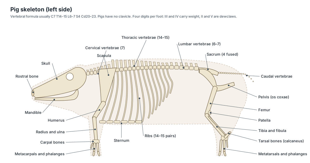
The usual vertebral formula of the pig is 7 cervical, 14–15 thoracic, 6 lumbar, 4 sacral and 20–23 caudal vertebrae (C7 T14–15 L6 S4 Cd20–23). Other references give 6–7 lumbar vertebrae. As in nearly all mammals there are seven neck vertebrae. (IMAIOS vet-Anatomy: vertebral column (vertebral formulae of domestic mammals).)
The count really does vary. Wild boar have 19 thoracic plus lumbar vertebrae, while European commercial breeds have 21–23, probably because breeders selected for longer bodies. A gene variant in NR6A1 accounts for part of this difference. (Mikawa et al. (2007), Fine mapping of a swine QTL for number of vertebrae, Genome Research.)
Each thoracic vertebra carries a pair of ribs, so pigs usually have 14 or 15 pairs. The snout is stiffened by a special prenasal (rostral) bone, which lets pigs dig with it. Adult pigs have 44 teeth (dental formula 3.1.4.3 in each half of each jaw), and the canines of boars grow continuously into tusks. (Pig, Wikipedia.)
Pigs have no clavicle: the forelimb is held to the trunk by muscles. The 3D model shows C7 T14 L6 S4 and about 20 caudal vertebrae.
Pig hoof and foot anatomy
Each pig foot has four hoofed toes. The two large central toes, digits III and IV, carry most of the weight and form the cloven hoof; the two outer toes, digits II and V, are the dewclaws, which sit higher and help on soft ground. (Pig, Wikipedia.)
Underneath, the toe bones follow the same pattern: four metacarpals or metatarsals, each with its phalanges. The joint people call a pig's 'knee' on the front leg is really the carpus (wrist), and the hock on the hind leg is the tarsus (ankle).
Male and female pig reproductive anatomy
The sow has a bicornuate uterus with two long uterine horns, where the litter develops, and pregnancy only continues if embryos are present in both horns. Her cervix has a series of interlocking pads that grip the boar's corkscrew-shaped penis during mating. Sows typically have 12–14 teats, and gestation lasts about 114 days (roughly 112–120). (Pig, Wikipedia.)
The pig placenta is diffuse and epitheliochorial: the fetal membranes contact a large area of the uterine lining, but six tissue layers separate maternal and fetal blood. Term is around day 114 of pregnancy. (Bazer et al. (2012), Uterine biology in pigs and sheep, Journal of Animal Science and Biotechnology.)
In the boar, the testes lie in a scrotum high in the perineum, just below the anus. The sperm ducts (ductus deferentia) run to the urethra, where the vesicular glands, prostate and very large bulbourethral glands add fluid. The penis runs forward under the belly skin to the preputial opening just behind the navel.
Male vs female pig anatomy
| Male (boar) | Female (sow or gilt) | |
|---|---|---|
| Gonads | Testes in the scrotum below the anus | Ovaries just behind the kidneys |
| Ducts | Epididymis, ductus deferens, urethra | Oviducts, two long uterine horns, uterine body, cervix, vagina |
| Accessory glands | Vesicular glands, prostate, very large bulbourethral glands | None comparable |
| External opening | Preputial opening just behind the navel | Vulva (genital papilla in the fetus) just below the anus |
| Teats | Present but not functional | Typically 12–14, for nursing the litter |
Use the Female/Male switch on the 3D model to compare the two reproductive tracts.
Fetal pig anatomy compared with the adult
A fetal pig has the same organ systems as an adult, so it is a good guide to pig anatomy, but several features exist only before birth or change size after it.
- Umbilical cord with two umbilical arteries, one umbilical vein and the allantoic duct, and an elongated allantoic bladder.
- Fetal shunts: the foramen ovale, ductus arteriosus and ductus venosus, which close soon after birth.
- Lungs are fluid-filled and solid; the thymus and liver are relatively huge.
- The skeleton is partly cartilage, and the testes may still be on their way through the inguinal canal.
- The head is large and the legs short compared with an adult; the gut contains meconium instead of food.
Pig meat anatomy: which muscles are the cuts?
Butchers first divide a pig into four primal cuts: the shoulder, the loin, the belly and the leg. Pork butt (Boston butt) comes from the upper shoulder, and only the hind leg is properly called a ham. Spare ribs are cut from the ribs with the meat between them. (Cuts of pork, Wikipedia.)
Pork tenderloin is the psoas major muscle, which lies under the lumbar vertebrae. Because it does little heavy work, it is the most tender cut. (Pork tenderloin, Wikipedia.)
The loin chops and loin roast are mainly the longissimus muscle along the back. The ham is the large muscle mass of the thigh (biceps femoris, semimembranosus, semitendinosus and quadriceps). The shoulder includes the triceps and other muscles around the shoulder blade, and bacon comes from the belly: the layered abdominal wall with its fat.
Pig muscles
- Masseter: The thick chewing muscle on the cheek. Closes the jaw.
- Triceps brachii: The large muscle mass behind the upper forelimb. Extends (straightens) the elbow so the leg can carry weight.
- Longissimus dorsi: The long back muscle beside the spine: the pork loin. Extends and stiffens the back.
- Psoas major: A long, soft muscle under the lumbar vertebrae: the pork tenderloin. Flexes the hip.
- Ham muscles (biceps femoris): The large muscles of the rump and thigh, led by the biceps femoris. Extend the hip and drive the pig forward.
- Abdominal wall muscles: Thin layered muscles of the belly: external and internal obliques, transversus and rectus abdominis. Hold in and compress the abdominal organs.
Is a guinea pig a pig?
No. The guinea pig, Cavia porcellus, is a rodent (order Rodentia, family Caviidae) from South America. Despite its name it is not native to Guinea and is not closely related to pigs. Its anatomy is a rodent's: ever-growing incisors, clawed feet instead of hooves, and a large caecum for digesting plants. (Guinea pig, Wikipedia.)
Pig anatomy quiz: test yourself
Use the model or diagrams to answer these. Answers are in the FAQ and on the dissection page.
- How many toes does a pig have on each foot, and which ones carry the weight?
- Which bone supports the pig's snout disk?
- Where in the abdomen is the spiral colon, and what is its job?
- About how long is the large intestine of an adult pig?
- Name the four main lobes of the pig liver.
- What is unusual about the bronchus to the right cranial lung lobe?
- How many thoracic vertebrae does a pig usually have?
- Name two differences between the pig heart and the human heart.
Full lab questions with an answer key are in the fetal pig dissection guide.
Frequently asked questions
Do pigs have similar anatomy to humans?
In many ways, yes. Pigs and humans are both omnivorous mammals of similar size, with a single-chambered stomach, a heart and coronary arteries of similar size and layout, multi-lobed kidneys and similar skin. That is why pigs are a major model in medical research (Swindle et al., 2012). The main differences are the pig's four-legged posture, spiral colon, large caecum, snout and rostral bone, cloven hooves, more vertebrae, and a different placenta.
How long is a pig's large intestine?
An adult pig has about 4–6 metres of large intestine, compared with about 1.5 m in an adult human. Its small intestine is about 15–22 m long. Relative to body weight, though, the total length is similar in the two species (Gonzalez et al., 2015).
How many lobes does a pig liver have?
School lab manuals usually count five. Veterinary and surgical anatomy describes four main lobes (left lateral, left medial, right medial and right lateral) plus a small quadrate lobe around the gallbladder and a caudate lobe, so you may see five or six named depending on the source.
What is a spiral colon?
It is the pig's ascending colon, coiled into a cone. Food passes inward along centripetal coils to the tip of the cone, then turns and comes back out along centrifugal coils. It sits on the left side of the abdomen behind the stomach.
Is a pig heart the same as a human heart?
It is very similar in size and has the same four chambers, valves and great vessels, which is why pig hearts and valves are used in research and medicine. There are differences: a more rounded 'Valentine' shape, a tubular right auricle, a large left azygos vein draining into the coronary sinus, and only two pulmonary vein openings into the left atrium (Crick et al., 1998).
How many ribs does a pig have?
Usually 14 or 15 pairs, one pair per thoracic vertebra. The number varies between individuals and breeds; long modern breeds tend to have more vertebrae than wild boar.
How many toes does a pig have?
Four on each foot. The two large central toes (digits III and IV) form the cloven hoof and carry the weight; the two smaller dewclaws (digits II and V) sit higher at the back.
Is a guinea pig a pig?
No. The guinea pig (Cavia porcellus) is a South American rodent in the family Caviidae. It is not closely related to pigs and its anatomy is that of a rodent, with ever-growing incisors and no hooves.
References
- Swindle et al. (2012), Swine as models in biomedical research and toxicology testing, Veterinary Pathology
- Gonzalez, Moeser & Blikslager (2015), Porcine models of digestive disease, Translational Research
- Crick et al. (1998), Anatomy of the pig heart: comparisons with normal human cardiac structure, Journal of Anatomy
- Griffith et al. (2022), Genetically modified porcine-to-human cardiac xenotransplantation, NEJM
- Nykonenko, Vávra & Zonča (2017), Anatomic peculiarities of pig and human liver, Experimental and Clinical Transplantation
- Court et al. (2003), Segmental nature of the porcine liver, British Journal of Surgery
- IMAIOS vet-Anatomy: vertebral column (vertebral formulae of domestic mammals)
- Mikawa et al. (2007), Fine mapping of a swine QTL for number of vertebrae, Genome Research
- Bazer et al. (2012), Uterine biology in pigs and sheep, Journal of Animal Science and Biotechnology
- Mammalian kidney, Wikipedia
- Tracheal bronchus, Radiopaedia
- Pig, Wikipedia
- Cuts of pork, Wikipedia
- Guinea pig, Wikipedia
- Whitman College, Virtual Pig Dissection
More interactive anatomy models
- Fetal pig dissectionGuided fetal pig dissection with umbilical vessels and organs labeled.
- Human anatomy1,000+ bones, muscles, organs, vessels and nerves from real anatomical data.
- Octopus anatomyThree hearts, a ring-shaped brain, eight arms and a beak, in 3D.
- Squid anatomyPen, eight arms, two tentacles, giant axons and three hearts of the longfin squid.
- Squid dissectionStep-by-step guided squid dissection with every structure labeled.
- Fish anatomyExternal and internal anatomy of a bony fish: fins, gills, swim bladder, heart and brain.