

Anatomy of a butterfly at a glance
A butterfly is an insect in the order Lepidoptera, a name that means “scale wings”. Its body has three regions. The head carries two large compound eyes, two clubbed antennae, two labial palps and a coiled feeding tube, the proboscis. The thorax carries three pairs of legs and two pairs of wings and is packed with flight muscle. The abdomen holds most of the gut, the heart and the reproductive organs. The four wings are thin sheets of cuticle supported by hollow veins and covered on both sides with overlapping scales.
This page uses the monarch butterfly, Danaus plexippus, as the reference species. It is a large brush-footed butterfly (family Nymphalidae) with a wingspan of about 8.9–10.2 cm (3.5–4 in), orange wings with black veins and borders, and white spots. Its anatomy is typical of butterflies, so most of what you learn applies to painted ladies, swallowtails and blue morphos as well. Where they differ, this page says so.
Directions: anterior is toward the head, posterior toward the tip of the abdomen, dorsal is the upper side and ventral the underside. Wing surfaces are described as the upperside and underside. On a wing, proximal means near the body (the base) and distal means toward the edge.
Butterfly anatomy diagram: labeled external parts
The external anatomy of a butterfly includes the head (compound eyes, antennae with clubs, labial palps, proboscis), the thorax (prothorax, mesothorax, metathorax, six legs, two forewings and two hindwings) and the segmented abdomen with its spiracles and genitalia. The labeled diagram below shows a male monarch from above.
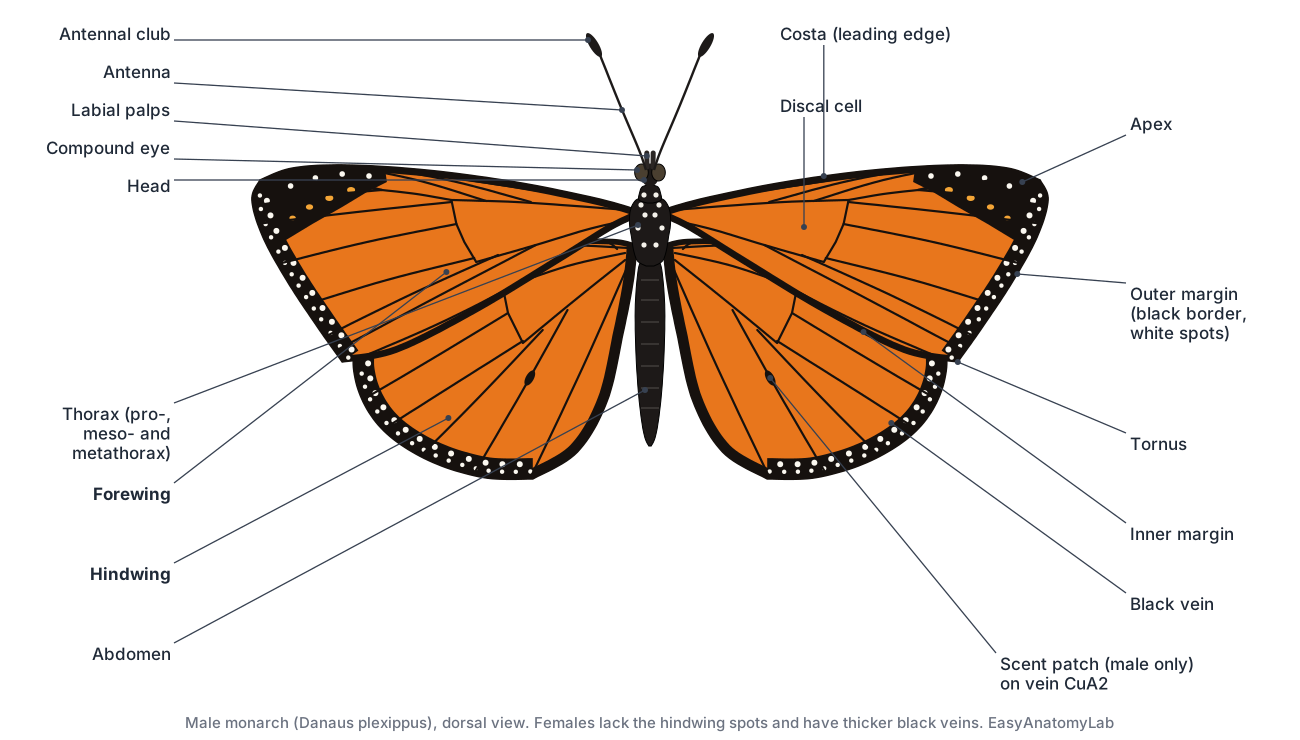
Three features identify a monarch at a glance: orange wings with black veins, a black outer border with two rows of small white spots, and a black forewing tip with a band of orange and white spots. The body is black with white spots on the head and thorax. The viceroy, which looks similar, has an extra black line across the hindwing that the monarch lacks.
External vs internal anatomy of a butterfly
External anatomy is everything you can see on the outside of the exoskeleton; internal anatomy is the organs inside the body cavity. Use this table to sort the parts into the two groups and to match each to its body region.
| Region | External parts | Internal parts |
|---|---|---|
| Head | Compound eyes, antennae (scape, pedicel, flagellum, club), labial palps, proboscis | Brain and optic lobes, subesophageal ganglion, sucking pump, start of the oesophagus, Johnston's organ (inside the pedicel) |
| Thorax | Prothorax, mesothorax, metathorax, tegulae, 3 pairs of legs, forewings, hindwings, thoracic spiracles | Flight muscles, oesophagus, salivary glands, aorta, thoracic ganglia, tracheae and air sacs |
| Abdomen | Segments, spiracles, claspers (male) or ovipositor (female) | Crop, midgut, Malpighian tubules, hindgut, rectum, heart, ventral nerve cord, ovaries or testes, bursa copulatrix |
Butterfly anatomy labeled: every part and its function
This is the text version of the 3D model: every labeled structure of the monarch, grouped by body system, with what it does. Structures marked “males only” or “females only” appear when you switch sex in the viewer.
Body & legs
- Head (Head capsule)
- The small, rounded first body region. It carries the eyes, antennae, palps and proboscis. Holds the main sense organs and the feeding tube, and encloses the brain and the sucking pump that draws nectar up the proboscis.
- Prothorax (First thoracic segment, collar)
- The short, collar-like first segment of the thorax, just behind the head. Carries the first pair of legs (the reduced forelegs in the monarch) and lets the head move.
- Mesothorax (Second thoracic segment)
- The largest thoracic segment. It carries the forewings and the middle legs. Houses most of the flight muscles that power the forewings, and anchors the middle legs.
- Metathorax (Third thoracic segment)
- The third thoracic segment. It carries the hindwings and the hind legs. Anchors the hindwings and hind legs.
- Tegula (Tegulae (plural), shoulder covers)
- A small scaly flap that covers the base of each forewing. Shields the delicate wing hinge and its small hinge plates (axillary sclerites).
- Abdomen
- The long, soft, segmented rear body region. It holds most of the gut, the heart and the reproductive organs. Digests food, stores reserves, pumps blood forward, and produces eggs or sperm. Breathing pores (spiracles) line its sides.
- Forelegs (Front legs, reduced in brush-footed butterflies)
- The first pair of legs. In the monarch they are tiny, hairy and held folded against the body. Not used for walking. In females of many brush-footed butterflies they carry taste sensors used to test plants.
- Middle legs (Mesothoracic legs)
- The second pair of legs. In the monarch they are one of the two pairs used for walking and perching. Walking, gripping flowers and leaves, and tasting what the butterfly stands on.
- Hind legs (Metathoracic legs)
- The third pair of legs, used with the middle legs for walking and perching. Support the body when perched and help grip.
- Tarsi and tarsal taste receptors (Feet, tarsal chemoreceptors)
- The jointed 'feet' at the end of each leg. They carry taste receptors. Grip surfaces and taste them. Sugar touching the tarsi triggers the proboscis to uncoil and feed.
- Tarsal claws (Pretarsal claws)
- A pair of small hooks at the tip of each walking leg. Hook onto petals, bark and leaves so the butterfly can hang on in wind.
Wings & scales
- Forewing (Front wing)
- The larger, more triangular front wing, attached to the mesothorax. Produces most of the lift and thrust and carries most of the warning colour pattern.
- Hindwing (Back wing)
- The rounder rear wing, attached to the metathorax. Adds lift and makes turning easier; it also carries colour patterns used for display.
- Costa and costal vein (Costal margin, leading edge)
- The thickened front (leading) edge of the forewing. Stiffens the leading edge, which takes the largest forces in flight.
- Apex (Wing tip)
- The outer tip of the forewing, where the costa meets the outer margin. In the monarch it carries a black patch with orange and white spots.
- Outer margin (Termen, marginal border)
- The outer edge of the wing, between the apex and the rear corner. In the monarch it carries a black band with two rows of small white spots.
- Inner margin (Dorsum, anal margin)
- The rear edge of the forewing, which overlaps the hindwing, and the matching edge of the hindwing beside the abdomen. Where the two wings overlap so that they beat as one.
- Discal cell (Cell, discoidal cell)
- The large closed cell in the middle of each wing, enclosed by veins. A key landmark: the veins that reach the wing edge branch from its border.
- Subcosta (Sc)
- The second main vein, running just behind the costa. Helps stiffen the leading part of the wing.
- Radial veins (R) (R1–R5 on the forewing, Rs on the hindwing)
- A set of veins that branch from the front edge of the discal cell toward the costa and apex. Support the front and tip of the wing.
- Medial veins (M) (M1, M2, M3)
- Three veins that run from the outer end of the discal cell to the outer margin. Support the middle of the wing.
- Cubital veins (CuA1, CuA2) (Cubitus, Cu1 and Cu2 in older books)
- Two veins that branch from the rear edge of the discal cell to the outer margin. Support the rear part of the wing.
- Anal veins (A) (1A+2A, 3A)
- Veins near the inner margin, behind the discal cell. Support the rear edge of the wing.
- Humeral lobe (wing coupling) (Amplexiform wing coupling)
- The enlarged front base of the hindwing, which lies under the forewing so the two wings move together. Couples forewing and hindwing by broad overlap and friction, not by hooks (amplexiform coupling).
- Wing scales (Lepidoptera = 'scale wings')
- Tiny flat plates that overlap like roof tiles and give the wing its colour. Make colour patterns, shed water, insulate, and help control wing temperature. They rub off as 'dust' when a wing is handled.
- Androconial scent patch (Male hindwing spot, scent pouch) · males only
- A small black spot on a vein near the middle of each hindwing, present only in males. Made of specialised scent scales (androconia). In many butterflies such scales release courtship scent; in monarchs it does not seem to be needed for mating.
Head & senses
- Compound eye
- Each large eye is made of thousands of tiny units called ommatidia. Gives a wide field of view with good colour and motion vision, including ultraviolet and polarised light.
- Antennae (Feelers)
- Two long, thin feelers on top of the head, used to smell and to sense balance and air flow. Smell nectar and pheromones, sense touch, and help keep time for the sun compass used during migration.
- Antennal club (Club)
- The thickened tip of each antenna, typical of butterflies. Carries dense clusters of smell receptors (chemoreceptors).
- Johnston's organ
- A ring of stretch-sensing cells inside the second antennal segment. Senses how the antenna's shaft moves, which reports air flow, gravity and body orientation in flight.
- Labial palps (Palpi)
- Two upturned, densely scaled lobes in front of the face. Shield and clean the proboscis and carry sensory receptors.
Digestive
- Proboscis (Tongue, two joined galeae)
- The long feeding tube, coiled like a watch spring under the head when not in use. Sucks up nectar, water and other liquids.
- Food canal
- The channel between the two galeae through which liquid food travels. Carries nectar from the flower to the mouth.
- Sucking pump (Cibarial and pharyngeal pump)
- A muscular chamber in the head that sucks liquid up the proboscis. Strong dilator muscles expand it to create suction, then it squeezes the food back into the oesophagus.
- Salivary glands (Labial glands)
- A pair of long, thin glands in the thorax that make saliva. Saliva wets and dissolves dried sugars so they can be sucked up; Heliconius butterflies use it to soak nutrients out of pollen.
- Oesophagus (Esophagus)
- A thin tube that carries food from the sucking pump through the thorax. Carries liquid food to the crop and midgut.
- Crop (Food reservoir)
- A thin-walled storage sac that branches off the oesophagus. Stores nectar until it can pass into the midgut.
- Midgut (Ventriculus, stomach)
- The main tube of the gut where food is digested and absorbed. Absorbs sugars and other nutrients into the hemolymph.
- Malpighian tubules
- Thin tubes that act as the butterfly's kidneys. Filter waste from the hemolymph and pass it into the hindgut as uric acid.
- Hindgut (Ileum and colon)
- The narrow rear part of the gut. Reabsorbs water and salts and carries waste to the rectum.
- Rectum (Rectal sac)
- A wider sac at the end of the gut. Stores waste and reclaims water before it is expelled.
Circulatory
- Heart (Dorsal vessel (abdominal part))
- A long, thin tube along the top of the abdomen that pumps hemolymph forward. Draws hemolymph in through valved openings (ostia) and pumps it toward the head.
- Aorta (Dorsal vessel (thoracic part))
- The front, narrower part of the dorsal vessel that runs through the thorax to the head. Delivers hemolymph to the head, from where it flows back through the body cavity.
Respiratory
- Spiracles (Breathing pores)
- Small breathing holes in a row along each side of the thorax and abdomen. Let air into the tracheal tubes. Valves can close them to save water.
- Tracheae (Air tubes, tracheal trunks)
- A network of air tubes that carries oxygen directly to every tissue. Delivers oxygen and removes carbon dioxide without using the blood.
- Air sacs
- Thin, balloon-like enlargements of the tracheae. Store air, help ventilate the tracheae during flight, and lighten the body.
Nervous
- Brain (Supraesophageal ganglion)
- The main nerve centre, above the oesophagus in the head. Processes vision, smell and learning, and drives behaviour such as migration.
- Optic lobes
- Large side lobes of the brain that process input from the compound eyes. Process colour, motion and polarised light from the eyes.
- Subesophageal ganglion
- The nerve centre under the oesophagus that controls the mouthparts. Controls the proboscis and the sucking pump.
- Thoracic ganglia
- Nerve centres on the floor of the thorax that control the legs and wings. Generate the rhythms of walking and flight.
- Ventral nerve cord
- A paired nerve cord with small ganglia that runs along the underside of the body. Carries signals between the brain and the body segments.
Flight muscles
- Dorsal longitudinal flight muscles
- Large muscles running front to back along the top of the thorax. Shorten the thorax so the roof bulges up and the wings flip down: the downstroke.
- Dorsoventral flight muscles
- Upright muscles running from the roof to the floor of the thorax. Pull the roof of the thorax down, which levers the wings up: the upstroke.
Reproductive
- Ovaries · females only
- Paired egg-making organs, each made of four long coiled tubes (ovarioles). Produce eggs, which mature in a chain along each ovariole.
- Oviducts · females only
- Tubes that carry eggs from the ovaries to the egg-laying opening. Carry eggs past the spermatheca, where they are fertilised, to the ovipore.
- Bursa copulatrix · females only
- A sac that receives the male's sperm package (spermatophore) during mating. Holds and digests the spermatophore; the sperm move on to the spermatheca.
- Spermatheca · females only
- A small sac that stores sperm after mating. Releases sperm to fertilise each egg as it is laid.
- Ovipositor (Papillae anales, egg-laying tip) · females only
- The soft, padded tip of the female abdomen that places each egg. Glues single eggs to the underside of milkweed leaves.
- Testes · males only
- The sperm-making organs, close together on top of the gut. Produce sperm, which pass down the sperm ducts.
- Sperm ducts and accessory glands (Vasa deferentia, accessory glands) · males only
- Ducts that carry sperm, and long glands that make the spermatophore. Build the spermatophore, a package of sperm and nutrients passed to the female.
- Ejaculatory duct and aedeagus · males only
- A long duct ending in the aedeagus, the male's tubular copulatory organ. Delivers the spermatophore into the female's bursa copulatrix.
- Claspers (Valvae) · males only
- A pair of hard plates at the tip of the male abdomen. Grasp the female's abdomen during mating.
Butterfly wing anatomy: veins, scales and colour
A butterfly wing is a double layer of cuticle stretched over a framework of hollow veins and covered on both surfaces with tiny overlapping scales. Each vein carries hemolymph, a trachea and a nerve, so parts of the wing stay alive. Wings are named by their edges and veins: the front edge is the costa, the tip is the apex, the outer edge is the outer margin (termen), the rear corner of the forewing is the tornus, and the edge nearest the body is the inner margin.
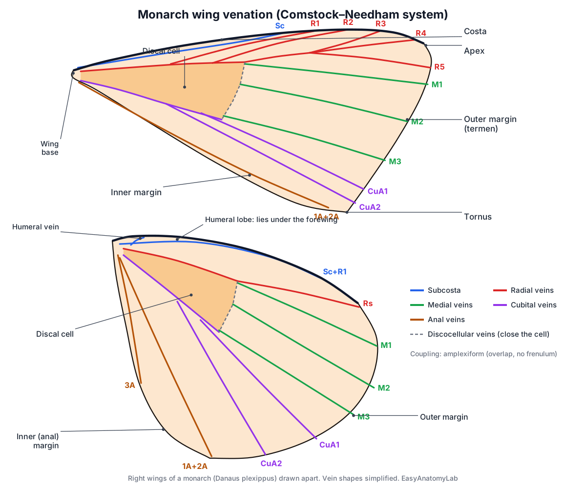
Entomologists name veins with the Comstock–Needham system. From front to back they are the costa (C), subcosta (Sc), radius (R), media (M), cubitus (Cu) and anal veins (A). In the middle of each wing, veins enclose the large discal cell; most of the veins that reach the margin branch from its edge. On the monarch forewing, R3, R4 and R5 share a stalk before they split near the apex, and the two cubital branches (CuA1 and CuA2) leave the rear edge of the cell. Field guides and older books sometimes number veins instead (veins 1–12), so check which system a key uses.
- Costa and costal vein: The thickened front (leading) edge of the forewing. Stiffens the leading edge, which takes the largest forces in flight.
- Subcosta (Sc): The second main vein, running just behind the costa. Helps stiffen the leading part of the wing.
- Radial veins (R): A set of veins that branch from the front edge of the discal cell toward the costa and apex. Support the front and tip of the wing.
- Medial veins (M): Three veins that run from the outer end of the discal cell to the outer margin. Support the middle of the wing.
- Cubital veins (CuA1, CuA2): Two veins that branch from the rear edge of the discal cell to the outer margin. Support the rear part of the wing.
- Anal veins (A): Veins near the inner margin, behind the discal cell. Support the rear edge of the wing.
- Discal cell: The large closed cell in the middle of each wing, enclosed by veins. A key landmark: the veins that reach the wing edge branch from its border.
Wing coupling. Butterflies have no frenulum. The forewing and hindwing are held together by broad overlap: the enlarged base of the hindwing (the humeral lobe) lies under the rear edge of the forewing, so the two beat as one. This is called amplexiform coupling. Most moths instead hook the wings together with a bristle (frenulum) that catches under a retinaculum on the forewing.
Scales. Each scale is a flattened, modified hair that grows from a single cell and is attached by a short stalk into a socket. Scales overlap like roof tiles in rows across the wing. They make the colour pattern, help shed water, insulate and help control wing temperature, and they rub off as “dust” on your fingers. Male monarchs also carry a patch of special scent scales (androconia) on a vein in the middle of each hindwing.
Pigment vs structural colour. Butterfly colours come from pigments, from structures, or from both. Pigments such as melanin absorb some light: they give the monarch its black veins and borders. Structural colour comes from microscopic ridges and layers in the scales that reflect certain wavelengths by interference. The blue morpho's brilliant blue is structural: its scales carry ridges of stacked cuticle layers separated by air. Structural colours often shimmer and change with viewing angle; pigment colours do not.
Butterfly head anatomy: eyes, antennae and proboscis
A butterfly's head carries two large compound eyes, two clubbed antennae, two hairy labial palps and the coiled proboscis. Inside are the brain and the muscular sucking pump that draws liquid food up the proboscis. Adult butterflies have no working jaws.
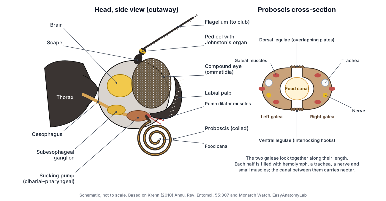
Butterfly eye anatomy. Each compound eye is made of thousands of units called ommatidia, each with its own lens. Together they give a wide view and excellent detection of motion and colour, including ultraviolet light. The monarch eye has three kinds of light-sensitive pigment (ultraviolet, blue and long-wavelength), and ultraviolet receptors at the top edge of the eye may help it read polarised skylight.
Antennae. Each antenna has a base segment (scape), a second segment (pedicel) and a long, many-segmented shaft (flagellum) ending in a club. The club is densely covered with smell receptors. Inside the pedicel, Johnston's organ senses how the shaft bends, reporting air flow and orientation. Monarch antennae also contain clock cells used for navigation.
Butterfly proboscis anatomy. The proboscis is made of two very long galeae (parts of the maxillae) zipped together by tiny plates and hooks called legulae. The space between them is the food canal. The proboscis uncoils when hemolymph pressure rises inside it and recoils by its own springiness. A muscular sucking pump in the head (formed by the cibarium and pharynx) draws nectar up the canal.
- Compound eye: Each large eye is made of thousands of tiny units called ommatidia. Gives a wide field of view with good colour and motion vision, including ultraviolet and polarised light.
- Antennae: Two long, thin feelers on top of the head, used to smell and to sense balance and air flow. Smell nectar and pheromones, sense touch, and help keep time for the sun compass used during migration.
- Antennal club: The thickened tip of each antenna, typical of butterflies. Carries dense clusters of smell receptors (chemoreceptors).
- Johnston's organ: A ring of stretch-sensing cells inside the second antennal segment. Senses how the antenna's shaft moves, which reports air flow, gravity and body orientation in flight.
- Labial palps: Two upturned, densely scaled lobes in front of the face. Shield and clean the proboscis and carry sensory receptors.
- Proboscis: The long feeding tube, coiled like a watch spring under the head when not in use. Sucks up nectar, water and other liquids.
- Food canal: The channel between the two galeae through which liquid food travels. Carries nectar from the flower to the mouth.
- Sucking pump: A muscular chamber in the head that sucks liquid up the proboscis. Strong dilator muscles expand it to create suction, then it squeezes the food back into the oesophagus.
Butterfly legs anatomy
Every butterfly has six legs, one pair on each thoracic segment. In brush-footed butterflies such as the monarch and painted lady, the front pair is greatly reduced, hairy and held folded against the body, so the butterfly appears to stand on four legs. Swallowtails and whites use all six legs for walking.
Each walking leg has a coxa, trochanter, femur, tibia and a five-segmented tarsus ending in a pair of claws. The tarsi are not just feet: they carry taste receptors. Monarch Watch notes that sugar touching the tarsi triggers the proboscis to uncoil, and that females test milkweed with chemoreceptors on their legs before laying eggs.
- Forelegs: The first pair of legs. In the monarch they are tiny, hairy and held folded against the body. Not used for walking. In females of many brush-footed butterflies they carry taste sensors used to test plants.
- Middle legs: The second pair of legs. In the monarch they are one of the two pairs used for walking and perching. Walking, gripping flowers and leaves, and tasting what the butterfly stands on.
- Hind legs: The third pair of legs, used with the middle legs for walking and perching. Support the body when perched and help grip.
- Tarsi and tarsal taste receptors: The jointed 'feet' at the end of each leg. They carry taste receptors. Grip surfaces and taste them. Sugar touching the tarsi triggers the proboscis to uncoil and feed.
- Tarsal claws: A pair of small hooks at the tip of each walking leg. Hook onto petals, bark and leaves so the butterfly can hang on in wind.
Butterfly abdomen anatomy
The butterfly abdomen is a soft, segmented tube behind the thorax. It has no legs or wings. Along its sides is a row of spiracles, the breathing holes; inside are the crop, midgut, Malpighian tubules, hindgut, heart, ventral nerve cord and the reproductive organs. The tip carries the genitalia: a pair of claspers in males and the soft egg-laying tip (ovipositor) in females.
Sources count the segments differently: Monarch Watch describes eleven abdominal segments with the last two or three fused, while other references count ten. Either way, only seven or eight are easy to see from outside. A female's abdomen is usually plumper, especially when full of eggs.
Butterfly internal anatomy
Inside a butterfly, a straight gut runs from the head to the tip of the abdomen, the heart runs along the back, the nerve cord runs along the belly, and air tubes branch everywhere. The thorax is filled with flight muscles, and the abdomen holds the reproductive organs around the gut.
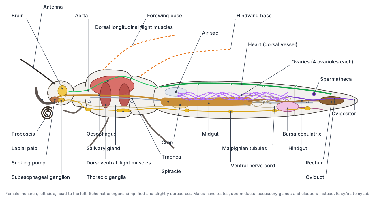
Digestive system. Food is pulled up the proboscis by the sucking pump, passes down the oesophagus through the neck and thorax, and can be stored in the crop, a thin-walled sac. The midgut digests and absorbs sugars. Six Malpighian tubules join the gut where the midgut meets the hindgut; they remove waste from the hemolymph. The hindgut and rectum reclaim water before waste leaves through the anus. Salivary (labial) glands lie in the thorax; in caterpillars the same glands make silk.
- Oesophagus: A thin tube that carries food from the sucking pump through the thorax. Carries liquid food to the crop and midgut.
- Crop: A thin-walled storage sac that branches off the oesophagus. Stores nectar until it can pass into the midgut.
- Midgut: The main tube of the gut where food is digested and absorbed. Absorbs sugars and other nutrients into the hemolymph.
- Malpighian tubules: Thin tubes that act as the butterfly's kidneys. Filter waste from the hemolymph and pass it into the hindgut as uric acid.
- Hindgut: The narrow rear part of the gut. Reabsorbs water and salts and carries waste to the rectum.
- Rectum: A wider sac at the end of the gut. Stores waste and reclaims water before it is expelled.
- Salivary glands: A pair of long, thin glands in the thorax that make saliva. Saliva wets and dissolves dried sugars so they can be sucked up; Heliconius butterflies use it to soak nutrients out of pollen.
Nervous system. The brain sits above the oesophagus, with large optic lobes serving the eyes. The subesophageal ganglion below the oesophagus controls the mouthparts. A ventral nerve cord with thoracic and abdominal ganglia runs along the floor of the body.
Respiratory system. Air enters through the spiracles and travels through branching tracheae and balloon-like air sacs straight to the tissues. Butterflies do not have lungs.
Flight muscles. The wings have no muscles of their own. Large dorsal longitudinal muscles shorten the thorax and flip the wings down; dorsoventral muscles pull the roof of the thorax down and lever the wings up. Because they move the wings by deforming the thorax, they are called indirect flight muscles.
Butterfly heart anatomy and circulation
A butterfly's heart is a long, thin tube along the top of the abdomen. It draws hemolymph in through valved slits (ostia) and pumps it forward into the aorta, which runs through the thorax and empties near the brain. From there the hemolymph flows back through the body cavity, bathing the organs. This is an open circulation: there are no capillaries or veins.
Insect blood does not carry oxygen. The tracheal system does that, so hemolymph has no hemoglobin and is clear to yellowish green. Small pulsatile organs near the wing bases help push hemolymph through the wing veins.
- Heart: A long, thin tube along the top of the abdomen that pumps hemolymph forward. Draws hemolymph in through valved openings (ostia) and pumps it toward the head.
- Aorta: The front, narrower part of the dorsal vessel that runs through the thorax to the head. Delivers hemolymph to the head, from where it flows back through the body cavity.
Male vs female monarch butterfly
You can tell a male monarch from a female by its hindwings. Males have a small black spot on a vein in the middle of each hindwing and thinner black veins. Females have no spots and thicker, darker veins. Males also have a pair of claspers at the tip of the abdomen. Use the sex toggle in the viewer to compare them.
| Feature | Male | Female |
|---|---|---|
| Hindwing spot | Black scent patch (androconia) on vein CuA2 | None |
| Wing veins | Thinner black veins | Thicker, darker veins |
| Abdomen tip | Claspers (valvae) around the aedeagus | Soft ovipositor; separate mating opening |
| Internal organs | Testes, sperm ducts, accessory glands, ejaculatory duct | Ovaries (4 ovarioles each), oviducts, bursa copulatrix, spermatheca |
- Androconial scent patch: A small black spot on a vein near the middle of each hindwing, present only in males. Made of specialised scent scales (androconia). In many butterflies such scales release courtship scent; in monarchs it does not seem to be needed for mating.
- Claspers: A pair of hard plates at the tip of the male abdomen. Grasp the female's abdomen during mating.
- Testes: The sperm-making organs, close together on top of the gut. Produce sperm, which pass down the sperm ducts.
- Ovaries: Paired egg-making organs, each made of four long coiled tubes (ovarioles). Produce eggs, which mature in a chain along each ovariole.
- Bursa copulatrix: A sac that receives the male's sperm package (spermatophore) during mating. Holds and digests the spermatophore; the sperm move on to the spermatheca.
- Spermatheca: A small sac that stores sperm after mating. Releases sperm to fertilise each egg as it is laid.
Compare the anatomy of the butterfly and bird wing
Butterfly wings and bird wings are analogous structures: they do the same job (flight) but evolved separately and are built in completely different ways. A bird wing is a modified forelimb, with bones, muscles and feathers. A butterfly wing is an outgrowth of the exoskeleton, with no bones and no muscles inside it, and the insect still has all six legs. Analogies like this are the result of convergent evolution.
| Feature | Butterfly wing | Bird wing |
|---|---|---|
| Origin | Outgrowth of the thorax wall (exoskeleton); not a leg | Modified forelimb (arm) |
| Number | Four (two pairs) | Two (one pair) |
| Support | Hollow veins of cuticle carrying hemolymph, tracheae and nerves | Bones: humerus, radius, ulna, fused hand bones and digits |
| Surface | Thin membrane covered with chitin scales | Feathers made of keratin |
| Flight muscles | Inside the thorax; move the wings indirectly by deforming the thorax | Breast muscles attached to the keeled sternum pull directly on the wing bones |
| Living tissue | Mostly non-living cuticle; veins and scent organs stay alive | Living bone, muscle, skin and blood vessels throughout |
| Repair | Cannot grow or repair once the adult emerges | Feathers are moulted and replaced |
| Homologous or analogous? | Analogous to bird wings | Analogous to butterfly wings; homologous to a human arm |
Compare this with a bat wing, which is also a modified forelimb but uses skin stretched between long finger bones. Bird and bat wings are analogous as wings (they evolved flight separately) but homologous as forelimbs, because both are built from the same arm bones inherited from a common ancestor. A butterfly wing is neither: it evolved flight on its own and contains no bones at all.
Butterfly vs moth anatomy
Butterflies and moths belong to the same order, Lepidoptera, and share scaled wings, a coiled proboscis and complete metamorphosis. Most butterflies have clubbed antennae, fly by day and rest with their wings held upright over the back. Most moths have thread-like, comb-like or feathery antennae, fly at night and rest with their wings flat or folded. No single trait works for every species, so use several together.
| Feature | Butterfly (e.g. monarch) | Moth (most species) |
|---|---|---|
| Antennae | Thin with a club (skippers: club with a hook) | Thread-like, spindle-shaped or feathery |
| Wing coupling | Amplexiform overlap; no frenulum | Usually a frenulum and retinaculum |
| Resting posture | Wings closed upright over the back | Wings flat, tent-like or wrapped around the body |
| Activity | Mostly day-flying | Mostly night-flying |
| Pupa | Bare chrysalis hanging from a silk pad | Often inside a silk cocoon or in the soil |
| Body | Usually slender | Often stout and hairy |
Butterfly life cycle and caterpillar anatomy
Butterflies go through complete metamorphosis, with four stages: egg, larva (caterpillar), pupa (chrysalis) and adult. A female monarch lays her eggs one at a time on milkweed; the USDA Forest Service gives 100–300 per female, and some studies report several hundred more. The eggs hatch in about four days, the caterpillar grows through five instars (stages between moults), and the adult emerges from the chrysalis after about one to two weeks, sooner in hot weather.
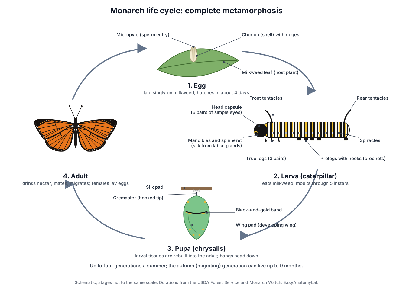
Caterpillar anatomy. A monarch caterpillar has a hard head capsule with very short antennae, chewing mandibles and six pairs of simple eyes (ocelli or stemmata). Under the head is the spinneret, which releases silk made by the labial glands. The three thoracic segments each carry a pair of jointed true legs; the abdomen carries usually five pairs of fleshy prolegs with tiny hooks (crochets) that grip silk and leaves. A row of spiracles runs along each side. The monarch caterpillar is banded white, yellow and black and carries a pair of fleshy tentacles at the front and another at the rear. They are sense organs, not antennae.
Pupa (chrysalis). Before pupating, the caterpillar spins a silk pad and hangs head down. The chrysalis hooks into the pad with the cremaster, a spiny tip at the end of the abdomen. A monarch chrysalis is pale green with gold dots and a black-and-gold band. Inside, larval tissues are broken down and rebuilt into the adult; the wing pads, legs, eyes and abdomen can be seen through the pupal skin. Newly emerged adults pump hemolymph into their crumpled wings to expand them.
Monarch butterfly anatomy: migration and milkweed toxins
The monarch is famous for two things its anatomy makes possible: a long migration and a chemical defence. Eastern North American monarchs fly roughly 2,000–3,000 miles (about 3,200–4,800 km) to overwinter in mountain forests of central Mexico; western monarchs overwinter mainly along the California coast. Summer adults live about two to five weeks, but the autumn generation delays breeding and can live up to nine months. Up to four generations are produced each summer.
For navigation, monarchs use a time-compensated sun compass. The compound eyes read the sun and polarised light, and the antennae contain the clocks that correct for the time of day. Large forewings and an efficient gliding flight let them ride rising air for long distances.
For defence, monarch caterpillars store cardenolides (cardiac glycosides) from milkweed, which make both the caterpillar and the adult distasteful to many predators. Changes in the monarch's sodium pump make it resistant to these toxins. The orange-and-black wing pattern advertises the warning.
Painted lady, swallowtail and blue morpho anatomy
All butterflies share the same basic body plan, but families differ in legs, wing shape and colour. The table compares the monarch with three butterflies that students often ask about.
| Species | Family | Key anatomical differences from the monarch |
|---|---|---|
| Monarch (Danaus plexippus) | Nymphalidae (Danainae) | Reference species: reduced forelegs, black veins, male hindwing scent patch, toxic from milkweed |
| Painted lady (Vanessa cardui) | Nymphalidae | Also has reduced forelegs. Smaller, mottled orange-brown with black and white markings and eyespots under the hindwing. Caterpillars feed mainly on thistles and other Asteraceae; adults migrate in multigenerational steps between Africa and Europe. |
| Swallowtails (Papilionidae) | Papilionidae | All six legs work for walking. Many have long hindwing tails. The forewing keeps an extra short anal vein and the hindwing has only one anal vein. Caterpillars have an osmeterium, a forked scent organ pushed out from behind the head when threatened. The chrysalis is held upright by a silk girdle. |
| Blue morpho (Morpho) | Nymphalidae | Reduced forelegs like the monarch. The bright blue upperside is structural colour from ridged, layered scales; the underside is brown with eyespots for camouflage. |
Not this butterfly: butterflyfish are reef fish, “sea butterflies” are swimming sea snails, and the butterfly needle, knife, valve and yoga pose are named only for their shape.
Butterfly wings are living tissue, not dead film
A butterfly wing looks like a thin sheet of coloured paper, but parts of it stay alive for the whole adult life. Tsai and colleagues showed that the wing veins and the male scent organs (androconia) are supplied by the circulatory, nervous and tracheal systems. Because a wing is so thin, it has very little capacity to store heat and can overheat within seconds in strong sunlight. The scales and the uneven thickness of the wing cuticle cool the living parts, such as the veins and scent organs, more than the rest of the wing. The butterflies also sensed light and infrared radiation through their wings and moved to avoid overheating. (Tsai et al. (2020): adaptations that prevent overheating of the living wings of butterflies, Nature Communications 11.)
This changes how you should read a wing diagram. The black lines on a monarch wing are not only colour: each vein is a hollow tube carrying hemolymph (insect blood), a trachea (air tube) and a nerve. The membrane between the veins is mostly non-living cuticle covered in scales. When you study the 3D model, select a vein group and then the discal cell. Ask yourself which parts would be harmed by heat, and why a scent patch that sits on a vein, as in the male monarch, has a supply line close by.
How the monarch's antennae help it navigate
Migrating monarchs use the sun as a compass. Because the sun moves across the sky during the day, the butterfly must correct its heading for the time of day, which needs a clock. Merlin, Gegear and Reppert found that monarchs whose antennae were removed could no longer hold the correct south-west heading in a flight simulator, and that the antennae contain their own circadian clocks. This overturned the idea that the timing for the sun compass came only from the brain. (Merlin, Gegear & Reppert (2009): antennal circadian clocks and the monarch sun compass, Science 325.)
So the antenna is not just a nose. It carries smell receptors on the club, the Johnston's organ in the pedicel that senses movement of the shaft, and clock cells that take part in navigation. The compound eyes supply the other half of the system: they detect the sun's position and the pattern of polarised light in the sky. On the model, compare the long antennae with the large eyes and remember that migration depends on both.
How a butterfly proboscis uncoils, drinks and recoils
The proboscis is made of two greatly lengthened galeae, parts of the maxillae, which are joined by rows of tiny linking structures called legulae. Together they form a fluid-tight food canal. Krenn's review explains that the proboscis uncoils when hemolymph pressure is raised inside it, and coils up again because of the elasticity of its cuticle and small muscles inside each galea. Liquid is pulled up the canal by a muscular sucking pump in the head, which lets butterflies take up many kinds of fluid, from nectar to water. (Krenn (2010): Feeding mechanisms of adult Lepidoptera, Annual Review of Entomology 55.)
A practical way to remember this is 'pressure out, spring back, pump up'. Pressure unrolls the tube, the springy wall rolls it back, and the pump in the head does the sucking. The proboscis is not a hollow single straw grown from one piece, and butterflies do not 'lick' nectar. On a labeled diagram, draw the two galeae in cross-section with the canal between them; this is the drawing most exam questions on butterfly mouthparts expect.
Why milkweed toxins do not poison monarchs
Milkweeds make cardiac glycosides (cardenolides), toxins that block the sodium pump (Na⁺/K⁺-ATPase) found in animal cells. Monarch caterpillars eat milkweed and store these toxins, which makes both caterpillars and adults distasteful to many predators. Karageorgi and colleagues identified three amino-acid changes in the monarch's sodium pump that reduce its sensitivity to the toxins. When they edited the same changes into fruit flies, the 'monarch flies' became nearly as insensitive as monarchs and kept small amounts of toxin through metamorphosis. (Karageorgi et al. (2019): evolution of toxin resistance in the monarch butterfly, Nature 574.)
This links anatomy and behaviour. The bright orange-and-black wings advertise that the butterfly is unpleasant to eat (warning colouration), the female's leg chemoreceptors help her find milkweed, and the caterpillar's gut and tissues handle the toxins. When you describe 'monarch butterfly anatomy', mention that the defence comes from the host plant, not from a venom gland: monarchs have no sting.
Pigment colour vs structural colour on butterfly wings
Butterfly colours come from two sources. Pigments absorb some wavelengths of light and reflect the rest; the black of a monarch's veins and borders comes from melanin. Structural colours are made by tiny shapes in the scales that reflect certain wavelengths through interference. In blue morpho butterflies, each scale carries vertical ridges built from stacked layers of cuticle separated by air. Potyrailo and colleagues showed that the tops and bottoms of these ridges also differ chemically, which is why the colour reacts differently to different vapours. (Potyrailo et al. (2013): surface chemistry of iridescent Morpho scales, PNAS 110.)
You can tell the two apart by how they behave. Structural colour often changes with viewing angle (iridescence), and it can shift or fade if the air spaces are filled with a liquid, because that changes how light is reflected. Pigment colour looks the same from most angles. The blue morpho's wing underside is brown with eyespots, which is why a resting morpho seems to vanish when it closes its wings.
The bursa copulatrix: a stomach for the spermatophore
During mating, a male butterfly passes a spermatophore, a package of sperm and nutrients, into the female's bursa copulatrix. The sperm move on to the spermatheca, where they are stored. Plakke and colleagues describe the bursa of the cabbage white butterfly as a stomach-like organ that digests the male ejaculate with protein-digesting enzymes, and showed that its digestive activity changes after mating. (Plakke et al. (2015): digestive physiology of the bursa copulatrix in a butterfly, Journal of Experimental Biology 218.)
This explains why female butterflies have two separate genital openings: one leads to the bursa for mating, and the ovipore at the abdomen tip is used for laying eggs. When you label the female reproductive system, follow the sperm route (ostium, bursa, ductus seminalis, spermatheca) separately from the egg route (ovarioles, oviducts, common oviduct, ovipore).
Open circulation and air tubes: why insect blood is not red
Insects have an open circulation. The dorsal vessel pumps hemolymph forward from the abdomen; the abdominal part is the heart, with valved openings (ostia), and the thoracic part is the aorta, which empties near the brain. From there the hemolymph flows back through the body cavity, bathing the organs directly. Accessory pulsatile organs near the wing bases help push hemolymph into the wings, and the circulation also helps with defence, immunity, heat balance and ventilating the tracheae in strong fliers. (Hillyer & Pass (2020): The insect circulatory system, Annual Review of Entomology 65.)
Oxygen travels by a different route. Spiracles on the sides of the body open into tracheae, which branch into tracheoles that reach individual cells, so insect blood does not need hemoglobin and is usually clear, yellowish or greenish. In the model, select the heart, aorta and tracheae one after another: they run along different sides of the body because they do different jobs. (NC State University, General Entomology: insect respiratory system.)
Frequently asked questions
What are the main parts of a butterfly?
A butterfly has three body parts: the head (eyes, antennae, palps and a coiled proboscis), the thorax (three pairs of legs and two pairs of wings) and the abdomen (gut, heart and reproductive organs). The four wings are covered in tiny coloured scales.
How many legs does a butterfly have?
Six, like every insect. But in brush-footed butterflies such as the monarch and painted lady, the front pair is tiny and folded against the body, so the butterfly appears to stand on four legs.
Do butterflies have a heart?
Yes. A butterfly's heart is a long tube along the top of the abdomen that pumps clear-to-greenish hemolymph toward the head. The circulation is open, and the blood does not carry oxygen: air tubes called tracheae deliver oxygen instead.
How does a butterfly drink with its proboscis?
The proboscis is made of two halves locked together to form a food canal. Near the tip it soaks up liquid between its plates, and a muscular sucking pump in the head pulls the liquid up the canal. When finished, the proboscis coils back up under the head.
How can you tell a male monarch from a female?
Males have a small black spot (a patch of scent scales) on a vein in the middle of each hindwing, and thinner black veins. Females have no spots and thicker, darker veins. Males also have claspers at the tip of the abdomen.
Are butterfly wings and bird wings homologous or analogous?
Analogous. Both are used for flight, but they evolved separately. A bird wing is a modified forelimb with bones, muscles and feathers; a butterfly wing is a thin sheet of cuticle with veins, moved by muscles inside the thorax.
Why are butterfly wings covered in scales?
Scales give the wings their colour patterns, shed water, help regulate temperature and can slip free from spider webs. Colours come from pigments in the scales or from microscopic structures that reflect light, as in the blue morpho.
What is the difference between a butterfly and a moth?
Most butterflies have clubbed antennae, fly by day, rest with wings held upright and form a bare chrysalis. Most moths have thread-like or feathery antennae, fly at night, rest with wings flat, link their wings with a frenulum and often spin a silk cocoon. There are exceptions on both sides.
References
- Monarch Watch, University of Kansas. Monarch biology: anatomy of the egg, larva, pupa and adult; sexing monarchs; sensory systems.
- USDA Forest Service. Monarch butterfly biology: life cycle, generations and migration.
- Krenn, H. W. (2010). Feeding mechanisms of adult Lepidoptera: structure, function, and evolution of the mouthparts. Annual Review of Entomology 55, 307–327.
- Merlin, C., Gegear, R. J. & Reppert, S. M. (2009). Antennal circadian clocks coordinate sun compass orientation in migratory monarch butterflies. Science 325, 1700–1704.
- Karageorgi, M. et al. (2019). Genome editing retraces the evolution of toxin resistance in the monarch butterfly. Nature 574, 409–412.
- Tsai, C.-C. et al. (2020). Physical and behavioral adaptations to prevent overheating of the living wings of butterflies. Nature Communications 11, 551.
- Potyrailo, R. A. et al. (2013). Discovery of the surface polarity gradient on iridescent Morpho butterfly scales reveals a mechanism of their selective vapor response. PNAS 110, 15567–15572.
- Hillyer, J. F. & Pass, G. (2020). The insect circulatory system: structure, function, and evolution. Annual Review of Entomology 65, 121–143.
- Plakke, M. S. et al. (2015). Dynamic digestive physiology of a female reproductive organ in a polyandrous butterfly. Journal of Experimental Biology 218, 1548–1555.
- NC State University, General Entomology (ENT 425). Digestive, circulatory, respiratory and nervous systems of insects.
- NC State University, General Entomology (ENT 425). Order Lepidoptera: butterflies and moths.
- University of California Museum of Paleontology. Understanding Evolution: homologies and analogies.
More interactive anatomy models
- Insect body partsHead, thorax and abdomen: the three body parts every insect shares.
- Grasshopper anatomyExternal and internal anatomy of the grasshopper, with mouthparts.
- Grasshopper dissectionGuided grasshopper dissection: crop, gastric caeca, heart and tracheae.
- Ant anatomyAnt body parts: elbowed antennae, mandibles, petiole and stinger.
- Honeybee anatomyWorker bee anatomy: pollen baskets, honey stomach, sting and wax glands.
- Human anatomy1,000+ bones, muscles, organs, vessels and nerves from real anatomical data.