

Sheep heart dissection at a glance
A sheep heart dissection shows the four chambers, four valves and great vessels of a mammal heart that is close to a human heart in size and layout. You first identify the outside: the apex, the auricles, the coronary vessels and the vessels at the base. Then you open the right side through the pulmonary trunk, the right atrium through the cranial vena cava, and the left side through a pulmonary vein and the aorta. Most labs finish by comparing the thick left ventricle wall with the thin right one.
Sheep hearts are about the size of a fist and weigh roughly 150 to 200 g. A pig heart is dissected the same way; the differences are covered in the pig heart section and the sheep vs pig vs human table.
Materials and safety
Materials
- Preserved sheep heart (or a fresh pig or sheep heart from a butcher), ideally with some pericardium and long great vessels
- Dissecting tray or cutting board and paper towels
- Scalpel or single-edged razor blade, and sharp scissors (one blunt-tipped blade helps)
- Blunt probe, forceps and a few dissecting pins
- Two different-coloured pipe cleaners, drinking straws or wooden splints to mark vessels
- Ruler (to measure wall thickness) and a hand lens
- Nitrile gloves, safety glasses and an apron or lab coat
- Sealable bag or container for waste, as your school directs
Safety
- Wear gloves and eye protection. Preserved hearts are kept in a preservative fluid; rinse the heart under running water and work in a ventilated room.
- Cut away from your body and your partner. Hold the heart with forceps or your non-cutting hand behind the blade, never in front of it.
- Use scissors, not a scalpel, for long cuts through vessel and chamber walls: they are easier to control.
- Fresh hearts from a butcher are food-grade raw meat: wash hands and surfaces as you would after handling raw meat, and never eat them after dissection.
- No eating or drinking in the lab. Dispose of tissue and blades as your teacher directs, and wash your hands when you finish.
How to orient a sheep heart: anterior, posterior, left and right
The ventral (anterior) surface is the one with the diagonal groove of fat and vessels and the pulmonary trunk in front. The left side is the firm side that holds the whole apex. Work through these checks before you cut:
- Apex and base. The pointed end is the apex; the broad end with vessels is the base.
- Left vs right. Squeeze both sides. The left ventricle is thick and firm; the right ventricle is soft. The apex belongs to the left ventricle.
- Front vs back. Find the diagonal paraconal (anterior interventricular) groove and the pulmonary trunk. If you can see both, you are looking at the ventral surface. The dorsal surface shows the collapsed venae cavae, the pulmonary vein openings and the coronary sinus.
- Arteries vs veins. Arteries (aorta, pulmonary trunk) have thick walls and stay open. Veins (venae cavae, pulmonary veins) are thin and collapse flat.
- Pericardium. If a loose, whitish sac is attached at the base, that is the pericardium. Many suppliers remove most of it.
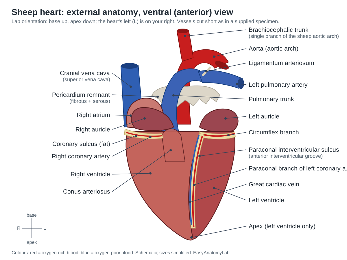
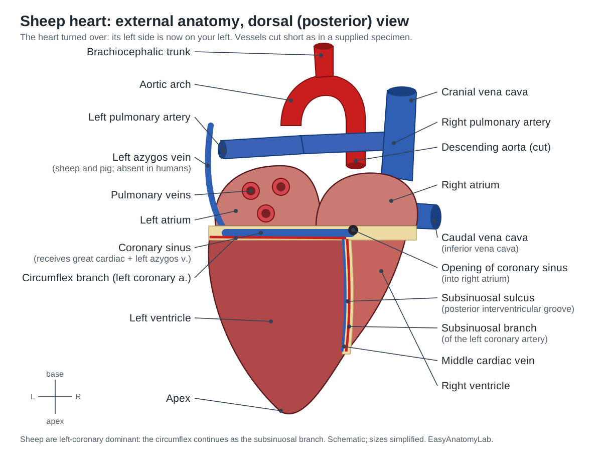
Where the heart sits in a sheep, and why lab 'anterior' is really its left side
A sheep's heart lies low in the chest between about the 2nd and 5th intercostal spaces, with its base toward the spine and its apex pointing down toward the sternum. Tıpırdamaz measured 38 Akkaraman sheep hearts at about 161 g on average, and the pericardial sac was tied to the sternum by a sternopericardiac ligament at the level of the 6th costal cartilage. The heart of a four-legged animal hangs more vertically and more in the midline than a human heart, which is tilted to the left. (Tıpırdamaz (1987), comparative studies on the hearts and coronary arteries of Akkaraman sheep and goats, Selçuk Üniv. Vet. Fak. Derg. 3:179–191.)
This changes the names of the surfaces. Veterinary anatomy calls the right face of the heart the atrial surface (facies atrialis) and the left face the auricular surface (facies auricularis), because both auricles point to the left. The left (paraconal) interventricular groove runs over the auricular surface, and the right (subsinuosal) groove runs over the atrial surface. Most school handouts borrow the human words instead: the face with the pulmonary trunk and the diagonal groove is 'anterior' or 'ventral', and the other face is 'posterior' or 'dorsal'. Both naming systems describe the same specimen. This page uses the lab-manual words and gives the veterinary terms alongside them. (Université Constantine 1, veterinary anatomy course notes: organs of the cardiovascular system.)
The practical rule is the same in every manual: find the diagonal groove of vessels and fat; the half of the heart that holds the whole apex is the left side. With the groove facing you and the apex pointing down, the heart's left is on your right, just as when you face another person. (Biology Corner, Sheep heart dissection: a step-by-step guide.)
Sheep heart dissection procedure, step by step
These steps match the 3D viewer: open a step there to see the right layers and the structures to find. Use scissors for long cuts and keep the blunt blade inside the vessel or chamber.
1. Orient the heart: front, back, left and right
- Rinse the heart and pat it dry. Note any pericardium still attached: a loose, whitish bag around the base.
- Find the apex (pointed tip) and the base (broad top with the vessels).
- Squeeze each side. The firm, thick side that contains the whole apex is the left ventricle; the softer side is the right ventricle.
- Find the diagonal groove of fat and vessels on one face: the paraconal (anterior) interventricular sulcus. The face with this groove and with the pulmonary trunk in front is the ventral (anterior) surface. Turn the heart so that face looks at you and the apex points down: the heart's left is now on your right.
- Identify the two auricles: ear-like flaps at the top on each side of the great arteries.
Identify: Apex, Base of the heart, Left ventricle, Right ventricle, Paraconal interventricular sulcus, Right auricle, Left auricle, Fibrous pericardium, Epicardial fat.
2. Identify the great vessels
- The front-most vessel, rising from the top of the right ventricle, is the pulmonary trunk. Push a probe down it: it enters the right ventricle.
- Just behind it is the aorta, the thickest and most rubbery vessel. Probe it: it leads into the left ventricle. Look for the brachiocephalic trunk branching from its arch (sheep normally have only this one branch).
- Look between the pulmonary trunk and aorta for the ligamentum arteriosum, a short fibrous cord.
- Turn the heart over (dorsal surface). Find the thin, collapsed cranial and caudal venae cavae entering the right atrium, and the pulmonary vein openings on the left atrium.
- Look for the left azygos vein curving over the left atrium into the coronary groove.
- Mark the aorta and pulmonary trunk with different-coloured pipe cleaners.
Identify: Pulmonary trunk, Aorta, Brachiocephalic trunk, Ligamentum arteriosum, Left pulmonary artery, Right pulmonary artery, Cranial vena cava, Caudal vena cava, Pulmonary veins, Left azygos vein.
3. Trace the coronary vessels
- Pick away fat in the coronary sulcus and the paraconal groove with forceps.
- Follow the paraconal branch of the left coronary artery and the great cardiac vein beside it down the ventral groove.
- Lift the left auricle to see the left coronary artery dividing into the paraconal and circumflex branches.
- On the dorsal surface, follow the subsinuosal groove up to the wide coronary sinus in the coronary groove.
Identify: Coronary sulcus, Left coronary artery, Paraconal interventricular branch, Circumflex branch, Great cardiac vein, Right coronary artery, Subsinuosal interventricular sulcus, Subsinuosal interventricular branch, Middle cardiac vein, Coronary sinus.
4. Open the right ventricle through the pulmonary trunk
- Slide the blunt blade of your scissors into the pulmonary trunk and cut down its ventral wall into the conus arteriosus.
- Stop to look at the three pocket-like cusps of the pulmonary semilunar valve before you cut through them; cut between two cusps.
- Continue the cut down the right ventricle, parallel to the paraconal groove and about 1 cm to its right, toward the apex. Spread the walls open.
- Find the papillary muscles, the chordae tendineae and the moderator band crossing the cavity. Note the crescent shape of the cavity and the thin wall.
Caution: Keep the scissors' blunt blade inside the vessel so you cut only the wall you can see.
Identify: Conus arteriosus, Pulmonary semilunar valve, Right ventricle, Papillary muscles (right ventricle), Chordae tendineae, Moderator band, Trabeculae carneae, Interventricular septum.
5. Open the right atrium
- Insert scissors into the cranial vena cava and cut down through the lateral wall of the right atrium toward the tricuspid valve.
- Look at the ridged pectinate muscles in the atrial wall and auricle.
- On the septal wall find the fossa ovalis, and near the caudal vena cava the opening of the coronary sinus.
- Look down into the ventricle through the three cusps of the tricuspid valve.
Identify: Right atrium, Pectinate muscles, Fossa ovalis, Interatrial septum, Opening of the coronary sinus, Tricuspid valve.
6. Open the left side through the aorta and left atrium
- Push the blunt scissor blade into a pulmonary vein and cut open the left atrium and left auricle.
- Look through the two cusps of the mitral (bicuspid) valve into the left ventricle.
- Now cut down the left ventricle from the atrium toward the apex, along its left side.
- Open the aorta from its cut end down to the aortic valve. Find the three semilunar cusps and the two coronary ostia in the pockets behind them.
- If you feel a hard grain near the aortic root, you may have found an os cordis.
Identify: Left atrium, Mitral (bicuspid) valve, Left ventricle, Papillary muscles (left ventricle), Aortic semilunar valve, Coronary ostia, Os cordis (heart bone).
7. Compare the walls, valves and septum
- Measure the thickness of the left ventricle wall, the right ventricle wall and an atrial wall with a ruler. Record the values.
- Compare the atrioventricular valves (cusps, chordae, papillary muscles) with the semilunar valves (pockets, no chordae).
- Feel the interventricular septum between the two ventricles: it is about as thick as the left ventricle wall.
Identify: Myocardium (cut wall), Interventricular septum, Left ventricle, Right ventricle, Tricuspid valve, Mitral (bicuspid) valve, Aortic semilunar valve, Pulmonary semilunar valve.
8. Optional: frontal section
- On a second heart, cut through the whole heart in a frontal (coronal) plane, from the apex up through both ventricles and atria, separating the ventral half from the dorsal half.
- Lay both halves cut side up. Trace blood flow through all four chambers and valves.
- Use the slider on the model to fade the ventral half in and out and compare with your specimen.
Identify: Myocardium (cut wall), Right atrium, Tricuspid valve, Right ventricle, Left atrium, Mitral (bicuspid) valve, Left ventricle, Interventricular septum.
Sheep heart dissection labeled: internal anatomy
After a frontal section you should see the four chambers side by side. The right atrium and right ventricle (on your left) receive oxygen-poor blood; the left atrium and the thick-walled left ventricle (on your right) handle oxygen-rich blood. The interventricular septum separates the ventricles.
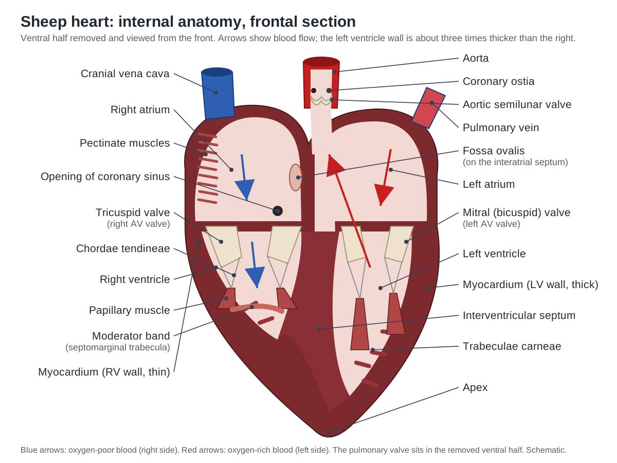
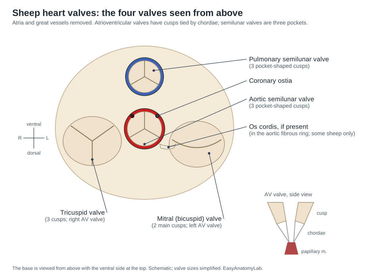
Blood flow through the heart: trace it on your specimen
Blood passes through the heart twice in each circuit: once through the right side to the lungs and once through the left side to the body. Push a probe along each step of this path on your specimen:
- Cranial and caudal venae cavae (and the coronary sinus) → right atrium
- Right atrium → tricuspid valve → right ventricle
- Right ventricle → conus arteriosus → pulmonary semilunar valve → pulmonary trunk
- Pulmonary trunk → right and left pulmonary arteries → lungs (gas exchange)
- Lungs → pulmonary veins → left atrium
- Left atrium → mitral (bicuspid) valve → left ventricle
- Left ventricle → aortic semilunar valve → aorta → brachiocephalic trunk and descending aorta → body
Two vessels break the "arteries carry oxygen-rich blood" rule: the pulmonary trunk and arteries carry oxygen-poor blood, and the pulmonary veins carry oxygen-rich blood. The coronary arteries, which leave the aorta just above the aortic valve, feed the heart muscle itself; their blood returns through the cardiac veins and coronary sinus into the right atrium.
| Chamber | Receives blood from | Sends blood to | Blood |
|---|---|---|---|
| Right atrium | Venae cavae, coronary sinus | Right ventricle | Oxygen-poor |
| Right ventricle | Right atrium | Pulmonary trunk | Oxygen-poor |
| Left atrium | Pulmonary veins | Left ventricle | Oxygen-rich |
| Left ventricle | Left atrium | Aorta | Oxygen-rich |
Why the left ventricle wall is thicker
The left ventricle has the thickest wall because it pumps blood through the systemic circuit to the whole body, which needs far more pressure than the short pulmonary circuit to the lungs. OpenStax describes the left ventricular muscle as much thicker and better developed than the right. Both ventricles eject the same volume of blood with each beat; only the pressure differs. (OpenStax Anatomy and Physiology 2e, 19.1 Heart Anatomy.)
You can see this in any sectioned sheep heart. The right ventricle's cavity is a thin crescent wrapped around the round left ventricle, and its free wall is only a few millimetres thick. The interventricular septum bulges into the right ventricle and behaves as part of the left ventricle's wall. The Visible Heart Lab notes the same pattern in all large mammals: ventricular walls are much more muscular than atrial walls, and the left ventricle is much thicker than the right. (Visible Heart Lab comparative anatomy tutorial: ventricles.)
Heart valve structure and function
The heart has two kinds of valves. The atrioventricular (AV) valves, the tricuspid on the right and the mitral (bicuspid) on the left, have flap-like cusps tied by chordae tendineae to papillary muscles. When the ventricles contract, the papillary muscles contract too and keep the cords tight, so the cusps cannot swing back into the atria. The semilunar valves, the pulmonary and the aortic, are three pocket-shaped cusps at the roots of the great arteries. They have no chordae and no papillary muscles; they close when blood falls back and fills the pockets. (OpenStax Anatomy and Physiology 2e, 19.1 Heart Anatomy.)
Sheep valves are close to human valves in size, but there are small, testable differences. The sheep mitral valve's two main cusps can carry small extra commissural cusps that look like extra leaflets. The sheep aortic cusps are substantially thinner than those of humans, pigs or dogs, and in pigs the non-coronary aortic cusp is smaller than the other two. All three animal aortic valves have more muscle around them than the human valve. (Visible Heart Lab comparative anatomy tutorial: valves.)
In sheep the tricuspid valve is supported by one anterior, one or two posterior and one or two septal papillary muscles. The left ventricle has one anterior and one (occasionally two) posterior papillary muscles. A moderator band crosses the right ventricle as a free muscular arch and inserts on the body or head of the anterior papillary muscle. Trabeculae carneae are fewer and coarser than in a human heart. (Visible Heart Lab comparative anatomy tutorial: ventricles.)
Coronary vessels of the sheep heart: a left-dominant supply
Two coronary arteries leave the aorta just above the aortic valve and supply the heart muscle. In sheep the left one does most of the work. The Visible Heart Lab states that the ovine myocardium is supplied mainly by branches of the left coronary artery, with limited collateral links between arteries. Pig hearts are supplied mainly by the right coronary artery or equally by both, which is closer to the usual human pattern. (Visible Heart Lab comparative anatomy tutorial: coronary vasculature.)
Tıpırdamaz found the right coronary artery to be much smaller than the left in sheep, and Gómez and colleagues describe the middle cardiac vein running up the subsinuosal groove beside 'the subsinusal interventricular branch of the left coronary artery'. In other words, in sheep the left circumflex branch continues round to the dorsal surface and supplies the dorsal groove, the job the right coronary artery does in most people. (Tıpırdamaz (1987), comparative studies on the hearts and coronary arteries of Akkaraman sheep and goats, Selçuk Üniv. Vet. Fak. Derg. 3:179–191.)
The veins follow the arteries. The great cardiac vein rises in the paraconal groove, then turns left in the coronary groove. In 62 sheep hearts it began at the apex in 71% of specimens, and the middle cardiac vein began at the apex in 91.9%. Both drain into the coronary sinus, which measured 24.85 ± 4.94 mm long, close to the human value. None of the sheep hearts had a valve at the coronary sinus opening. (Gómez, Ballesteros & Estupiñan (2018), coronary sinus and its tributaries in short hair sheep, Ital. J. Anat. Embryol. 123:81–90.)
Great vessels of the sheep heart: one aortic branch, a left azygos vein
Many worksheets ask students to find three branches on the aortic arch: the brachiocephalic, the left common carotid and the left subclavian. That is the human pattern. Veterinary texts describe a different one in ruminants: only the brachiocephalic trunk leaves the arch, and it later gives off the left and right subclavian arteries and continues as a bicarotid trunk. Pigs have two arch branches, the brachiocephalic trunk and the left subclavian artery. (Aydin et al. (2011), arteries originating from the aortic arch in ground squirrels (discussion of domestic species), Vet. Med. (Praha) 56:469–472.)
A 2023 study that used sheep as a surgical model put it the same way: ovine thoracic vessels follow the bovine arch pattern, and the arteries do not branch until the thoracic inlet. The same study noted a prominent left azygos vein and ligamentum arteriosum. In cattle, Layton and colleagues describe a single great vessel from the arch that gives rise to both subclavian arteries and a bicarotid trunk; the human 'bovine arch' variant is not the same thing. (Chu (2023), Development of an ovine cardiac model for the modified Blalock-Thomas-Taussig shunt, Colorado State University.)
Variation exists. A Brazilian case report described a Santa Inês sheep, found during a dissection class, whose left subclavian artery arose directly from the arch next to the brachiocephalic trunk. So if your specimen shows two branches, it is not necessarily mislabelled. Most school specimens have the vessels cut too short to check either way. (Rosa et al. (2011), variation of the aortic arch branches in a Santa Inês sheep, Bioscience Journal 27(1).)
On the dorsal surface, sheep and pigs have a left azygos vein that drains blood from the left chest wall into the coronary sinus. In humans the azygos vein drains into the superior vena cava instead. In sheep the coronary sinus begins where the left azygos vein joins the great cardiac vein. Also note that the two venae cavae enter the sheep right atrium at roughly a right angle, not in line as in an upright human. (Gómez, Ballesteros & Estupiñan (2018), coronary sinus and its tributaries in short hair sheep, Ital. J. Anat. Embryol. 123:81–90.)
Does a sheep heart have a bone? The os cordis
Cattle regularly have a small bone, the os cordis, in the fibrous skeleton of the heart. In sheep the evidence is mixed. Tıpırdamaz reported a single os cordis inside the fibrous ring around the aortic opening in Akkaraman sheep, while goats had both a cartilage and a bone there. The fibrous skeleton is made of fibrocartilage islands in which bone nodules can form. (Tıpırdamaz (1987), comparative studies on the hearts and coronary arteries of Akkaraman sheep and goats, Selçuk Üniv. Vet. Fak. Derg. 3:179–191.)
A study of 70 sheep hearts from Iran found an os cordis in only 11 (about 16%), averaging about 12 mm long, 3 mm wide and 1 mm thick, and called it a rare finding. Breed and age probably explain the difference. If you feel a hard grain when cutting near the aortic root, you may have found one; most students will not. (A statistical and morphological study of os cordis in sheep, Pajoohandeh 5(2):95–97 (2000).)
Pig heart dissection: what is the same and what is different
Dissecting a pig's heart follows exactly the same steps as a sheep heart: orient it by the diagonal paraconal groove and the firm left ventricle, open the right side through the pulmonary trunk, the left side through the aorta or a pulmonary vein, and finish with a frontal section. The chambers, valves, chordae, papillary muscles, moderator band and great vessels are all in the same places. Crick and colleagues, who dissected pig hearts to compare them with human hearts, found the pig heart has a rounded 'Valentine' shape, while the human heart is more trapezoidal. (Crick et al. (1998), Anatomy of the pig heart: comparisons with normal human cardiac structure, J. Anat. 193:105–119.)
Four differences are easy to see in a pig heart dissection. The pig left atrium usually receives only 2 pulmonary veins (humans usually 4). A prominent left azygos vein enters the left side of the base and drains through the coronary sinus. The moderator band sits much higher in the right ventricle than in a human heart. And the trabeculae near the apex of both ventricles are much coarser than human trabeculae. The two venae cavae enter the right atrium at right angles, unlike the in-line human arrangement. (Crick et al. (1998), Anatomy of the pig heart: comparisons with normal human cardiac structure, J. Anat. 193:105–119.)
Compared with a sheep heart, the pig heart has a blunter apex, a smaller right auricle and a larger left auricle (the reverse of the sheep), a deeper fossa ovalis placed higher on the septum, and a right-dominant or balanced coronary supply. The sheep apex is the most pointed of the common lab hearts. Pig hearts are about 2.9 g per kg of body weight, sheep hearts about 3.1 g/kg and human hearts about 5 g/kg, though these ratios vary between breeds. (University of Minnesota Visible Heart Lab, comparative anatomy tutorial: external anatomy.)
The pig aortic arch has two branches: the brachiocephalic trunk, which gives off the right subclavian artery and the carotids, and the left subclavian artery, which leaves the arch directly. A sheep usually has one arch branch and a human three, so the arch is a good place to tell the three hearts apart if the vessels are long enough. (Damian et al. (2015), Features of the subclavian arteries and their branching in the domestic pig, Scientific Works Series C Vet. Med. 61(1).)
How similar is a pig heart to a human heart?
The pig heart is the animal heart most often compared with the human one. A 2022 review of large animal models concluded that pig and sheep valves are comparable to human valves and that pig coronary vessels are similar to human coronaries, so pigs were judged best suited as a model for aortic-root surgery. The same review notes that differences between species in the ascending aorta have not been studied in much detail. (Srinivas, Yii & Smith (2022), Comparative review of large animal models for proximal aortic endovascular repair, Int. J. Med. Students 10(2).)
Similarity is not identity. Crick and colleagues started their study because pig hearts were assumed to resemble human hearts but had been poorly documented. They found real differences in the atria (a tubular right appendage, two pulmonary veins, the left azygos vein) and in the ventricles (a high moderator band and less fibrous continuity between the aortic and mitral valves). Descriptions of the auricles vary between sources: the Visible Heart Lab calls the pig right appendage half-moon shaped, so judge your own specimen. (Crick et al. (1998), Anatomy of the pig heart: comparisons with normal human cardiac structure, J. Anat. 193:105–119.)
In January 2022, surgeons at the University of Maryland transplanted a heart from a pig with 10 gene edits into a 57-year-old man with end-stage heart failure who could not receive a human heart. The pig heart worked well for weeks with no sign of typical rejection. It then thickened and failed suddenly on day 49, and life support was withdrawn on day 60. Porcine cytomegalovirus was later detected in the donor tissue and the patient's blood. The case showed that a pig heart can support a human circulation, and why that is still experimental. (Griffith et al. (2022), Genetically modified porcine-to-human cardiac xenotransplantation, N. Engl. J. Med. 387:35–44 (PMC author manuscript).)
Sheep vs pig vs human heart: comparison table
The three hearts share the same chambers, valves and blood flow. The differences below are the ones you can check in a lab or that exam questions ask about. Values come from the Visible Heart Lab comparative tutorial, Crick et al. (1998) and the vascular studies cited above; individual animals vary.
| Feature | Sheep | Pig | Human |
|---|---|---|---|
| Overall shape | Conical to Valentine; most pointed apex | Valentine (rounded); blunter apex | Variable, often trapezoidal |
| Apex formed by | Left ventricle only | Left ventricle only | Left ventricle, or both ventricles |
| Heart weight : body weight | About 3.1 g/kg | About 2.9 g/kg | About 5 g/kg |
| Branches of the aortic arch | 1: brachiocephalic trunk (then bicarotid trunk + both subclavians) | 2: brachiocephalic trunk, left subclavian artery | 3: brachiocephalic trunk, left common carotid, left subclavian |
| Venae cavae entering the right atrium | Cranial and caudal, at a right angle | At a right angle | Superior and inferior, in line |
| Left azygos vein to coronary sinus | Yes | Yes, prominent | No (azygos drains to superior vena cava) |
| Pulmonary veins entering left atrium | 2 to 4 | Usually 2 | Usually 4 (4 to 5) |
| Auricles (atrial appendages) | Right larger, half-moon; left smaller, triangular | Right smaller; left larger, triangular | Right triangular; left tubular |
| Coronary dominance | Left (circumflex supplies the dorsal groove) | Right, or balanced | Mostly right |
| Moderator band | Free muscular arch inserting on the anterior papillary muscle | Free arch, set high in the right ventricle | Arises lower (more apically) on the septum |
| Trabeculae carneae | Fewer, coarse | Fewer, coarse | Numerous, fine |
| Aortic valve cusps | Equal size, notably thin | Non-coronary cusp smaller | Equal size |
| Valve over coronary sinus opening | None | None | Thebesian valve partly covers it |
| Os cordis (heart bone) | In some sheep, in the aortic fibrous ring | Not reported in these sources | Normally absent |
Pre-lab questions and answers
Answer these before you start. Click a question to reveal the answer.
Which side of the heart pumps blood to the lungs, and which to the body?
The right side (right atrium and right ventricle) pumps oxygen-poor blood to the lungs through the pulmonary trunk. The left side pumps oxygen-rich blood to the body through the aorta.
How can you tell the left side of a preserved heart from the right before cutting?
The left ventricle is firmer and forms the whole apex. With the ventral face toward you (pulmonary trunk in front, diagonal groove visible) and the apex down, the heart's left side is on your right.
What is the difference between an atrioventricular valve and a semilunar valve?
Atrioventricular valves (tricuspid and mitral) sit between atria and ventricles and have flap-like cusps held by chordae tendineae and papillary muscles. Semilunar valves (pulmonary and aortic) sit at the roots of the great arteries and have three pocket-shaped cusps with no chordae.
Why will the arteries on your specimen stay open while the veins collapse?
Arteries have thick, elastic, muscular walls to withstand high pressure. Veins have thin walls under low pressure, so they flatten when empty.
What fetal structures leave remnants you may see in the heart?
The ductus arteriosus becomes the ligamentum arteriosum between the pulmonary trunk and aorta. The foramen ovale becomes the fossa ovalis in the interatrial septum.
Sheep heart dissection lab questions and answer key
Answer these during the dissection.
Which chamber has the thickest wall, and why?
The left ventricle. It must generate enough pressure to push blood through the whole systemic circulation, a much longer, higher-resistance circuit than the lungs.
If you put a probe into the aorta, which chamber does it enter? What about the pulmonary trunk?
The aorta leads into the left ventricle. The pulmonary trunk leads into the right ventricle (through the conus arteriosus).
Into which chamber do the cranial and caudal venae cavae open? The pulmonary veins?
The venae cavae (and the coronary sinus) open into the right atrium. The pulmonary veins open into the left atrium.
What attaches the cusps of the tricuspid and mitral valves to the ventricle wall?
Chordae tendineae, which run from the free edges of the cusps to papillary muscles projecting from the ventricle wall.
What is the moderator band, and in which chamber did you find it?
A muscular band (septomarginal trabecula) crossing the right ventricle from the septum to the anterior papillary muscle. It carries conduction fibres so that muscle contracts on time.
How many branches leave the aortic arch in a sheep? How does this compare with a human?
Normally one, the brachiocephalic trunk, which later divides into both subclavian arteries and a bicarotid trunk. Humans have three (brachiocephalic trunk, left common carotid, left subclavian); pigs have two (brachiocephalic trunk and left subclavian).
Name the vein that drains into the coronary sinus in sheep but not in humans.
The left azygos vein. In humans the azygos vein drains into the superior vena cava instead.
Why is the pulmonary trunk coloured blue in diagrams even though it is an artery?
Arteries carry blood away from the heart. The pulmonary trunk carries oxygen-poor blood, and diagram colours show oxygen content, not vessel type.
Which coronary artery supplies most of the sheep heart muscle?
The left coronary artery. In sheep its circumflex branch continues as the subsinuosal (posterior) interventricular branch, so the heart is left-dominant. Pigs and most humans are right-dominant.
What is the fossa ovalis, and where is it?
A shallow oval dip on the right-atrial side of the interatrial septum. It is the closed remnant of the foramen ovale, which let blood pass from the right to the left atrium before birth.
Post-lab analysis questions and answers
These anatomy and physiology lab analysis questions ask you to connect structure with function.
List, in order, the chambers, valves and vessels a red blood cell passes through from the cranial vena cava back to the aorta.
Cranial vena cava → right atrium → tricuspid valve → right ventricle → pulmonary semilunar valve → pulmonary trunk → pulmonary arteries → lungs → pulmonary veins → left atrium → mitral (bicuspid) valve → left ventricle → aortic semilunar valve → aorta.
What would happen if the chordae tendineae of the mitral valve broke?
The cusps could flip back into the left atrium when the ventricle contracts (prolapse), so blood would leak backward (mitral regurgitation) and less blood would reach the aorta.
Your measured left ventricle wall was about three times as thick as the right. Relate this to blood pressure.
Pressure in the systemic circuit is several times higher than in the pulmonary circuit, so the left ventricle needs more muscle. Both ventricles pump the same volume per beat.
Give two ways the sheep heart differs from the human heart.
Any two of: the sheep aortic arch has one branch (human three); sheep have a left azygos vein draining into the coronary sinus; the sheep heart is left-coronary dominant (human usually right); the sheep apex is more pointed and formed only by the left ventricle; the venae cavae meet the right atrium at a right angle rather than in line; some sheep have an os cordis.
Why are pig hearts studied for transplantation into humans?
Pig hearts are similar in size and in coronary and valve anatomy to human hearts. In 2022, surgeons in Maryland transplanted a gene-edited pig heart into a 57-year-old man; it worked for weeks but failed after day 49, and life support was withdrawn on day 60.
Virtual sheep heart dissection
The 3D viewer at the top of this page is a complete virtual sheep heart dissection. Each of the 8 steps sets up the heart, hides the layers you would have removed and lists the structures to find; click any part for its location, function and lab notes. The slider performs the frontal section, so you can compare the wall thickness of all four chambers. Students who opt out of a physical dissection can complete the same steps and answer the same lab questions. For a whole-animal view of the heart in the chest, see the fetal pig dissection.
Where do sheep and pig hearts for dissection come from?
Hearts for dissection come from sheep and pigs slaughtered for meat. The heart is a by-product of the meat industry. Biological supply companies preserve sheep hearts (with or without the pericardium) and sell them to schools; fresh lamb and pig hearts are sold by butchers as food and work just as well. Research labs use the same source: the 62 sheep hearts in the coronary sinus study by Gómez and colleagues came from animals destined for slaughter.
Fresh hearts often have longer great vessels and more pericardium than preserved ones, and their valves are more flexible, but they must be handled like raw meat. Preserved hearts are firmer and greyer and keep for longer. Ask your supplier for hearts with the pericardium and long vessels if you want to see the brachiocephalic trunk and ligamentum arteriosum.
Sheep heart structures: labeled reference
Every structure in the 3D model, grouped by system. Veterinary terms are given alongside the lab-manual names.
Surface landmarks & fat
- Apex (Apex cordis)
- The pointed tip of the heart. In the sheep it is formed only by the left ventricle. A landmark: the half of the heart that contains the apex is the left side.
- Base of the heart (Basis cordis)
- The broad top of the heart, where the atria lie and the great vessels enter and leave. Anchors the heart. The great vessels and the pericardium attach here.
- Coronary sulcus (Coronary groove, atrioventricular groove)
- A fat-filled groove that circles the heart between the atria and the ventricles. Carries the main coronary vessels: the circumflex branch, the right coronary artery and the coronary sinus.
- Paraconal interventricular sulcus (Anterior (ventral) interventricular groove; left interventricular groove)
- The diagonal groove on the ventral face that marks the septum between the right and left ventricles. Carries the paraconal interventricular branch of the left coronary artery and the great cardiac vein.
- Subsinuosal interventricular sulcus (Posterior (dorsal) interventricular groove; right interventricular groove)
- The groove on the dorsal face that marks the septum, running from below the coronary sinus toward the apex. Carries the subsinuosal interventricular branch and the middle cardiac vein.
- Epicardial fat (Subepicardial adipose tissue)
- Fat under the epicardium, concentrated in the grooves and around the base. Cushions and surrounds the coronary vessels and stores energy.
Pericardium
- Fibrous pericardium (Pericardial sac (outer layer))
- The tough outer layer of the pericardial sac. Specimens usually keep only a torn remnant around the great vessels. Protects the heart, anchors it in the chest and stops it over-filling.
- Serous pericardium (parietal layer) (Inner lining of the pericardial sac; its visceral layer is the epicardium)
- The thin, shiny membrane lining the inside of the fibrous sac. It folds back onto the heart as the epicardium. Makes a little serous fluid so the beating heart slides without friction inside the pericardial cavity.
Chambers & heart wall
- Right atrium
- The thin-walled chamber that receives oxygen-poor blood from the body and the heart wall. Collects blood and passes it through the tricuspid valve into the right ventricle.
- Right auricle (Right atrial appendage)
- The ear-shaped flap of the right atrium that wraps forward around the root of the aorta. Adds volume to the right atrium.
- Left atrium
- The chamber that receives oxygen-rich blood from the lungs through the pulmonary veins. Passes blood through the mitral (bicuspid) valve into the left ventricle.
- Left auricle (Left atrial appendage)
- The flap of the left atrium that curls forward on the left side of the pulmonary trunk. Adds volume to the left atrium.
- Right ventricle
- The chamber that pumps oxygen-poor blood into the pulmonary trunk and to the lungs. Pumps blood through the low-pressure pulmonary circuit.
- Conus arteriosus (Infundibulum, right ventricular outflow tract)
- The cone-shaped upper part of the right ventricle that leads into the pulmonary trunk. Channels blood from the right ventricle to the pulmonary valve.
- Left ventricle
- The thick-walled chamber that pumps oxygen-rich blood into the aorta and around the whole body. Generates the high pressure needed for the systemic circulation.
- Interventricular septum
- The thick muscular wall between the right and left ventricles. Separates oxygen-poor from oxygen-rich blood and contracts with the left ventricle.
- Interatrial septum
- The thin wall between the right and left atria. Separates the two atria. Before birth an opening in it (foramen ovale) let blood bypass the lungs.
- Myocardium (cut wall) (Heart muscle; outer epicardium and inner endocardium)
- The cardiac muscle layer of the heart wall, seen here as the cut surface of the frontal section. Contracts to pump blood. It is thickest in the left ventricle and thinnest in the atria.
Great vessels
- Aorta (Ascending aorta and aortic arch)
- The body's main artery. It leaves the left ventricle, rises behind the pulmonary trunk and arches dorsally. Carries oxygen-rich blood from the left ventricle to the body.
- Brachiocephalic trunk (Brachiocephalic artery, innominate artery)
- The single large branch of the sheep aortic arch, supplying both forelimbs, the neck and the head. Divides near the thoracic inlet into the left and right subclavian arteries and a bicarotid trunk.
- Pulmonary trunk (Pulmonary artery (lab handouts))
- The large artery that leaves the right ventricle and carries oxygen-poor blood toward the lungs. Carries blood from the right ventricle to the lungs.
- Right pulmonary artery
- The branch of the pulmonary trunk to the right lung. It passes under the aortic arch. Carries oxygen-poor blood to the right lung.
- Left pulmonary artery
- The branch of the pulmonary trunk to the left lung. Carries oxygen-poor blood to the left lung.
- Ligamentum arteriosum
- A short fibrous cord joining the pulmonary trunk to the underside of the aortic arch. None after birth. It is the closed remnant of the fetal ductus arteriosus.
- Cranial vena cava (Superior vena cava (human term), precava)
- The thin-walled vein bringing oxygen-poor blood from the head, neck and forelimbs into the right atrium. Returns blood from the front of the body.
- Caudal vena cava (Inferior vena cava (human term), postcava)
- The large vein bringing oxygen-poor blood from the abdomen and hind limbs into the right atrium. Returns blood from the back half of the body.
- Left azygos vein
- A vein from the left chest wall that runs over the left atrium and empties into the coronary sinus. Drains blood from the intercostal spaces straight into the coronary sinus.
- Pulmonary veins
- Short, thin veins that bring oxygen-rich blood from the lungs into the left atrium. Return blood from the lungs to the left side of the heart.
Coronary vessels
- Left coronary artery
- The larger coronary artery in sheep. It leaves the left aortic sinus and passes behind the pulmonary trunk. Splits into the paraconal interventricular branch and the circumflex branch.
- Paraconal interventricular branch (Anterior interventricular artery (human: LAD))
- The coronary artery running down the ventral interventricular groove toward the apex. Supplies the ventral walls of both ventricles and most of the septum.
- Circumflex branch
- The branch of the left coronary artery that runs around the left side in the coronary groove. Supplies the left atrium and the left ventricular wall.
- Subsinuosal interventricular branch (Posterior interventricular artery (human: posterior descending artery))
- The coronary artery in the dorsal interventricular groove. In sheep it comes from the left coronary artery. Supplies the dorsal walls of both ventricles and the septum.
- Right coronary artery
- The smaller coronary artery. It leaves the right aortic sinus and runs round the right side in the coronary groove. Supplies the right atrium and the right ventricular free wall.
- Great cardiac vein
- The vein that rises in the paraconal groove and turns left in the coronary groove to become the coronary sinus. Drains most of the left ventricle and septum.
- Middle cardiac vein
- The vein in the subsinuosal groove that empties into the coronary sinus. Drains the dorsal walls of both ventricles.
- Coronary sinus
- A short, wide vein in the dorsal coronary groove that collects blood from the heart wall and empties it into the right atrium. Returns coronary venous blood (and, in sheep, left azygos blood) to the right atrium.
Valves & supports
- Tricuspid valve (Right atrioventricular valve)
- The three-cusped valve between the right atrium and right ventricle. Opens to let blood into the right ventricle and closes when it contracts, so blood does not flow back.
- Mitral (bicuspid) valve (Left atrioventricular valve)
- The two-cusped valve between the left atrium and left ventricle. Lets blood into the left ventricle and seals during contraction.
- Pulmonary semilunar valve (Pulmonary valve)
- Three pocket-shaped cusps at the root of the pulmonary trunk. Stops blood flowing back from the pulmonary trunk into the right ventricle.
- Aortic semilunar valve (Aortic valve)
- Three pocket-shaped cusps at the root of the aorta. Stops blood flowing back from the aorta into the left ventricle.
- Chordae tendineae ('Heart strings')
- Thin white cords that tie the free edges of the atrioventricular valve cusps to the papillary muscles. Stop the cusps turning inside out into the atria when the ventricles contract.
- Papillary muscles (right ventricle)
- Small cone-shaped muscles in the right ventricle that anchor the tricuspid chordae. Contract with the ventricle to keep the chordae tight.
- Papillary muscles (left ventricle)
- Two large papillary muscles in the left ventricle that anchor the mitral chordae. Keep the mitral cusps from bulging back into the left atrium.
Inner wall features
- Trabeculae carneae
- Irregular ridges of muscle on the inner walls of the ventricles. Probably reduce suction against the smooth wall and help the ventricles empty.
- Moderator band (Septomarginal trabecula)
- A muscular band that crosses the right ventricle from the septum to the anterior papillary muscle. Carries part of the conduction system (right bundle branch) to the free wall so the papillary muscle contracts on time.
- Pectinate muscles (Musculi pectinati)
- Parallel ridges of muscle, like the teeth of a comb, on the inside of the atrial wall and auricles. Strengthen the thin atrial wall and let it stretch as it fills.
- Fossa ovalis
- An oval, thin-floored dip in the interatrial septum. The closed remnant of the foramen ovale, the fetal opening between the atria.
- Opening of the coronary sinus (Ostium of the coronary sinus)
- The round opening in the right atrium where the coronary sinus empties. Lets coronary venous blood enter the right atrium.
- Coronary ostia (Openings of the coronary arteries)
- Two small holes in the aortic wall, just above the aortic valve cusps, where the coronary arteries begin. Feed the coronary arteries, which supply the heart muscle itself.
- Os cordis (heart bone)
- A small bone or cartilage that can form in the fibrous ring around the aortic opening. Stiffens the fibrous skeleton that anchors the valves.
Frequently asked questions
How do you tell the front of a sheep heart from the back?
Look for the diagonal groove of fat and blood vessels (the paraconal or anterior interventricular sulcus) and the pulmonary trunk, the front-most vessel at the base. The face that shows both is the ventral (anterior) surface. The back (dorsal) surface shows the thin-walled venae cavae, the pulmonary vein openings and the coronary sinus in the groove.
How do you tell the left side of a sheep heart from the right?
Squeeze it. The left ventricle has a thick, firm wall and forms the whole apex; the right ventricle is soft and thin. With the ventral face toward you and the apex down, the heart's left side is on your right.
Why is the left ventricle wall thicker than the right?
The left ventricle pumps blood around the whole body, which needs much higher pressure than pumping blood through the lungs. More pressure needs more muscle, so its wall is about two to three times as thick as the right ventricle's.
Is a pig heart dissection the same as a sheep heart dissection?
Almost. The steps, chambers, valves and vessels are the same. Pig hearts are more rounded (Valentine-shaped) with a blunter apex, usually have only two pulmonary veins, two branches on the aortic arch instead of one, a moderator band placed higher in the right ventricle, and a right-dominant coronary supply.
How similar is a pig heart to a human heart?
Similar in size, valves and coronary arteries, which is why pig hearts are used in research and have been transplanted into people experimentally. There are clear differences too: pigs have a left azygos vein draining into the coronary sinus, usually two pulmonary veins instead of four, two aortic arch branches instead of three, and coarser trabeculae.
What are the white strings inside the heart?
They are chordae tendineae, strong collagen cords that tie the cusps of the tricuspid and mitral valves to the papillary muscles. They stop the valves turning inside out into the atria when the ventricles contract.
Why is there so much fat on a sheep heart?
Fat (epicardial fat) normally fills the coronary groove and the interventricular grooves around the coronary vessels, and sheep hearts often carry a lot of it. Pick it away with forceps to find the coronary arteries and veins underneath.
How many branches come off the aorta in a sheep heart?
Normally one: the brachiocephalic trunk, which later divides into both subclavian arteries and a bicarotid trunk. Humans have three arch branches and pigs two. Individual sheep can vary, and the branches are often cut off in supplied specimens.
Where do sheep hearts for dissection come from?
From sheep slaughtered for meat. The heart is a by-product of the meat industry: supply companies preserve hearts for schools, and fresh lamb and pig hearts are sold by butchers as food. Research studies also collect hearts from abattoirs.
References
- University of Minnesota Visible Heart Lab. Comparative anatomy tutorial: external anatomy, atria, ventricles, coronary vasculature and valves of human, dog, sheep and pig hearts.
- Crick, S. J., Sheppard, M. N., Ho, S. Y., Gebstein, L. & Anderson, R. H. (1998). Anatomy of the pig heart: comparisons with normal human cardiac structure. Journal of Anatomy 193, 105–119.
- Gómez, F. A., Ballesteros, L. E. & Estupiñan, H. Y. (2018). Morphological characterization of the coronary sinus and its tributaries in short hair sheep. Italian Journal of Anatomy and Embryology 123, 81–90.
- Tıpırdamaz, S. (1987). Comparative studies on the hearts and coronary arteries of Akkaraman sheep and ordinary goats. Selçuk Üniversitesi Veteriner Fakültesi Dergisi 3, 179–191.
- A statistical and morphological study of os cordis in sheep (2000). Pajoohandeh 5(2), 95–97.
- Aydin, A., Ozkan, Z. E., Yilmaz, S. & Ilgun, R. (2011). The arteries originating from the aortic arch in ground squirrels, with comparison to domestic species. Veterinarni Medicina 56, 469–472.
- Rosa, M. C. B. et al. (2011). Variação dos ramos do arco aórtico em um ovino da raça Santa Inês: relato de caso. Bioscience Journal 27(1).
- Chu, M. L. (2023). Development of an ovine cardiac model for the modified Blalock-Thomas-Taussig shunt. Colorado State University.
- Damian, A. et al. (2015). Features of the subclavian arteries and their branching in the domestic pig. Scientific Works Series C, Veterinary Medicine 61(1).
- Griffith, B. P. et al. (2022). Genetically modified porcine-to-human cardiac xenotransplantation. New England Journal of Medicine 387, 35–44.
- Srinivas, A., Yii, M. & Smith, J. A. (2022). Comparative review of large animal models for suitability of proximal aortic endovascular repair. International Journal of Medical Students 10(2).
- OpenStax. Anatomy and Physiology 2e, section 19.1: Heart anatomy.
- Biology Corner. Sheep heart dissection: a step-by-step guide.
More interactive anatomy models
- Cow eye dissectionGuided cow eye dissection: cornea, lens, retina and tapetum lucidum.
- Sheep brain dissectionGuided sheep brain dissection with cerebrum, cerebellum and cranial nerves labeled.
- Human anatomy1,000+ bones, muscles, organs, vessels and nerves from real anatomical data.
- Octopus anatomyThree hearts, a ring-shaped brain, eight arms and a beak, in 3D.
- Squid anatomyPen, eight arms, two tentacles, giant axons and three hearts of the longfin squid.
- Squid dissectionStep-by-step guided squid dissection with every structure labeled.