

Action potential at a glance
An action potential is the electrical signal that neurons, muscle cells and some other cells use to send messages. It is a rapid, self-renewing change in membrane potential: the inside of the cell flips from negative to positive and back in a few milliseconds, and the change travels along the cell membrane without getting smaller.
The whole event is caused by ions moving through protein channels. Sodium ions (Na+) rush into the cell to make the inside positive (depolarization), then potassium ions (K+) flow out to make it negative again (repolarization). The two ions are kept at very different concentrations inside and outside the cell by the Na+/K+ pump, and those concentration gradients are the battery that powers every action potential.
| Feature | Typical neuron value |
|---|---|
| Resting membrane potential | About −70 mV (inside negative) |
| Threshold | About −55 mV |
| Peak (overshoot) | About +30 mV (OpenStax A&P) to +40 mV (OpenStax Biology) |
| Undershoot (hyperpolarization) | About −80 mV |
| Total swing, rest to peak | About 100 mV |
| Duration of the spike | About 1–2 ms |
| Absolute refractory period | About 1–2 ms |
| Conduction speed | From less than 0.1 m/s to about 100 m/s, depending on diameter and myelin |
These are the numbers most textbooks use. They are averages for teaching: real neurons rest anywhere from about −40 to −80 mV and have thresholds from about −40 to −55 mV. The table near the end of this page lists how the values change between cell types.
Action potential diagram labeled: the phases
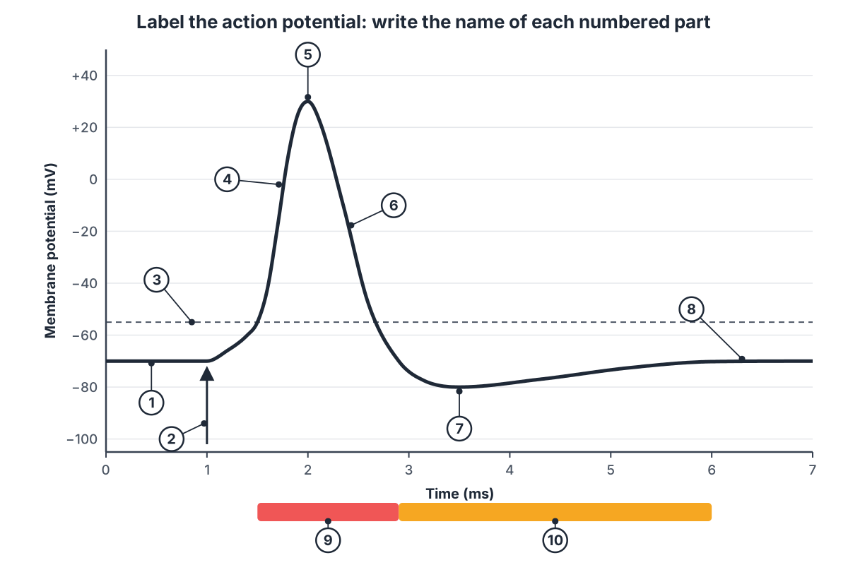
A labeled action potential diagram plots membrane potential (mV) against time (ms) and shows seven parts: the resting potential, the stimulus, the threshold, depolarization (the rising phase), the peak or overshoot, repolarization (the falling phase) and hyperpolarization (the undershoot), followed by the return to resting potential. The two refractory periods are usually drawn as bars under the curve.
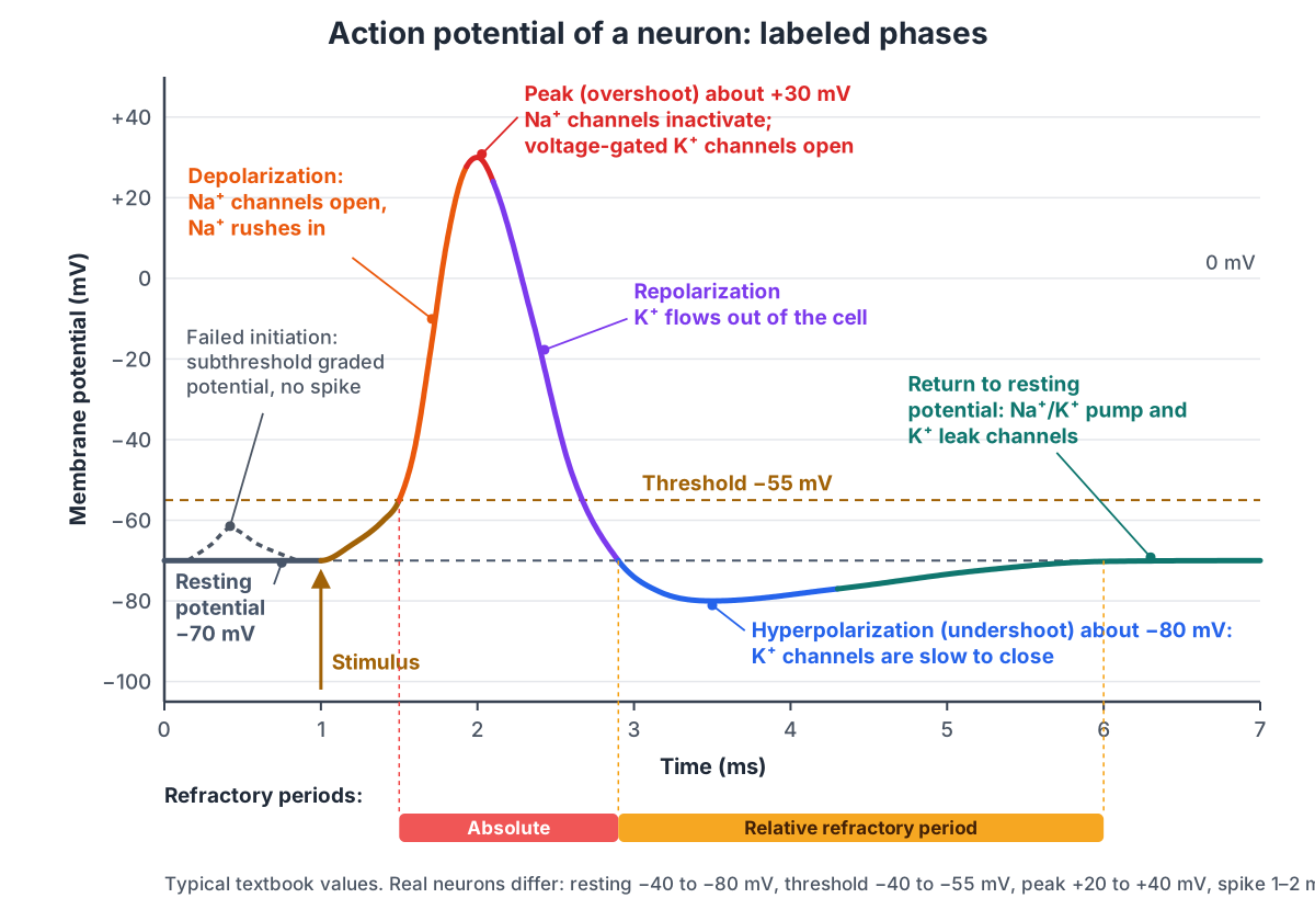
| Phase | Voltage | What the channels do | Ion movement |
|---|---|---|---|
| 1. Resting potential | −70 mV | Voltage-gated Na+ and K+ channels closed; K+ leak channels open | Small K+ leak out; pump moves 3 Na+ out, 2 K+ in |
| 2. Stimulus to threshold | −70 → −55 mV | Ligand-gated channels in dendrites open; a few voltage-gated Na+ channels open | Na+ in (graded) |
| 3. Depolarization | −55 → +30 mV | Voltage-gated Na+ channels open (positive feedback) | Large Na+ influx |
| 4. Peak / overshoot | About +30 mV | Na+ channels inactivate; voltage-gated K+ channels open | Na+ entry stops; K+ starts to leave |
| 5. Repolarization | +30 → −70 mV | Na+ channels inactivated; K+ channels open | Large K+ efflux |
| 6. Hyperpolarization | About −80 mV | K+ channels slow to close; Na+ channels reset | Extra K+ efflux |
| 7. Return to rest | −80 → −70 mV | K+ channels closed; leak channels and pump hold the rest | Back to resting fluxes |
The dashed hump on the left of the labeled diagram is a failed initiation: a small graded depolarization that does not reach threshold, so no action potential follows. It is a common item on labeled diagrams because it shows that threshold, not stimulus size, decides whether the neuron fires.
Action potential steps: what happens, in order
The steps of an action potential are: (1) the membrane rests at −70 mV; (2) a stimulus depolarizes it to threshold; (3) voltage-gated Na+ channels open and Na+ rushes in; (4) at the peak, Na+ channels inactivate and voltage-gated K+ channels open; (5) K+ flows out and the membrane repolarizes; (6) K+ channels close slowly, so the membrane overshoots into hyperpolarization; (7) the membrane returns to rest.
- Rest. The voltage-gated Na+ channel has two gates. At rest its activation gate is shut and its inactivation gate is open, so it is closed but ready. The voltage-gated K+ channel is closed. K+ leaks out through leak channels.
- Stimulus. Neurotransmitter opens ligand-gated channels on the dendrites. Positive ions enter and the depolarization spreads to the axon initial segment. If the summed depolarization reaches about −55 mV, the neuron fires.
- Depolarization. At threshold, the activation gates of many Na+ channels open. Na+ is pulled in by both its concentration gradient and the negative inside. Each Na+ ion that enters depolarizes the membrane further and opens more channels.
- Peak. A fraction of a millisecond after opening, each Na+ channel's inactivation gate swings shut. At about the same time, the slower voltage-gated K+ channels finish opening.
- Repolarization. K+ leaves the cell, taking positive charge with it, and the inside becomes negative again.
- Hyperpolarization. The K+ channels close with a delay, so for a moment more K+ leaves than at rest and the voltage dips below −70 mV. During repolarization the Na+ channels reset: the activation gate closes and the inactivation gate reopens.
- Return to rest. When the K+ channels have closed, the leak channels and the Na+/K+ pump hold the membrane at −70 mV, ready for the next action potential.
A common mistake is to say the Na+/K+ pump repolarizes the membrane. It does not. Repolarization is caused by K+ flowing out through channels, which is thousands of times faster than pumping. One action potential moves only a tiny fraction of the cell's ions, so the pump works in the background to keep the gradients topped up.
Ionic basis of the action potential
The ionic basis of the action potential is the unequal distribution of Na+ and K+ across the membrane, plus channels that let each ion through at a different time. Na+ is about 12 times more concentrated outside the cell, and K+ is about 40 times more concentrated inside.
| Ion | Outside (mM) | Inside (mM) | Role in the action potential |
|---|---|---|---|
| Na+ | 145 | 12 | Flows in to depolarize; equilibrium potential about +55 mV |
| K+ | 4 | 155 | Leaks out at rest; flows out to repolarize; equilibrium potential about −75 to −90 mV |
| Cl− | 120 | 4 | Mostly passive; entering Cl− hyperpolarizes (inhibitory synapses) |
| Organic anions (A−) | — | 100 | Trapped inside; add to the negative charge on the inner face |
| Ca2+ | High | Very low | Enters at axon terminals to trigger transmitter release; carries the heart's plateau |
Concentrations are typical mammalian values from OpenStax Biology 2e (table 35.1). Every ion has an equilibrium potential, the voltage at which its electrical pull exactly balances its concentration gradient (given by the Nernst equation; a tenfold concentration difference is worth about 60 mV). The membrane potential at any moment lies between the K+ and Na+ equilibrium potentials, closer to whichever ion the membrane is more permeable to. At rest the membrane is far more permeable to K+, so it sits near EK; at the peak it is far more permeable to Na+, so it swings toward ENa. That shift in permeability is the whole action potential.
| Membrane protein | Gate | When open | Ion |
|---|---|---|---|
| Voltage-gated Na+ channel | Activation gate + inactivation gate | From threshold until a fraction of a ms later | Na+ in |
| Voltage-gated K+ channel | One slower gate | From the peak through the undershoot | K+ out |
| K+ leak channel | None (mostly open) | All the time | K+ out |
| Ligand-gated channel | Opened by a neurotransmitter | During synaptic input | Na+, K+, Ca2+ or Cl− |
| Na+/K+-ATPase (pump) | Uses ATP | All the time | 3 Na+ out, 2 K+ in |
Resting membrane potential
The resting membrane potential is the voltage across the membrane of a cell that is not firing, about −70 mV in a typical neuron. It is negative because the resting membrane is much more permeable to K+ than to Na+: neurons have far more K+ leak channels than Na+ leak channels. K+ leaks out, down its concentration gradient, and the large negative ions it leaves behind cannot follow.
The leak stops getting bigger because the inside becomes negative and starts to pull K+ back. If K+ were the only ion that could cross, the membrane would settle at EK, about −75 to −90 mV. A small Na+ leak drags it a little more positive, to about −70 mV. In the classic example from Neuroscience Online the resting K+ permeability is about 100 times the Na+ permeability.
The Na+/K+ pump uses one ATP to move 3 Na+ out and 2 K+ in. It sets up the gradients in the first place and keeps them there. Because it moves more positive charge out than in, it also makes the inside a few millivolts more negative directly. At the muscle end-plate, for example, pump activity makes the membrane 2–4 mV more negative than the rest of the fibre.
Resting potential is not the same in every cell: ventricular heart muscle rests near −90 mV, atrial muscle near −80 mV, skeletal muscle fibres around −60 to −90 mV, and SA node cells never rest at all. See the cardiac sections below.
Graded potentials vs action potentials (all-or-none)
A graded potential is a small, local change in membrane potential whose size depends on the strength of the stimulus; an action potential is a large, fixed-size, all-or-none signal that travels the length of the axon without fading. Graded potentials happen in dendrites and the cell body; action potentials start at the axon initial segment.
| Graded potential | Action potential | |
|---|---|---|
| Size | Varies with stimulus strength (a few mV) | Always about 100 mV, rest to peak |
| Direction | Depolarizing or hyperpolarizing | Always depolarizing, then repolarizing |
| Threshold | None | About −55 mV |
| Spread | Passive; fades with distance | Regenerated; does not fade |
| Summation | Yes, spatial and temporal | No; refractory period prevents it |
| Refractory period | None | Absolute and relative |
| Channels | Ligand-gated, mechanically gated or sensory | Voltage-gated Na+ and K+ |
| Where | Dendrites, cell body, sensory receptors, motor end-plate | Axon initial segment, axon, nodes of Ranvier |
| Examples | EPSP, IPSP, receptor potential, end-plate potential | Nerve impulse, muscle action potential |
All-or-none means that once threshold is reached, the action potential always runs to the same size, however strong the stimulus. A stronger stimulus does not make bigger action potentials; it makes them come more often. That is how neurons code stimulus strength: by firing rate, not by spike size.
Excitatory postsynaptic potentials (EPSPs) move the membrane toward threshold and inhibitory ones (IPSPs) move it away. Inputs at different places add up (spatial summation) and inputs that arrive close together in time add up (temporal summation). In OpenStax's example, inputs that together depolarize the membrane by 15 mV, from −70 to −55 mV, reach threshold and fire the neuron.
Propagation of the action potential: continuous vs saltatory conduction
An action potential propagates because the Na+ entering one patch of membrane spreads sideways inside the axon and depolarizes the next patch to threshold, which then fires its own action potential. In unmyelinated axons this happens patch by patch along the whole membrane (continuous conduction). In myelinated axons it happens only at the nodes of Ranvier, and the signal seems to jump from node to node (saltatory conduction).
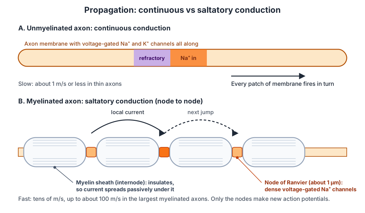
Under the myelin, the current spreads passively and gets weaker, but it is still strong enough to bring the next node to threshold, where a fresh, full-size action potential is made. Each node is like a relay station. The spacing matters: if nodes were too far apart, the current would fall below threshold before reaching the next one; if they were too close, conduction would be slower than it needs to be.
| Continuous conduction | Saltatory conduction | |
|---|---|---|
| Axon | Unmyelinated | Myelinated (Schwann cells in nerves, oligodendrocytes in the brain and spinal cord) |
| Where action potentials are made | Every patch of membrane | Only at nodes of Ranvier (about 1 µm long) |
| Speed | Slow, about 1 m/s or less in thin axons | Fast, tens of m/s, up to about 100 m/s |
| Energy cost | Higher: ions cross along the whole length | Lower: ions cross only at the nodes |
| Examples | Many pain and autonomic fibres | Motor neurons, touch fibres |
Conduction speed depends on axon diameter and myelin. Wider axons conduct faster, but the gain is poor: doubling the speed needs about four times the diameter. Squid solve this with a giant axon; vertebrates use myelin. Measured speeds range from less than 0.1 m/s to about 100 m/s. In one recent study, single nodes of a rat touch-sensory nerve conducted at about 48 m/s.
Action potentials travel only one way, away from the cell body, because the membrane they have just left is in its absolute refractory period. Its Na+ channels are inactivated and cannot be reopened by the current spreading back toward them.
Refractory periods
The refractory period is the time after an action potential during which the membrane cannot fire normally. It has two parts. In the absolute refractory period no stimulus, however strong, can start a second action potential. In the relative refractory period a stronger-than-normal stimulus can.
| Absolute refractory period | Relative refractory period | |
|---|---|---|
| When | From threshold through most of repolarization | Late repolarization through the undershoot |
| Duration (neuron) | About 1–2 ms | A few ms; up to about 15 ms in neurons with a long undershoot |
| Cause | Na+ channels are open or inactivated | Some Na+ channels still inactivated, and extra K+ is still flowing out |
| Can a stimulus fire the cell? | No | Yes, if it is stronger than normal |
Refractory periods do two jobs. They make propagation one-way, and they set a maximum firing rate: a neuron with a 1–2 ms absolute refractory period cannot fire faster than roughly 500–1,000 times a second, and most fire far more slowly. Na+ channels need about 3–4 ms to recover fully from inactivation. In the heart the refractory period lasts about 250 ms, almost as long as the contraction, which is why heart muscle cannot be tetanized.
Action potential neuron diagram: where it happens in the cell
In a neuron, inputs arrive at the dendrites and cell body as graded potentials, add up at the axon hillock and initial segment, where the action potential starts, and the action potential then travels down the axon, jumping between nodes of Ranvier under the myelin sheath, to the axon terminals and synaptic boutons. The 3D model above shows each of these, plus a magnified slice of the axon membrane with its channels, pump and ions. Here is every labeled structure of the model, by group.
Neuron
- Dendrites
- Branched extensions of the cell body that receive signals from other neurons. Collect synaptic inputs. Neurotransmitters open ligand-gated channels here, making small graded potentials that spread toward the cell body.
- Cell body (soma) (Soma, perikaryon)
- The rounded part of the neuron that holds the nucleus and most organelles. Keeps the cell alive and makes its proteins, including the ion channels and pumps that are shipped down the axon. Graded potentials from the dendrites pass through it to the axon hillock.
- Nucleus
- Holds the cell's DNA; seen through the cut-away wedge in the cell body. Contains the genes for the channels, pumps and other proteins the neuron needs.
- Axon hillock
- The cone-shaped part of the cell body where the axon begins. Funnels the summed graded potentials of the cell body into the axon. Many textbooks call it the trigger zone; strictly, the spike starts just beyond it, in the initial segment.
- Axon initial segment (AIS, trigger zone)
- The first, unmyelinated stretch of the axon, packed with voltage-gated Na+ channels. This is where action potentials begin. Has the lowest threshold in the neuron, so when the summed graded potentials reach about −55 mV here, the action potential starts and then travels down the axon.
- Axon
- The long, thin output fibre of the neuron that carries action potentials away from the cell body. Conducts the action potential without loss of size to the axon terminals, where it triggers transmitter release.
- Nodes of Ranvier
- Short gaps between myelin segments where the axon membrane is exposed to the extracellular fluid. The only places on a myelinated axon that generate action potentials. The signal 'jumps' from node to node.
- Axon terminals (Telodendria, terminal branches)
- The fine branches at the end of the axon. Carry the action potential to many synaptic boutons, so one neuron can contact many target cells (here, many muscle fibres or neurons).
- Synaptic boutons (Terminal buttons, synaptic end bulbs, synaptic knobs)
- The swollen tips of the axon terminals, filled with neurotransmitter vesicles. When an action potential arrives, voltage-gated Ca2+ channels open, Ca2+ enters, and vesicles release neurotransmitter into the synaptic cleft by exocytosis.
Myelin & Schwann cells
- Myelin sheath
- Segments of fatty insulation wrapped around the axon by Schwann cells (in nerves) or oligodendrocytes (in the brain and spinal cord). Insulates the axon so that current spreads quickly under it to the next node, where a new action potential is made. This is saltatory conduction.
- Schwann cell nucleus
- The nucleus of the Schwann cell that made the myelin segment, pushed to the outside of the sheath. Shows that each myelin segment is part of a living glial cell, not just a coating.
- Compact myelin lamellae
- Layers of Schwann cell membrane wrapped tightly around the axon, seen here cut lengthwise. Many layers of lipid-rich membrane give high resistance and low capacitance, so little current leaks out of the internode.
- Paranodal loops (Paranode)
- The ends of each myelin layer, which curl into cytoplasm-filled loops that attach to the axon membrane beside the node. Seal the edges of the myelin to the axon with septate-like junctions, and act as a fence that keeps the node's Na+ channels and the juxtaparanode's K+ channels apart.
- Schwann cell outer cytoplasm (Neurilemma)
- The thin outer layer of the Schwann cell, containing its cytoplasm and nucleus. Keeps the myelin-making cell alive; in peripheral nerves it guides axon regrowth after injury.
Membrane & axoplasm
- Axon membrane (axolemma) (Phospholipid bilayer, plasma membrane)
- The axon's plasma membrane: two layers of phospholipids whose water-loving (hydrophilic) heads face the fluid on each side. Separates the ion-rich fluids inside and outside. Ions cannot cross the oily core, so they move only through channels and pumps; this is what lets the membrane hold a voltage.
- Hydrophobic core (Fatty acid tails of the phospholipids)
- The oily middle of the bilayer, made of the phospholipids' fatty acid tails. Blocks charged particles. Na+, K+ and Cl− cannot dissolve in it, so they can only cross through channel and pump proteins. Because it is a thin insulator between two salty fluids, the membrane works like a capacitor that stores charge.
- Axoplasm (Axon cytoplasm)
- The cytoplasm inside the axon, rich in K+ and in negatively charged proteins. Holds the K+ that flows out during repolarization and the large anions that cannot leave.
- Microtubules
- Long protein tubes running along the axon. Tracks for motor proteins that carry new channels, pumps and vesicles from the cell body to the terminals.
- Mitochondrion
- An organelle that makes ATP. Supplies the ATP the Na+/K+ pumps use to restore the ion gradients.
Channels & pump
- Voltage-gated Na+ channels (Nav1.6, sodium channels)
- Proteins with a Na+-selective pore that opens when the membrane depolarizes to threshold. Packed densely at the node. Open at threshold (about −55 mV) and let Na+ rush in, which depolarizes the membrane further and opens more channels: the rising phase of the action potential.
- Na+ channel inactivation gate (h gate, 'ball and chain')
- A loop of the Na+ channel protein on the cytoplasmic side that swings in and plugs the open pore. Closes the channel at the peak of the action potential, even though the membrane is still depolarized. Until it reopens after repolarization, the channel cannot open again: the absolute refractory period.
- Nodal K+ channels (KCNQ) (Voltage-gated K+ channels at the node, Kv7, KCNQ2/3, M-type channels)
- Slow voltage-gated K+ channels that sit with the Na+ channels at nodes and initial segments. Open slowly with depolarization and let K+ out. They help keep the node from firing repeatedly and help repolarize it.
- Juxtaparanodal Kv1 channels (Kv1.1/Kv1.2, fast voltage-gated K+ channels)
- Fast voltage-gated K+ channels hidden under the myelin just beyond the paranodes. Help stabilize the internodal membrane and damp extra firing; they are not the main repolarizing channels of a healthy node.
- K+ leak channels (Two-pore-domain (K2P) channels, TREK-1, TRAAK)
- K+ channels that are open most of the time, whatever the voltage. Let a steady trickle of K+ out of the cell. This is the main reason the inside is negative at rest. At mammalian nodes, TREK-1 and TRAAK leak channels also do most of the fast repolarizing.
- Na+/K+ pump (Na+/K+-ATPase, sodium–potassium pump)
- A pump protein that uses ATP to move Na+ out and K+ in against their gradients. Moves 3 Na+ out and 2 K+ in for each ATP. It keeps the concentration gradients that every action potential runs down. It is not what repolarizes the membrane after a spike; the K+ channels do that.
Ions
- Sodium ions (Na+)
- Small positive ions, about 12 times more concentrated outside the cell than inside. Flow in through voltage-gated Na+ channels during depolarization, making the inside positive for a moment.
- Potassium ions (K+)
- Positive ions about 40 times more concentrated inside the cell than outside. Leak out at rest to set the resting potential, and flow out through voltage-gated K+ channels to repolarize the membrane.
- Chloride ions (Cl−)
- Negative ions concentrated outside the cell. Balance the positive charge of Na+ outside. Cl− entering through inhibitory channels hyperpolarizes neurons (IPSPs).
- Organic anions (A−) (Negatively charged proteins and phosphates)
- Large negatively charged molecules trapped inside the cell. Cannot cross the membrane, so they add to the negative charge on the inside of the membrane at rest.
At the synapse: handing the signal on
When an action potential reaches an axon terminal, it opens voltage-gated Ca2+ channels. Ca2+ flows in and makes synaptic vesicles fuse with the membrane and release neurotransmitter into the synaptic cleft. The transmitter binds receptors on the next cell and produces a graded postsynaptic potential there, about 1 ms after the action potential arrived. The transmitter is then broken down by enzymes or taken back up by the neuron or by glia.
So the signal changes form twice: electrical (action potential) to chemical (neurotransmitter) at the terminal, and chemical back to electrical (graded potential) at the next cell. Electrical synapses, where ions pass directly through gap junctions, skip the chemical step and have almost no delay.
Cardiac action potential diagram: ventricular muscle phases 0–4
The cardiac action potential of a ventricular muscle cell has five phases: phase 4 (rest at about −90 mV), phase 0 (rapid depolarization by Na+ entry), phase 1 (a small notch as transient K+ channels open), phase 2 (a long plateau as L-type Ca2+ channels let Ca2+ in) and phase 3 (repolarization as Ca2+ channels close and K+ flows out). The whole event lasts 250–300 ms.
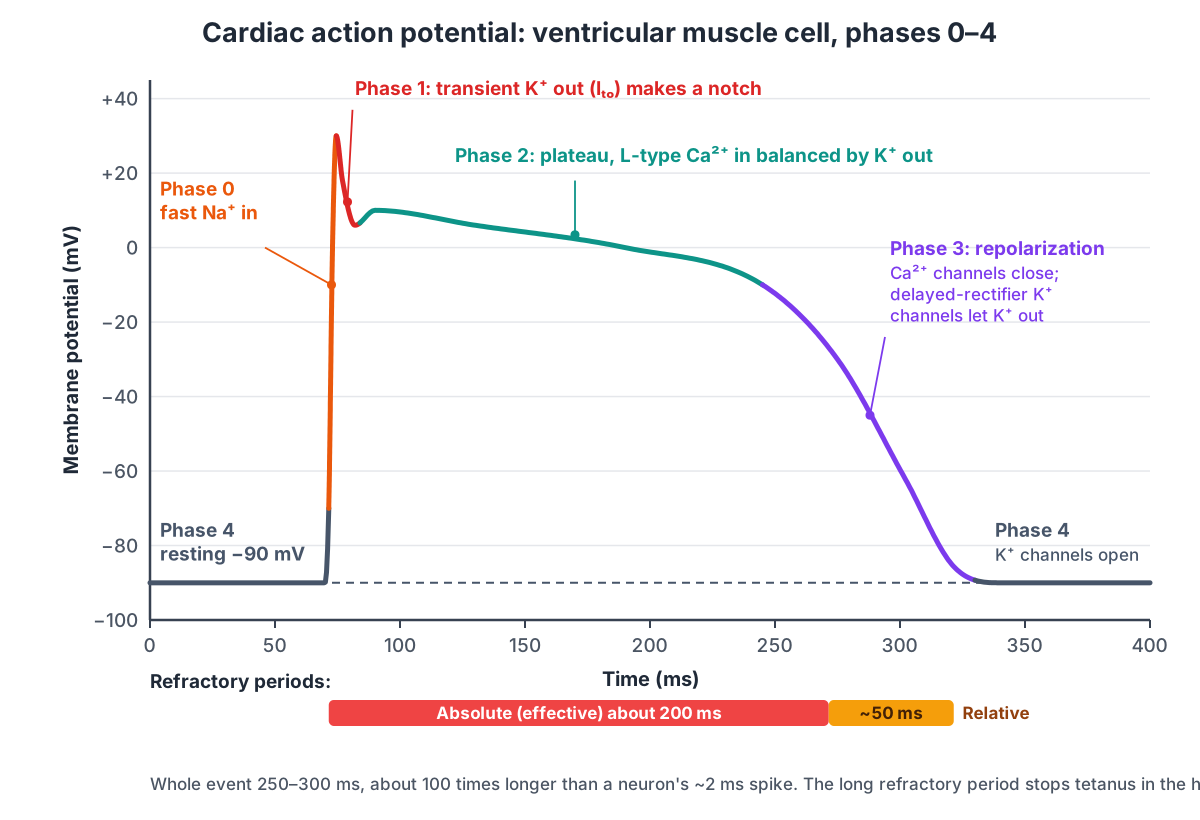
| Phase | Name | Main current | Voltage and timing |
|---|---|---|---|
| 4 | Resting potential | K+ channels open at rest (high K+ conductance) | About −90 mV (atria about −80 mV) |
| 0 | Rapid depolarization | Fast Na+ in (I_Na) | Up to about +30 mV in 3–5 ms |
| 1 | Early repolarization (notch) | Transient outward K+ (I_to) | Brief dip |
| 2 | Plateau | L-type Ca2+ in (I_Ca,L) balanced by K+ out | Near 0 mV for about 175 ms |
| 3 | Repolarization | Delayed-rectifier K+ out (I_Kr); Ca2+ channels inactivate | Back to rest in about 75 ms |
The plateau is the key difference from a neuron. The Ca2+ that enters during the plateau supplies about 20% of the Ca2+ the cell needs to contract; the rest comes from the sarcoplasmic reticulum. Because the fast Na+ channels stay inactivated until the membrane has fully repolarized, the absolute (effective) refractory period lasts about 200 ms, and the relative refractory period about 50 ms more. A new action potential cannot start until the heart has nearly finished contracting, so the heart always relaxes and refills between beats.
SA node pacemaker potential and the funny current
The SA node pacemaker potential is a slow, spontaneous depolarization from about −60 mV to a threshold of about −40 mV, followed by a Ca2+-driven upstroke to about +15 mV and K+-driven repolarization. Because SA node cells have no stable resting potential, they fire on their own, and they set the heart rate.
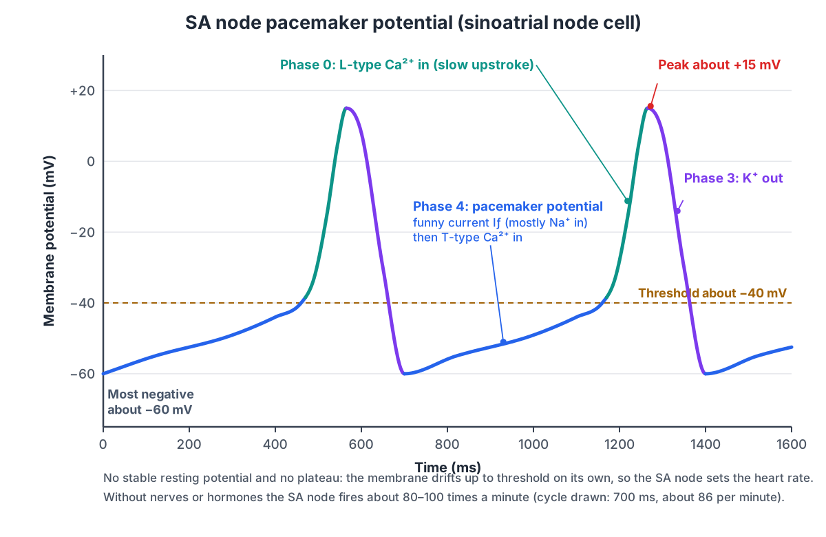
- Phase 4 (pacemaker potential or prepotential). Starting near −60 mV, a slow inward current called the funny current (If) begins. It flows through HCN channels (mainly HCN4), which open when the membrane is hyperpolarized, the opposite of most voltage-gated channels; that is why it was called "funny". The net inward current is carried mostly by Na+. Late in phase 4, T-type Ca2+ channels open at about −50 mV and speed the climb.
- Phase 0 (upstroke). At about −40 mV, L-type Ca2+ channels open and Ca2+ entry drives the upstroke. It is much slower than a neuron's or a ventricular cell's, because Ca2+ channels are slower than fast Na+ channels.
- Phase 3 (repolarization). Ca2+ channels inactivate and K+ channels open; K+ leaves until the cell is back near −60 mV, and the cycle restarts.
Pacemaker cells have no phase 1 or phase 2. Left alone, the SA node fires about 80–100 times a minute; the vagus nerve slows it and sympathetic nerves speed it up by changing the slope of phase 4.
Skeletal muscle action potential and the end-plate potential
A skeletal muscle action potential starts at the neuromuscular junction. The motor neuron releases acetylcholine, which opens ligand-gated channels in the muscle fibre's motor end-plate. Positive ions flow in and make a local graded depolarization, the end-plate potential (EPP). The EPP opens voltage-gated Na+ channels next to the end-plate, and a muscle action potential spreads along the sarcolemma and down the T-tubules.
In the T-tubules the action potential triggers Ca2+ release from the sarcoplasmic reticulum, and Ca2+ starts the sarcomeres contracting (see our sarcomere diagram). Acetylcholinesterase breaks down acetylcholine in the cleft so that each nerve impulse gives one muscle action potential.
The end-plate potential is normally 2–5 times bigger than it needs to be to reach threshold. This margin is the safety factor of neuromuscular transmission, and it falls with fatigue, injury and diseases such as myasthenia gravis. A single vesicle (quantum) of acetylcholine gives only a tiny, about 1 mV, miniature end-plate potential; a normal EPP is the sum of many quanta released together.
Neuron vs cardiac vs skeletal muscle action potentials
Neurons, skeletal muscle fibres and heart cells all use action potentials, but the shapes differ because each cell uses a different set of channels. The table compares the textbook values.
| Neuron | Skeletal muscle fibre | Ventricular myocyte | SA node cell | |
|---|---|---|---|---|
| Resting potential | About −70 mV | About −60 to −90 mV | About −90 mV | None (drifts from about −60 mV) |
| Threshold | About −55 mV | Reached via the end-plate potential | About −70 mV | About −40 mV |
| Upstroke ion | Na+ | Na+ | Na+ | Ca2+ |
| Peak | +30 to +40 mV | Positive overshoot | About +30 mV | About +15 mV |
| Plateau | No | No | Yes, L-type Ca2+ | No |
| Duration | About 1–2 ms | A few ms | 250–300 ms | Cycle about 600–750 ms at 80–100 per minute |
| Started by | Graded potentials at the initial segment | Acetylcholine at the end-plate | Current from neighbouring cells | Itself (funny current) |
How much the values vary between cells
The textbook numbers on this page, −70, −55 and +30 mV, are averages. Different books and different cells give different values, and students often meet more than one set.
| Value | Common textbook figure | Range or alternatives you may see |
|---|---|---|
| Resting potential | −70 mV | −60 mV is typical in some courses; −40 to −80 mV across cell types; −90 mV in heart and many muscle fibres |
| Threshold | −55 mV | About −45 mV in Neuroscience Online; lowest at the axon initial segment |
| Peak | +30 mV | +40 mV in OpenStax Biology 2e; always below E_Na (about +55 mV) |
| Undershoot | −80 mV | Approaches E_K, about −75 to −90 mV; absent or small in some neurons |
| Spike duration | About 2 ms | About 1 ms; nodal spikes in rat nerve about 0.7 ms wide |
| Ion ratio | Na+ about 10× higher outside | 145 vs 12 mM, so about 12× (OpenStax Biology 2e) |
Invertebrate axons such as the squid giant axon, used in the classic experiments of Hodgkin and Huxley, have different ion concentrations again (for example about 400 mM K+ inside and 20 mM outside). The principles are the same in all of them: rest near EK, swing toward ENa, and return.
Where the action potential starts: the axon initial segment
Most textbook diagrams say the action potential starts at the axon hillock, the cone where the axon leaves the cell body. Measurements in real neurons put the trigger zone a little further along, in the axon initial segment (AIS), the first 10–60 µm of the axon before the myelin begins. The AIS has a lower threshold than the cell body because it holds far more voltage-gated Na+ channels: about 30–50 times more Nav1.6 channels than the soma. (Solé & Tamkun (2019): Trafficking mechanisms underlying Nav channel subcellular localization in neurons, Channels 14:1.)
These channels are not scattered at random. A large scaffolding protein, ankyrin-G, holds them in place, and the same ankyrin-G system clusters Na+ channels at every node of Ranvier. That is why the AIS and the nodes look so alike: both are short stretches of axon packed with Na+ channels, built to make action potentials. OpenStax describes the same idea in simpler terms: summation happens at the axon hillock or initial segment, where voltage-gated Na+ channels are dense. (OpenStax Anatomy & Physiology 2e, §12.5 Communication Between Neurons.)
What is really at a node of Ranvier
In most diagrams, every patch of membrane that fires an action potential has voltage-gated Na+ channels and voltage-gated K+ channels side by side. That is true for unmyelinated axons and for the classic squid giant axon, but mammalian myelinated axons are organized differently. The node itself is packed with Na+ channels. The fast voltage-gated K+ channels, Kv1.1 and Kv1.2, sit under the myelin on each side of the node, in a region called the juxtaparanode, separated from the node by the paranodal junctions where the myelin seals onto the axon. (Pinatel & Faivre-Sarrailh (2020): Assembly and function of the juxtaparanodal Kv1 complex, Life 11:8.)
So what repolarizes a mammalian node? A 2019 study recorded directly from nodes of rat sensory nerves and found that blocking voltage-gated K+ channels had little effect. Instead, two leak-type channels from the two-pore-domain (K2P) family, TREK-1 and TRAAK, were clustered at the nodes at more than 3,000 times their density on the cell body. Knocking them down slowed repolarization and cut the conduction speed from about 48 m/s to 25–30 m/s. (Kanda et al. (2019): TREK-1 and TRAAK are principal K+ channels at the nodes of Ranvier, Neuron 104:960.)
Nodes also contain slow voltage-gated K+ channels of the KCNQ (Kv7) family. KCNQ2 sits with Na+ channels and ankyrin-G at nodes and initial segments throughout the brain, spinal cord and peripheral nerves. Drugs that open KCNQ channels make axons less excitable, and mutations in KCNQ2 cause neonatal epilepsy and myokymia, a rippling twitch of muscles caused by extra firing in motor axons. The 3D model shows all three: Na+ channels and KCNQ channels at the node, K2P leak channels at the node, and Kv1 channels hidden under the myelin. (Devaux, Kleopa, Cooper & Scherer (2004): KCNQ2 is a nodal K+ channel, Journal of Neuroscience 24:1236.)
How nerves and hormones change the heart rate
The SA node sets the heart rate, but the rate it sets is not fixed. Sympathetic nerves release norepinephrine, which acts on beta-adrenoceptors and steepens phase 4, mainly by boosting the funny current and the slow inward Ca2+ currents, so threshold is reached sooner. The vagus nerve releases acetylcholine, which acts on muscarinic receptors, increases K+ conductance and hyperpolarizes the cell, and reduces the funny and Ca2+ currents, so the cell takes longer to reach threshold. (Klabunde, CV Physiology: Regulation of Pacemaker Activity.)
Without any nerve input the SA node fires at about 100–110 beats per minute in Klabunde's figures (80–100 in OpenStax); resting vagal tone is what brings a typical resting heart rate down to 60–80. The channels that carry the funny current are HCN4 channels. They are concentrated in the SA node and conduction system, and inherited loss-of-function HCN4 mutations mainly cause a slow heart rate (sinus bradycardia). One mutation that shifts their activation only about 5 mV more negative was calculated to slow the heart by about 29%. (DiFrancesco (2015): HCN4, sinus bradycardia and atrial fibrillation, Arrhythmia & Electrophysiology Review 4:9.)
Action potential for psychology students: a short summary
In psychology courses the action potential is taught as the way a neuron "fires". The key points are:
- At rest, the inside of a neuron is negative, about −70 mV: the resting potential.
- Messages from other neurons push the charge up or down. If it reaches the threshold of excitation, about −55 mV, the neuron fires.
- Firing is all-or-none: the action potential is either fully sent or not sent at all. Stronger stimuli make a neuron fire more often, not more strongly.
- During firing, Na+ rushes in and the inside briefly becomes positive (about +30 mV); then K+ flows out and the charge drops, briefly overshooting to about −80 mV, before returning to rest.
- The myelin sheath, made by glial cells, insulates the axon and speeds the signal, which jumps between gaps called nodes of Ranvier.
- At the end of the axon, the terminal buttons release neurotransmitters from synaptic vesicles into the synaptic cleft. Leftover transmitter is removed by reuptake into the neuron that released it.
- The sodium–potassium pump moves 3 Na+ out for every 2 K+ it brings in, keeping the neuron ready to fire.
Label the action potential: unlabeled diagram and answer key
Test yourself with this unlabeled action potential diagram. Write the name of each numbered part, and the main ion movement where there is one, then open the answer key.
Answer key
- Resting membrane potential, about −70 mV (K+ leaks out; pump runs)
- Stimulus
- Threshold, about −55 mV
- Depolarization (rising phase): voltage-gated Na+ channels open, Na+ flows in
- Peak or overshoot, about +30 mV: Na+ channels inactivate, K+ channels open
- Repolarization (falling phase): K+ flows out
- Hyperpolarization (undershoot or after-hyperpolarization), about −80 mV
- Return to resting potential
- Absolute refractory period
- Relative refractory period
Extra questions:
- Which ion enters the cell during depolarization, and through which channel?
- Why can't a second action potential start during the absolute refractory period?
- Why does a stronger stimulus not produce a bigger action potential?
- Name two ways an action potential in a ventricular muscle cell differs from one in a neuron.
- What carries the upstroke of an SA node action potential?
Answers to the extra questions
- Na+, through voltage-gated Na+ channels.
- The Na+ channels are open or inactivated, and inactivated channels cannot reopen until the membrane repolarizes.
- The action potential is all-or-none: positive feedback through Na+ channels always runs to the same peak. Strength is coded by firing frequency.
- It rests lower (about −90 mV), has a Ca2+ plateau (phase 2), lasts 250–300 ms instead of about 2 ms, and has a refractory period of about 250 ms.
- Ca2+ entering through L-type Ca2+ channels.
Related pages: sheep brain dissection, heart blood flow and human anatomy.
Frequently asked questions
What are the phases of an action potential?
Resting potential (about −70 mV), a stimulus that depolarizes the membrane to threshold (about −55 mV), rapid depolarization as voltage-gated Na+ channels open, the peak or overshoot (about +30 to +40 mV), repolarization as Na+ channels inactivate and voltage-gated K+ channels let K+ out, hyperpolarization (undershoot, about −80 mV) while the slow K+ channels close, and the return to resting potential.
What causes depolarization and repolarization?
Depolarization is caused by Na+ flowing into the cell through voltage-gated Na+ channels. Repolarization is caused by those Na+ channels inactivating and by K+ flowing out through voltage-gated K+ channels. The Na+/K+ pump is not what repolarizes the membrane; it slowly restores the ion gradients.
Why is the action potential all-or-none?
Once the membrane reaches threshold, opening Na+ channels lets in Na+, which depolarizes the membrane and opens more Na+ channels. This positive feedback always runs to about the same peak. A stimulus below threshold produces no action potential; a stronger stimulus above threshold gives more frequent action potentials, not bigger ones.
What is the difference between the absolute and relative refractory period?
During the absolute refractory period (about 1–2 ms in neurons) no stimulus, however strong, can start another action potential, because the Na+ channels are open or inactivated. During the relative refractory period, which lasts through the undershoot, a stronger-than-normal stimulus can start one, because some Na+ channels have recovered but extra K+ is still flowing out.
What is the resting membrane potential and why is it negative?
The resting membrane potential is the voltage across a cell membrane at rest, about −70 mV in a typical neuron (inside negative). It is negative mainly because the membrane at rest is far more permeable to K+ than to Na+, so K+ leaks out down its concentration gradient and leaves the large anions inside unbalanced. The Na+/K+ pump keeps the gradients in place and adds a few millivolts.
How does an action potential travel down a myelinated axon?
By saltatory conduction. Current from an action potential at one node spreads quickly and passively under the insulating myelin to the next node of Ranvier, depolarizes it to threshold, and a new action potential is made there. Because only the nodes fire, myelinated axons conduct much faster than unmyelinated ones of the same size.
How is the cardiac action potential different from a neuron action potential?
A ventricular muscle cell rests at about −90 mV and its action potential lasts 250–300 ms, not about 2 ms, because L-type Ca2+ channels hold the membrane near 0 mV in a plateau (phase 2). Its refractory period, about 250 ms, lasts almost as long as the contraction, so heart muscle cannot be tetanized. SA node pacemaker cells have no stable resting potential: the funny current slowly depolarizes them to threshold, and their upstroke is carried by Ca2+, not Na+.
What happens when the action potential reaches the axon terminal?
It opens voltage-gated Ca2+ channels in the synaptic bouton. Ca2+ enters and triggers synaptic vesicles to fuse with the membrane and release neurotransmitter into the synaptic cleft. The transmitter binds receptors on the next cell and causes a graded postsynaptic potential there, about 1 ms after the action potential arrived.
What is the threshold of an action potential?
The threshold is the membrane voltage at which enough voltage-gated Na+ channels open for the inward Na+ current to win over the outward K+ current, so the action potential runs on by itself. Textbooks usually give −55 mV for a neuron. Real values vary between cells and are lowest at the axon initial segment, which has the most Na+ channels.
References
- OpenStax. Anatomy and Physiology 2e, §12.4 The Action Potential. Rice University, 2022.
- OpenStax. Anatomy and Physiology 2e, §12.5 Communication Between Neurons.
- OpenStax. Anatomy and Physiology 2e, §19.2 Cardiac Muscle and Electrical Activity.
- OpenStax. Anatomy and Physiology 2e, §10.2 Skeletal Muscle.
- OpenStax. Biology 2e, §35.2 How Neurons Communicate (table 35.1, ion concentrations).
- OpenStax. Psychology 2e, §3.2 Cells of the Nervous System.
- Byrne, J. H. Neuroscience Online, Section 1 ch. 1: Resting Potentials and Action Potentials. UTHealth McGovern Medical School.
- Byrne, J. H. Neuroscience Online, Section 1 ch. 3: Propagation of the Action Potential. UTHealth McGovern Medical School.
- PhysiologyWeb. Refractory periods: neuronal action potential.
- Klabunde, R. E. CV Physiology: Non-pacemaker action potentials; Sinoatrial node action potentials; Regulation of pacemaker activity.
- Kanda, H. et al. (2019). TREK-1 and TRAAK are principal K+ channels at the nodes of Ranvier for rapid action potential conduction on mammalian myelinated afferent nerves. Neuron 104, 960–971.
- Devaux, J. J., Kleopa, K. A., Cooper, E. C. & Scherer, S. S. (2004). KCNQ2 is a nodal K+ channel. Journal of Neuroscience 24, 1236–1244.
- Pinatel, D. & Faivre-Sarrailh, C. (2020). Assembly and function of the juxtaparanodal Kv1 complex in health and disease. Life 11, 8.
- Solé, L. & Tamkun, M. M. (2019). Trafficking mechanisms underlying Nav channel subcellular localization in neurons. Channels 14, 1–17.
- Krivoi, I. I. & Petrov, A. M. (2019). Cholesterol and the safety factor for neuromuscular transmission. International Journal of Molecular Sciences 20, 1046.
- DiFrancesco, D. (2015). HCN4, sinus bradycardia and atrial fibrillation. Arrhythmia & Electrophysiology Review 4, 9–13.
More interactive anatomy models
- Skin layersEpidermis, dermis and hypodermis with hair follicles, glands and receptors, in 3D.
- Sarcomere diagramZ discs, A and I bands, H zone, actin and myosin of a sarcomere, in 3D.
- Nephron anatomyGlomerulus, Bowman's capsule, tubules, loop of Henle and collecting duct, in 3D.
- Heart blood flowStep-by-step path of blood through the heart's chambers and valves, in 3D.
- Human anatomy1,000+ bones, muscles, organs, vessels and nerves from real anatomical data.
- Octopus anatomyThree hearts, a ring-shaped brain, eight arms and a beak, in 3D.