

Bacteriophage diagram: the essentials
A bacteriophage (phage) is a virus that infects bacteria. The best-known phage, T4, has an icosahedral head made of protein that holds its double-stranded DNA, a neck and collar with six whiskers, a tail made of a hollow tube inside a contractile sheath, a hexagonal baseplate, and twelve tail fibres: six long and six short. Those are the parts labeled on almost every bacteriophage diagram.
"Bacteriophage" means "bacteria eater". Phages have no cell, no ribosomes and no metabolism of their own. Outside a host they are inert particles; inside one, they take over the cell's machinery to make copies of themselves. NCERT describes viruses in just these terms: non-cellular, inert outside a living cell, and obligate parasites, with bacteriophages usually being double-stranded DNA viruses. (NCERT Biology Class XI, Chapter 2.)
This page uses Enterobacteria phage T4 as the reference virus. T4 infects Escherichia coli and is one of the most studied biological objects on Earth; its structure has been solved almost atom by atom with cryo-electron microscopy and X-ray crystallography. Where other phages differ, the text says so. For viruses of plants and animals, see the virus structure page, and for the host cell itself, the bacterial cell diagram.
Labeled diagram of bacteriophage
A labeled diagram of bacteriophage T4 shows, from top to bottom: the head (capsid) with the DNA inside and the portal at its lower corner; the neck, ringed by the collar and its whiskers; the contractile sheath around the tail tube; and the baseplate, from which the short tail fibres, the central needle and six kinked long tail fibres hang.
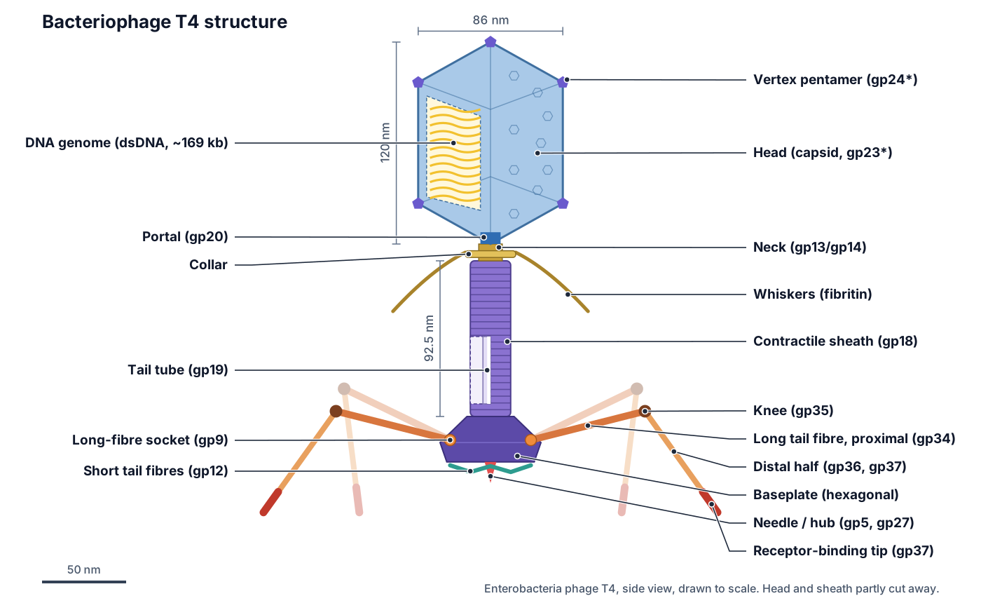
School diagrams often use simpler labels. The NCERT Class 11 figure of a bacteriophage, for example, labels just the head, collar, sheath and tail fibres; some older diagrams add the base plate and tail pins (an old name for the short fibres and spikes under the baseplate). In the 3D model, switch on All labels to see every part at once. (NCERT Biology Class XI, Figure 2.6.)
Bacteriophage structure and functions
Each part of a bacteriophage has a job in one of two tasks: protecting the genome outside the cell, or finding a host and getting the genome into it. The table covers every part a school or university diagram asks for.
| Part | What it is | Function |
|---|---|---|
| Head (capsid) | Prolate icosahedron of gp23*, gp24* and the portal | Protects the DNA; holds it under pressure |
| DNA genome | One linear dsDNA molecule, 168,903 bp | Carries about 300 genes for making new phages |
| Portal | Ring of 12 gp20 at the lower vertex | DNA goes in (assembly) and out (infection) here |
| Neck and collar | Protein rings (gp13, gp14) and a disc | Join head to tail; anchor the whiskers |
| Whiskers | Six fibritin fibres, about 53 nm | Hold the long fibres folded; help attach them |
| Tail sheath | 23 rings of gp18 around the tube | Contracts to drive the tube into the cell |
| Tail tube | Rigid hollow tube of gp19, 4 nm channel | Channel for the DNA |
| Baseplate | Hexagonal dome of six wedges and a hub | Senses binding; triggers contraction |
| Needle (gp5, gp27) | Spike with a lysozyme domain | Pierces the outer membrane; digests cell wall |
| Long tail fibres | Six kinked fibres, 144 nm each | Find the host; bind OmpC or LPS reversibly |
| Short tail fibres | Six gp12 fibres under the baseplate | Bind LPS irreversibly; anchor the phage |
Parts of a bacteriophage: every structure in the 3D model
This is the text version of the 3D model: every labeled structure, grouped by region, with what it does. The E. coli envelope parts are included because they are what the phage interacts with. Use the list as a study sheet or to check a labeling worksheet.
Head (capsid & DNA)
- Capsid (head) (Protein coat; major capsid protein gp23*)
- The protein shell of the head: an elongated (prolate) icosahedron about 120 nm long and 86 nm wide, built mostly from 960 copies of the major capsid protein gp23*. Protects the DNA genome outside the host cell and holds it under high pressure, ready to be injected.
- Vertex pentamers (gp24*) (Vertex protein)
- Five-subunit rings of the protein gp24* that sit at 11 of the 12 corners (vertices) of the head. Close the curved corners of the shell, where a six-sided hexamer cannot fit.
- Hoc (highly immunogenic outer capsid protein) (Decoration protein Hoc)
- A small, fibre-like decoration protein that sticks out from the centre of the gp23* hexamers; about 155 copies per head. Not essential for infection. Because it sits on the outside of the head, it is widely used in biotechnology to display foreign proteins on the phage.
- Soc (small outer capsid protein) (Decoration protein Soc)
- A small 'clamp' protein, about 870 copies per head, that binds between neighbouring gp23* hexamers. Reinforces the shell by clamping neighbouring hexamers together. Like Hoc, it is not essential.
- Portal (gp20) (Portal vertex, connector)
- A ring of 12 gp20 subunits (a dodecamer) that fills the twelfth, lower vertex of the head. The channel through which the DNA is pumped into the empty head during assembly, and through which it leaves at infection. The tail is attached here.
- DNA genome (Double-stranded DNA, about 169 kb)
- One linear double-stranded DNA molecule of 168,903 base pairs, coding for about 300 genes, packed tightly inside the head. Carries the genes that take over the host and make about 300 phage proteins, including all the parts of new phages.
Neck, collar & whiskers
- Neck (gp13 / gp14) (Head–tail connector)
- Short stacked protein rings of gp13 and gp14 that join the portal to the top of the tail. Completes the head and forms the docking site for the finished tail.
- Tail terminator (gp3 / gp15) (Tail-tube terminator and sheath terminator)
- Six-subunit rings that cap the top of the tail tube (gp3) and of the sheath (gp15). Stop the tube and sheath growing at the right length and form the joint that binds the tail to the head.
- Collar (Neck collar)
- A flat disc about 30 nm across and 4 nm thick around the neck, built from the ends of the six whiskers. Anchors the six whiskers to the phage. Textbook diagrams (e.g. NCERT) label this region the collar.
- Whiskers (fibritin, gp wac) (Neck whiskers)
- Six thin protein fibres, each about 53 nm long and 2 nm wide, that stick out from the collar. Act as an environment sensor and chaperone: they help attach the long tail fibres during assembly and hold the fibres folded back against the tail until the phage meets a host.
Tail & baseplate
- Contractile tail sheath (gp18) (Tail sheath)
- A hollow, spring-loaded cylinder of 138 gp18 subunits in 23 stacked six-subunit rings, 92.5 nm long and 24 nm wide when extended. When the baseplate is triggered, the sheath snaps into a shorter, fatter form (42 nm long, 33 nm wide), driving the inner tube through the host's outer membrane like a syringe.
- Tail tube (gp19) (Tail core, inner tube)
- A rigid hollow tube of 138 gp19 subunits, 9 nm across outside and 4 nm inside, running down the centre of the tail. The 'needle' through which the DNA travels from the head into the bacterium.
- Baseplate (Hexagonal baseplate; star-shaped after triggering)
- A hexagonal dome about 52 nm across and 27 nm high at the bottom of the tail, made of six wedges around a central hub: at least 16 different proteins and about 140 chains. The phage's trigger. When the tail fibres bind the host, the baseplate opens from a hexagon into a flat star (61 nm across, 12 nm thick), which releases the short tail fibres and sets off sheath contraction.
- Central hub and needle (gp27, gp5) (Cell-puncturing device, tail lysozyme gp5)
- A 19 nm 'torch'-shaped spike at the centre of the baseplate: a gp27 cylinder holding a gp5 lysozyme and a sharp triple β-helix needle (gp5C) with a tip protein, gp5.4. Punctures the outer membrane, then the lysozyme part of gp5 digests a small hole in the peptidoglycan cell wall so the tube can pass.
- Long-fibre sockets (gp9) (gp9 trimers)
- Six small trimers of gp9 at the corners of the baseplate, one for each long tail fibre. Connect each long tail fibre to the baseplate and pass the 'host found' signal from the fibre to the baseplate.
Tail fibres
- Long tail fibre, proximal half (gp34) (Upper half-fibre)
- The upper half of each of the six long tail fibres: a 70 nm rod of three gp34 chains. Acts as a lever: when the far end binds the host, it swings and signals the baseplate.
- Knee (gp35) (Hinge, kneecap)
- The hinge in the middle of each long tail fibre, formed by a single gp35 protein. Lets the fibre bend, so it can fold back along the tail or reach down to the cell surface.
- Long tail fibre, distal half (gp36, gp37) (Lower half-fibre)
- The lower half of each long fibre, about 74 nm long, made of gp36 near the knee and gp37 towards the tip. Reaches out to find the host and carries the receptor-binding tip.
- Receptor-binding tip (gp37) (Distal tip, adhesin)
- The end of each long fibre, a 15 nm needle-like domain of gp37 that recognises the host. Reversibly binds the receptor: the outer-membrane porin OmpC in E. coli K-12, or the glucosyl-α-1,3-glucose end of rough LPS in E. coli B.
- Short tail fibres (gp12) (Tail pins (older term), short fibres)
- Six short fibres of gp12, each kinked by about 90°, that lie in a head-to-tail 'garland' tucked under the baseplate until the phage is triggered. Make the irreversible attachment: they bind tightly to the inner core of the host LPS, anchoring the baseplate before the sheath contracts.
E. coli envelope
- Lipopolysaccharide (LPS) (Endotoxin layer)
- Sugar-and-lipid molecules that make up the outer leaflet of the E. coli outer membrane (drawn as short bristles). Protects the bacterium from detergents and antibiotics; for T4 its inner core is the binding site of the short tail fibres, and in E. coli B its outer core is the long-fibre receptor.
- OmpC porin (receptor) (Outer membrane protein C)
- A barrel-shaped channel protein (a trimer) in the outer membrane that lets small molecules through. Lets nutrients diffuse into the periplasm; T4 uses it as its primary receptor on E. coli K-12.
- Outer membrane (OM)
- The outer lipid bilayer of the Gram-negative cell envelope, with LPS on the outside and phospholipid on the inside. A barrier the phage must cross; the tail tube is punched through it by sheath contraction.
- Peptidoglycan cell wall (Murein)
- A thin mesh of sugar chains cross-linked by short peptides, lying in the periplasm. Gives the cell its shape and strength; the gp5 lysozyme cuts a small hole in it for the tube.
- Periplasm (Periplasmic space)
- The gel-filled compartment between the outer and inner membranes (drawn see-through). Holds the peptidoglycan wall and many enzymes; the tail tube tip ends here.
- Inner (cytoplasmic) membrane (Plasma membrane)
- The phospholipid bilayer that encloses the cytoplasm. Its proton motive force (the voltage and pH difference across it) is needed to pull the phage DNA into the cell, although fusion with the tube does not need it.
- Host cytoplasm (E. coli cytoplasm)
- The inside of the bacterium, with its own chromosome, ribosomes and enzymes (drawn see-through). Where the phage DNA is copied and new phage parts are made, using the host's ribosomes and energy.
- Injected phage DNA (Entering genome)
- The phage genome entering the cytoplasm through the tail tube after the sheath has contracted. Once inside, its early genes are switched on by the host RNA polymerase and the lytic cycle begins.
T4 bacteriophage structure in numbers
Phage T4 is about 250 nm from the top of its head to the bottom of its baseplate, with a 120 nm × 86 nm head and a tail about 130 nm long including the baseplate. These measurements come from cryo-electron microscopy and crystal structures; each row gives its source.
| Feature | Value | Source |
|---|---|---|
| Head (capsid) | 120 nm long, 86 nm wide; prolate icosahedron, T(end) = 13, T(mid) = 20 | Rao & Black 2010 |
| Capsid proteins per head | 960 gp23*, 55 gp24* (11 pentamers), 12 gp20 (portal), 155 Hoc, 870 Soc | Rao & Black 2010 |
| Genome | Linear dsDNA, 168,903 bp; about 300 genes (289 protein genes, 8 tRNAs) | Miller et al. 2003 |
| DNA packing | About 1.02 genome lengths per head, about 500 mg/ml, 2.5 nm between duplexes | Rao & Black 2010 |
| Packaging motor | Up to 2,000 bp/s; whole genome in about 5 min; forces near 60 pN | Rao & Black 2010 |
| Extended sheath | 92.5 nm long, 24 nm wide; 138 gp18 in 23 rings of 6; 40.6 Å and 17.2° per ring | Leiman et al. 2010 |
| Contracted sheath | 42 nm long, 33 nm wide; 32.9° per ring | Leiman et al. 2010 |
| Tail tube | 138 gp19; 9 nm outside, 4 nm inside | Leiman et al. 2010 |
| Baseplate | Hexagon about 52 nm across, 27 nm high; star 61 nm across, 12 nm thick; ≥16 proteins, about 140 chains | Leiman et al. 2010 |
| Needle (gp5–gp27) | 19 nm long; the gp5 β-helix spike is 11 nm | Leiman et al. 2010 |
| Long tail fibre | About 144 nm (70 nm proximal + 74 nm distal), up to 5 nm wide | Leiman et al. 2010 |
| Whiskers (fibritin) | 53 nm long, 2 nm wide; collar 30 nm across | Leiman et al. 2010 |
| Whole tail before / after firing | About 132 nm before; the tube complex about 114 nm after, its last 16 nm inside the cell | Hu et al. 2015 |
Two measurements differ between sources. Leiman et al. give the hexagonal baseplate as about 52 nm and the star as 61 nm across, while Hu et al. describe the change as a widening from about 40 to 60 nm. Rao and Black give the genome as 171 kb in places, while the reference sequence is 168,903 bp; ViralZone rounds it to about 169 kb. (Leiman et al. (2010): Morphogenesis of the T4 tail and tail fibers, Virology Journal; Hu, Margolin, Molineux & Liu (2015): Structural remodeling of bacteriophage T4 and host membranes during infection initiation, PNAS; ViralZone: Tequatrovirus (T4).)
How a bacteriophage infects a bacterium
A T4 phage infects E. coli in five steps: its long tail fibres bind receptors on the cell surface, the baseplate changes shape and the short tail fibres lock on, the sheath contracts and drives the tail tube through the outer membrane, the needle's lysozyme opens the cell wall, and the DNA passes through the tube into the cytoplasm. Step through it below, and compare the two states of the 3D model above.
1. A free phage meets a cell
Outside a host, T4 is an inert particle. Most of its six long tail fibres are folded up against the sheath, held at the knee by the whiskers. The phage drifts by diffusion until it bumps into an E. coli cell. When fibres flick out and touch the surface, attachment can begin.
Side view, drawn to scale from cryo-EM and cryo-electron tomography measurements (OM = outer membrane). Only the two long fibres in the plane of the page are drawn in full; two of the others are shown paler.
Adsorption (attachment). The tip of each long tail fibre, gp37, recognises the outer-membrane porin OmpC on E. coli K-12, or the glucosyl-α-1,3-glucose end of rough lipopolysaccharide (LPS) on E. coli B. A phage needs at least three long fibres to be infectious. This first binding is reversible. (Leiman et al. 2010.)
Irreversible binding. When the baseplate changes from hexagon to star, the short tail fibres swing down and bind the innermost part of LPS. Miller et al. place this second receptor at the heptose of the LPS inner core; Hu et al. describe it as the lipid A–KDO region. Either way, the phage is now anchored. (Miller et al. (2003): Bacteriophage T4 genome, Microbiology and Molecular Biology Reviews; Hu, Margolin, Molineux & Liu (2015): Structural remodeling of bacteriophage T4 and host membranes during infection initiation, PNAS.)
Sheath contraction and DNA injection. The sheath fires, the tube crosses the outer membrane, and gp5's lysozyme digests the peptidoglycan. Cryo-electron tomograms show that the tube tip stops about 24 nm into the periplasm and the inner membrane bulges out by about 16 nm to fuse with it. Fusion does not need energy from the cell, but moving the DNA across does: it needs the proton motive force of the inner membrane. (Hu et al. 2015.)
Bacteriophage tail sheath: extended vs contracted
The T4 tail sheath is a spring-loaded protein tube: extended, it is 92.5 nm long and 24 nm wide; contracted, it is 42 nm long and 33 nm wide. The extended sheath, like the hexagonal baseplate, is a high-energy, metastable assembly: once triggered, its 138 subunits slide into a tighter packing with about four times more contact between them, releasing the stored energy in one go. A phage can fire only once.
| Feature | Extended (before firing) | Contracted (after firing) |
|---|---|---|
| Sheath length | 92.5 nm | 42 nm |
| Sheath width | 24 nm | 33 nm |
| Twist per ring of 6 subunits | 17.2° | 32.9° |
| Baseplate shape | Hexagonal dome, about 52 nm | Flat star, about 61 nm |
| Short tail fibres | Tucked under the baseplate | Extended, bound to LPS |
| Tail tube | Hidden inside the sheath | Sticks out below the baseplate into the cell |
| Needle (gp5C, gp5.4) | At the tip of the hub | Lost, probably pushed into the cell |
- Contractile tail sheath (gp18): When the baseplate is triggered, the sheath snaps into a shorter, fatter form (42 nm long, 33 nm wide), driving the inner tube through the host's outer membrane like a syringe.
- Tail tube (gp19): The 'needle' through which the DNA travels from the head into the bacterium.
- Baseplate: The phage's trigger. When the tail fibres bind the host, the baseplate opens from a hexagon into a flat star (61 nm across, 12 nm thick), which releases the short tail fibres and sets off sheath contraction.
- Central hub and needle (gp27, gp5): Punctures the outer membrane, then the lysozyme part of gp5 digests a small hole in the peptidoglycan cell wall so the tube can pass.
The same kind of contractile tail is used by many tailed phages, and bacteria have borrowed it: the type VI secretion system that some bacteria use to inject toxins into neighbouring cells appears to be related to a phage tail. (Leiman et al. 2010.)
Bacteriophage life cycle diagram: lytic and lysogenic
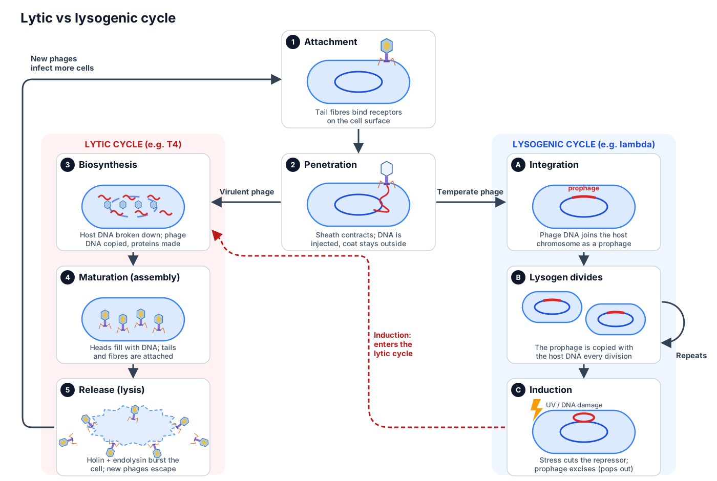
Bacteriophages reproduce by one of two life cycles. In the lytic cycle the phage multiplies at once and bursts (lyses) the cell to release new phages. In the lysogenic cycle a temperate phage inserts its genome into the host chromosome as a prophage and is passed on quietly when the cell divides, until it is induced to switch to the lytic cycle. (OpenStax Microbiology 6.2.)
Lytic cycle of bacteriophage T4
The lytic cycle of T4 has five stages: attachment, penetration, biosynthesis, maturation and release. T4 is a virulent phage: it can only follow the lytic cycle, and every infected cell dies.
- Attachment. The tail fibres bind specific receptors on the cell surface (OmpC or LPS for T4). Because receptors differ between bacteria, most phages have a narrow host range.
- Penetration. The sheath contracts and the DNA is injected through the tail tube, which acts like a hypodermic needle. The empty protein coat stays outside.
- Biosynthesis. Phage genes are switched on in three waves (early, middle and late). Early proteins shut down host gene expression and break down the host chromosome; the cytosine-free T4 DNA, made with 5-hydroxymethylcytosine, is protected. The phage DNA is then copied many times, and late genes make the head, tail and fibre proteins.
- Maturation (assembly). Heads, tails and fibres are built on separate assembly lines. Each empty head is filled with DNA by the packaging motor at the portal; then the tail joins at the neck and the fibres are added last.
- Release. A phage holin (gene t) makes holes in the inner membrane at a set time, letting the phage endolysin (T4 lysozyme, gene e) reach and digest the peptidoglycan wall. The cell bursts and the new phages escape. Without either protein, lysis does not happen.
(OpenStax Microbiology, 6.2 The viral life cycle; Miller et al. (2003): Bacteriophage T4 genome, Microbiology and Molecular Biology Reviews.)
The number of phages released from one cell is the burst size. Classic measurements for the T phages give bursts of roughly 120 to 300 per cell. (BioNumbers BNID 105870.) T-even phages can also delay lysis when many other phages are around (lysis inhibition), making more phages from each cell while hosts are scarce. (Miller et al. 2003.)
Lysogenic cycle: phage lambda
In the lysogenic cycle a temperate phage such as lambda (λ) joins its DNA to the host chromosome instead of killing the cell. The integrated phage genome is called a prophage, and a bacterium carrying one is a lysogen. The prophage is copied along with the host chromosome every time the cell divides. (OpenStax Microbiology 6.2.)
- Entry. Lambda binds LamB, an outer-membrane porin used for maltose uptake, and injects its 48,502 bp genome through its long, flexible tail. The genome circularises by its 12-base sticky ends (cos sites).
- The decision. The phage protein CII tips the cell towards lysogeny; the competing regulator Cro favours the lytic cycle.
- Integration. The phage enzyme integrase (Int) joins the phage circle to the chromosome by site-specific recombination.
- Staying quiet. The CI repressor keeps the lytic genes switched off, so the lysogen grows normally.
- Induction. When the host DNA is damaged, for example by ultraviolet light, the host protein RecA is activated by single-stranded DNA. Activated RecA makes CI cut itself, the repressor falls away, Int and Xis cut the prophage out (excision), and the phage enters the lytic cycle.
- Release. Lambda lyses the cell with three kinds of protein: the holin S105 makes large holes in the inner membrane, the endolysin R digests the cell wall, and the spanins Rz and Rz1 break the outer membrane.
A prophage can change the bacterium it lives in, which is called phage conversion. The genes for the cholera toxin of Vibrio cholerae and the botulinum toxin of Clostridium botulinum are carried by phages. Phages can also move bacterial genes between cells: in generalized transduction a random piece of host DNA is packed into a phage head during the lytic cycle, and in specialized transduction an excising prophage takes neighbouring host genes with it. (OpenStax Microbiology 6.2.)
| Feature | Lytic cycle | Lysogenic cycle |
|---|---|---|
| Example phage | T4, T7 (virulent) | Lambda (temperate) |
| Phage DNA | Stays separate; copied many times | Integrated into the host chromosome as a prophage |
| Host cell | Killed (lysed) | Survives and divides |
| New phages made | Yes, released in a burst | No, until induction |
| Duration | One infection, then lysis | Many host generations |
| Ends with | Lysis by holin and endolysin | Induction (e.g. DNA damage), then the lytic cycle |
| Can cause transduction | Generalized | Specialized |
Types of bacteriophage: T4, lambda, T7, M13 and MS2
Bacteriophages come in several shapes. The best-known phages are tailed phages with an icosahedral head and double-stranded DNA; their tails can be contractile (T4), long and flexible (lambda) or short (T7). Others have no tail at all: filamentous phages such as M13 are long protein rods, and small RNA phages such as MS2 are tiny icosahedra.
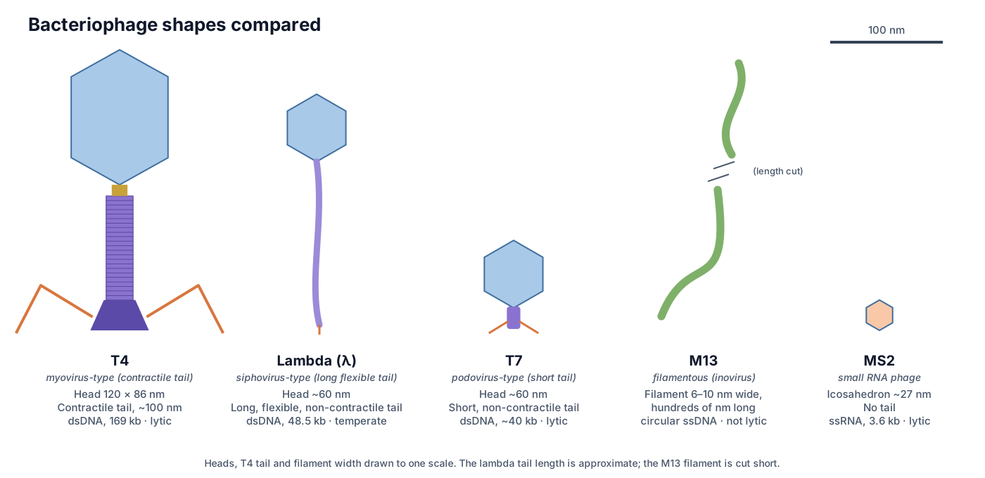
| Phage | Shape | Genome | Notes |
|---|---|---|---|
| T4 | Prolate head 120 × 86 nm; contractile tail | Linear dsDNA, 169 kb | Virulent; injects DNA by sheath contraction |
| Lambda (λ) | Head about 60 nm (T = 7); long, flexible, non-contractile tail | Linear dsDNA, 48.5 kb | Temperate; integrates as a prophage |
| T7 | Head about 60 nm (T = 7); short tail with six short fibres | Linear dsDNA, 40–42 kb | Virulent; short-tail DNA ejection; released by lysis |
| M13 | Flexible filament, 6–10 nm wide, 600–2,500 nm long (family range) | Circular ssDNA | Enters via the tip of a pilus; leaves by extrusion without killing the cell |
| MS2 | Icosahedron about 26 nm (T = 3), no tail | Linear (+) ssRNA, about 3.5 kb | Attaches to the F pilus; released by lysis |
(ViralZone: Tequatrovirus (T4); ViralZone: Lambdavirus; ViralZone: Teseptimavirus (T7); ICTV Report: family Inoviridae (M13); ViralZone: Emesvirus (MS2).)
Filamentous bacteriophage. An M13 particle is a helical tube of about 2,700 copies of the major coat protein p8 around a single-stranded DNA circle, plus five copies each of four minor coat proteins (p3, p6, p7 and p9). The p3 protein at one end binds the tip of a pilus, which retracts and brings the phage to the TolQRA entry receptor. New particles are pushed out through the membrane while the cell keeps growing, so M13 causes a chronic infection rather than lysis. This is why filamentous phages became the workhorse of phage display. (ICTV: Inoviridae.)
Myoviridae, Siphoviridae and Podoviridae: the 2022 taxonomy change
Older textbooks classify tailed phages into three families by tail shape: Myoviridae (contractile tail, like T4), Siphoviridae (long, flexible tail, like lambda) and Podoviridae (short tail, like T7), all in the order Caudovirales. In 2022 the International Committee on Taxonomy of Viruses (ICTV) abolished all three families and the order. (Turner et al. 2023.)
The reason is evolution. Genome comparisons showed that each of the old families was polyphyletic: phages with similar tails did not share one common ancestor, so grouping them by shape did not reflect their history. All tailed phages with an icosahedral head and dsDNA genome now sit in the class Caudoviricetes, divided into families defined by shared genes. The T4-like phages are in the family Straboviridae, and the species once called Escherichia virus T4 is now Tequatrovirus T4 (genus Tequatrovirus, subfamily Tevenvirinae). The same update brought in two-part (binomial) species names.
The words myovirus, siphovirus and podovirus are still fine to use as plain descriptions of a phage's shape. The ICTV says they can be used freely, but no longer have any formal taxonomic meaning. Exam syllabi may still use the old family names, so it is worth knowing both. (Turner et al. 2023.)
T-even phages: T2, T4 and T6
The T-even phages are T2, T4 and T6, three closely related E. coli phages with the same head, contractile tail and fibres. The name comes from the 1940s, when phage researchers agreed to work on one standard set of seven phages that all grow on E. coli B, numbered T1 to T7 ("T" for type). The even-numbered ones turned out to be one family, distinct from the "T-odd" phages such as T7. (BioNumbers BNID 106725.)
A T2 diagram therefore looks almost exactly like a T4 diagram: the same prolate head, collar, contractile sheath, baseplate and fibres. T2 is the phage used in the Hershey–Chase experiment, and T4 became the main model for phage structure and genetics.
Phage therapy
Phage therapy is the use of lytic bacteriophages to treat bacterial infections. A phage attacks only the bacteria it can bind, so a well-chosen phage kills the pathogen without harming human cells or most of the normal microbiota, and it multiplies where its host is. (Gordillo Altamirano & Barr 2019.)
Félix d'Hérelle coined the word "bacteriophage" and began treating patients with phages in the early twentieth century. Enthusiasm faded in the West in the 1940s, when antibiotics arrived and reviews criticised early studies for weak methods, but the work continued, notably at the Eliava Institute in Tbilisi, Georgia (founded 1923) and the Hirszfeld Institute in Wrocław, Poland (founded 1952). (Gordillo Altamirano & Barr 2019.)
Rising antibiotic resistance has brought phages back. In a widely reported 2017 case in the United States, a 68-year-old man with a multidrug-resistant Acinetobacter baumannii infection improved after receiving phage cocktails matched to his strain. Treatments today are usually personalised: the patient's bacterium is isolated, phages that kill it are chosen from a library, and they are often given together with antibiotics, which can work in synergy. The main hurdles are the need to identify the exact bacterium, bacteria evolving resistance to the phage, the immune response, and the lack of a regulatory framework designed for living, evolving medicines. (Gordillo Altamirano & Barr 2019.)
Phages in biotechnology: phage display and the Hershey–Chase experiment
Bacteriophages are tools as well as viruses. Two uses appear in almost every biology course: the Hershey–Chase experiment, which showed that genes are made of DNA, and phage display, which won a share of the 2018 Nobel Prize in Chemistry.
The Hershey–Chase experiment
In 1952 Alfred Hershey and Martha Chase grew phage T2 with radioactive sulfur (³⁵S), which labels protein, or radioactive phosphorus (³²P), which labels DNA. They let each batch infect E. coli, shook the cells in a kitchen blender to knock off the phage coats, and spun the mixture in a centrifuge. The ³⁵S stayed in the liquid with the empty coats, while the ³²P went into the pellet of infected cells. So the part of the phage that enters the cell and directs the making of new phages is the DNA. (OpenStax Biology 2e 14.1.)
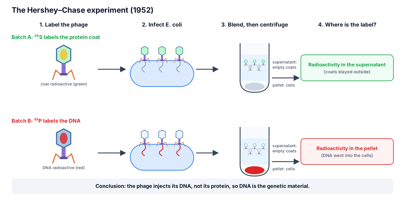
The original paper was more cautious than textbook summaries. Blending removed about 75% of the phage sulfur but only about 15% of the phosphorus, and the authors concluded that the protein "probably has no function in the growth of intracellular phage" and that "the DNA has some function". Even so, the experiment quickly became the classic evidence that DNA, not protein, is the genetic material. (Hershey & Chase 1952.)
Phage display
In phage display, a gene for a foreign peptide or protein is joined to the gene for a phage coat protein, so the new protein is displayed on the outside of the phage while its gene is carried inside. A library of billions of such phages can be washed over a target molecule; the phages that stick are kept, multiplied in bacteria, and selected again. George Smith developed the method in 1985 with filamentous phages, and Gregory Winter used it to evolve antibodies for medicine. The first drug made this way, adalimumab, was approved in 2002 for rheumatoid arthritis, psoriasis and inflammatory bowel diseases. Smith and Winter shared half of the 2018 Nobel Prize in Chemistry "for the phage display of peptides and antibodies". (Nobel Prize in Chemistry 2018.)
T4 is used for display too: its Hoc and Soc head proteins can be fused to pieces of other proteins, and have been used to display antigens from HIV, Bacillus anthracis and foot-and-mouth disease virus for vaccine research. (Rao & Black 2010.)
Bacteriophage for Class 8 and Class 9 (NCERT and state boards): simple summary
For Class 8 and 9 science, a bacteriophage is simply a virus that infects bacteria. Viruses are microscopic, smaller than bacteria, and can only reproduce inside the cells of a host, which may be a bacterium, a plant or an animal. (NCERT Science Class VIII, Chapter 2.)
- Head: a protein box (capsid) with many sides. It holds the genetic material, DNA.
- Collar: the narrow ring that joins the head to the tail.
- Tail (sheath): a hollow tube that works like an injection syringe.
- Base plate: the flat plate at the end of the tail.
- Tail fibres: thin "legs" that hold on to the bacterium.
How it multiplies: the phage sits on a bacterium with its tail fibres, injects its DNA through the tail, and leaves its protein coat outside. The DNA makes the bacterium build many new phages. Then the bacterium bursts open and the new phages are released to infect other bacteria.
Is it living? Outside a cell a virus does nothing at all and can even be crystallised. Inside a cell it reproduces. That is why viruses are said to lie on the border between living and non-living things. A virus has either DNA or RNA, never both; bacteriophages usually have double-stranded DNA. (NCERT Biology Class XI, Chapter 2.)
Drawing tip for exams: draw a six-sided head on top, a short collar, a long rectangular tail with horizontal lines for the sheath, a flat base plate, and six bent tail fibres spreading out below. Label head, DNA, collar, sheath, base plate and tail fibres.
Label the bacteriophage diagram yourself
Test yourself on the parts of a bacteriophage. Write the name of each numbered part, then check your answers below. Numbers run down the left side first, then down the right.
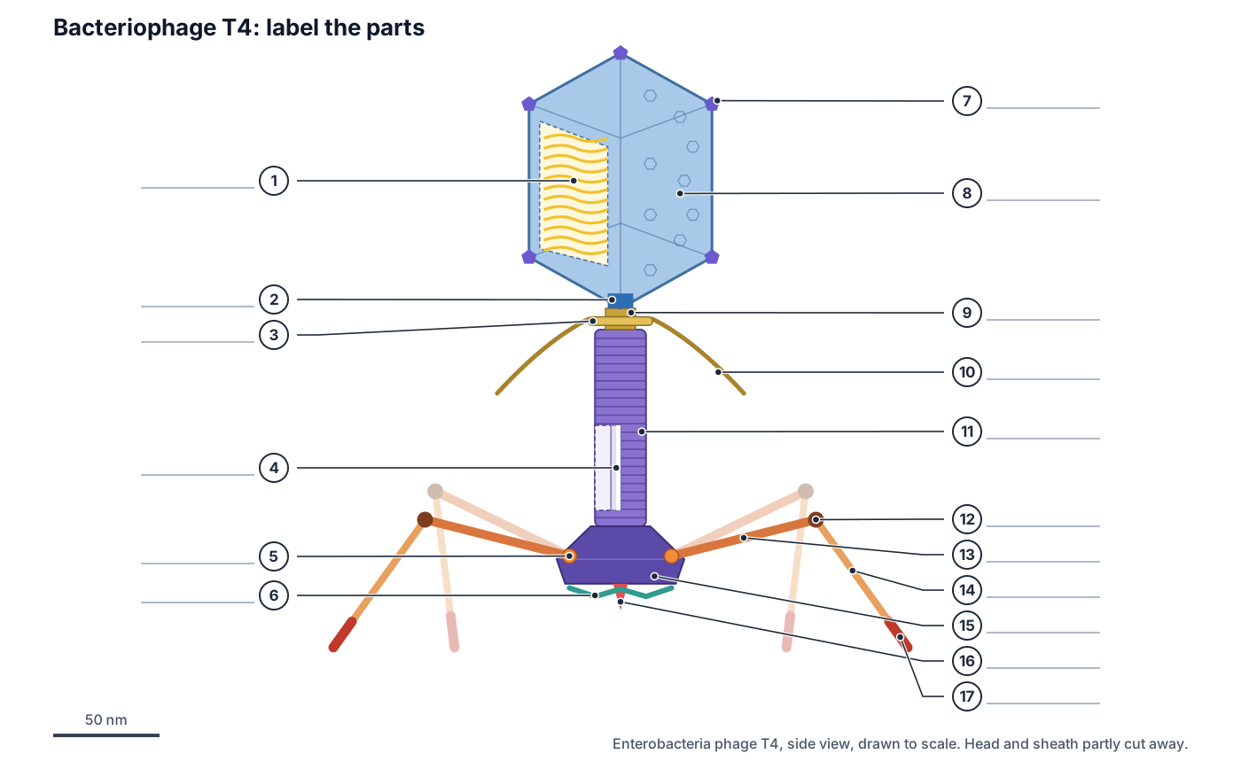
Show the answer key
- DNA genome (double-stranded DNA, about 169 kb)
- Portal (gp20)
- Collar
- Tail tube (gp19), seen through a cut in the sheath
- Long-fibre socket (gp9)
- Short tail fibres (gp12)
- Vertex pentamer (gp24*)
- Head / capsid (gp23*)
- Neck (gp13/gp14)
- Whiskers (fibritin)
- Contractile tail sheath (gp18)
- Knee (gp35)
- Long tail fibre, proximal half (gp34)
- Long tail fibre, distal half (gp36, gp37)
- Baseplate (hexagonal)
- Central hub and needle (gp5, gp27)
- Receptor-binding tip of the long tail fibre (gp37)
Short-answer questions: (1) Which part of the phage enters the bacterium? Only the DNA (and a few internal proteins); the coat stays outside. (2) What makes the tail contract? The baseplate changes from a hexagon to a star after the tail fibres bind the host. (3) Name the two proteins that burst the cell at the end of the lytic cycle. Holin and endolysin (T4 lysozyme). (4) What is a prophage? Phage DNA integrated into the bacterial chromosome during the lysogenic cycle.
Research notes: how the baseplate pulls the trigger
The T4 baseplate is a spring-loaded trap. In the free phage it is held in a high-energy, 'metastable' hexagonal shape, and the sheath above it is held stretched. The trigger is mechanical. When a long tail fibre binds the host, the fibre swings like a lever, moving the gp9 socket and the gp11 trimer it is attached to. Each gp11–gp10 unit then rotates by about 100°, which loosens the garland of short tail fibres and lets the whole baseplate unlock into its low-energy star shape. (Leiman et al. (2010): Morphogenesis of the T4 tail and tail fibers, Virology Journal.)
Because the long and short fibres compete for the same binding site on gp11, only one kind can be locked in place at a time. That makes the switch all-or-nothing: either the baseplate stays hexagonal, or it opens fully and fires the sheath. The same kind of machine turns up outside phages: the type VI secretion system, found in at least a quarter of Gram-negative bacteria, appears to be related to a phage tail. (Leiman et al. (2010): Morphogenesis of the T4 tail and tail fibers, Virology Journal.)
Cryo-electron tomography of real infections added a twist to the textbook picture. Most long fibres on a free phage are folded back against the sheath, and probably only one to three of them bind receptors before the short fibres grab on. Only after contraction do the whiskers bend and release all six long fibres. (Hu, Margolin, Molineux & Liu (2015): Structural remodeling of bacteriophage T4 and host membranes during infection initiation, PNAS.)
Research notes: why T4 DNA is chemically different
T4 DNA contains no ordinary cytosine. Phage enzymes make 5-hydroxymethylcytosine (HMC) instead, and other phage enzymes then attach glucose to it. The sugar coat protects T4 DNA from many host restriction enzymes, and the difference lets T4 nucleases chop up the host's cytosine-containing DNA while leaving its own alone. (Miller et al. (2003): Bacteriophage T4 genome, Microbiology and Molecular Biology Reviews.)
The T4 genome is 168,903 base pairs and codes for about 300 gene products: 289 probable protein-coding genes, 8 tRNA genes and a few other small RNAs. Its genes are switched on in three waves, from 39 early, 30 middle and 50 late promoters. Middle genes need the phage activator MotA; late genes, which code for the head, tail and fibres, need a phage sigma factor, gp55. (Miller et al. (2003): Bacteriophage T4 genome, Microbiology and Molecular Biology Reviews.)
Research notes: the DNA-packaging motor
An empty T4 head is filled by a motor that docks on the portal vertex. The large terminase, gp17, forms a five-subunit ring that burns ATP to thread the DNA in at up to 2,000 base pairs per second, so the whole genome goes in in about 5 minutes. The motor can push against forces of about 60 piconewtons, which makes it, scaled up, about twice as powerful as a car engine. (Rao & Black (2010): Structure and assembly of bacteriophage T4 head, Virology Journal.)
Packing stops when the head is full, not when a particular DNA sequence is reached. That 'headful' rule means each head holds about 1.02 genome lengths. The extra bit is a repeat of the start of the genome at the end (terminal redundancy), and because each new head starts packing where the last one stopped, every phage's DNA starts at a different place (circular permutation). The DNA ends up at about 500 mg/ml, with neighbouring duplexes 2.5 nm apart, like a liquid crystal. (Rao & Black (2010): Structure and assembly of bacteriophage T4 head, Virology Journal.)
Frequently asked questions
What are the parts of a bacteriophage?
A T4 bacteriophage has a head (an icosahedral protein capsid holding double-stranded DNA), a neck and collar with whiskers, a tail made of a hollow tube inside a contractile sheath, a hexagonal baseplate, six long tail fibres and six short tail fibres. Simple school diagrams label the head, collar, sheath, base plate and tail fibres.
How big is a bacteriophage?
Phage T4 is about 250 nm from the top of its head to the bottom of its baseplate: the head is 120 nm long and 86 nm wide, and the extended tail is about 130 nm long including the baseplate. Each long tail fibre is about 144 nm. The whole phage is about an eighth of the length of the 2 µm E. coli cell it infects, and too small to see with a light microscope.
What is the function of the tail fibres of a bacteriophage?
The six long tail fibres find and reversibly bind receptors on the host (OmpC or LPS on E. coli). Their binding triggers the baseplate, which releases six short tail fibres that bind the LPS core irreversibly and anchor the phage before it injects its DNA.
Is a bacteriophage living or non-living?
A bacteriophage has genes and evolves, but it has no cell, no metabolism and cannot reproduce on its own. Outside a host it is an inert particle. Most biologists describe viruses as on the border between living and non-living, and textbooks such as NCERT note they are not truly living in the cellular sense.
What is the difference between the lytic and lysogenic cycle?
In the lytic cycle the phage takes over the cell at once, makes new phages and bursts (lyses) the cell to release them. In the lysogenic cycle a temperate phage such as lambda inserts its DNA into the host chromosome as a prophage, which is copied quietly with the host DNA until something such as DNA damage induces it to enter the lytic cycle. T4 is lytic only.
What type of genetic material does a bacteriophage have?
It depends on the phage. T4, lambda and T7 have double-stranded DNA; M13 has single-stranded circular DNA; MS2 has single-stranded RNA. T4's genome is linear dsDNA of 168,903 base pairs with about 300 genes.
What does the tail sheath of a bacteriophage do?
The contractile sheath is a spring-loaded protein tube around the tail tube. When the baseplate is triggered, it shortens from 92.5 nm to 42 nm and widens, pushing the rigid inner tube through the bacterium's outer membrane so the DNA can be injected.
Why is T4 called a T-even phage?
In the 1940s a group of seven E. coli phages were named T1 to T7 ('T' for type). T2, T4 and T6, the even-numbered ones, turned out to be closely related, with the same head-tail shape and contractile tails, so they are called the T-even phages. T2 was the phage used in the Hershey–Chase experiment.
What is phage therapy?
Phage therapy is the use of lytic bacteriophages to treat bacterial infections. Because each phage infects only certain bacteria, it can kill a pathogen while sparing the normal microbiota. It is being revived for infections that resist antibiotics, usually as personalised phage cocktails used alongside antibiotics.
References
- Rao, V. B. & Black, L. W. (2010). Structure and assembly of bacteriophage T4 head. Virology Journal 7, 356.
- Leiman, P. G., Arisaka, F., van Raaij, M. J., Kostyuchenko, V. A., Aksyuk, A. A., Kanamaru, S. & Rossmann, M. G. (2010). Morphogenesis of the T4 tail and tail fibers. Virology Journal 7, 355.
- Hu, B., Margolin, W., Molineux, I. J. & Liu, J. (2015). Structural remodeling of bacteriophage T4 and host membranes during infection initiation. PNAS 112, E4919–E4928.
- Miller, E. S., Kutter, E., Mosig, G., Arisaka, F., Kunisawa, T. & Rüger, W. (2003). Bacteriophage T4 genome. Microbiology and Molecular Biology Reviews 67, 86–156.
- ViralZone (SIB Swiss Institute of Bioinformatics). Tequatrovirus.
- ViralZone. Lambdavirus; Teseptimavirus; Emesvirus.
- ICTV Report on Virus Taxonomy: family Inoviridae.
- Casjens, S. R. & Hendrix, R. W. (2015). Bacteriophage lambda: early pioneer and still relevant. Virology 479–480, 310–330.
- Turner, D. et al. (2023). Abolishment of morphology-based taxa and change to binomial species names: 2022 taxonomy update of the ICTV bacterial viruses subcommittee. Archives of Virology 168, 74.
- OpenStax Microbiology, sections 6.1 Viruses and 6.2 The viral life cycle.
- Hershey, A. D. & Chase, M. (1952). Independent functions of viral protein and nucleic acid in growth of bacteriophage. Journal of General Physiology 36, 39–56.
- OpenStax Biology 2e, section 14.1 Historical basis of modern understanding.
- Gordillo Altamirano, F. L. & Barr, J. J. (2019). Phage therapy in the postantibiotic era. Clinical Microbiology Reviews 32, e00066-18.
- The Royal Swedish Academy of Sciences (2018). The Nobel Prize in Chemistry 2018, press release.
- NCERT (2026–27 reprint). Biology, Class XI, Chapter 2: Biological classification; Science, Class VIII, Chapter 2: Microorganisms: friend and foe.
- BioNumbers (Milo et al. 2010): BNID 106725 and 105870, the T-system phages, from Brock (1990) and Demerec & Fano (1945).
More interactive anatomy models
- Bacterial cell diagramLabeled prokaryotic cell: cell wall, membrane, nucleoid, ribosomes, pili and flagellum.
- Amoeba diagramLabeled Amoeba proteus: pseudopodia, nucleus, contractile and food vacuoles.
- Euglena diagramLabeled Euglena: flagellum, eyespot, chloroplasts, pellicle and contractile vacuole.
- Virus structureCapsid, envelope, spikes and genome of helical, icosahedral and enveloped viruses.
- Human anatomy1,000+ bones, muscles, organs, vessels and nerves from real anatomical data.
- Octopus anatomyThree hearts, a ring-shaped brain, eight arms and a beak, in 3D.