

What is a virus made of? The basic structure of a virus
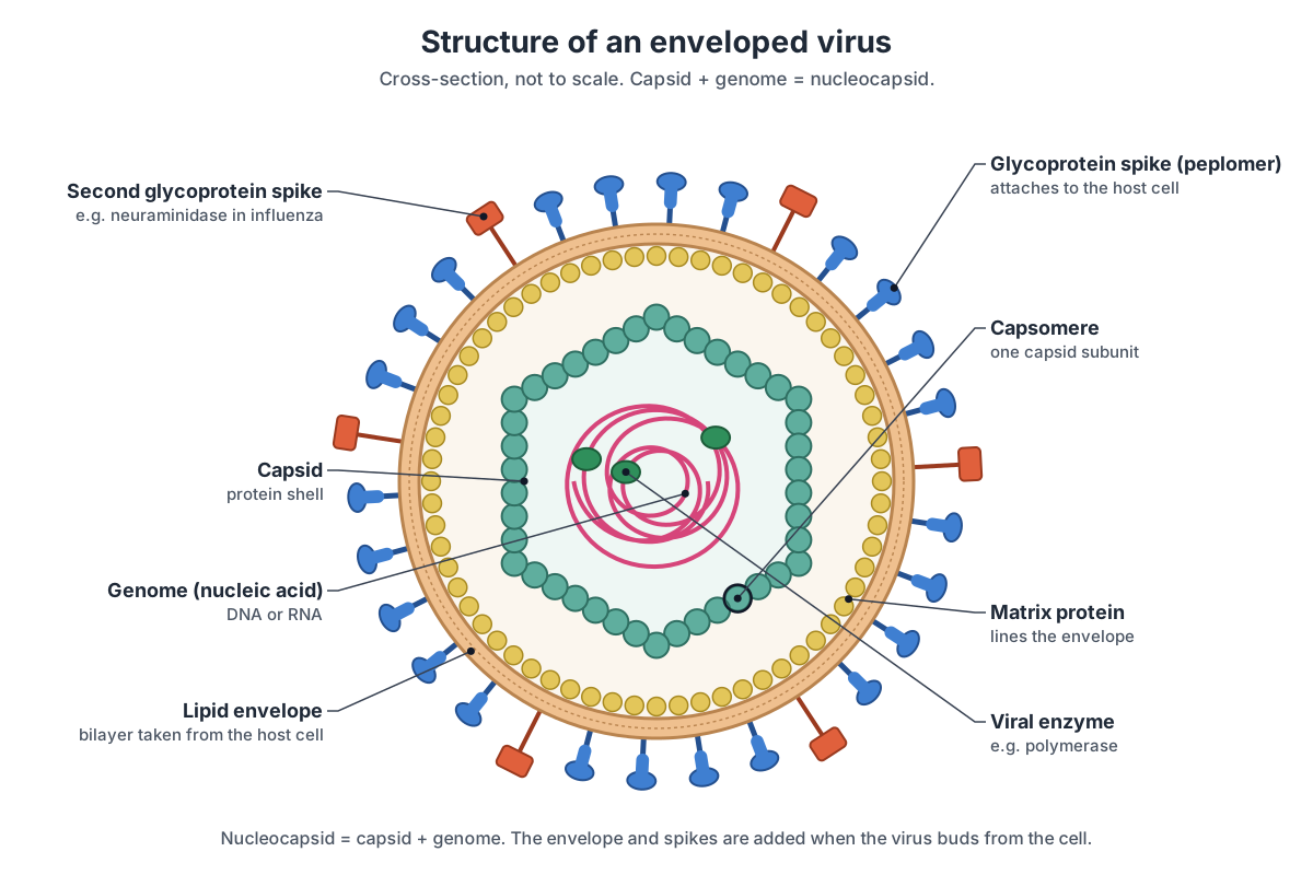
A virus particle, called a virion, is made of a genome (DNA or RNA, never both) enclosed in a protein coat called the capsid. The capsid is built from many repeating protein subunits called capsomeres. The genome and capsid together are the nucleocapsid. Many viruses also have a lipid envelope around the nucleocapsid, studded with glycoprotein spikes (peplomers), and some have a matrix protein layer under the envelope and enzymes packed inside.
That is the whole list. A virion has no cytoplasm, no ribosomes, no mitochondria and no metabolism. Its job is to protect the genome outside a cell, find the right host cell, and deliver the genome inside. Everything else happens inside the host cell, using the host's machinery.
| Part | What it is | Found in |
|---|---|---|
| Genome | DNA or RNA; single- or double-stranded; one piece or several segments | Every virus |
| Capsid | Protein shell around the genome, coded by the virus | Every virus |
| Capsomere | One repeating protein unit of the capsid (visible bumps in electron micrographs) | Every virus |
| Nucleocapsid | Capsid plus genome; in helical viruses, the protein-coated RNA strand itself | Every virus |
| Envelope | Lipid bilayer taken from a host-cell membrane during budding | Enveloped viruses only |
| Spikes (peplomers) | Viral glycoproteins in the envelope or on the capsid that attach to cells and fuse membranes | Most viruses; on the capsid in naked viruses such as adenovirus |
| Matrix protein | Protein layer between the envelope and the nucleocapsid | Many enveloped viruses (influenza, HIV, rabies, measles) |
| Tegument | Protein-filled space between capsid and envelope | Herpesviruses |
| Enzymes | Viral polymerase, reverse transcriptase, integrase, protease or neuraminidase carried in the virion | Some viruses, especially RNA viruses and retroviruses |
Two simple rules help when you label a diagram. First, if a part is made of lipid, it came from the host cell; if it is protein or nucleic acid, it is encoded by the virus. Second, only the envelope is optional for the virus to count as complete: some viruses are "naked" (non-enveloped), but no virus is without a capsid and a genome.
Virus structure diagram, labeled
A labeled diagram of an enveloped virus shows, from outside in: the glycoprotein spikes, the lipid envelope, the matrix protein layer, the capsid made of capsomeres, and the nucleic acid genome inside, often with a viral enzyme bound to it. This is the generic textbook picture; real viruses vary the theme, as the 3D models above show.
The influenza model in the viewer is the real-life version of this diagram. Its envelope carries two kinds of spike, hemagglutinin (HA, blue) and neuraminidase (NA, red), plus a few M2 ion channels. The matrix protein M1 lines the envelope. Instead of one icosahedral capsid, influenza has eight separate helical nucleocapsids (RNPs), each an RNA segment wrapped around nucleoprotein, with a polymerase at one end. Use the tabs to compare it with HIV, SARS-CoV-2, a naked adenovirus and the helical tobacco mosaic virus.
Structure of viruses with diagram: every labeled part
This is the text version of the five 3D models: every labeled structure, grouped by virus and by layer, with what it does. Use it to revise or to check a labeled virus structure diagram.
Influenza A virus: a typical enveloped virus
Envelope & spikes
- Lipid envelope (Viral membrane, lipid bilayer)
- The outer membrane of the virion: a lipid bilayer taken from the host cell's plasma membrane as the virus buds out. Holds the virion together and carries the HA, NA and M2 proteins. It fuses with the membrane of an endosome to release the genome into the cell.
- Hemagglutinin (HA) (HA spike, H antigen)
- A rod-shaped glycoprotein spike, a trimer, and the most common protein on the virion surface. Binds sialic acid on the host cell (attachment), then changes shape at low pH to fuse the viral envelope with the cell membrane.
- Neuraminidase (NA) (NA spike, N antigen)
- A mushroom-shaped glycoprotein spike: a box-shaped enzyme head on a thin stalk, made of four identical subunits (a tetramer). An enzyme that cuts sialic acid, so new virions are released from the cell and do not clump together.
- M2 ion channel (Matrix protein 2, proton channel)
- A small four-part (tetrameric) channel protein that crosses the envelope. Only a few copies are present. Lets protons (H⁺ ions) into the virion inside the endosome. The acid loosens M1 from the RNPs so the genome can be released (uncoating).
Matrix (M1)
- Matrix protein (M1) (Matrix layer)
- A protein layer lining the inside of the envelope, like scaffolding under a tent. Gives the virion its shape, links the spikes' tails to the RNPs, and organizes budding.
Genome (RNPs)
- Nucleoprotein (NP) and RNPs (Ribonucleoprotein (RNP) segments, nucleocapsid)
- Eight rod-shaped ribonucleoprotein complexes (RNPs). Each is one RNA segment coated with many copies of nucleoprotein, folded back on itself into a twisted hairpin. NP coats and protects the RNA and keeps it in a form the polymerase can copy. The RNPs are the nucleocapsid of influenza.
- Viral RNA genome (8 segments) (vRNA, negative-sense single-stranded RNA)
- Eight separate pieces of negative-sense single-stranded RNA, 13.5 kb in total, wrapped around the nucleoprotein. Carries the genes for 12–14 proteins. Being negative-sense, it must be copied into mRNA by the viral polymerase before ribosomes can read it.
- RNA polymerase (PB1, PB2, PA) (RNA-dependent RNA polymerase, RdRp)
- A three-protein enzyme complex, one bound to the end of each RNP. Copies the negative-sense RNA into mRNA and into new genome copies. PA 'cap-snatches' the caps of host mRNAs to prime viral mRNA.
Adenovirus: an icosahedral, non-enveloped virus
Capsid
- Hexon (Polypeptide II, hexon capsomere)
- The main building block (capsomere) of the capsid: a trimer of hexon protein with a six-sided base. There are 240 hexons. Forms the protective protein shell around the DNA and carries the main type-specific antigens.
Vertices & fibres
- Penton base (Polypeptide III)
- A five-sided capsomere, a pentamer, at each of the 12 corners (vertices) of the icosahedron. Anchors the fibre and binds integrins on the host cell, which triggers uptake of the virus.
- Fibre (Fiber, polypeptide IV)
- A long, thin trimeric spike projecting from each penton base. Reaches out to the host cell; its knob makes the first contact.
- Fibre knob (Fiber head)
- The rounded head at the tip of each fibre. Binds the coxsackievirus–adenovirus receptor (CAR) on the host cell: the attachment step.
Minor proteins
- Protein IX (Cement protein IX)
- A small 'cement' protein that sits in the grooves between hexons on the outer surface. Glues the hexons together and stabilizes the capsid.
- Protein IIIa
- A minor capsid protein on the inner surface, five copies under each vertex. Helps hold the vertex region together.
- Protein VI
- A minor protein inside the hexon cavities, linking the capsid to the core. Binds hexons and DNA. After entry, it helps the virus break out of the endosome.
Core & genome
- Double-stranded DNA genome (dsDNA, about 36 kb)
- One linear molecule of double-stranded DNA, about 35–36 kb, packed tightly inside the capsid. Carries about 40 genes. It is delivered into the cell nucleus, where host enzymes transcribe it.
- Core protein VII (Histone-like protein VII)
- A small, basic core protein that binds and condenses the DNA, a little like histones in our chromosomes. Packs the genome tightly enough to fit in the capsid.
- Core protein V
- A core protein on the outside of the DNA–protein VII core. Thought to bridge the core to the capsid (via protein VI).
- Terminal protein (TP)
- A protein covalently linked to each end of the DNA. Acts as the primer when the DNA is copied in the next cell.
- Adenovirus protease (AVP) (L3 23K protease)
- The virus's own enzyme, packaged inside the virion. Cuts the precursor proteins (IIIa, VI, VIII, VII, μ, TP) to make a mature, infectious virion.
Tobacco mosaic virus: a helical virus
Helical capsid
- Helical capsid (Protein coat, rod)
- A rigid, hollow rod about 300 nm long and 18 nm wide, built from about 2,130 identical coat-protein subunits stacked in a helix. Protects the RNA, and falls apart in the plant cell to release it.
- Coat protein subunit (capsomere) (Capsid protein)
- One coat-protein molecule, a small wedge-shaped protein. Thousands of copies make the rod; one is highlighted here. Each subunit binds three nucleotides of the RNA, so the protein and RNA assemble together.
- Central channel (Axial canal)
- A narrow hollow running along the axis of the rod. Empty space; it shows that the RNA is not in the middle but wound inside the protein.
RNA genome
- Single-stranded RNA genome (ssRNA(+), about 6.4 kb)
- One positive-sense RNA strand, 6.3–6.5 kb, wound in a helix between the turns of protein. Acts directly as mRNA in the plant cell; it encodes the replicase, a movement protein and the coat protein.
HIV-1: an enveloped retrovirus
Envelope & Env
- Lipid envelope (Viral membrane)
- A lipid bilayer taken from the plasma membrane of the infected cell when the virion buds. Carries the Env spikes and fuses with the target cell membrane to deliver the core.
- gp120 (surface glycoprotein) (SU, Env surface subunit)
- The heavily glycosylated outer part of each Env spike; three gp120s form the spike's head. Binds the CD4 receptor and then a co-receptor (CCR5 or CXCR4) on helper T cells and macrophages.
- gp41 (transmembrane glycoprotein) (TM, Env transmembrane subunit)
- The stalk of each Env spike: three gp41 molecules that cross the envelope. Anchors the spike; after gp120 binds the receptors, gp41 refolds and pulls the viral and cell membranes together so they fuse.
Matrix (p17)
- Matrix protein (p17, MA)
- A protein layer lining the inside of the envelope. Targets the Gag protein to the membrane during assembly and helps bring Env into the virion.
Capsid core (p24)
- Conical capsid (p24, CA) (Core, cone-shaped capsid)
- A cone-shaped protein shell, the core, made of p24 capsid protein arranged in hexamers, closed by 12 pentamers. Protects the RNA and enzymes, and carries them to the nucleus, where reverse transcription completes.
Genome & enzymes
- Genomic RNA (two copies) (Dimeric ssRNA(+), 9.75 kb)
- Two identical copies of positive-sense single-stranded RNA, about 9.75 kb each, linked near one end. Template for reverse transcriptase to make the DNA copy that is integrated into the host genome.
- Nucleocapsid protein (p7, NC)
- A small, basic protein that coats the RNA inside the core. Packs and protects the RNA and helps reverse transcription.
- Reverse transcriptase (RT) (p66/p51)
- The enzyme that copies RNA into DNA, the step that gives retroviruses their name. Makes double-stranded DNA from the RNA genome; its RNase H part removes the RNA strand.
- Integrase (IN) (p31)
- The enzyme that cuts the host DNA and inserts the viral DNA. Integrates the viral DNA into a host chromosome, where it stays as a provirus.
- Protease (PR) (p11)
- The enzyme that cuts the Gag and Gag-Pol polyproteins into their working parts. Triggers maturation: without it, the cone-shaped core does not form and the virion is not infectious.
SARS-CoV-2: a coronavirus
Envelope proteins
- Lipid envelope (Viral membrane)
- A lipid bilayer taken from internal membranes of the host cell (coronaviruses bud into the ER–Golgi compartment). Holds the S, M and E proteins; it fuses with the host cell membrane.
- Spike (S) glycoprotein (Peplomer, S trimer)
- Club-shaped spikes, each a trimer of S protein: the 'crown' (corona) seen in electron micrographs. The S1 part binds the ACE2 receptor on human cells; the S2 part then drives fusion of the viral and cell membranes.
- Membrane (M) protein
- The most abundant protein in the envelope, small and mostly embedded in the membrane. Shapes the envelope, gathers the other structural proteins, and binds the nucleocapsid during assembly.
- Envelope (E) protein
- A tiny membrane protein present in only a few copies, which forms an ion channel (a pentamer). Helps assembly and budding of new virions.
Nucleocapsid
- Nucleocapsid (N) protein (Ribonucleoprotein (RNP) complexes)
- An RNA-binding protein that packs the long genome into many small ribonucleoprotein particles. Coats and condenses the RNA and links it to M for packaging.
- Single-stranded RNA genome (ssRNA(+), about 30 kb)
- One positive-sense RNA molecule of about 30 kb, threaded through the N particles like beads on a string. Acts directly as mRNA in the cell; it encodes the replicase and the structural proteins.
Capsid symmetry: helical, icosahedral and complex viruses
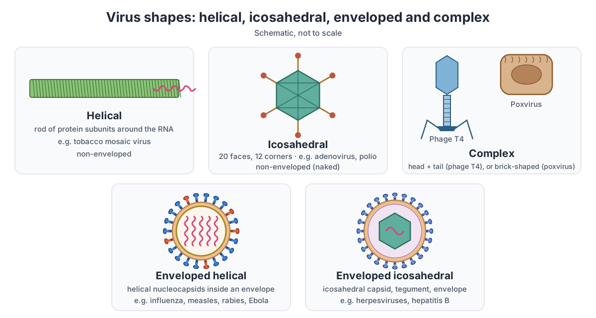
Virus capsids come in three basic architectures: helical (a rod or filament), icosahedral (a closed, roughly spherical shell with 20 triangular faces and 12 corners), and complex (anything that is not simply one or the other). OpenStax uses tobacco mosaic virus as the helical example and bacteriophage T4 and poxviruses as complex examples.
Helical capsids
In a helical capsid, identical protein subunits stack in a spiral around the genome, like steps of a spiral staircase, and the nucleic acid runs in a groove between the turns. The length of the rod is set by the length of the genome. Tobacco mosaic virus is the classic case: a rigid rod about 300 nm long and 18 nm wide, built from about 2,130 coat-protein subunits, 16⅓ per turn, each binding three nucleotides of RNA. Many enveloped RNA viruses (influenza, measles, rabies, Ebola) also have helical nucleocapsids, but these are flexible and coiled up inside the envelope, so the virion itself is not rod-shaped.
Icosahedral capsids and the triangulation number
An icosahedron is the most efficient way to build a closed shell from many copies of one small protein. The simplest one has 60 identical subunits, three per face. Bigger capsids use more subunits in slightly different (quasi-equivalent) positions. The triangulation number, T, counts how many of these positions there are per face: T = h² + hk + k², where h and k are whole numbers that describe how many steps you take on the surface lattice from one corner (pentamer) to the next. Allowed values are 1, 3, 4, 7, 9, 12, 13 and so on. A capsid with triangulation number T is made of 60T subunits, arranged as 12 pentamers (always 12, at the corners) plus 10(T − 1) hexamers.
| Virus | T number | What it means |
|---|---|---|
| Poliovirus, hepatitis A | pseudo T = 3 | 60 protomers, each of three different proteins (VP1, VP2, VP3), in place of 180 identical subunits |
| Hepatitis B core | T = 4 | Icosahedral core inside the 42 nm Dane particle |
| Herpes simplex virus | T = 16 | 162 capsomeres: 150 hexons and 12 pentons |
| Adenovirus | pseudo T = 25 | 240 hexon trimers and 12 penton bases, with a fibre at each corner |
"Pseudo" means that the subunits are not all the same protein but the lattice has the same geometry. In adenovirus, each hexon is a trimer of one large protein with a six-sided base, so it fills a hexamer position without being six copies of one subunit.
Complex viruses
Complex viruses do not fit either plan. Bacteriophage T4 has an icosahedral (elongated) head attached to a helical tail with a baseplate and tail fibres; see our bacteriophage diagram for its structure. Poxviruses, including vaccinia and mpox, are large brick-shaped particles, about 250 × 200 nm, with surface tubules and several membranes, and no simple capsid symmetry.
Enveloped vs non-enveloped (naked) viruses
An enveloped virus has a lipid membrane around its nucleocapsid; a non-enveloped or naked virus does not, so its capsid is the outermost layer. The envelope is acquired when the virus buds through a host-cell membrane, usually the plasma membrane (influenza, HIV) or an internal membrane, such as the ER–Golgi compartment used by coronaviruses.
| Enveloped virus | Non-enveloped (naked) virus | |
|---|---|---|
| Outer layer | Lipid bilayer from the host cell, with viral glycoprotein spikes | Protein capsid |
| Attachment proteins | Envelope glycoproteins (e.g. HA, gp120, S) | Capsid proteins or fibres (e.g. adenovirus fibre knob) |
| Matrix layer | Often present under the envelope | Absent |
| How it gets out of the cell | Usually by budding, often without killing the cell at once | Usually when the cell bursts (lysis) |
| How it gets into the cell | Envelope fuses with a cell membrane | Capsid crosses or disrupts a membrane |
| Hardiness | Damaged by soap, alcohol, detergents and drying | Usually more resistant to drying, acid and detergents |
| Examples | Influenza, HIV, SARS-CoV-2, herpes, measles, rabies, Ebola, hepatitis B and C | Adenovirus, poliovirus, hepatitis A, norovirus, TMV, most phages |
Envelope and capsid shape are independent features. Influenza and measles are enveloped viruses with helical nucleocapsids; herpesviruses and hepatitis B are enveloped viruses with icosahedral capsids; adenovirus and poliovirus are naked icosahedral viruses; tobacco mosaic virus is a naked helical virus.
Virus genomes: the Baltimore classification
The Baltimore classification sorts viruses into seven groups by the kind of genome they carry and how they turn it into mRNA: double-stranded DNA, single-stranded DNA, double-stranded RNA, positive-sense RNA, negative-sense RNA, and two groups that use reverse transcription. Positive-sense (+) RNA can be read directly by ribosomes; negative-sense (−) RNA is the complement of mRNA and must be copied first.
| Group | Genome | Enzyme the virus must bring or make | Examples on this page |
|---|---|---|---|
| I | dsDNA | DNA polymerase (often the host's) | Adenovirus, herpes simplex, Epstein–Barr virus, poxviruses (mpox), phage T4 |
| II | ssDNA | Rep endonuclease | Parvoviruses (not modelled here) |
| III | dsRNA | RNA-dependent RNA polymerase, carried in the virion | Rotavirus (not modelled here) |
| IV | ssRNA(+) | RNA-dependent RNA polymerase, made after entry | Poliovirus, hepatitis A and C, SARS-CoV-2, dengue, Zika, TMV |
| V | ssRNA(−) | RNA-dependent RNA polymerase, carried in the virion | Influenza (incl. H1N1), measles, RSV, Nipah, rabies, Ebola |
| VI | ssRNA(+) with a DNA step (RT) | Reverse transcriptase, RNase H, integrase | HIV-1 |
| VII | dsDNA with an RNA step (RT) | Reverse transcriptase, RNase H | Hepatitis B virus |
The enzyme column explains which enzymes appear in a structure diagram. Negative-sense RNA viruses such as influenza must pack their polymerase in the virion, because a host cell has no enzyme that copies RNA from an RNA template. HIV packs reverse transcriptase and integrase for the same reason. Positive-sense viruses such as poliovirus carry no enzyme: their RNA is translated straight away to make one.
How big is a virus? Virus size compared with a bacterium and a ribosome
Most viruses are 20–300 nm across, roughly 10 to 100 times smaller than a bacterium, and too small to see with a light microscope. OpenStax gives a typical range of about 20 nm to 900 nm, with recently found giant viruses approaching the size of small bacteria. For comparison, an E. coli cell measured by scanning electron microscopy was 1.78 µm long and 0.49 µm wide, and its 70S ribosome is about 21 nm across, almost as big as a poliovirus.
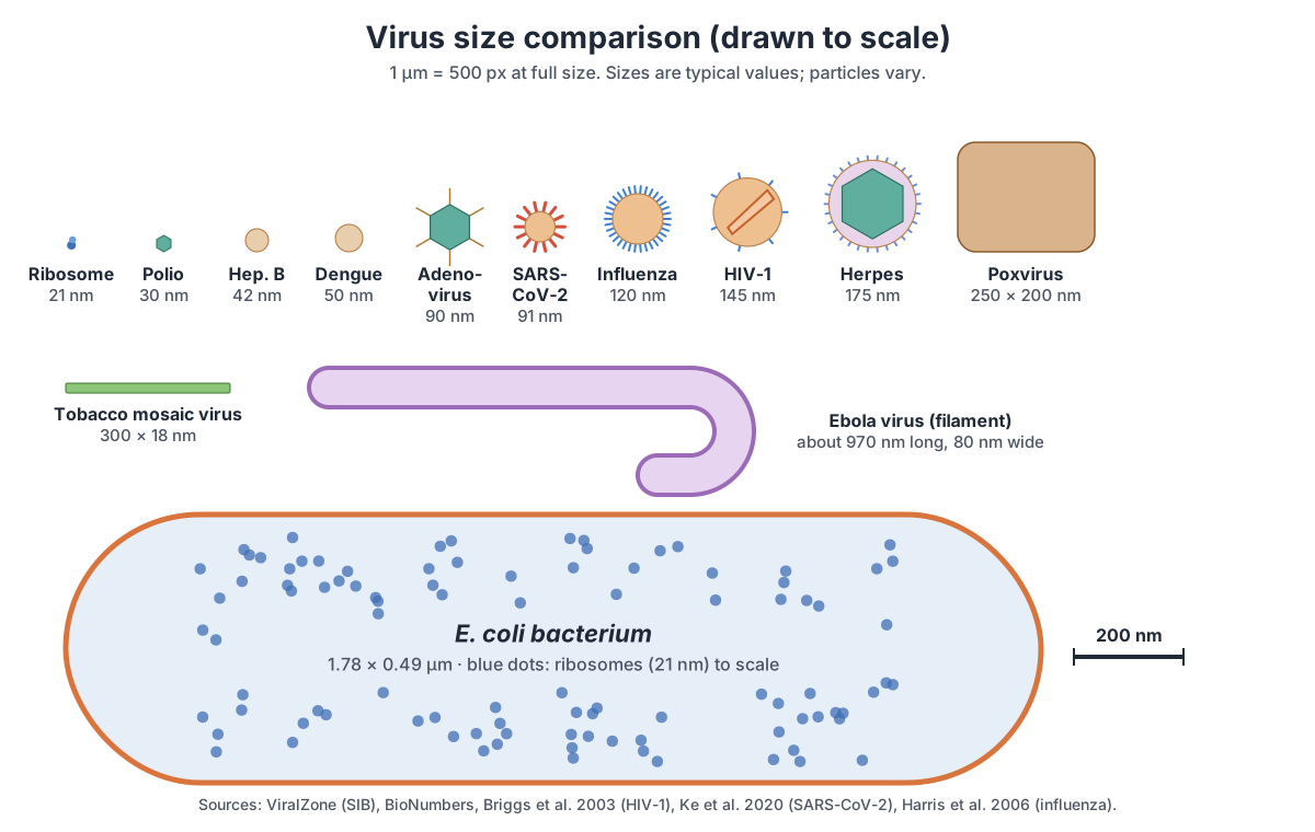
Sizes differ between sources because virions vary from particle to particle and because methods differ: negative-stain electron microscopy dries and flattens particles, while cryo-electron microscopy images them frozen in water. For HIV-1, ViralZone gives 80–100 nm for lentiviruses in general, while cryo-electron tomography of HIV-1 measured 145 ± 25 nm. When a textbook gives one number, treat it as a typical value.
Structure of HIV: labeled diagram
HIV-1 is a roughly spherical enveloped retrovirus. From outside in it has: a lipid envelope carrying Env spikes (gp120 on top of gp41), a matrix layer of p17 under the envelope, a cone-shaped capsid of p24 in the middle, and inside the cone two copies of the single-stranded RNA genome coated with nucleocapsid protein p7, together with the enzymes reverse transcriptase, integrase and protease.
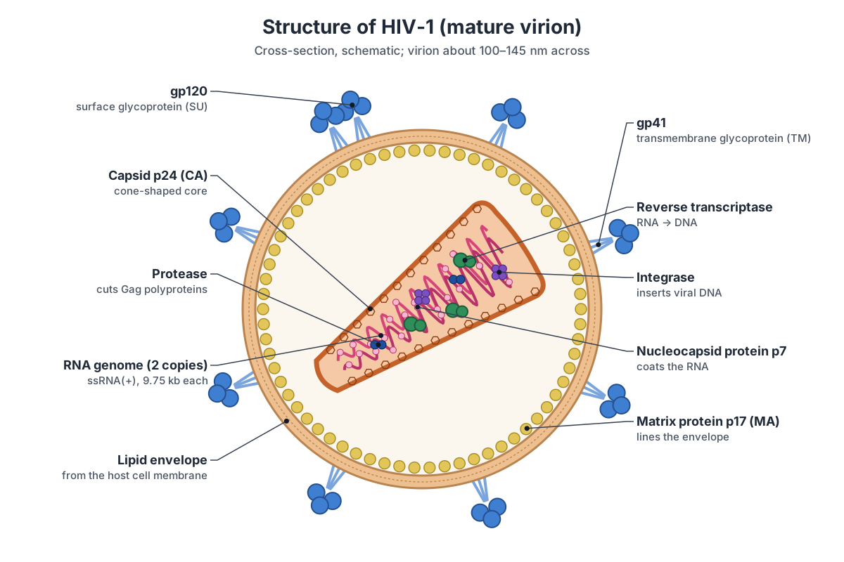
| Part | Protein | What it does |
|---|---|---|
| Envelope spike, outer part | gp120 (SU) | Binds CD4 and a co-receptor (CCR5 or CXCR4) |
| Envelope spike, stalk | gp41 (TM) | Crosses the envelope; drives membrane fusion |
| Matrix | p17 (MA) | Lines the envelope; directs assembly at the membrane |
| Capsid (core) | p24 (CA) | Cone of hexamers closed by 12 pentamers; 1,000–1,500 CA molecules |
| Nucleocapsid | p7 (NC) | Coats and packs the RNA |
| Genome | 2 × ssRNA(+), 9.75 kb | Template for reverse transcription |
| Enzymes | RT, IN, PR | Copy RNA to DNA, insert it into the host genome, and cut Gag and Gag-Pol |
Two numbers make HIV unusual. It carries only about 14 Env spikes per virion, compared with hundreds on an influenza virion, which is thought to help it avoid antibodies. And its mature cores are about 119 nm long and up to 60 nm wide, nearly as long as the virion itself, so the cone sits diagonally across the particle. Switch the viewer to HIV-1 to see the cone inside the cut-away envelope.
Structure of influenza virus (influenza A and H1N1)
Influenza A virus is an enveloped virus, usually rounded and 80–120 nm across, sometimes filamentous. Its envelope carries two spike glycoproteins, hemagglutinin (HA) and neuraminidase (NA), and a few M2 ion channels. The matrix protein M1 lines the envelope, and inside are eight ribonucleoprotein (RNP) segments: negative-sense RNA wrapped around nucleoprotein (NP), each with a polymerase made of PB1, PB2 and PA. The genome totals 13.5 kb.
| Segment | Length (approx.) | Main protein(s) encoded |
|---|---|---|
| 1 | 2,341 nt | PB2 (polymerase subunit; binds mRNA caps) |
| 2 | 2,341 nt | PB1 (catalytic polymerase subunit) |
| 3 | 2,233 nt | PA (polymerase subunit; cap-snatching endonuclease) |
| 4 | about 1,780 nt | HA (hemagglutinin) |
| 5 | about 1,565 nt | NP (nucleoprotein) |
| 6 | about 1,410 nt | NA (neuraminidase) |
| 7 | about 1,027 nt | M1 (matrix) and M2 (ion channel) |
| 8 | 890 nt | NS1 and NEP/NS2 (nuclear export protein) |
Segment lengths vary slightly between strains; ViralZone gives a range of 890 to 2,341 nucleotides. The subtype name comes from the spikes: H1N1 carries HA type 1 and NA type 1, H3N2 carries HA 3 and NA 2. So the structure of H1N1 is the structure shown in the model; only the HA and NA proteins differ in sequence. Because the genome is segmented, two influenza strains in one cell can swap segments, which is how new pandemic strains arise.
Hepatitis B virus structure: the Dane particle (and hepatitis A and C)
The infectious hepatitis B virion, called the Dane particle, is an enveloped sphere about 42 nm across. Its envelope carries the surface antigen (HBsAg). Inside is an icosahedral core with T = 4 symmetry, made of core protein (HBcAg), which holds a small, partially double-stranded circular DNA genome of about 3.2 kb and the viral polymerase. Hepatitis B copies its genome through an RNA intermediate, using reverse transcription, which puts it in Baltimore group VII.
The three hepatitis viruses students usually compare are unrelated and built quite differently. Hepatitis A is a small, non-enveloped picornavirus, about 30 nm across, with a pseudo T = 3 capsid and a 7.5 kb positive-sense RNA genome. Hepatitis C is an enveloped virus about 50 nm across, with E1 and E2 glycoproteins and a positive-sense RNA genome of about 10 kb.
Herpesvirus structure: capsid, tegument and envelope (including Epstein–Barr virus)
Herpesviruses have four layers: a large double-stranded DNA genome, an icosahedral capsid with T = 16 symmetry made of 162 capsomeres, a protein-filled layer called the tegument, and an outer envelope carrying glycoproteins. Herpes simplex virus virions are 150–200 nm across, and the genome is about 152 kb.
The tegument is the feature unique to herpesviruses. Its proteins are delivered into the cell as soon as the envelope fuses, ready to act before any viral gene is switched on. Epstein–Barr virus (EBV) is a herpesvirus with the same layered structure, 150–200 nm across, and a dsDNA genome of about 180 kb according to ViralZone.
Ebola virus structure: a filamentous virus
Ebola virus is filamentous: long, thread-like virions about 80 nm wide and, on average, about 970 nm long, sometimes branched or curled into a "6" or a loop. It is enveloped, with glycoprotein (GP) spikes, and has a helical nucleocapsid running the length of the filament. The genome is negative-sense RNA of about 18–19 kb that encodes seven proteins.
Tobacco mosaic virus structure: a helical virus
Tobacco mosaic virus (TMV) is a rigid, non-enveloped helical rod about 300 nm long and 18 nm wide. About 2,130 identical coat-protein subunits stack in a right-handed helix, 16⅓ per turn, and the single positive-sense RNA genome of 6.3–6.5 kb winds between the turns, three nucleotides per subunit. A narrow channel runs down the middle.
TMV was the first virus to be discovered and one of the first whose structure was worked out, and it is the standard example of a helical virus. Its rod builds itself: coat protein forms two-layer disks, the RNA's assembly signal threads into the hole in the middle of a disk, and more protein adds along the RNA until it is fully covered, so the RNA sets the rod's length. In the TMV model, one end is cut open to show the RNA helix inside the protein, and one subunit is highlighted.
Poliovirus structure: a small icosahedral virus
Poliovirus is one of the smallest and simplest viruses: a non-enveloped icosahedral particle about 30 nm across. Its capsid has pseudo T = 3 symmetry and is made of 60 protomers, each containing the four proteins VP1, VP2, VP3 and VP4, with VP4 on the inside. It encloses a single positive-sense RNA genome of 7.2–8.5 kb with a small viral protein, VPg, attached to its 5' end instead of a cap. Hepatitis A, rhinoviruses (common cold) and enteroviruses share this design.
Rabies virus structure: bullet-shaped
Rabies virus is bullet-shaped: an enveloped virion about 180 nm long and 75 nm wide, rounded at one end and flat at the other. Its envelope carries glycoprotein (G) spikes, a matrix protein lies underneath, and inside a helical nucleocapsid of nucleoprotein-coated RNA is coiled tightly to fill the bullet. The genome is negative-sense RNA of about 11 kb, encoding five main proteins.
Measles, RSV and Nipah virus structure
Measles virus, respiratory syncytial virus (RSV) and Nipah virus are related negative-sense RNA viruses with the same basic plan: a pleomorphic (variably shaped) envelope with two kinds of glycoprotein spike, an attachment protein and a fusion (F) protein, a matrix layer under the envelope, and a long, flexible helical nucleocapsid inside.
| Virus | Virion | Genome | Source |
|---|---|---|---|
| Measles (Morbillivirus) | Enveloped, spherical, 150–300 nm | ssRNA(−), 15–16 kb, 8 proteins | ViralZone Morbillivirus (measles) |
| RSV (Orthopneumovirus) | Enveloped, about 150 nm; also forms long filaments | ssRNA(−), about 15 kb, 11 proteins | ViralZone Orthopneumovirus (RSV) |
| Nipah (Henipavirus) | Enveloped, about 150 nm, pleomorphic | ssRNA(−), about 18 kb, 9 proteins | ViralZone Henipavirus (Nipah) |
Poxvirus structure: brick-shaped (mpox and vaccinia)
Poxviruses are among the largest viruses: brick-shaped particles about 250 nm long and 200 nm wide, big enough to just make out with a good light microscope. The surface is covered with tubules or filaments. Inside is a dumbbell-shaped core holding a linear double-stranded DNA genome of about 170–250 kb, with hairpin ends.
Mpox (monkeypox) virus is an orthopoxvirus, like vaccinia and variola, so it has this same brick-shaped structure. Poxviruses exist in two infectious forms: the intracellular mature virus (one membrane) and the extracellular enveloped virus, which has an extra membrane wrapped around it as it leaves the cell.
Zika and dengue virus structure (flaviviruses)
Zika and dengue viruses are flaviviruses: small enveloped viruses about 50 nm across with a smooth surface. Instead of sticking out as spikes, the envelope (E) protein lies flat on the membrane, together with the small membrane (M) protein, in an icosahedral-like arrangement. Inside is a single positive-sense RNA genome of 10–11 kb. Immature virions carry the precursor prM instead of M and look spiky until the protein is cut.
Bacteriophage structure in brief
Bacteriophages (phages) are viruses that infect bacteria. The best known, phage T4, has a complex structure: an elongated icosahedral head holding double-stranded DNA, a collar, a contractile tail sheath around a hollow tube, a baseplate and six long tail fibres that attach to the bacterium. It injects its DNA rather than entering the cell whole. For a labeled 3D model of T4, see the bacteriophage diagram.
Virus structures compared: shape, envelope, genome and size
This table compares the structure of the viruses students are most often asked about. Sizes are typical values from ViralZone unless stated; individual particles vary.
| Virus | Shape / capsid | Envelope | Genome | Size | Source |
|---|---|---|---|---|---|
| Influenza A (incl. H1N1) | Rounded or filamentous; 8 helical RNPs | Yes (HA, NA, M2) | ssRNA(−), 8 segments, 13.5 kb | 80–120 nm | ViralZone Alphainfluenzavirus (influenza A) |
| HIV-1 | Spherical; conical capsid | Yes (gp120/gp41) | 2 × ssRNA(+), 9.75 kb (RT) | 80–145 nm | ViralZone Lentivirus (HIV) |
| SARS-CoV-2 (COVID-19) | Spherical; helical N–RNA | Yes (S, M, E) | ssRNA(+), about 30 kb | about 91 nm | Ke et al. (2020), Structures and distributions of SARS-CoV-2 spike proteins on intact virions, Nature |
| Hepatitis A | Icosahedral, pseudo T = 3 | No | ssRNA(+), 7.5 kb | about 30 nm | ViralZone Hepatovirus (hepatitis A) |
| Hepatitis B | Icosahedral core, T = 4 (Dane particle) | Yes (HBsAg) | Partly dsDNA, circular, 3.2 kb (RT) | about 42 nm | ViralZone Hepadnaviridae (hepatitis B) |
| Hepatitis C | Spherical | Yes (E1, E2) | ssRNA(+), about 10 kb | about 50 nm | ViralZone Hepacivirus (hepatitis C) |
| Herpes simplex | Icosahedral, T = 16, tegument | Yes | dsDNA, 152 kb | 150–200 nm | ViralZone Simplexvirus (herpes simplex) |
| Epstein–Barr | Icosahedral, T = 16, tegument | Yes | dsDNA, about 180 kb | 150–200 nm | ViralZone Lymphocryptovirus (Epstein–Barr virus) |
| Ebola | Filamentous; helical nucleocapsid | Yes (GP) | ssRNA(−), 18–19 kb | 80 nm × about 970 nm | ViralZone Orthoebolavirus |
| Tobacco mosaic virus | Helical rod | No | ssRNA(+), 6.3–6.5 kb | 300 × 18 nm | ViralZone Tobamovirus (tobacco mosaic virus) |
| Poliovirus | Icosahedral, pseudo T = 3 | No | ssRNA(+), 7.2–8.5 kb | about 30 nm | ViralZone Enterovirus (poliovirus) |
| Adenovirus | Icosahedral, pseudo T = 25, fibres | No | dsDNA, 35–36 kb | about 90 nm | ViralZone Mastadenovirus |
| Rabies | Bullet-shaped; helical nucleocapsid | Yes (G) | ssRNA(−), about 11 kb | 180 × 75 nm | ViralZone Lyssavirus (rabies) |
| Measles | Pleomorphic; helical nucleocapsid | Yes (H, F) | ssRNA(−), 15–16 kb | 150–300 nm | ViralZone Morbillivirus (measles) |
| RSV | Spherical or filamentous | Yes (G, F) | ssRNA(−), about 15 kb | about 150 nm | ViralZone Orthopneumovirus (RSV) |
| Nipah | Pleomorphic | Yes (G, F) | ssRNA(−), about 18 kb | about 150 nm | ViralZone Henipavirus (Nipah) |
| Poxvirus / mpox | Brick-shaped, complex | Yes (extra membrane on EEV form) | dsDNA, 170–250 kb | about 250 × 200 nm | ViralZone Orthopoxvirus (mpox, vaccinia) |
| Zika / dengue | Spherical; flat E proteins | Yes (E, M) | ssRNA(+), 10–11 kb | about 50 nm | ViralZone Orthoflavivirus (dengue, Zika) |
Are viruses alive?
Viruses are usually described as non-living, or as being on the border of life. A virus particle has no cells, no ribosomes and no metabolism; it cannot make energy or proteins by itself. OpenStax calls viruses acellular, leaves them out of the tree of life, and describes them as obligate intracellular parasites: they must infect a host cell to reproduce. (OpenStax Microbiology, 6.1 Viruses.)
On the other hand, viruses have genes made of DNA or RNA, those genes mutate, and viruses evolve by natural selection, as the steady arrival of new influenza and SARS-CoV-2 variants shows. Inside an infected cell, a virus redirects the cell's ribosomes and enzymes to make thousands of copies of itself.
A useful way to think about it for an exam: outside a cell, a virion is an inert package, a bit like a seed or a spore but with no metabolism at all. Inside a cell, the virus behaves like a set of instructions that the cell carries out. Whether that counts as alive depends on which definition of life you use. Most biology courses list viruses separately from the living kingdoms for this reason.
How a virion packs its genome: what cryo-electron tomography showed
Cryo-electron tomography images single frozen virions in 3D, without stain, so it shows how real particles are built rather than an idealised average. For influenza, tomograms of 110 X-31 virions showed that most were spherical, 84 to 170 nm across, and carried about 375 spikes, about six to eight HA for every NA. NA tended to sit in clusters, and the RNPs appeared as rods about 14 nm thick. (Harris et al. (2006), Influenza virus pleiomorphy characterized by cryoelectron tomography, PNAS.)
Electron microscopy of budding influenza particles showed that the eight RNA segments are not packed at random: each particle contains a central rod surrounded by seven others in a reproducible pattern. The 3D model on this page uses that 7 + 1 arrangement. (Noda et al. (2006), Architecture of ribonucleoprotein complexes in influenza A virus particles, Nature.)
HIV-1 is built very differently. Its virions carry only about 14 Env spikes each, far fewer than influenza, and some of them cluster. In the related SIV, the spike's stalk is made of three separate legs, like a tripod. (Zhu et al. (2006), Distribution and three-dimensional structure of AIDS virus envelope spikes, Nature.)
Inside, mature HIV-1 virions averaged 145 ± 25 nm across, and their isolated cores were 119 ± 11 nm long and up to 60 ± 8 nm wide. About a third of virions held two or more cores, and about 7% of cores were tubes rather than cones, so the textbook single cone is the most common form, not the only one. (Briggs et al. (2003), Structural organization of authentic, mature HIV-1 virions and cores, EMBO Journal.)
SARS-CoV-2 virions grown in Vero E6 cells measured 91 ± 11 nm across and carried 24 ± 9 spike trimers each. The spikes stand on flexible stalks and tilt at different angles, and the interior shows granular densities of nucleocapsid protein. (Ke et al. (2020), Structures and distributions of SARS-CoV-2 spike proteins on intact virions, Nature.)
Viruses vs bacteria: how their structure differs
A bacterium is a living prokaryotic cell; a virus is a non-cellular particle that can only reproduce inside a host cell. Bacteria are usually 1–5 µm long, while most viruses are 20–300 nm, small enough that a single bacterium can be infected by hundreds of phages. For the parts of a bacterial cell, see the bacterial cell diagram.
| Feature | Virus | Bacterium |
|---|---|---|
| Cell? | No; an acellular particle (virion) | Yes; a prokaryotic cell |
| Size | Mostly 20–300 nm | Mostly 1–5 µm (E. coli about 2 µm long) |
| Outer layer | Protein capsid, sometimes a lipid envelope | Plasma membrane and usually a cell wall (peptidoglycan) |
| Genetic material | DNA or RNA, never both | Both: a DNA chromosome, often plasmids, and many RNAs |
| Ribosomes | None | Yes, 70S (about 21 nm) |
| Metabolism | None outside a host cell | Own metabolism; makes its own energy and proteins |
| Reproduction | Assembled from parts made by the host cell | Binary fission |
| Grows on its own? | No; needs living host cells | Yes, in a suitable medium |
| Treated with | Antivirals and vaccines; antibiotics do not work | Antibiotics |
Label the diagram showing the structure of an enveloped virus
Test yourself: name the eight numbered parts of this enveloped virus, then check the answer key. You can also print the diagram as a virus structure worksheet.
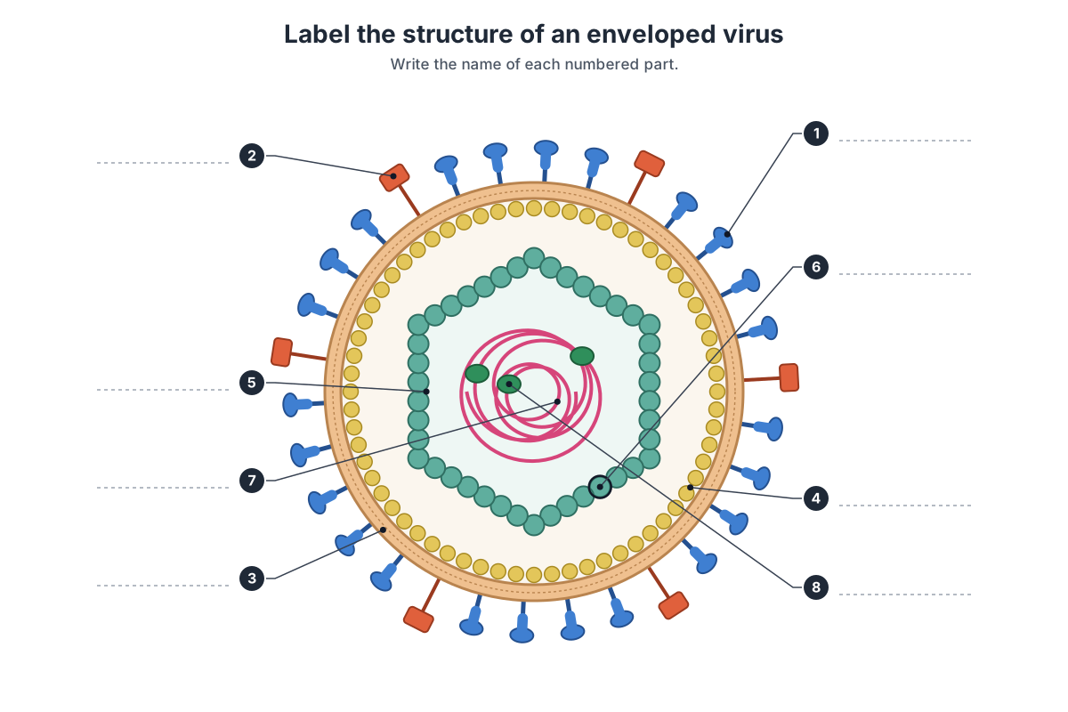
Show the answer key
- Glycoprotein spike (peplomer): attaches the virus to receptors on the host cell.
- Second glycoprotein spike: many enveloped viruses have two kinds, for example HA and NA on influenza.
- Lipid envelope: a bilayer taken from the host cell membrane.
- Matrix protein: links the envelope to the nucleocapsid.
- Capsid: the protein shell around the genome.
- Capsomere: one protein subunit of the capsid.
- Genome (nucleic acid): DNA or RNA.
- Viral enzyme: for example a polymerase or reverse transcriptase.
Bonus: the capsid together with the genome (parts 5–7) is called the nucleocapsid.
More practice questions, using the 3D models:
- Which part of an enveloped virus comes from the host cell, and how is it acquired?
- Name the two spike proteins of influenza A and say what each does.
- Why must influenza and HIV carry enzymes inside the virion, but poliovirus does not?
- What is the shape of the HIV capsid, and which protein makes it?
- How many pentons and hexons does an adenovirus capsid have?
- Why is tobacco mosaic virus called a helical virus?
Frequently asked questions
What are the main parts of a virus?
Every virus particle (virion) has a genome of DNA or RNA and a protein coat called the capsid, built from repeating subunits called capsomeres. The genome plus capsid is the nucleocapsid. Many viruses also have a lipid envelope with glycoprotein spikes, and some have a matrix protein layer under the envelope and enzymes packed inside.
What is the difference between a capsid and an envelope?
The capsid is a shell of virus-encoded protein that encloses the genome; every virus has one. The envelope is a lipid membrane taken from the host cell as the virus buds out, studded with viral glycoproteins. Only some viruses have an envelope (for example influenza, HIV and SARS-CoV-2); naked viruses such as poliovirus and adenovirus do not.
What are the three shapes of virus capsids?
Helical (a rod or filament of protein wound around the genome, as in tobacco mosaic virus), icosahedral (a closed 20-faced shell with 12 corners, as in poliovirus and adenovirus) and complex (combinations or other shapes, as in bacteriophage T4 and poxviruses).
How big is a virus?
Most viruses are about 20–300 nm across, far below the resolution of a light microscope. Poliovirus is about 30 nm, influenza 80–120 nm, SARS-CoV-2 about 91 nm and poxviruses about 250 × 200 nm. Filamentous Ebola virions are about 80 nm wide but around 1 µm long. A typical E. coli bacterium is about 2 µm long.
Are viruses living or non-living?
Viruses are on the border. They have genes, evolve and reproduce, but they are not cells, have no metabolism of their own and can only multiply inside a host cell. Most textbooks describe them as acellular, obligate intracellular parasites rather than living organisms.
What is the structure of HIV?
HIV-1 is a roughly spherical enveloped virus about 100–145 nm across. Its lipid envelope carries a few Env spikes (gp120 on gp41), a matrix layer of p17 lines the envelope, and a cone-shaped capsid of p24 holds two copies of the RNA genome coated with nucleocapsid protein p7, together with reverse transcriptase, integrase and protease.
What are the spikes on a virus called?
Spikes are viral glycoproteins, also called peplomers. Examples are hemagglutinin and neuraminidase on influenza, gp120/gp41 on HIV and the S protein on coronaviruses. They attach the virus to receptors on the host cell and drive membrane fusion.
What is the difference between a virus and a bacterium?
A bacterium is a living prokaryotic cell, about 1–5 µm long, with its own ribosomes and metabolism, and it divides by binary fission. A virus is much smaller, has no ribosomes or metabolism, contains either DNA or RNA but not both, and can only be copied inside a host cell. Antibiotics act on bacteria, not viruses.
Why is the influenza genome in eight pieces?
The influenza A genome is eight separate negative-sense RNA segments, each packaged as its own ribonucleoprotein with its own polymerase. Segments can be swapped when two strains infect the same cell (reassortment), which is how new pandemic strains such as the 2009 H1N1 virus arose.
References
- ViralZone, SIB Swiss Institute of Bioinformatics. Alphainfluenzavirus (influenza A virus).
- ViralZone. Lentivirus (HIV) and HIV-1 capsid protein.
- ViralZone. Betacoronavirus (SARS-CoV-2).
- ViralZone. Mastadenovirus (human adenovirus).
- ViralZone. Tobamovirus (tobacco mosaic virus).
- ViralZone. Baltimore classification.
- ViralZone. Virus symmetry and T number.
- Parker, N., Schneegurt, M. et al. OpenStax Microbiology, section 6.1: Viruses. Rice University.
- Harris, A. et al. (2006). Influenza virus pleiomorphy characterized by cryoelectron tomography. PNAS 103, 19123–19127.
- Noda, T. et al. (2006). Architecture of ribonucleoprotein complexes in influenza A virus particles. Nature 439, 490–492.
- Briggs, J. A. G. et al. (2003). Structural organization of authentic, mature HIV-1 virions and cores. EMBO Journal 22, 1707–1715.
- Zhu, P. et al. (2006). Distribution and three-dimensional structure of AIDS virus envelope spikes. Nature 441, 847–852.
- Ke, Z. et al. (2020). Structures and distributions of SARS-CoV-2 spike proteins on intact virions. Nature 588, 498–502.
- San Martín, C. (2012). Latest insights on adenovirus structure and assembly. Viruses 4, 847–877.
- Goodsell, D. S. (2009). Tobacco mosaic virus. RCSB PDB-101 Molecule of the Month.
- BioNumbers (Harvard). Size of the E. coli 70S ribosome (ID 102320) and E. coli cell dimensions (ID 117344).
More interactive anatomy models
- Bacterial cell diagramLabeled prokaryotic cell: cell wall, membrane, nucleoid, ribosomes, pili and flagellum.
- Amoeba diagramLabeled Amoeba proteus: pseudopodia, nucleus, contractile and food vacuoles.
- Euglena diagramLabeled Euglena: flagellum, eyespot, chloroplasts, pellicle and contractile vacuole.
- Bacteriophage diagramT4 phage structure in 3D: head, collar, tail sheath, baseplate and tail fibres.
- Human anatomy1,000+ bones, muscles, organs, vessels and nerves from real anatomical data.
- Octopus anatomyThree hearts, a ring-shaped brain, eight arms and a beak, in 3D.